Mitochondrial dysfunction in human pathologies
María Monsalve 1,2,3,Sara Borniquel 1,2,3,Inmaculada Valle 1,2,3, and Santiago Lamas 1,2,3
1 Centro de Investigaciones Biológicas (Consejo Superior de Investigaciones Científicas). Ramiro de Maeztu 9, 28040, Madrid
Spain, 2Centro Nacional de Investigaciones Cardiovasculares (Instituto de Salud Carlos III), Melchor Fernandez Almagro 3,
28029, Madrid, Spain,3Instituto “Reina Sofía” de Investigaciones Nefrológicas, Spain
TABLE OF CONTENTS
1. Abstract 2. Introduction
3. Mitochondrial oxidative stress protection system.
3.1. Detoxification enzymes
3.1.1. Mn-SOD
3.1.2. Catalase
3.1.3. Peroxiredoxins
3.1.4. Thioredoxin and thioredoxin reductase
3.1.5. Glutathione peroxidase and other glutathione related systems
3.2. Other protection systems
3.2.1. UCP-2
3.2.2. Heat shock proteins (HSP) 4. Transcription factors 4.1. Nrf1 and Nrf2 4.2. AP-1 4.3. NFkappaB 4.4. HIF-1 4.5. PPARs 4.6. Foxo3a 4.7. PGC-1alpha 5. Nitric oxide 5.1. Direct effects 5.2. Indirect effects 6. Diseases 6.1. Neurodegenerative diseases 6.1.1. Parkinson’s disease 6.1.2. Huntington’s disease 6.1.3. Alzheimer’s disease 6.1.4. Epilepsy
6.1.5. Friedreich ataxia (FRDA)
6.2. Cancer and ageing
6.2.1. Cancer
6.2.2. Aging
6.3. Diabetes, atherosclerosis and ischemia reperfusion injury
6.3.1. Diabetes
6.3.2. Atherosclerosis
6.3.3. Ischemia reperfusion injury 7. Conclusions and perspectives
8. Acknowledgments 9. References
1. ABSTRACT
The integrity of mitochondrial function is fundamental to cell life. The cell demands for mitochondria and their complex integration into cell biology, extends far beyond the provision of ATP. It follows that disturbances of mitochondrial function lead to disruption of cell function, expressed as disease or even death. Mitochondria are major producers of free radical species and also
possibly of nitric oxide, and are, at the same time, major targets for oxidative damage. In this review we consider recent developments in our knowledge of how the mitochondrial production of reactive oxygen species (ROS) plays a critical role in several major human pathologies. We will also consider recent advances in our understanding of the molecular mechanisms involved in mitochondrial ROS detoxification.
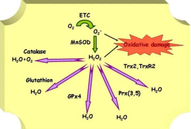
Figure 1. Main mitochondrial ROS detoxification enzymes.
2. INTRODUCTION
Mitochondria have been considered the powerhouse of the living cell. Their main function is the generation of energy in the form of the molecule ATP, a process that takes place through oxidative phosphorylation (1, 2). But, mitochondria are not just providers of energy, they are involved in many other aspects of cell physiology, the best known being calcium signaling, and programmed cell death (apoptosis) (3).
Generation of reactive oxygen species (ROS) in mitochondria is a consequence of the action of the electron transfer associated with oxidative phosphorylation that generates unpaired electrons. It has been estimated that 1-2% of the electrons that travel along the electron transfer (ETC) react with molecular oxygen (O2) to produce the anion superoxide (O2-) a highly reactive free radical species. The superoxide anion-radical is readily converted to ROS, such as hydrogen peroxide (H2O2) and hydroxyl radicals (HO.). There is no question that mitochondria are the major source of oxygen free radical generation in most cell types, except perhaps for macrophages, which express an NADPH oxidase that generates massive amounts of O2 -after stimulation.
The chemical species called ROS are able to cause lipid peroxidation and damage to cell membranes, proteins and DNA, so that mitochondria represent not only a major source of ROS generation, but also a major target of ROS induced damage. The high rate of mitochondrial DNA (mtDNA) mutation (10-fold higher than in the nuclear DNA (nDNA)) will eventually result in the accumulation of mutations in mitochondrial proteins. Defective proteins will in turn exacerbate mitochondrial dysfunction, increase production of ROS, and reduce mitochondrial energy production.
Mitochondria are equipped with an armamentarium of antioxidant defenses (Figure 1) and contain a high concentration of glutathione, mainly reduced glutathione (GSH) fundamental to cell survival. Although still not thoroughly investigated, important members of this system are Mn superoxide dismutase (Mn-SOD),
peroxiredoxins III and V (Prx3, Prx5), mitochondrial thioredoxin (Trx2) and, mitochondrial thioredoxin reductase (TrxR2), mitochondrial glutaredoxin (Grx2a), mitochondrial glutaredoxin reductase (mtGrxR), glutathion peroxidase 4 (GPX4), uncoupling protein 2 (UCP-2), and a specific set of heat shock proteins.
In general, the leak of electrons from the electron transfer chain seems to be increased by an increase in mitochondrial potential and decreased with mitochondrial depolarization. Provided that mitochondrial activity can vary greatly, as well as electron transfer efficiency as phosphorylation coupling, mitochondrial ROS production can change rapidly in various physiological and pathological conditions. Therefore the oxidative protection system must be under tight regulatory control. Several transcription factors, and biomolecules like nitric oxide (NO) have been proposed to modulate the expression of at least one member of the system, but until recently a coordinated regulation had not been described. Peroxisome proliferator activated receptor gamma coactivator 1 alpha (PGC-1alpha), a transcriptional coactivator well known as a key regulator of mitochondrial biogenesis and lipid catabolism, has been shown to be a master regulator of the mitochondrial oxidative stress protection system in endothelial cells, a role that is likely to play in other cells systems, particularly those with high metabolic rates where PGC-1alpha has a crucial role in the control of energy metabolism.
Mutations in mitochondrial genes have been shown to cause many different genetic diseases (i.e. LHON dystony, Leigh´s disease) (4). Phenotypes of mitochondrial diseases can be both diverse and overlapping, that is, the same mutation can produce quite different phenotypes, and different mutations can produce similar phenotypes. Variations in the percentage of mutant mtDNAs between patients must change the ATP output and cause the variation in clinical symptoms. Hence patients are generally classified by genetic defect rather than by clinical manifestation (5). However, the most common pathologies associated with mitochondria are not hereditary but associated with age and with the physiological or pathological production of mitochondrial ROS.
Major pathologies for which it the role played by mitochondrial dysfunction and ROS production has been clearly established include all the common neurodegenerative diseases (Parkinson´s disease, Alzheimer´s disease, Huntington´s disease, epileptic seizures, FRDA), atherosclerosis, diabetes, ischemia-reperfusion injury, cancer and aging.
Therefore, the knowledge of the causes that result in an excessive production of mitochondrial ROS and/or the failure of its detoxification systems are necessary for the development of new therapeutic strategies. In order to understand the pathologies associated with mitochondrial dysfunction, one of the fundamental milestones is to underscore the differences in the susceptibility of different cell types to mitochondrial oxidative stress. The factors responsible for this variability include the inner
mitochondrial membrane lipid composition and /or the oxidant/antioxidant balance, (i.e. superoxide dismutase) and/or heat shock protein activity and expression as well as the glutathione status. It is also important to point out that most of these pathologies involve tissues with high metabolic rates and mitochondrial content.
3. MITOCHONDRIAL OXIDATIVE STRESS PROTECTION SYSTEM
ROS include a family of chemically reactive molecules derived from O2. Some of these molecules are extremely reactive, such as HO., while some others are less reactive like O2- and H2O2. Intracellular free radicals (free, low molecular weight molecules with an unpaired electron, are often ROS and vice versa, and the two terms are commonly exchanged). Free radicals and ROS can readily react with most biomolecules, starting a chain reaction of free radical formation. In order to interrupt this chain reaction, a newly formed radical must either react with another free radical, with cancellation of the unpaired electrons, or react with a free radical scavenger, also known as a chain-breaker or primary antioxidant.
3.1 Detoxification enzymes 3.1.1. Mn-SOD
Superoxide anion is generated from molecular O2 by the addition of one electron. It is not highly reactive, and it lacks the ability to penetrate lipid membranes and is therefore enclosed in the compartment where it was produced. The formation of O2- takes place spontaneously, especially in the electron-rich aerobic environment in the vicinity of the inner mitochondrial membrane containing the respiratory chain. Although O2- is not an effective oxidant, it impairs mitochondrial function by oxidizing the Fe-S centers of various enzymes. In addition, O2- can react with NO to produce peroxynitrite, an extremely powerful oxidant.
Two molecules of O2- rapidly dismutate to H2O2 in the presence of the superoxide dismutase enzymes (SOD). Mn-SOD is the SOD isoform that localizes in the mitochondrial matrix. It is an essential protein (sod2 knock-out mice die soon after birth) (7). Several lines of evidence support the idea that Mn-SOD has tumor suppressor activity. In general, reduced levels of Mn-SOD are observed in many types of human tumors, whereas overexpression of Mn-SOD suppresses tumorigenicity. Life-long reduction in Mn-SOD activity (in heterozygotic
sod2+/- mice) results in a much higher incidence of cancer (7). It has been postulated that Mn-SOD anti tumorigenic activity is a direct consequence of the relieved mitochondrial oxidative stress caused by O2-, however it must be taken into account that Mn-SOD generates H2O2 and therefore can generate a different type of oxidative stress in the absence of the corresponding H2O2 scavengers. Nevertheless, Mn-SOD seems to behave mainly as an antioxidant in most biological settings. Evidence supporting an antioxidant role for Mn-SOD include studies with transgenic mice over-expressing Mn-SOD in various tissues, they show increased tolerance to several oxidative stress situations including ischemia-reperfusion injury (8),
streptozotocin induced beta-cell injury (9), 6-hydroxydropamine injury to dopaminergic neurons (10), and adriamycin and paraquat toxicity (11). Importantly, the
sod2 gene is transcriptionaly up-regulated by oxidative stress.
3.1.2. Catalase
Hydrogen peroxide is not a free radical, but it is highly important much because it is an essential intermediate in the formation of more reactive ROS molecules, particularly of hydroxyl radical (HO.) via Fenton reaction with transition metals. It also has the ability to penetrate biological membranes, and therefore H2O2 generated in the mitochondria can readily diffuse into the cytosol. Hydrogen peroxide is removed by at least three antioxidant enzyme systems, catalases, glutathione peroxidases and peroxiredoxins.
Catalase is mainly located in peroxisomes, where it catalyzes the dismutation of H2O2 to H2O and O2. It is likely to be responsible at least in part of the clearance of the H2O2 that is produced in the mitochondria and leaks out into the cytoplasm. The oxidative stress protective role of catalase is well established. Deficiencies in catalase activity are associated with oxidative stress related pathologies, and increased oxidant sensitivity. Cancer cells typically show reduced catalase levels. Inherited deficiency in catalase is associated with diabetes mellitus, hypertension and altered lipid, carbohydrate and homocysteine metabolism (12). Mice lacking catalase have increased sensitivity to various oxidants (13). In contrast, increased catalase levels are generally protective. Transgenic mice over-expressing catalase in various tissues show increased tolerance to ischemia-reperfusion injury and oxidative stress inducing agents such as adriamycin, paraquat (14) and streptozotocin induced beta-cell injury (9). Apolipoprotein E deficient (ApoE-/-) mice that over-express catalase show reduced atherosclerosis plaque formation (15), probably because over-expression of catalase in vascular smooth muscle cells (VSMC) inhibits H2O2 induced cell proliferation (a process that takes place during plaque formation) (16). Most importantly targeting of catalase to mitochondria extended life span in mice by 20% (17).
3.1.3. Peroxiredoxins
It is a family of enzymes capable of directly reducing hydroperoxides (i.e. H2O2 and different alkyl hydroperoxides). In the mitochondria of mammalian cells there are at least two members, Prx3, and Prx5. Prx3 contains a mitochondrial localization sequence, is found exclusively in the mitochondrion and uses mitochondrial thioredoxin-2 (Trx2) as the electron donor for its peroxidase activity (18). Prx3 is induced by oxidants in the vascular endothelial cell system and is thought to play a role in the antioxidant defense system and homeostasis within the mitochondria (19). Prx5 has three isoforms originated by alternative splicing, one is present in the mitochondria, another in the nucleus and the third in peroxisomes. mtPrx5 is comparatively poorly characterized than Prx3, however the information available so far point to a very similar role to that of Prx3. It uses Trx2 as electron donor (20-24).
Both Prx3 and Prx5 are over-expressed in different forms of cancer (25, 26), including the best characterized breast cancer (27, 28). This observation contrast with the above mentioned reduced activity of both Mn-SOD and catalase in human malignancies. It has been proposed that both Prx3 and Prx5 protect malignant cells from H2O2 toxicity.
Whereas low levels of H2O2 are essential for cell growth, elevated levels of H2O2 are toxic to the cell and can lead to apoptosis. Human solid tumors frequently show regions of hypoxia, and suffer elevated levels of H2O2. This situation can explain why Prx over-expression can be beneficial to cancer cells. Elevated Prx3 levels help to scavenge the excess H2O2 and protect cells from H2O2 induced apoptosis (29). Similarly Prx3 protects cells during hypoxia (30), a situation that induces the mitochondrial production of ROS by the disruption of oxidative phosphorylation (31).
However, in physiological conditions, low levels of H2O2 are an important cell proliferation signal. In these conditions Prx3 inhibits the growth stimulatory effects of H2O2. This effect can be crucial in the cardiovascular system where metabolic dysfunction in the endothelium generates an oxidative stress situation that initiates atherosclerosis processes. In this scenario excessive production of H2O2 by stressed endothelial cells promotes proliferation of VSMC, a process that could be prevented by Prx3. 3.1.4. Thioredoxin and thioredoxin reductase
The thioredoxin system consists of the two antioxidant oxidoreductase enzymes thioredoxin (Trx) and thioredoxin reductase (TrxR). The latter catalyzes the reduction of the active site disulfide in Trx using NADPH2 and, among other functions, reduced Trx is a general protein disulfide reductant, and a specific electron donor for many Prx.
Trx2 is located in mitochondria (24). The Trx2 system includes Prx3, Trx2 and TrxR2. Prx3 utilizes Trx2 as electron donor, and TrxR2 uses NADPH2 to regenerate reduced Trx. All tree are ubiquitously expressed, but are found at the highest levels in metabolically active tissues, such as heart (32), brain and liver (33, 34). Yeast mutants of any of these three proteins support their role in the protection against oxidative stress (35). Trx2 over-expression can protect cells from oxidant-induced apoptosis and increase the mitochondrial membrane potential (36, 37). Trx2-/- mice show early embryonic lethality. The timing of embryonic lethality coincides with the maturation of the mitochondria, since they begin oxidative phosphorylation during that stage of embryogenesis (38). Conditional Trx2 deficient cells show that upon loss of Trx2 cells show and increased accumulation of ROS, decreased mitochondrial GSH levels and induction of apoptosis (39). Trx2 is up regulated by ROS (40-42), and this regulation could be involved in preconditioning-induced protection from cardiac and neuronal ischemia.
TrxR reduces not only the disulfide in oxidized Trx, but also some other protein disulfides or a wide spectrum of oxidized low molecular weight compounds, playing an important role in the regeneration of antioxidants. TrxR2 is located in mitochondria (43, 44). Deletion of TrxR2 results in embryonic lethality at day 13. These embryos have hematopoiesis deficiencies and are severely anemic, show increased apoptosis in the liver, and reduced cardiomyocyte proliferation, the cultured fibroblast are extremely sensitive to oxygen radicals. In order to investigate the putative role of mitochondrial dysfunction in the embryonic lethality, TrxR2 was specifically ablated in mice heart. These mice die shortly after birth of fatal dilated cardiomyopathy, a condition observed in Friedreich´s ataxia and associated with mitochondrial oxidative stress (45). Over-expression of a dominant negative form of TrxR2 in HeLa cells results in a higher concentration of H2O2 (46), while over-expression of TrxR2 increases resistance to cytotoxicity induced by inhibition of the mitochondrial complex III.
3.1.5. Glutathione peroxidase and other glutathione related systems
The tri-peptide glutathione (g-glutamyl-cyteinyl-glycine) is an endogenous antioxidant of great importance. GSH is required for the maintenance of the thiol redox status of the cell, protection against oxidative damage, detoxification of reactive metals and electrophiles, storage and transport of cysteine, DNA synthesis, cell cycle regulation and cell differentiation (47). The key functional element of glutathione is the cysteinyl moiety, which provides the reactive thiol group. ROS are reduced by GSH in the presence of GSH peroxidase (GPx). As a result GSH is oxidized to GSSG, which in turn is rapidly reduced back to GSH by GSSG reductase (GR) at the expense of NADPH. The redox cycle also aids in maintaining reduced protein and enzyme thiols. Without it, vulnerable cysteinyl residues of essential enzymes might remain oxidized, leading to changes in catalytic activity. Glutathione is also an excellent scavenger of lipid peroxidation products such as HNE and acrolein. Glutathione reacts with carbon atoms via nucleophilic attack on an electrophilic carbon, in a reaction catalyzed by glutathione S-tranferase. GSH also has a high affinity for metal ions, and acts as a reductant in redox reactions involving metals. It also forms stable complexes with metals (mercury, lead, etc) and products of lipid peroxidation that can then be eliminated from the body.
Another class of proteins intimately related to GSH are the glutaredoxins (Grx) and glutaredoxin reductase (GrxR), with functions overlapping those of thioredoxin system, the major difference being that Grx can be reduced directly by GSH and is capable of reducing GSH mixed protein disulfides formed at oxidative stress.
Mitochondria have a high concentration of glutathione that must be imported from the cytosol via an specific carrier because it lacks the enzymes required for glutathione synthesis. Ethanol intake reduces the activity of the carriers (dicarboxylate and 2-oxoglutarate) in hepatic cells, sensitizing hepatocytes to oxygen radicals (48),
mitochondrial GSH depletion by beta-amyloid is associated with Alzheimer’s disease (AD) (49, 50). Changes in redox balance in the mitochondria are directly associated with alterations in the GSH/GSS ratio, and the induction of mitochondria-mediated apoptosis (51).
The mitochondrial Grx2a (52) can use both GSH and TrxR2 as electron donors. It has been shown to play a role in the protection of thiols of mitochondrial proteins from formation of disulfides when the redox balance is altered in oxidative stress situations (53), and could also help to prevent cardiolipin oxidation and the initiation of the mitochondrial apoptotic cascade induced by agents like doxorubicin and 2-deoxy-D-glucose (54). It has also been proposed to act as a mitochondrial redox sensor that is activated when the glutathione pool becomes oxidized. (55). The mitochondrial glutathione reductase (mtGrxR) in an splice variant isoform of the gene coding for the cytoplasmic enzyme (56).
Phospholipids that are rich in unsaturated fatty acids, are particularly susceptible to ROS attack. The result is the formation of lipid hydroperoxides. This process is particularly relevant in mitochondria where the electron transfer chain produces O2- and H2O2 at the mitochondrial inner membrane (57). GPx1 is the major GPx isoform, and it is present in all tissues; 10% of GPx1 localizes to mitochondria (mtGPx1) where it has been considered the most important H2O2 metabolizing enzyme in mitochondria, a view that was recently challenged (58). GPx4 is also called PHGPx and has three isoforms originated by alternative splicing, one present in the mitochondria (L-form), where it markedly reduces the lipid hydroperoxides generated in biomembranes. GPx4 over-expression effectively prevents apoptosis induced by different stimuli, including oxidized low density lipoproteins (oxLDL), and cholesterol hydroperoxide (both important risk factors in atherosclerosis). It protects the mitochondrial lipid cardiolipin from oxidation and hence prevents the release of cytochome c from the mitochondrial inner membrane. It also prevents the inactivation of the adenine nucleotide transporter that leads to the opening of the membrane transition pore (MTP), exit of cytochrome c and initiation of apoptosis (59-62). GPx4 also reduces the accumulation of mitochondrial H2O2 in cells treated with agents that generate mitochondrial stress. It has been proposed that Gpx4 is important for the protection of the damage associated with ischemia-reperfusion injury (63). 3.2 Other protection systems
3.2.1. UCP-2
It is a gated proton channel located in the mitochondria inner membrane, it decreases metabolic efficiency by dissociating substrate oxidation in the mitochondria from ATP synthesis.(64). Therefore, it can reduce the inner mitochondria membrane potential (delta-Psi), reduce ATP synthesis, dissipate energy in the form of heat, and diminish the production of O2-. It was initially proposed to play a role in adaptive thermogenesis, however accumulated data support the notion that UCP-2 prevents the excessive production of O2- in mitochondria. In fact, O2 -has been shown to induce the expression of UCP-2 (65). It
is also directly induced by H2O2 and under pathological conditions where excessive ROS production occurs (66-69). However, the transcriptional regulatory mechanisms are not fully elucidated, its expression is induced by PPARalpha, and PGC-1alpha and it seems that activation requires high glucose and FFA, a putative protective mechanism since both are likely to increase ROS production. UCP-2-/- animals show increased free radical production in monocytes (70), UCP-2 deficiency in bone marrow precursors results in a significant increase in atherosclerotic plaque formation in mice (71). Its over-expression has been proposed to be protective from ROS in various cell types and tissues. Cells that over-express UCP-2 are more resistant to HUCP-2OUCP-2 treatments (7UCP-2-74). UCP-UCP-2 over-expression also reduces lipid peroxidation at least in the brain (75), where it protects neurons against seizure and neuronal damage (76, 77). Over-expression in dopaminergic neurons protects from Parkinson-inducing agents (78). Furthermore, increased expression of human UCP-2 in the adult fly nervous system extends fly life span (77). Importantly, UCP-2 has also been proposed to control glucose dependent insulin secretion in beta-cells, through the reduction of ATP synthesis, and concomitantly has been proposed to play a role in the pathogenesis of diet-related type 2 diabetes.
3.2.2. Heat shock proteins (HSP)
In mammalian cells HSP synthesis is induced not only after hyperthermia, but also following a wide variety of stress conditions including oxidative stress (79), in fact it constitutes a fundamental mechanism necessary for cell survival under a wide array of toxic conditions that include mitochondrial oxidative stress (80). The induction of the heat shock response requires the activation and translocation to the nucleus of one or more heat shock transcription factors which control the expression of a set of protective heat shock proteins (81). The transcription factor HSF plays a crucial role in the transcriptional induction of HSP in stress conditions (82-84). Some of the best known HSPs include ubiquitin, Hsp10, Hsp27, Hsp32 (HO-1), Hsp47, Hsp60, Hsc70, Hsp70 (Hsp72), Hsp90 and Hsp100/105 (85). The important and well-characterized Hsp70 family includes Hsc70, Hsp70, and GRP75. They are protein chaperones, that bind to unfolded proteins and return them to their native conformation trough an ATP dependent mechanism. Ubiquitin targets proteins to be degraded by the proteosome (86).
Mitochondrial oxidative stress has been shown to induce a heat shock response (87). Several HSP have been found in the mitochondria where they could play a protective role. Hsp60 directs the entry of proteins into the mitochondria (88), its yeast homologue has been proposed to protect from oxidative stress (89). Glucose-regulated protein 75 (GRP75) (90), prevents the accumulation of ROS in mitochondria (91). Hsp10 exerts its chaperone function with proteins within mitochondria (92). Hsp10 is required for the folding and assembly of proteins imported into the matrix compartment, and is involved in the sorting of certain proteins, such as the Rieske Fe/S protein, passing through the matrix en route to the intermembrane space. Both Hsp10 and Hsp60 have been shown to protect
mitochondrial function in ischemia-reperfusion (93). Hsp70 (Ssc1) regulates the entry and adequate folding in the mitochondria of proteins with Fe-S centers (94, 95) and is activated by Hsp40.
Hsp32 has three isoforms, 1, 2 and HO-3. HO-1 is the rate limiting enzyme in the production of bilirubin, a potent antioxidant. It catalyzes the specific oxidative cleavage of the heme molecule to form equimolar amounts of biliverdin and carbon monoxide (CO). HO-1 is rapidly up-regulated by oxidative and nitrosative stresses, as well as by glutathione depletion. Its promoter has an antioxidant responsive element (ARE) similar to other antioxidant enzymes (96).
4. TRANSCRIPTION FACTORS
All the above mentioned antioxidant defenses are induced by different stimuli in response to stress situations. Several transcriptions factors have been proposed to be activated in response to oxidative stress and/or modulate the expression of one or more protection genes. However until recently a specific and coordinated regulation of the mitochondrial system had not been described. The transcriptional coactivator PGC-1alpha has been shown to play that role and to be directly regulated by the intracellular levels of NO. The activity of the transcription factors is also tightly controlled by the cellular redox state as evidenced by the protein Raf-1 that is necessary to keep several transcription factors in a reduced/active conformation (97).
4.1. Nrf1 and Nrf2
Basal and inducible expression of a number of antioxidant defense genes are in part regulated by a cis-acting DNA element known as the antioxidant responsive element, ARE, (98) that should be more properly called electrophyl response element, since it is present in genes that detoxify carcinogens, suggesting that induction trough ARE is chemopreventive, and not closely related to mitochondrial processes (99). However, activation through ARE appears to be driven by conditions that promote intracellular oxidative stress and therefore must be taken into consideration. A number of transcription factors have been proposed to bind to ARE, but the most recent studies point to the CNC subfamily of bZIP proteins as mediators of ARE function (100). They appear to function as obligate hererodimers with other bZIP proteins.
Forced expression experiments of the bZIP proteins Nrf1 and Nrf2 suggest that they mediate ARE function (101). Nrf1-/- mice show embryonic lethality but Nrf1 null cells have been isolated and shown to have an increased sensitivity to the toxic effects of oxidants than their wild type controls (102). Nrf2-/- mice are viable but they show diminished expression of several phase 2 enzymes (detoxification of xenobiotics) and are sensitive to treatments that cause oxidative stress (103, 104). The analysis of double knock-out animals shows they have overlapping functions and are functionally redundant in mediation ARE function and oxidative stress defense (105). The mechanism of signal transduction from xenobiotics to
Nrf1 and Nrf2 has not been elucidated although it has been proposed to involve the activation and phosphorylation by PKC (106).
4.2. AP-1
The immediate early response expression of Jun and Fos (AP-1) controls the expression of many genes, and plays a central role in the control of cell proliferation and transformation. Among other stimuli, it is rapidly induced by H2O2, UV-C, UV-A, ionizing radiation etc. Its DNA binding activity is also induced by UV and H2O2 (99). Several signal transduction pathways and the direct action of oxidants on the proteins have been proposed to mediate their induction and activation. AP-1 has been shown to induce the expression of Mn-SOD (107), but the biological role of this regulation seems to be mainly related to the role played by Mn-SOD in tumor suppression (108, 109), and although it is possible that it is also relevant in the control of the celullar redox state, to date this possibility has not yet been tested.
4.3. NFkappaB
Nuclear factor kappa B (NFkappaB) plays a central role in the regulation of many genes involved in cellular defense mechanisms, apoptosis, pathogen defenses, immunological responses and expression of cytokines and cell adhesion molecules. ROS, generated in the mitochondria, have been proposed as the intermediate second messengers to the activation of NFkappaB by tumor necrosis factor alpha (TNFalpha) and interleukin 1 (IL-1). NFkappaB activation can in fact be directly mediated by H2O2 and hydroperoxides (99). NFkappaB has been shown to induce Mn-SOD (110, 111), the biological role of this induction is not completely understood but it might be related with the induction of pro-survival signals in inflammatory situations where high levels of ROS are generally produced (112-114).
It is a well-established fact that females live longer than males. Mitochondria from females produce less H2O2 and have higher levels of Mn-SOD and GPx. Estrogen is responsible for these differences. Interestingly, its effects do not seem to be mediated by the nuclear receptor ER, but by its interaction with a membrane receptor that signals to exert NFkappaB activation (115). 4.4. HIF-1
Hypoxia-inducible factor-1 (HIF-1) is a transcription factor that governs cellular responses to reduced O2 availability, a situation that is also prone to production of mitochondrial ROS. Upon O2 deprivation the degradation rate of the HIF-1 alpha subunit turn-over is reduced and the protein stabilized. Importantly it has been recently shown that HIF-1 stabilization requires mitochondrial ROS and is inhibited in the presence of high levels of GPx or catalase (116, 117). HIF-1 has also been proposed to up-regulate the Mn-SOD promoter although the biological significance of this regulation has not been established (118).
4.5. PPARs
The connection between peroxisome proliferator activated receptors (PPARs) and oxidative stress is still a
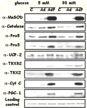
Figure 2. High glucose increases mitochondrial ROS production leading to oxidative stress and endothelial dysfunction. PGC-1alpha induction activates mitochondrial function and increases its ROS detoxification capacity. Over-expression of PGC-1alpha in high glucose conditions prevents oxidative stress and endothelial dysfunction. non solved issue. PPARalpha has been shown to induce ROS production in liver where it can result in increased carcinogenesis risk (119). However several reports also point to PPARs as involved in the regulation of the inflammatory response, and possibly in the control of ROS production (120). Reports suggesting that PPARs can induce UCP-2 promoter are also suggestive of the involvement of PPARs in the control of cellular ROS production (121, 122).
4.6. Foxo3a
Foxo3a is the DNA binding transcription factor that up to date has been more clearly and directly involved in the protection against mitochondrial oxidative stress. Foxo3a is the human homolog of Daf-16 from C. Elegans, genetic data in C. elegans show that Daf-16 increases life span in response to reduced insulin/IGF-1 signaling (123), and has also been shown to modulate, metabolism and fertility in the worm. Foxo3a can protect from H2O2 toxicity by directly inducing Mn-SOD and calalase genes (124). Regulation by insulin/IGF-1 activates protein kinase B (AKT) that phosphorylates Foxo3a and directs its exclusion from the nucleus (125). Therefore, Foxo3a is inactivated in high insulin (high glucose) conditions where lipid metabolism and mitochondrial activity are low. Foxo3a is also sensitive to redox status through its interaction with p66shcA (126). ERK directly phosphorylates p66shcA. In response to H2O2, inhibition of ERK activation represses p66shcA-dependent phosphorylation of Foxo3a, and facilitates Foxo3a entry into the nucleus (127). Importantly Foxo3a is also able to
induce cell cycle arrest in stress conditions supporting the role of Foxo3a in cell survival under stress conditions. 4.7. PGC-1alpha
The discovery that caloric restriction is associated with increased longevity, was the first indicator of the important connection between the regulation of oxidative metabolism or in other words, mitochondrial function and the oxidative stress protection systems. The idea is at first sight paradoxical, since increased mitochondrial activity should be associated with an elevated production of ROS in the mitochondria, however, cells that are more dependent on oxidative phosphorylation than on glycolysis, were more protected against oxidative stress, had lower ROS levels and hence aged more slowly. The clue to this apparent paradox is very likely that the activation of oxidative metabolism and the mitochondrial function goes hand in hand with the induction of the ROS protection system. The transcription factor responsible for this coordinated regulation is PGC-1alpha. PGC-1alpha was originally characterized as an inducer of mitochondrial proliferation (128), it was later found to be also and activator of lipid catabolism (129) and liver gluconeogenesis (130).
Two important characteristics of PGC-1alpha are its tissue-specific expression pattern and its inducibility by particular signals (131, 132). PGC-1alpha is selectively expressed in tissues that have high energy demands and are rich in mitochondria, such as heart, skeletal muscle, brown adipose, kidney, liver and brain (128, 130, 133, 134). Its activity and expression levels are induced by cold exposure, fasting and, exercise (133, 135, 136). Signaling pathways associated with these stimuli (p38 MAP kinase, beta-adrenergic/cAMP, nitric oxide, AMP kinase and Ca2+ -calmodulin kinase) activate PGC-1alpha and its downstream target genes by increasing PGC-1alpha expression or transactivation function (137-140). Previous studies using gain-of-function strategies (adenoviral-mediated or transgenic expression of PGC-1alpha in cultured cells and in vivo) have demonstrated that the transcriptional coactivator PGC-1alpha promotes the activation of mitochondrial biogenesis and increases cellular respiration (128, 135, 141).
On the other hand, studies in PGC-1alpha deficient mice have demonstrated the critical role for PGC-1alpha in the control of cellular energy metabolism. Oxidative metabolism and mitochondrial function are impaired in several tissues in mice lacking PGC-1alpha, fewer and smaller mitochondria were found in slow twitch muscles. Accordingly, a reduction in the expression of nuclear genes encoding proteins involved in mitochondrial electron transfer chain (cytochrome c and cytochrome oxidase), the beta subunit of ATP synthase, as well as a reduction of the expression of Tfam A (that directs the replication of the mitochondrial DNA) were found in knockout PGC-1alpha animals (142, 143).
Our group has recently proposed that PGC-1alpha coordinately regulates several key genes involved in the protection against mitochondrial oxidative stress, Mn-SOD, catalase, UCP-2, Prx3, Prx5, Trx2 y TrxR2 (Figure 2).
Cells that over-express PGC-1alpha produce less ROS and are protected against oxidative stress agents like high glucose and H2O2 (144).
5. NITRIC OXIDE
NO is a diatomic free radical which is a gas at room temperature, making it highly diffusible within the cell. Its major target is the soluble guanylate cyclase (sGC), an enzyme whose activation increases the concentration of cGMP, and the activation of the cGMP dependent kinase G (PKG). NO/cGMP/PKG signaling pathway was originally identified as playing a key role in blood pressure regulation promoting smooth muscle relaxation, (145). It was later found to play other important roles in endothelial physiology and maintenance, as well as in regulation of platelet aggregation, and peripheral and central neurotransmission (146). More recently it has been proposed to control mitochondrial number (147, 148).
NO is produced from arginine and O2 through a reaction catalyzed by nitric oxide synthases (NOS). Traditionally, three types of NOS have been described: iNOS (inducible), nNOS (neuronal) and eNOS (endothelial). It has also been proposed the existence of a mitochondrial NOS (mtNOS), that is not encoded by an independent gene but could be an alternative spliced product or specifically modified version of one or more of the classical enzymes (149). This hypothesis is intriguing since NO has multiple targets within the organelle. Although the existence of mtNOS is still under debate there are interesting findings. First of all, it seems that eNOS is attached to the outer mitochondrial membrane, which indicates that mitochondria may regulates NOS activity and conversely, eNOS may regulate mitochondrial respiration. The idea that mitochondria themselves are capable of producing NO is an important concept in several physiological and pathological mechanisms. Although mitochondria are not expected to release physiological relevant levels of NO, NO readily reacts with O2- to produce peroxynitrite a highly reactive compound with important biological activities (150).
A large body of evidence suggest that NO plays a dual role acting both as a pro-oxidant and as antioxidant in different biological settings. Most evidence points to peroxynitrite as the mayor mediator of NO prooxidant effects while how NO mediates its antioxidant effects has been a serious matter of controversy, although most evidence points to a putative role played by the mitochondria. We have recently shown that NO can modulate PGC-1alpha expression both positively and negatively and hence regulate the expression levels of the mitochondrial detoxification system (our unpublished results). Both the prooxidant and antioxidant effects of NO have been shown to be highly relevant in pathologies such as atherosclerosis, neurodenenerative diseases, and ischemia reperfusion. The dual effects of NO depend on one hand on the levels of NO, since low levels tend to be protective, while high concentrations are normally toxic to the cells. On the other hand, NO has direct effects (those that are direct consequence of its chemistry) and the most
important, indirect effects (those mediated by the activation of the sGC/PKG pathway).
5.1. Direct effects
One of the most significant biological reactions of NO is with transition metals like Fe resulting in NO-metal complexes. NO can react with heme iron containing proteins, a reaction that is highly reversible, or with non heme iron, a situation that results in the permanent inactivation of the target enzyme. Putative heme protein targets for NO include catalase, cytochrome oxidase (151), hemoglobin and peroxidase. NO reaction with non-heme iron, such as iron-sulfur clusters has been described with enzymes like NADH-ubiquinone oxidoreductase, cis-aconitase and NADH: succinate oxidoreductase. However the biological relevance of these reactions is still to be determined, particularly for the non-heme reactions with low Km that depend on extremely high NO concentrations.
Nitric oxide affects the mitochondrial respiratory chain through: a) inhibition of the cytochrome oxidase activity, b) inhibition of the electron transfer between cytochromes b and c and c) inhibition of NADH-dehydrogenase activity (152).
Inhibition of cytochrome oxidase activity. Cytochrome oxidase or complex IV is the terminal enzyme of the mitochondrial respiratory chain that is responsible for over 95% of cellular O2 consumption in mammals and that is inhibitable by NO in a direct interaction with the enzyme (153, 154). The inhibition takes place as NO competes with O2 for the binding site of the enzyme at the reactive center, increasing the operative Km of O2 for the enzyme. A consequence of cytochrome oxidase inhibition is a reduction of the capacity to use O2 in cells, a phenomenon known as ‘metabolic hypoxia’ (155). Inhibition of mitochondrial respiration occurs in a reversible and, concentration-dependent manner (151, 156). The degree of inhibition of cytochrome oxidase activity by NO depends on the O2 concentration in the reaction medium (152). The function of the interaction of NO with cytochrome oxidase is still a matter of controversy. Brown (153) proposed that NO inhibition of cytochrome oxidase might be involved in the physiological regulation of O2 consumption rate. Shiva (157) has hypothesized that NO through the inhibition of complex IV increases the production of O2- by the electron transfer chain and the formation of H2O2. This increased ROS production can damage cellular systems (158), a phenomenon proposed as to take place in inflammatory conditions or in neurodegenerative diseases. Taking into account the affinity (association constant, Ka = 1-10 pM) of the reaction of NO with cytochrome oxidase, which is similar to the one for the NO interaction with sGC, it follows that both NO-heme interaction are likely to occur under physiological conditions.
Inhibition of electron transfer between cytochromes b and c. The inhibition of electron transfer in ubiquinol-cytochrome c reductase (complex III) by NO results in an increased rate of O2- production in submitochondrial particles and in an increased rate of H2O2
production in whole mitochondria. The phenomenon is reversible and is not affected by the O2/NO ratio (152).
Inhibition of NADH-dehydrogenase activity. Nitric oxide inhibits the mitochondrial respiration chain at this level through secondary mediators, likely through peroxinitrite (ONOO-) (159). The process results in the inhibition of the electron transfer activity of NADH-ubiquinone reductase (complex I), and of related activities, such as malate-glutamate dependent mitochondrial respiration or NADH-cytochrome c reductase activity. This phenomenon takes place after relatively prolonged exposure of cells to NO and in conditions of reduced glutathione levels (152).
5.2. Indirect effects
Several observations support the notion that NO antioxidant properties are not restricted to the direct action of NO but are likely to be largely dependent on changes in gene expression and/or protein activity elicited indirectly by NO. Indeed, the induction of eNOS expression by H2O2 (160) has been suggested to protect trained muscles from oxidative stress (161). Similarly, NO preconditioning prevents oxidative damage after myocardial infarction (162). These data suggest that a relatively limited ROS production is necessary to induce eNOS and the ROS detoxification systems.
It has been proposed that NO acts as a positive regulator for cells and tissue metabolism. Long-term exposure of cells to NO induces mitochondrial biogenesis. This process involves increased expression of PGC-1alpha, nuclear respiratory factor 1 (NRF-1) and mitochondrial transcription factor A (Tfam A) (147, 148).
Experiments from our laboratory have shown that PGC-1alpha over-expression not only promotes mitochondrial activity but also results in the induction of genes involved in mitochondrial oxidative stress protection. In turn, this results in reduced levels of ROS production in cells that express PGC-1alpha (144). These results led us to investigate if NO could regulate the mitochondrial oxidative protection system through the regulation of PGC-1alpha expression. We found that endothelial cells treated with NO donors showed an up-regulation in the mRNA levels of PGC-1alpha and its target genes, including the mitochondrial ROS protection system, and this regulation was directly mediated by PGC-1alpha. Moreover, analysis of tissues from eNOS-/- mice showed reduced levels of PGC-1alpha and the mitochondrial ROS protection system, supporting the physiological relevance of the regulation (our unpublished results).
6. PATHOLOGIES
6.1. Neurodegenerative diseases
There is significant evidence that the pathogenesis of several neurodegenerative diseases, including Parkinson’s disease, Alzheimer’s disease, Friedreich’s ataxia (FRDA), epileptic seizures, multiple sclerosis and amyotrophic lateral sclerosis, may involve the
generation of reactive oxygen species (ROS) and/or reactive nitrogen species (RNS) associated with mitochondrial dysfunction. The mitochondrial genome may play an essential role in the pathogenesis of these diseases, and evidence of mitochondria as a site of damage in neurodegenerative disorders is based in part on observed decreases in the respiratory chain complex activities in Parkinson’s, Alzheimer’s, and Huntington’s diseases. Such defects in respiratory complex activities, possibly associated with oxidant/antioxidant imbalance, are thought to underlie defects in energy metabolism and induce cellular degeneration.
Several factors have been suggested to explain the exacerbated sensitivity to mitochondrial dysfunction of the central nervous system (CNS). The CNS has a large potential oxidative capacity due to the high level of tissue O2 consumption. However, the ability of brain to withstand oxidative stress is limited because of: a high content of easily oxidizable substrates, such as polyunsaturated fatty acids and catecholamines, a relatively low levels of antioxidants and antioxidant enzymes, the endogenous generation of ROS in specific reactions, the elevated content of iron in specific areas of the human brain (such as globus pallidus and substantia nigra) on top of the very little iron-binding capacity of the cerebrospinal fluid (low content of transferin). Moreover, the CNS contains non-replicating neuronal cells which, once damaged, may be permanently dysfunctional or committed to apoptosis (163).
It is tempting to speculate that biochemical reactions based on free radical and reactive oxygen species play the pathogenetic role in all these neurodegenerative conditions, though it is yet undetermined what types of oxidative damage occur early in pathogenesis, and what types are secondary manifestations of the dying neurons. Delineation of the profile of oxidative damage in each disease will provide clues as to how the specific neuronal populations are differentially affected by individual disease conditions (164).
6.1.1. Parkinson’s disease
Parkinson’s disease is the second most prevalent neurological disorder, its major clinical manifestation are tremors in rest, bradykinesia, stiffness, and postural instability. Most patients also suffer cognitive problems. Pathologically it is characterized by the degeneration of the dopaminergic neurons located in the substantia nigra pars
compacta, and the presence in the affected neurons of
intracellular inclusions known as Lewy’s bodies, which are made up of insoluble tangles of the protein alpha-synuclein. Although the symptomatic treatment has progressively improved in the last years, at present it is not possible to slow down or prevent the death of the dopaminergic neurons (165).
The etiology of the disease is still unknown, but accumulated evidence, both clinical and experimental, points out to the involvement of mitochondrial dysfunction and of oxidative stress. Mitochondria isolated from patients with Parkinson’s disease shows a reduced activity in
complex I (NADH-ubiquinone reductase) and increased production of ROS. Decreased complex I activity was observed in the substantia nigra of postmortem samples obtained from patients with Parkinson’s disease (166). These observations suggest that these alterations precede the clinical manifestations (167).
Exposure to environmental toxins that in vitro
inhibit mitochondrial complex I activity, such as paraquat, MPTP and rotenone, have been associated with an increased risk to develop Parkinson’s disease and have been used to establish animal models of the disease. Accordingly, dopaminergic neurons treated in vitro with complex I inhibitors show a reduction in electron transfer rates, lowered ATP levels and increased ROS production (168, 169).
The excessive ROS production has multiple effects that eventually result in cell death. Mitochondrial ROS hampers the process of synthesis and accumulation of dopamine in dopaminergic vesicles. As a consequence there is an accumulation of free 6-hidroxydopamine (6-HD), which behaves as a redox-cycling quinone in the presence of transition metals like Fe or Mn. This process serves to further exacerbate the production of ROS, and reduces the levels of dopamine. The neuron metabolism becomes glycolytic to compensate the mitochondrial defect, but excessive ROS finally block the glycolytic metabolism and result in apoptotic cell death. Increased lipid peroxidation is also observed, along with reduced glutathione levels, inhibition of the proteasome, and mutations in mtDNA, and at late stages in nDNA, The latter described effect seems to be responsible for the accumulation of alpha-synuclein in insoluble aggregates that are toxic to the cell. Neuronal cell death, and free dopamine activate the glia and initiate an inflammatory cascade that amplifies the damage (170-172).
6.1.2. Huntington’s disease
Huntington’s disease (HD) is an autosomal dominant neurodegenerative disorder caused by a pathological expansion of exonic CAG triplet repeats in the gene encoding the protein known as huntingtin (Htt) (173, 174). Disease symptomatology and progression are due to massive neuronal dysfunction and death in the striatum and in the cerebral cortex later in the disease. There is significant evidence that energy production is impaired in HD (175). Panov (176) found that mitochondria isolated from patients with HD had lower membrane potential than mitochondria from control subjects and upon calcium addition they depolarized faster than controls. These defects precede by months the onset of pathological or behavioral abnormalities. Additional evidence of the role of mitochondrial dysfunction in the disease comes from the observation that treatment of rodents or primates with 3-NP, an inhibitor of complex II and activator of the mitochondrial ATP-sensitive K channel (that regulates mitochondrial Ca2+ homeostasis) causes selective damage in the striatum that resembles the pathology and symptomatology of HD (177, 178). It seems that mutant Htt associates with mitochondrial membranes and somehow induces mitochondrial dysfunction trough the
inhibition of succinate oxidation (179, 180). Mutant huntingtin causes decreased mitochondrial oxidative phosphorylation and ATP production (181) What is the molecular target, is still a matter of controversy. Apparently, mitochondrial dysfunction becomes relevant when glycolysis is inhibited, a process that has also been described in Parkinson’s disease patients (182), however it is not clear why glycolysis becomes deficient. Evidence for excessive ROS production and the role played by oxidative stress in the pathology is provided by the finding that the pathological deposits are immunoreactive to antibodies recognizing protein side-chains modified either directly by reactive oxygen or nitrogen species, or by products of lipid peroxidation or glycosylation. Although the source(s) of increased oxidative damage is not entirely clear, the findings of increased localization of redox-active transition metals in the brain regions most affected is consistent with their contribution to oxidative stress.
6.1.3. Alzheimer’s disease
Alzheimer’s disease (AD) is the most common form of dementia, and it is by definition characterized by the accumulation in the brain of extracellular neuritic plaques, together with the presence of intraneuronal neurofibrillary tangles (NFT) and progressive neurodegeneration. The plaques are composed of amyloid-beta, a peptide derived from the precursor protein (APP) (183).
Mitochondrial abnormalities, namely a decrease in mitochondrial mass and reduced mtDNA content, have been identified as a very early pathological sign in AD, preceding the appearance of NFT. The activity of key mitochondrial enzymes, mainly cytochrome oxidase is decreased in AD as well as mitochondrial membrane potential. Impaired complex IV activity has also been reported in Alzheimer’s disease (184). The suggestion has been that AD results from the accumulation of mitochondrial mutations affecting mainly the complex IV. It has been suspected for many years that the pathogenesis involves oxidative stress, and it has been proposed that mitochondrial dysfunction may be a primary disorder in AD patients. Crucial supporting evidence comes from experiments showing that mitochondria isolated from the platelets of AD patients and introduced into a rho0 neuronal cell line by cell fusion showed increased rates of ROS generation and disturbed Ca2+ balance compared to control cybrids.
Significant increases in the levels of hemoxygenase-1 (HO-1) have been observed in AD brains in association with neurofibrillary tangles (185), and HO-1 mRNA was found to be increased in AD neocortex and cerebral vessels (183). HO-1 increase was not only detected in association with neurofibrillary tangles, but also co-localized with senile plaques and glial fibrillary acidic protein-positive astrocytes in AD brains (187). It has been proposed that the dramatic increase in HO-1 in AD may be a direct response to increased free heme associated with high levels of mitochondrial turnover. Removal of defective mitochondria would therefore induce HO-1 expression as an attempt to convert the highly damaging
heme into the antioxidants biliverdin and bilirubin. This process could explain the high levels of free Fe and Cu found in affected neurons. As previously mentioned free Fe2+ and Cu2+ catalize the Fenton reaction that produces the highly reactive HO. from H
2O2 (188). The resulting damage facilitates the formation of beta-amyloid deposits (189).
Evidences for oxidative damage to proteins, DNA and increased lipid peroxidation have all been reported (190). Acrolein and HNE are increased in AD brain (191, 192), and HNE is covalently bound in excess to the glutamate transporter in AD (190). The latter finding, that could also be induced by addition of beta-amyloid to synaptosomes, coupled with the reported loss of glutamine synthetase activity in AD brain (190), suggest that glutamate-stimulated excitotoxic mechanisms could be important in the neurodegeneration in AD.
APP expression is induced in response to stress situations like hypoxia/ischemia. Therefore, it has been considered that APP could be a member of the cellular defense system to prevent neuronal death. However, accumulated beta-amyloid, forms Ca2+ channels that facilitate the massive entry of intracellular Ca2+ and the induction of apoptosis (194). Since the expression pattern of APP closely resembles that of heat shock proteins, HSPs have been intensely studied in brains of patients with Alzheimer’s disease (195-199).
Local hypoxia due to vasculature malfunction and impaired microcirculation has been proposed as the origin of the mitochondrial dysfunction associated with advanced age. Risk factors like diabetes and hypercholesterolemia can induce mitochondrial dysfunction and oxidative stress in the vascular endothelial cells. The resulting endothelial degeneration would lead to a situation of local hypoxia or chronically impaired O2 delivery to brain areas that will lead to mitochondrial oxidative stress in the glia, neurons and astrocytes (200).
6.1.4. Epilepsy
Epilepsies are a group of clinical syndromes that affect more than 50 million people worldwide. Epileptic seizures can be convulsive or nonconvulsive episodes characterized by synchronized abnormal electrical activity arising from a group of cerebral neurons. In contrast with genetic forms of epilepsy, acquired epilepsy accounts for a approximately 60% of all cases and is usually preceded by injury such as an episode of prolonged seizures or status epilepticus, febrile seizures, hypoxia, or trauma (201). These initial insults are thought to set in motion complex changes that result over time in the development of spontaneous recurring seizures. Neuronal death is considered by most the propagating factor being both the cause and the consequence of epileptic seizures. This idea is supported by the fact that surgical removal of damaged hippocampus improves the condition of epilepsy patients (202).
Evidence for mitochondrial dysfunction in epilepsy derives from the observed dramatic metabolic and bioenergetic changes that occur as consequence of both
acute seizure episodes and chronic epilepsy. In acute seizures there is a huge increase in glucose uptake, and glycolysis. However, the mitochondrial utilization of pyruvate is not increased, resulting in lactate buildup (203). During interstitial phases (between seizure episodes), hypometabolism is prevalent and associated with a low mitochondrial activity (204). These evidences have provided a basis for the management of epilepsies that can be treated by caloric restriction and/or a ketogenic diet (205). A prominent support of the role played by mitochondrial dysfunction in epilepsy comes from the occurrence of familiar epilepsy due to mutations in mtDNA or mutations in nuclear genes that alter mitochondrial function. Well characterized examples include, myoclone epilepsy and ragged-red fiber disease (MERRF), due to a point mutation in mitochondrial tRNA Lys (206).
Although a precise role for ROS in epilepsies remains to be defined, a general role for ROS in seizure-induced neuronal death is supported by several observations: repeated seizures result in increased oxidation of cellular macromolecules, lipid peroxidation (207), and oxidative DNA damage (208).
The mitochondrial origin of ROS has been demonstrated in rats. Kainate-induced seizures inactivate O2- sensitive mitochondrial aconitase but not the cytosolic isoform, indicating mitochondria as the major site of seizure induced O2- production The physiological relevance of mitochondrial ROS production is supported by the observation that in transgenic mice overexpressing Mn-SOD both aconitase inactivation and neuronal loss are attenuated (209), while both are exacerbated in mice partially deficient in Mn-SOD (210, 211). Mitochondrial production of O2- during seizures has also been demonstrated in an epilepsy rat model that is induced by lithium/pilocarpine (212, 213). Aconitase inactivation by O2- releases Fe2+ that reacts with H2O2 to produce OH -(211).
A relevant role played by ROS in the disease has been demonstrated. Seizure-induced oxidative damage correlates with neuronal vulnerability, and oxidative damage occurs in areas that are vulnerable to kainate-induced damage. Also indicative is the observed strong age-dependency associated with neuronal damage (211). On the other hand, high levels of UCP-2 expression protect from seizure-induced ROS production (214). The central role of mitochondria is also supported by the observed activation of the mitochondrial apoptotic pathway (215, 216).
6.1.5. Friedreich ataxia (FRDA)
Friedreich ataxia (reviewed in (217)) is the commonest form of inherited ataxia. FRDA is an autosomal recessive degenerative disorder characterized by progressive gait and limb ataxia, loss of limb deep tendon reflexes, spasticity and extensor plantar responses (218). The causative mutation of FRDA is an abnormally expanded GAA triplet repeat in the first intron of the FRDA gene (219). Mutations in the FRDA gene, result in reduced expression of a protein called frataxin, which has
been shown to be localized in mitochondria (220). It is clearly established that frataxin in yeast plays an important role in the maintenance of mitochondrial iron homeostasis and cellular respiration. Deletion of the yeast frataxin homolog YFH1 results in a 10-fold increase in free iron within mitochondria along with increased ROS production, loss of mitochondrial DNA, and inability to carry out oxidative phosphorylation (221, 222). Further studies showed that frataxin deficiency leads to excessive free radical production in mitochondria and dysfunction of iron-sulfur cluster (ISC) containing enzymes (complexes I, II and III, and aconitase). Importantly, human frataxin complements YFH1, suggesting that frataxin function is conserved.
Loss of mitochondrial function and impaired oxidative phosphorylation, with severe deficiencies of mitochondrial respiratory chain complexes I and II/III and aconitase activities, have been observed in post-mortem
samples from FRDA patients, associated with reduced levels of mitochondrial DNA and with increased iron deposition. Aconitase deficiency is suggestive that oxidative stress may induce a self-amplifying cycle of oxidative damage associated with mitochondrial dysfunction, which may also contribute to cellular toxicity and degeneration (223). There are also evidences of an impairment in vivo of glutathione homeostasis and antioxidant enzymes in patients with Friedreich’s ataxia, suggesting a relevant role of free radical cytotoxicity in the pathophysiology of the disease. The precise sequence of events in FRDA is uncertain. However, impaired intramitochondrial metabolism associated, with increased free iron, and the consequent oxidative stress, are being considered as a possible pathogenic mechanism. Several model systems have been developed to understand the disease. Both decreased expression of frataxin protein (224), and selective inactivation in neuronal tissues are associated with neurological symptoms, mitochondrial iron-sulfur cluster-containing enzyme deficiencies and time-dependent mitochondrial iron accumulation (225).
Although the precise function of frataxin remains unknown, there is evidence to suggest that frataxin acts as a chaperone for Fe2+ and a storage compartment for excess iron (226), Fe-S cluster assembly (227), and prevention of oxidative stress. It also might detoxify ROS via activation of glutathione peroxidase and elevation of thiols (228). An early step of ISC synthesis, which takes place on the scaffold protein Isu1, is greatly enhanced by frataxin. It has been proposed that frataxin directly binds iron, shielding it from H2O2 and making it available for ISC synthesis. It is unclear whether this postulated chaperone function is specific to ISC synthesis. Yeast data indicate that heme synthesis may also be stimulated by frataxin, suggesting a frataxin more general role in mitochondrial iron handling. Alternative hypotheses view frataxin as a stabilizer of a complex including Isu1 and the nascent ISC, as a protein with a primarily antioxidant function, and as an activator of the respiratory chain.
6.2. Cancer and ageing 6.2.1. Cancer
Even though there is a large body of literature linking free radicals and antioxidant enzymes to cancer,
most of the evidence is correlative, (i.e. cancer cells are nearly always low in Mn-SOD and catalase activity (229)). Evidence for a causal relationship is that in various model systems, ROS cause cancer (230). Moreover, antioxidants in general, and SOD and SOD-mimetics in particular, inhibit malignant transformation. Molecular biology techniques have been used to show a role for SOD in transformation. Over-expression of Mn-SOD by cDNA tranfection leads to inhibition of radiation-induced transformation in mouse fibroblasts (231), and over-expression of Mn-SOD in several cancer cell lines led to suppression of cell growth. Mn-SOD in combination with chemicals that inhibit H2O2 removal causes cell killing of cancer cells by H2O2 toxicity. Moreover, Mn-SOD suppresses tumor metastasis (232). On the other hand, life-long reduction in Mn-SOD activity (in heterozygotic sod2
+/- mice) results in a much higher incidence of cancer (233),
The origin of cancer is multifactorial, the best characterized carcinogens are those that damage DNA directly, followed by those that directly alter mitosis (i.e.
those that modify the structure of microtubules). However, the relevance of carcinogens whose main action is the increased production of ROS, is becoming evident (234, 235). ROS can mutate DNA both directly and indirectly. Free radicals, capable of both directly damaging DNA and affecting the DNA repair machinery, enhance genetic instability of affected cells, thus contributing to the first stage of neoplastic transformation also known as "initiation". The activation of pro-inflammatory factors like NFkappaB and AP-1 contribute to the setting of a oxidative stress situations, and deregulation of the machinery that controls cellular proliferation. In fact chronic inflammation has long been suggested to constitute a risk factor for a variety of epithelial cancers such as malignancies of prostate, cervix, esophagus, stomach, liver, colon, pancreas, and bladder. The inflammatory response is typically accompanied by an increased generation of free radicals and an increased production of cytokines, chemokines, growth factors and angiogenic factors. Cytokines and growth factors can further promote tumor growth by stimulating cell proliferation, adhesion, vascularization, and metastatic potential of later stage tumors. (236). ROS may have a multifactorial origin, mitochondria being one of the possible sources. Arsenic, a well characterized and common carcinogen, damages mitochondria and promotes mitochondrial ROS production (237, 238), and the carcinogen benzo(a)pyrene induces mitochondrial dysfunction (239). Importantly, the accumulation of mutations in people not exposed to carcinogens is associated with the presence of peroxidized lipids. Ultra sensitive methods for measuring DNA adducts allow the quantification and elucidation of DNA damage arising from oxidative stress and lipid peroxidation, which have been found to be the driving forces in several human malignancies. DNA damage in unexposed individuals has been shown unequivocally due to lipid peroxidation products (240). The accumulation of lipid peroxidation products is a normal consequence of mitochondrial dysfunction and ROS production in subjects with hyperlipidemia and/or hyperglycemia.
A number of studies have reported a high incidence of mtDNA mutations in cancer cells implicating these mutations in the process of carcinogenesis (241). Mitochondrial genomic aberrations have been reported in solid tumors of the breast, colon, stomach, liver, kidney, bladder, head/neck, and lung. Alterations in the expression of mtDNA transcripts in a variety of cancer types are also well described. In solid tumors, the observed elevated expression of mtDNA-genes coding for subunits of the mitochondrial electron respiratory chain may reflect mitochondrial adaptation to perturbations in cellular energy requirements (242). Mitochondrial DNA mutations can initiate a cascade of events leading to a continuous increase in the production of reactive oxygen species (persistent oxidative stress), a condition that probably favors tumor formation.
In general, cancer cells show an elevated production of mitochondrial ROS and a low metabolic capacity (243), associated to an exacerbated sensibility to ROS and inhibitors of mitochondrial functions (244). Therefore, the generation of ROS could be exploited therapeutically in the treatment of cancer. One of the first developed drugs that generate ROS was procarbazine, that is readily oxidized to its azo derivative generating ROS. Forty years ago, Berneis reported a synergistic effect in DNA degradation when procarbazine was combined with radiation; this was confirmed in preclinical in vivo models. Early uncontrolled clinical trials suggested an enhancement of the radiation effect with procarbazine, but two randomized trials failed to confirm this. The role of ROS in cancer treatments and in the development of chemotherapy resistance is now better understood. The possibility of exploiting drugs that by redox cycling generate O2- and H2O2 or other ROS as cancer treatment is re-emerging as a promising therapeutic option with the development of agents such as buthionine sulfoximine and motexafin gadolinium (245).
6.2.2. Aging
Harman in 1972 first proposed that mitochondria may have a central role in the process of ageing. According to this theory, free radicals generated through mitochondrial metabolism can act as causative factor of abnormal function and cell death. Mitochondria are the cellular most significant source of oxidants and in vitro
studies have indicated that approximately 1-2% of electron flow through the electron transfer chain results in the generation of O2-. Moreover, various toxins in the environment can injure mitochondrial enzymes, leading to increased generation of free radicals that over the lifespan would eventually play a role in aging (246, 247).
Mitochondrial generation of ROS is a major cause of cellular damage that accumulates over time and seems responsible for aging (248). During aging some of the free radical scavenging systems are decreased (249, 250) and as a consequence an increased escape of free radicals occurs, targeting lipids, proteins and DNA in proximity to the respiratory chain (251). Oxidized lipids decrease the fluidity and increase the permeability of the inner mitochondrial membrane (252). Oxidative damage
proteins also increase markedly with age (249). Toxic roles for oxidized proteins have been proposed (i.e. Alzheimer’s disease). Furthermore, levels of the oxidized nucleotide 8-hydroxy-deoxyguanosine (8-OH-dG), a biomarker of DNA damage has been show to increase in aging. In high metabolic tissues like brain and muscle, levels of 8-OH-dG in mtDNA exceed that of nuclear DNA nDNA some 16-fold (254). Finally, several age-related disorders (Parkinson, Alzheimer, etc.) have been shown to be linked to higher levels of mtDNA mutations than the age-matched controls (241, 255).
The levels of cardiolipin, an acidic phospholipid that occurs only in mitochondrial inner membrane, decrease with age. Cardiolipin is involved in protein translocation and has electrical insulating properties, contributing to the maintenance of transmembrane potential (256). Typically, mitochondria from aged subjects have decreased membrane potential (257), reduced cytochrome c levels (258), and are larger and less numerous. They also show vacuolization, cristae rupture and accumulation of paracrystalline inclusions, indicating that mitochondrial function is severely impaired (259, 260).
6.3. Diabetes, atherosclerosis and isquemia reperfusion injury
6.3.1. Diabetes
It is well established that mitochondrial function is required for normal glucose-stimulated insulin secretion from pancreatic beta cells. Several lines of evidence indicate that insulin resistance is an early feature of type 2 diabetes. As skeletal muscle and liver become more and more insulin resistant, beta cell production of insulin increases. When type 2 diabetes develops beta cells do not secrete enough insulin to compensate for the increased demand.
Beta cell dysfunction can be directly associated with a reduced mitochondrial function, as evidenced by the observation that increased UCP-2 levels that impair mitochondrial ATP synthesis also prevent insulin secretion (261). Both elevated glucose levels (262) and high concentration of triglycerides in plasma (263) that are important risk factors for type 2 diabetes have been proposed to be responsible for mitochondrial dysfunction, reduced ATP synthesis and elevated ROS levels in beta cells. A recent study demonstrates that hyperglycemia-induced mitochondrial O2- production activates uncoupling protein 2, which decreases the ATP/ADP ratio and thus reduces the insulin-secretory response. These data suggest that pharmacologic inhibition of mitochondrial O2- overproduction in beta cells exposed to hyperglycemia could prevent a positive feed-forward loop of glucotoxicity that drives impaired glucose tolerance toward frank type 2 diabetes (264).
6.3.2. Atherosclerosis
In atherothrombosis, ROS are responsible for the initiation and perpetuation of the pathological process. Each of the known risk factors for atherotrombosis promotes vascular oxidant stress, including hypercholesterolemia, hyperglicemia, hypertension, diabetes mellitus, tobacco use and hyperhomocysteinemia. Under normal circumstances, endothelial cells provide a permeability barrier to blood cells