Digital Commons@Georgia Southern
Electronic Theses and Dissertations Graduate Studies, Jack N. Averitt College ofSpring 2016
Optimization of Electroencephalograph-Based
Classification for Imaginary Motion Brain Computer
Interface Study
Sylvia Bhattacharya
Follow this and additional works at: https://digitalcommons.georgiasouthern.edu/etd
Part of the Electrical and Computer Engineering Commons
Recommended Citation
Bhattacharya, Sylvia, "Optimization of Electroencephalograph-Based Classification for Imaginary Motion Brain Computer Interface Study" (2016). Electronic Theses and Dissertations. 1427.
https://digitalcommons.georgiasouthern.edu/etd/1427
This thesis (open access) is brought to you for free and open access by the Graduate Studies, Jack N. Averitt College of at Digital Commons@Georgia Southern. It has been accepted for inclusion in Electronic Theses and Dissertations by an authorized administrator of Digital Commons@Georgia Southern. For more information, please contact [email protected].
FOR IMAGINARY MOTION BRAIN COMPUTER INTERFACE STUDY by
SYLVIA BHATTACHARYA (Under the Direction of Rami J. Haddad)
ABSTRACT
Using Electroencephalography (EEG) to detect imaginary motions from brain waves, is a very nascent and challenging field that started developing rapidly in the past few decades. EEG involves having some electrodes attached on the scalp of the patient to capture the brain signals generated through the patients thought process and record them in a computer. This technique of human and computer interfacing is called Brain Computer Interface (BCI). Disability is a serious problem of our nation and hence BCI can play an important role in facilitating the lives of people who are incapable of communicating due to spinal cord injuries. This technique uses the brain signals to make decisions, control objects and communicate with the world using brain integration with peripheral devices and systems. This requires some intelligence to classify these motions. Neural network have been used as a mean to classify motions, however, the accuracy of classification for certain motions was limited. The novelty of the proposed approach is in using a majority vote system for a network of artificial neural networks (ANNs) that is used to optimally classify imaginary motions performed by individual or multiple subjects. Three kinds of imaginary motions were classified which are imaginary left hand movement, imaginary right hand movement, and imagination of words starting with the same letter. Using an optimized set of electrodes, classification accuracy was optimized for the three users as a group and also individually. The optimization procedure was based on the ranking of the electrodes according to their
classification accuracy, electrodes with the lowest accuracies were eliminated to achieve the optimal accuracy. The group optimization of 3 subjects altogether resulted in an electrode structure consisting of 15 electrodes with a relatively high classification accuracy of almost 80%. The individual optimization for each subject resulted in structure of 20 electrodes for subject 1 and subject 3 with classification accuracies of 63.63% and 84.33%, respectively, and a single electrode structure for subject 2 with an accuracy of 94.01%. The overall average classification accuracy of all the users with the individual optimization of electrodes was as high as 82.32%.
Index Words: Artificial Neural Network, Brain Computer Interface, Majority Vote, Electroencephalography
FOR IMAGINARY MOTION BRAIN COMPUTER INTERFACE STUDY by
SYLVIA BHATTACHARYA
B.S, West Bengal University Of Technology, India, 2013
A Thesis Submitted to the Graduate Faculty of Georgia Southern University in Partial Fulfillment
of the Requirement for the Degree
MASTER OF SCIENCE
SYLVIA BHATTACHARYA All Rights Reserved
OPTIMIZATION OF ELECTROENCEPHALOGRAPH-BASED CLASSIFICATION FOR IMAGINARY MOTION BRAIN COMPUTER INTERFACE STUDY
by
SYLVIA BHATTACHARYA
Major Professor: Rami J. Haddad Committee: Mohammad Ahad
Rocio Alba-Flores
Electronic Version Approved: May 2016
DEDICATION
To my mentor and my parents, I couldn’t have done this without you. Thank you for all your support all along the way.
ACKNOWLEDGMENTS
First and foremost, I would like to express my sincere and heartfelt gratitude to my advisors Dr. Rami J. Haddad and Dr. Mohammad Ahad, whose encouragement, guidance and advice helped me finish my MS journey successfully, and to the rest of my committee members, Dr. Rocio Alba-Flores, who generously gave her time. I would also like to thank Dr. Youakim Kalaani, who served as the department chair during the time of study. I deeply appreciated the valuable help in all aspects that was offered by all the faculty members in the Electrical department, who never hesitated to create time for me in their very busy schedules. I am thankful to my friend Kaushik Bhimraj who I collaborated with supported strongly all along. Last but not the least, I would like to thank my family, I especially would like to mention my dearest parents whose understanding and support during this time kept me going, I am most grateful.
TABLE OF CONTENTS Page DEDICATION . . . 2 ACKNOWLEDGMENTS . . . 3 LIST OF TABLES . . . 6 LIST OF FIGURES . . . 7 CHAPTER 1 Introduction . . . 9
2 BACKGROUND AND LITERATURE REVIEW . . . 12
2.1 The Human Brain . . . 12
2.2 Electroencephalography (EEG) . . . 14
2.3 EEG Electrode Positioning . . . 15
2.4 Brain Computer Interface (BCI) . . . 16
2.5 Literature Review . . . 18
3 Individual user BCI using EEG electrode optimization . . . 21
3.1 Data Set Used . . . 21
3.2 Proposed Technique . . . 23
3.3 Result For Individual Subject Analysis . . . 25
4 Multiuser BCI using EEG electrode optimization . . . 30
4.1 Result For Individual Subject Analysis . . . 30
5 Conclusion and Future Work . . . 37 REFERENCES . . . 39
LIST OF TABLES
Table Page
3.1 10-20 system electrodes used in experiments . . . 22
3.2 Optimization of Electrodes for Individual Subjects . . . 29
4.1 Optimization of Electrodes . . . 33
LIST OF FIGURES
Figure Page
1.1 Brain Waves . . . 10
1.2 Taxonomy of Imaginary Motions . . . 11
2.1 Parts of the Brain . . . 12
2.2 Characteristics of left brain and right brain . . . 14
2.3 EEG Electrodes . . . 16
2.4 EEG Electrodes . . . 17
2.5 Means of conducting Imaginary Motions . . . 18
2.6 EEG Classifier Taxonomy . . . 20
2.7 ANN Algorithms . . . 20
3.1 A Single Artifical Neural Network . . . 24
3.2 ANN Architecture . . . 24
3.3 Optimization of electrodes for individual user . . . 26
3.4 Individual Electrode Classification Accuracy for a) Subject 1, b) Subject 2, c) Subject 3 . . . 27
3.5 Optimal set of electrodes for a) Subject 1, b) Subject 2, c) Subject 3 . . . 28
3.6 Classification Accuracy vs. No. of Channels for a) Subject 1, b) Subject 2, c) Subject 3 . . . 29
4.2 Optimization of Electrodes for Multiusers . . . 32 4.3 Individual Channel Classification Accuracy . . . 33 4.4 The Optimal Electrode Sets using a) 17 Channels, b) 16 Channels, c) 15
Channels . . . 34 4.5 Classification accuracy of each subject versus sessions for 17, 16, 15,
and 14 electrode structures . . . 35 4.6 Classification accuracy of each subject versus channel structure for 3
CHAPTER 1 INTRODUCTION
The World Health Organization (WHO) reported that about 15% of the world population (more than 1 billion persons) live with some form of disability with 110-190 million people having very significant difficulties in functioning. In the United States of America, at least 70% of adults with physical disabilities require some form of assistance to conduct their daily activities which culminates to restricted social and economical implications [1]. In severe cases such as spinal cord injuries, body paralysis is induced and Brain Computer Interface (BCI) is used to provide a non-muscular output for communication and control using raw signals from the human brain that reflects the user’s intention [2, 3, 4]. Hence, signal processing is used to translate brain signals directly into specific actions [5]. The machine used to record the electrical signals from the brain is called an Electroencephalograph and the method used to record these signals is called Electroencephalography (EEG).
EEG is a non-invasive electrophysiological monitoring method that records the brain’s analog electrical signals. This method comprises of several sets of electrodes, in which the 10-20 electrode system is regarded as the standard international electrode system with 32 electrodes. These 32 electrodes are positioned in specific locations all over the scalp according to the standard positioning system accepted internationally to record brain signals across different parts of the brain [6]. EEG is still considered the best known tool for BCI in terms of portability and cost benefit [7]. BCI using EEG is used to help facilitate the mobility of individuals who have severe disabilities like spinal cord injury or tetraplegia, brain stem stroke, and amyotrophic lateral sclerosis.
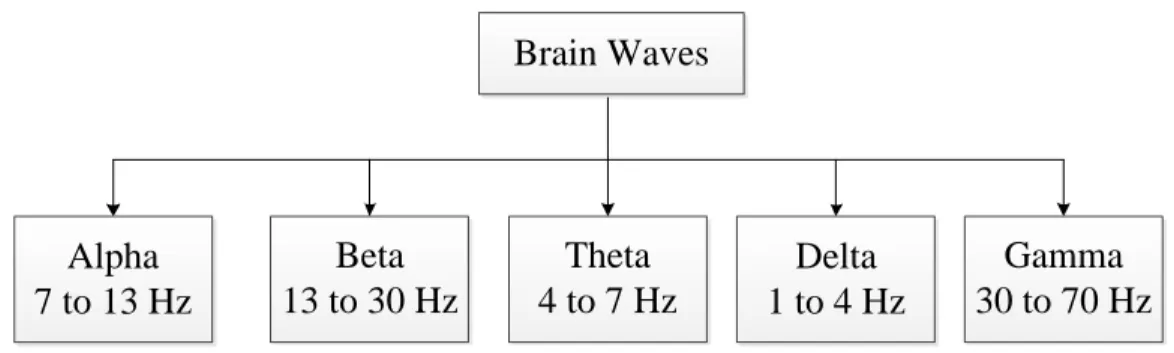
Brain signals are dynamic in nature and also vary across different people which tends to hinder the application of BCI. The human brain generally generates five different waves namely Alpha, Beta, Gamma, Delta and Theta moves. The bandwidth for the all the five waves combined is 1 to 70 Hz. Alpha waves have a frequency between 7 to 13 Hz and
are generated when a subject is relaxed and in an awakened state. Beta waves are present only when a subject is awake and it is always associated with one of the other four waves. Beta waves range from 13 to 30 Hz in frequency and are associated with attentiveness and concentration in mental activity. The frequency of Delta wave is between 1 to 4 Hz and is generated when a subject is in a deep sleep state [8]. The frequency of Gamma wave is 30 to 70 Hz and it is generated when processing various visual, auditory and touch responses. Frequency of Theta wave is 4 to 7 Hz and it is generated during deep meditation, and hypnosis. The Theta stage is called the twilight stage when a person is neither fully awake nor asleep. The various types of brain waves and their frequencies are illustrated in Figure 1.1 Brain Waves Beta 13 to 30 Hz Alpha 7 to 13 Hz Theta 4 to 7 Hz Delta 1 to 4 Hz Gamma 30 to 70 Hz Figure 1.1: Brain Waves
When sensing these signals, there can be more than one wave generated at any time depending on the thought process. Every mental task generates a particular wave and the strength of each wave varies depending on the individual [9]. This study deals with imaginary tasks without any actual physical movement. This is only one type of EEG-based signals used in BCI. Among the various EEG-based signals used in BCI (e.g. intentional change of brain rhythms, evoked potentials, anticipatory potentials, cognitive potentials, and imaginary movement). The brain signals become more complex when more than one signal is generated at the same time which might overlap, therefore, classifying these signals is a very difficult task.
To help study brain signals, researchers classified imaginary tasks into small groups of simple two or three tasks each time brain signals are recorded. The main types of motions includes imaginary motion of hand, leg, finger, tongue, and imaginary word structure. The various types of imaginary motions used in this research field are listed in the taxonomy in Figure 1.2. Imagination Tasks Hand Movement Foot Movement Finger Movement Head Movement Tongue Movement Imagination of Words
CHAPTER 2
BACKGROUND AND LITERATURE REVIEW
2.1 The Human Brain
The human brain is a very vital and complex organ of our body. It is the source of human intelligence. The weight of a normal human brain is about 3 pounds [10]. It controls behavior and body movement. The brain is protected inside a brain cage and brain fluid surrounding it. Our identities, qualities, and decisions are defined by our brain. The brain is divided into three parts namely Cerebrum, Cerebellum, and Brain Stem among which the Cerebrum is the largest part which controls various important brain functions including our thought process. The Cerebrum consists of four lobes. The names of these lobes are Frontal lobe, Occipital lobe, Parietal lobe, and Temporal lobe [11]. The function of the various lobes are highlighted in the Figure 2.1.
PARTS OF BRAIN OCCIPITAL LOBE (Eye) TEMPORAL LOBE (Memory, Sound) FRONTAL LOBE (Language, Reasoning) PARIETAL LOBE (Language, Memory)
Figure 2.1: Parts of the Brain
Each lobe has a specific function among which Frontal lobe is responsible for language, reasoning, higher level recognition, and motor skills [12]. The location of this lobe is at the front part of the brain. Any injury to the frontal lobe leads to change of socialization, attention, sexual habits etc. The Parietal lobe processes the information sent to the brain by the sense organs and it makes us feel pain, pressure, and touch. The Parietal lobe is located at the center of the brain. Injury to the Parietal lobe disturbs language, ability of controlling eye gaze, and verbal memory. The Occipital lobe is responsible for interpreting the signals sent by the eyes to the brain. Occipital lobe is located at the back of the brain. Any damage
to the occipital lobe affects the visual ability and hampers the recognition power of colors, words, and objects. The Temporal lobe is associated with memories and it processes the sounds that is received by the ears. It is located at the bottom of the brain. Temporal damage can cause problem with language skills, speech perception, and memory [13].
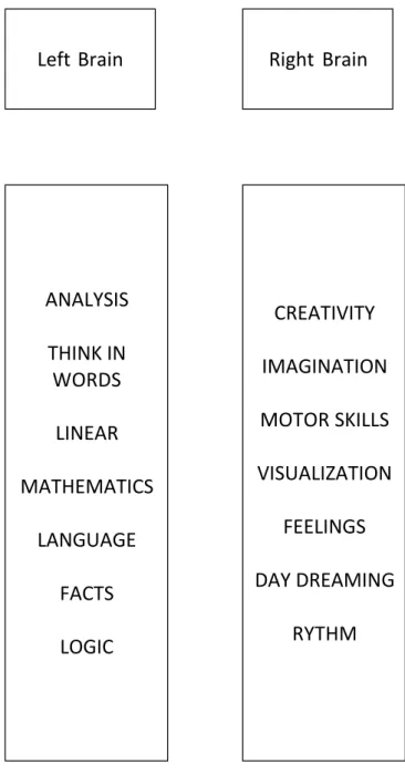
The human brain is divided into two halves. These halves are commonly called the right side and the left side of the brain or hemispheres. It is known that the right and left hemispheres control the opposite sides of the human body i.e, the right hemisphere controls the left side and possess the vision of left eye and controls our left hand and leg while the left hemisphere controls the right side of the body and possesses the right eye vision and controls right hand and leg. The concept of the left and right brain developed in the late 1960s of an psychobiologist Roger W. Sperry [14]. The human brain has two different ways of processing. The right side of the brain focuses on the overall image of an object first, then goes into the details of that image. While the left brain is analytical and hence captures every part of the image sequentially and then joins them altogether. The use of each of the brain sides varies from one person to another. Some people use their left brain while some use their right brain and it is involuntarily controlled. The difference in usage makes the personality of each person unique. The left brain is also called the digital brain as it notes minute details. It also controls reading and writing, calculation, and logical thinking. The right brain is known as the analog brain [15]. It controls 3D sense, creativity, and artistic senses [16]. The functionality of the left and right brain sides is summarized in Figure 2.2. In general, the left hemisphere of our brain conducts imaginative logical tasks [17]. The right hemisphere performs all creative tasks. The left brain is said to be more functional in hand motion and language in about 92% of people [18].
CREATIVITY
IMAGINATION
MOTOR SKILLS
VISUALIZATION
FEELINGS
DAY DREAMING
RYTHM
ANALYSIS
THINK IN
WORDS
LINEAR
MATHEMATICS
LANGUAGE
FACTS
LOGIC
Right Brain
Left Brain
Figure 2.2: Characteristics of left brain and right brain
2.2 Electroencephalography (EEG)
An electroencephalogram (EEG) is a medical diagnostic test which is used to evaluate the electrical activity within the brain. These electrical impulses connect the brain cells with each other. EEG is effective in detecting the abnormalities in brain activity of a
person. The test identifies and records the pattern of the brain wave. Small, flat metal discs called electrodes are used to capture the signals by placing them on the scalp. The electrodes capture the signals and transmit it to the computer to record. Any irregularity in the pattern of the signals is the reason of some brain disorder such as seizure disorders (Such as epilepsy), a head injury, encephalitis (an inflammation of the brain), a brain tumor, encephalopathy (a disease that causes brain dysfunction), memory problems, sleep disorders, stroke, and dementia. Magnetic Resonance Imaging (MRI) is another imaging technique used to detect brain disorder but they are more expensive to operate than EEG. EEG is a non-invasive and a very safe medical diagnostic test which has no side effects. An epileptic patient’s stimuli causes a seizure while doing the EEG. The technician who performs EEG on patients are specially trained to manage these kinds of situations.
2.3 EEG Electrode Positioning
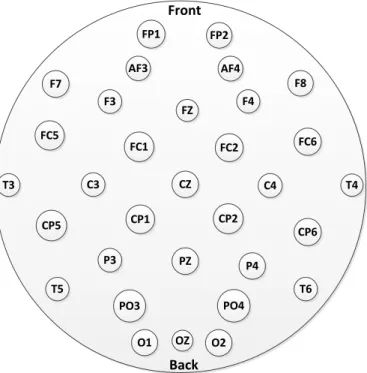
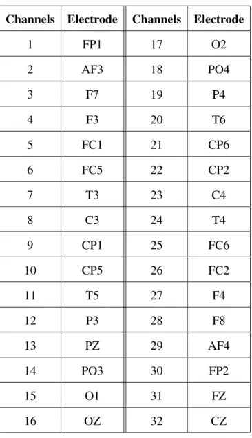
EEG requires the placement of a large number of electrodes placed on the scalp at specific positions. Based on the physiology of the brain, depending on the function of the specific brain regions scientists came up with a standard positioning system of EEG electrodes. This electrode positioning system is called 10-20 system which is adopted internationally [19]. This system comprises of 32 electrodes each of which is named according to the region of the brain it is placed on. F, T, C, P, and O are the letters used which represents Frontal, Temporal, Central, Parietal and Occipital lobes respectively. The letter ’z’ is used to denote the center of each lobe. ’Cz’ represents the center of the scalp. Along with letters, numbers are also designated for naming. Odd numbers are used to denote the electrodes on the left side of the brain and even numbers are used to name the electrodes on the right side of the brain [20]. The 10-20 electrode positioning system is shown in Figure 2.3.
In order to study some more advanced and detailed features of the brain waves, it is not sufficient to only use 32 electrodes. In such cases, more than 32 electrodes are used
Front Back CZ FP1 FP2 F3 AF3 AF4 FZ F7 FC5 T3 C3 FC1 FC2 F4 CP5 T5 P3 CP1 PZ F8 FC6 T4 C4 CP2 PO4 P4 CP6 T6 PO3 O1 OZ O2
Figure 2.3: EEG Electrodes
and these extra electrodes are placed in between the spaces of these 32 major positioned electrodes. This new system that added extra electrodes to the 10-20 system is called Modified Combinatorial Nomenclature (MCN) [21].
2.4 Brain Computer Interface (BCI)
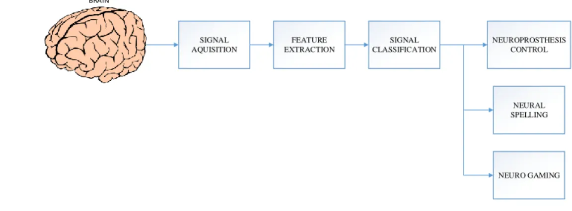
BCI is an interface system which utilizes the captured brain signal to control objects such as controlling a devices or prosthetic limbs [22]. This helps a paralyzed person to control a wheel chair, play video game or write a book. For controlling these objects, the brain signal are processed and then classified with the help of a classifier [8]. Traditionally, BCI was associated with implants but now the technique is completely non-invasive [23]. It was the scientists from Europe who first came up with the idea of using non-invasive electrode caps on the scalp of a human being to capture signals [24]. One of the area of that is still being investigated is how to increase the classification accuracy of this technique. An increased
accuracy would improve the overall performance, hence it is very important to find out the optimal classifier to classify a particular task. An overview of the overall BCI system is shown in Figure 2.4. SIGNAL AQUISITION FEATURE EXTRACTION SIGNAL CLASSIFICATION NEUROPROSTHESIS CONTROL NEURAL SPELLING NEURO GAMING BRAIN
Figure 2.4: EEG Electrodes
BCI system is almost always asscociated with EEG due to its high temporal resolution and its ease of use [25]. However, EEG signal tends to picksup noise which limits its usability. To overcome such limitation, EEG signals are usually pre-processed raw to eliminate this noise [26]. Pre-processing of raw EEG signals involve many techniques among which is P300 [27].
Presently, the work of Bin He [28] fused the imaging and signal processing together with BCI in order to get more detailed information to be utilized to control objects [29]. He used advanced functional neuroimaging including functional MRI and EEG source imag-ing. His work identified the co-variation and co-localization of electrophysiological and hemodynamic signals induced by motor imagination [30]. Using neuroimaging approach and applying a training protocol, the ability of a non-invasive EEG based brain-computer interface to control the flight of a virtual helicopter in 3-dimensional space, based upon motor imagination was demonstrated [31].
For BCI to work, the EEG signals are processed and then classified into different imaginary motions using pattern recognition based classifiers. When a subject performs
such imaginary motions, these motions are either self paced or changed according to the operator’s instruction. In the self-paced instructions, the subject proceeds from one imaginary task to the next based on his/her own pace. So it is completely based on the subject’s response and does not require any immediate instruction from an operator. As for the operator instructed tasks, the subject will have to respond to what imaginary task the operator is currently instructing. Hence, the means of recording imaginary motions are summarized in Figure 2.5. Motions Self paced instructions Operators Instructions
Figure 2.5: Means of conducting Imaginary Motions
2.5 Literature Review
A study using BCI data with two features classifier (right and left hand movement at oper-ator’s instruction) shows better performance when using Artificial neural network (ANN) compared to Hidden Markov Model (HMM) [32] [33]. The study also concludes that reducing the number of electrodes used in the BCI setup gives a much better accuracy. A similar study compared between Linear Discriminant Analysis (LDA), ANN, and Deci-sion Trees also showed that ANN outperformed the other classifiers with an accuracy of 81.6% [34, 35].
A different study was conducted to compare the performance of classifying imaginary motions such as hand, foot, and tongue. In this study, a nonlinear K-nearest neighbor algorithm based Support Vector Machine (KNN-SVM) was compared with two linear
classifiers; LDA and Naive Bayesian. The nonlinear SVM was reported to outperform the other classifiers with an accuracy of 82.14% [36]. Additionally, it was reported that when the size of the data set is reduced, then KNN-SVM accuracy increases by 5%. Fuzzy particle swarm optimization was also used with neural network to classify BCI data of imaginative head movement and it has achieved an accuracy of 84.4% [37, 38].
P300 classification which is based on Independent Component Analysis (ICA) and
Wavelet Transform proves to be a very good approach in selecting optimal features from the time domain and frequency domain. Based on specific subjects and it reduces the amount to data to improve the speed of classification but at the same time it increases the accuracy. This algorithm was tested on imagination of characters from words defined by the operator [36].
The University of Barcelona used Statistical Discriminator with the preprocessed EEG data samples to classify three types of imaginary motions that were conducted by the sub-jects in a self paced repetitive fashion. The imaginary motion classified were left-hand, right-hand movements, and the generation of words beginning with same random letter. Data was preprocessed offline at first and then processed using a statistical discrimination classifier [39]. The three mental tasks are classified after normalizing and then transforming the normalized data using canonical variate transform. This algorithm achieved an average accuracy of nearly 71%. Another interesting study was conducted by the Computer Vision and Multimedia Lab. at the University of Geneva, Switzerland. They tested four different classifiers which are Support Vector Machine (SVM), decision tree, Learning Vector Quan-tization (LVQ), and Naive Bayes classifier. Each of the datasets was used to train these classifiers and the results generated showed that SVM gives better accuracy than the rest of the three classifiers with a margin of 8% [40]. The dataset used in these studies was chosen to be used in this study. The reason behind choosing this particular dataset is the relatively low reported classification accuracy. However, we used a different method than what was
used by the University of Barcelona.
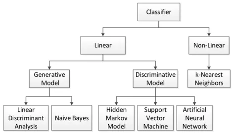
Imaginary movements can be classified using classifiers, linear and nonlinear [41]. Linear classifiers are said to be more suitable for this kind of data. Linear classifier is again broadly divided into two models namely generative and discriminative models. The various types of classifiers are illustrated in the Figure 2.6.
Classifier Linear Non-Linear Discriminative Model Linear Discriminant Analysis Naive Bayes Hidden Markov Model Support Vector Machine Artificial Neural Network Generative Model k-Nearest Neighbors
Figure 2.6: EEG Classifier Taxonomy
Most of the studies in this field showed that certain algorithms like Elman recurrent, wavelet transform, Fuzzy logic, and Principal component analysis are used along with ANN in order to classify the imaginary motions accurately. Some of the common algorithms that are used along with ANN are shown below in the Figure 2.7.
Commonly used Algorithms with ANN
Principal Component Analysis Fuzzy logic Wavelet Transform Elman recurrent
CHAPTER 3
INDIVIDUAL USER BCI USING EEG ELECTRODE OPTIMIZATION
Although the physiology of the brain is same for every human being, still there is a big difference between each person by the way they use their brain. Some people use their left side of the brain during their thought process while some use their right side of the brain. General tendency of any human being is to use both left and right side of the brain and this usage of brain is completely involuntary [42]. So, in order to get a good accuracy for object controlling, it is required to design the electrode positioning of each indiviual seperately. Keeping this in mind, we tried to find out the best set of probes of a person and study the difference of probes used across individuals. Our objective was also to study the brain usage pattern across different subjects.
3.1 Data Set Used
For the purpose of their work, the BCI Competition III dataset V were used as the exper-imental dataset. This dataset is generated by the IDIAP research institute in Switzerland [40]. In this dataset, data of 3 healthy subjects were recorded, with 3 sessions for each of them performing 3 Imaginary mental motion tasks (Imagination of Left hand movement, Right hand movement and word generation starting with the same letter). All of these 3 sessions for each subject were recorded on the same day. Each session lasted for 4 minutes with a break of 5-10 minutes in between each session. In each session, a subject performed all three kinds of Imaginary motions with each motion lasting for about 15 seconds and then switching to another motion at the operator’s request. The EEG potentials were recorded at 512 Hz sampling rate using a portable Biosemi EEG machine with 32 electrodes placed on the scalp according to 10-20 system of standard electrode positioning. Each electrode was assigned a channel number to simplify the experimentation as shown in Table 3.1.
Table 3.1: 10-20 system electrodes used in experiments Channels Electrode Channels Electrode
1 FP1 17 O2 2 AF3 18 PO4 3 F7 19 P4 4 F3 20 T6 5 FC1 21 CP6 6 FC5 22 CP2 7 T3 23 C4 8 C3 24 T4 9 CP1 25 FC6 10 CP5 26 FC2 11 T5 27 F4 12 P3 28 F8 13 PZ 29 AF4 14 PO3 30 FP2 15 O1 31 FZ 16 OZ 32 CZ
precomputed Power Spectral Density (PSD). In this thesis, the raw data (without any pre-processing) with over 288 data samples were used for training and testing of the proposed system of ANNs. This dataset is previously used by other universities during BCI competi-tion and University of Barcelona is ranked first for classifying the dataset with an accuracy of 71%. This accuracy was used as the benchmark to the performance of the proposed classifying technique.
3.2 Proposed Technique
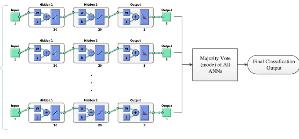
To classify this dataset of imaginary motions, ANN was the classifier of choice. Although, ANN was used in BCI Competition before by the University of Essex, they only ranked eighth with a reported accuracy of 63.91%. The goal of this work, is not only to improve the reported accuracy by the University of Essex, but also exceed the best reported accuracy of 71%. The proposed model involves using, separate ANN for each channel (electrode) and then combine all classification using a majority vote. Artificial neural networks are network models that are created with motivations from biological neural networks. These networks use an approximation function that assimilates a wide variety of inputs and targets and provides an output The model uses a system of neurons that are interconnected to learn the patterns underlying a large array of inputs. An artificial neural network operates by creating connections between many different processing elements, each analogous to a single neuron in a biological brain. These neurons may be physically constructed or simulated by a digital computer [43]. The proposed neural network consists of two hidden layers of 10 and 20 neuron in size to maintain a high classification accuracy for the network. The number of neurons in each layer is selected based on various experimentation. Back propagation method was used to train this network. Artificial neural networks have proved useful in a variety of real-world applications that deal with complex data such as visual pattern recognition and speech recognition. In addition, recent programs for text-to-speech have utilized ANNs. Many handwriting analysis programs are powered by ANNs [44]. The ANN structure used in this study for individual user accuracy classification is shown in Figure 3.1.
The novelty of this proposed system lies in using the Majority Vote system in conjunc-tion with a network of ANNs. Using majority vote system along with the network of ANNs improved the ability of the system to successfully detect motions since it was based voting which didn’t require high accuracy per channel. The structure of our proposed system is
Majority Vote (mode) of All ANNs
.
.
.
Final Classification Output Channel Inputs (ANN/Ch.)Figure 3.1: A Single Artifical Neural Network
detailed below 4.1. The process of classification utilizes the data acquired from an electrode and categorizes these values into three imaginary task categories or classes. This is done using the target specifications in the data-set.
Majority Vote (mode) of All ANNs . . . Final Classification Output Channel Inputs (ANN/Ch.)
Figure 3.2: ANN Architecture
Majority vote is the concept which chooses the decision of more than half hence, majority [45]. Majority voting is a very powerful tool in case of odd number of classes in any pattern recognition process [46]. It is seen that majority voting is a much faster way of decision making than other algorithms like Genetic Algorithm. In this study, hence we implemented the concept of majority voting to classify motions into three classes namely Class 7, Class 3, Class 2. The ANNs are trained with with the data of all the 32 channels as the first step. Then the outputs for the individual channels are combined using a majority vote system in order to find the final voted classification [47]. The outputs of the classes are represented in the form of bits(1 or 0) and the class which gets the highest number of
votes the final classification. To validate the results obtained, the final classifications are compared with the original motions recorded in the target files. If two of the classes have the same vote then the previous vote is also considered to break the tie.
The implementation of majority vote has been the key factor in enhancing the accuracy of the EEG classification. Besides, increasing the classification accuracy using the majority vote, the set of electrodes were optimized to get the optimal accuracy. For the process of optimization, the 32 ANNs for each subject were used. Based on the accuracy of each channel, all the channels were ranked to optimize the electrodes. The optimization process is shown in Figure 3.3. An optimized set of electrodes was obtained for every subject since human in general differ in the way they use their brains. But for each subject the brain behaves differently and there is no consistency and it is necessary to find optimized electrodes for each user. The electrodes were optimized for each user separately and then used to obtain the individual classification accuracy. finally these accuracies were averaged out to obtain the overall accuracy. To optimize the set of electrodes, first the EEG raw inputs of three session for every subject were used to train and test the 32 ANNs with 50% of data for training and 50% for testing. The accuracies of the 32 electrodes across three sessions are averaged and the channels are ranked then according to their accuracies. Using this rank, we eliminated the worst electrodes were eliminated one at a time to obtain the new accuracy of the majority vote. The process continues until the classification accuracy obtained is the maximum. The resulting set of electrodes that obtain the maximum classsification accuracy is the optimal set of electrodes.
3.3 Result For Individual Subject Analysis
At first, the classification accuracy was recorded for each electrode and subject. Figures 3.4-(a, b, c) illustrate the accuracies of all the electrodes for subject 1, subject 2, and subject 3, respectively.
RANK Channel X Channel Y Channel Z . . . Channel Inputs (ANN/Ch.) Accuracy Channel 1 Accuracy Channel 2 Accuracy Channel 32 . . . . . . Channel 1 Channel 2 Channel 32 . . .
Figure 3.3: Optimization of electrodes for individual user
Based on the average classification accuracy of each individual electrode for each subject, the electrodes were optimized. For the dataset used in this experiment, subject 1 had a set of 20 electrodes as the optimized electrode set which resulted in a classification accuracy of 63.33%, while subject 2 had a set of only 1 electrode with a classification accuracy of 94.01%. Subject 2 had only one electrode since this electrode classification accuracy was significantly higher compared to the rest of the electrodes. Finally, subject 3 optimal set of electrodes consisted of 14 electrodes with a classification accuracy of 84.33%. Figure 3.5-(a, b, c) illustrate the placement of the optimal set of electrodes for subject 1, subject 2, and subject 3, respectively. It is obvious from these results, that each subject utilized a different set of electrodes to obtain the optimal classification accuracy. For two subjects (subject 1 and 3), both sides of the brain worked together through Corpus Callosum, while subject 2 was left lateralized, since the left side of the brain is known for critical thinking. It is worth noting that subject 2 performed the best among all three.
Figure 3.6 illustrates the overall classification accuracy results of the optimization process as a function of the number of electrodes used for subject 1, subject 2, and subject
(a) Subject 1
(b) Subject 2
(c) Subject 3
Figure 3.4: Individual Electrode Classification Accuracy for a) Subject 1, b) Subject 2, c) Subject 3
Front Back FP1 FP2 AF3 AF4 FZ FC5 FC1 FC2 F4 T5 PZ FC6 T4 C4 PO4 P4 CP6 OZ O2 (a) Subject 1 Front Back C3 (b) Subject 2 Front Back FP1 FP2 AF3 AF4 FZ FC5 T3 FC1 FC2 F4 CP5 T5 P3 PZ T4 C4 T6 PO3 O1 OZ (c) Subject 3
Figure 3.5: Optimal set of electrodes for a) Subject 1, b) Subject 2, c) Subject 3 3, respectively.
Table 3.2 summarizes all the optimized classification accuracies of all the subjects. The reported overall accuracy calculated by averaging the classification accuracy of all subjects was 82.32% which is higher than the 71% accuracy reported by the University of Barcelona. It is also observed that subject 1 and subject 3 used both the left and the right side of their brains to perform the imaginary tasks given while subject 2 used only right side of the brain to do so.
5 10 15 20 25 30 62 63 64 65 66 67 68 69 Channels Accuracy Accuracy/Channel Maximum Accuracy
(a) Subject 1 Accuracy Optimization
5 10 15 20 25 30 90 90.5 91 91.5 92 92.5 93 93.5 94 94.5 Channels Accuracy Accuracy/Channel Maximum Accuracy
(b) Subject 2 Accuracy Optimization
5 10 15 20 25 30 72 74 76 78 80 82 84 86 Channels Accuracy Accuracy/Channel Maximum Accuracy
(c) Subject 3 Accuracy Optimization
Figure 3.6: Classification Accuracy vs. No. of Channels for a) Subject 1, b) Subject 2, c) Subject 3
Table 3.2: Optimization of Electrodes for Individual Subjects Subject Classification Accuracy
1 63.63%
2 94.01%
3 84.33%
CHAPTER 4
MULTIUSER BCI USING EEG ELECTRODE OPTIMIZATION
Even though human beings are unique in the way they use their brain, there are some applications where the BCI system would only be utilized for a small amount of time such as gaming. In such uses, to enhance the BCI system commercial viability and utilization a multiuser BCI system with an optimized set of electrodes can help achieve these goals. In the multiuser BCI system, multiple users will share this system at different times which help save the cost of purchasing dedicated systems for every user.
4.1 Result For Individual Subject Analysis
In this research work, a three-layer feed-forward neural network per electrode was used, with sigmoid hidden and output layers. This neural network can classify all the three different imaginary motions relatively well, given a 10 and 20 neurons in its two hidden layers. The network was trained using the scaled conjugate gradient back-propagation algorithm. The novelty of this proposed approach is in using a majority vote system for a network of artificial neural networks (ANNs) that is used to optimally classify imaginary motions performed by multiple subjects. 32 channels were used to train 32 ANNs in order to reduce the complexity of designing a single ANN with 32 inputs. The difference between the other reported studies and this one is that, they used a single ANN with 32 channels input which resulted in a complicated ANN structure and low overall accuracy. unlike the proposed model uses a simple ANN for every channel. Figure 4.1 illustrates the proposed model.
In general, all the EEG based BCI systems will target a specific class of imaginary motions to classify. This provides a priory information about which region of the scalp will be more effective which help reduce the number of channels used. However, this approach won’t be as effective when developing an optimal system for multiusers due to the individual difference among the users. Therefore, the number of electrode channels used are optimized
Majority Vote (mode) of All ANNs . . . Final Classification Output Channel Inputs (ANN/Ch.)
Figure 4.1: Structure of ANN
by maximizing the overall multiuser classification accuracy. The channel optimization is based on the elimination of channels with low average classification accuracy across all users. To do so, the channels are first ranked based on their average classification accuracy across all users as illustrated in Figure 4.2. The cost function for this optimization problem is to maximize the overall average classification accuracy of the system.
4.2 Result For Multiuser Subject Analysis
To validate the performance of the proposed method discussed in this chapter, the BCI Competition III dataset V was used to train and test the network of ANNs. First, we recorded the classification accuracy for each channel separately as reported in Figure 4.3. Based on the individual channel classification accuracy results obtained in the previous step, the 17 channels with the highest classification accuracies channels were chosen as the first initial optimized set of electrodes with minimum accuracy of 58.63%. The chosen electrodes are illustrated in Figure 4.4 (a). It is observed that the left side and the center of the scalp are the most effective common regions among the 3 users in this study. Most of our selected electrodes are on the left lateral frontal lobe region of the brain. This region is known to be responsible for motor functions and word generation [48].
. . . Channel Inputs (ANN/Ch.) Subject 1 Accuracy Channel 1 Accuracy Channel 2 Accuracy Channel 32 . . . . . . Channel Inputs (ANN/Ch.) Subject 2 Accuracy Channel 1 Accuracy Channel 2 Accuracy Channel 32 . . . . . . Channel Inputs (ANN/Ch.) Subject 3 Accuracy Channel 1 Accuracy Channel 2 Accuracy Channel 32 . . . Average Accuracy Channel 1 Average Accuracy Channel 2 Average Accuracy Channel 32 Channel X Channel Y Channel Z . . . . . . . . . . . . . . . . . . . . . RANK
Figure 4.2: Optimization of Electrodes for Multiusers
The overall system performance using the 17 channel structure resulted in an average classification accuracy of 78.71%, which is higher than the maximum reported accuracy of nearly 71% for this dataset [39]. A fine optimization stage was implemented, by eliminating channels 15, 31, and 7, respectively. The final optimal structure consisted of 15 electrodes, as illustrated with an average classification accuracy of 79.96%. The set of 16 and 15 chosen
1 2 3 4 5 6 7 8 9 10 11 12 13 14 15 16 17 18 19 20 21 22 23 24 25 26 27 28 29 30 31 32 50 55 60 65 70 Channels Classification Accuracy (%)
Figure 4.3: Individual Channel Classification Accuracy
electrodes are illustrated in Figures 4.4 (b) and (c), respectively. The results of optimization step is recorded in Table 4.1.
Table 4.1: Optimization of Electrodes No. of Channels Classification Accuracy
17 78.71%
16 79.21%
15 79.96%
14 78.91%
Figure 4.5 illustrates how the classification accuracy varies with time across sessions for the 17, 16, 15, and 14 electrode structures. It is observed that classification accuracy varies across the different users with subject 2 having the highest accuracy followed by subjects 3 and 1, respectively. In addition, it is also observed that the classification accuracy degrades with time for all users, which could be due to fatigue and lose of interest.
Figure 4.6 illustrates how the classification accuracy varies with different electrode structures in 3 different sessions. It is observed that the classification accuracy do not
Front Back CZ FP1 FP2 AF3 FZ FC5 T3 C3 FC1 CP5 P3 PZ C4 CP6 PO3 O1 P4 (a) 17 Ch. Set Front Back CZ FP1 FP2 AF3 FZ FC5 T3 C3 FC1 CP5 P3 PZ C4 CP6 PO3 O1 (b) 16 Ch. Set Front Back CZ FP1 FP2 AF3 FZ FC5 T3 C3 FC1 CP5 P3 PZ C4 CP6 PO3 O1 (c) 15 Ch. Set
Figure 4.4: The Optimal Electrode Sets using a) 17 Channels, b) 16 Channels, c) 15 Channels
vary significantly with the structure consisting of 15 electrodes providing the best accuracy. However, it is observed that the classification accuracy per user varies differently across sessions with subject 1 having the highest variation across sessions. This emphasize that EEG based BCI is very user dependent and this makes the optimization of a specific set of EEG electrodes to serve as a unified multiuser BCI system very difficult.
0.5 1 1.5 2 2.5 3 3.5 50 60 70 80 90 Sessions Classification Accuracy (%) Subject 1 Subject 2 Subject 3 (a) 17 Ch. Structure 0.5 1 1.5 2 2.5 3 3.5 50 60 70 80 90 Sessions Classification Accuracy (%) Subject 1 Subject 2 Subject 3 (b) 16 Ch. Structure 0.5 1 1.5 2 2.5 3 3.5 50 60 70 80 90 Sessions Classification Accuracy (%) Subject 1 Subject 2 Subject 3 (c) 15 Ch. Structure 0.5 1 1.5 2 2.5 3 3.5 50 60 70 80 90 Sessions Classification Accuracy (%) Subject 1 Subject 2 Subject 3 (d) 14 Ch. Structure
Figure 4.5: Classification accuracy of each subject versus sessions for 17, 16, 15, and 14 electrode structures
From Table 4.2, the University of Barcelona that ranked first reported an accuracy of 71.00% but our method of majority vote improved the accuracy to 79.96% for multiuser optimized BCI and 82.32% for individually optimized BCI.
Table 4.2: Result Comparison
Institution Classification Technique Overall Accuracy Georgia Southern Uni. MV with ANNs (Individual User Optimized) 82.32 % Georgia Southern Uni. MV with ANNs (Multiuser Optimized) 79.96 %
Uni. of Barcelona Statistical Discriminator 71.00 %
14 15 16 17 50 60 70 80 90 # of Channels Classification Accuracy (%) Subject 1 Subject 2 Subject 3 (a) Session 1 14 15 16 17 50 60 70 80 90 # of Channels Classification Accuracy (%) Subject 1 Subject 2 Subject 3 (b) Session 2 14 15 16 17 50 60 70 80 90 # of Channels Classification Accuracy (%) Subject 1 Subject 2 Subject 3 (c) Session 3
Figure 4.6: Classification accuracy of each subject versus channel structure for 3 different sessions
CHAPTER 5
CONCLUSION AND FUTURE WORK
A majority vote system for a network of artificial neural networks (ANN) to optimally classify imaginary motions performed by individual and multiple subjects were proposed. The proposed method optimizes the channels used for single user and multi-user classifi-cation by ranking the channels based on their classificlassifi-cation accuracy. The best performing electrodes are identified with the help of some statistical analysis. The performance of the proposed method was evaluated using the BCI competition III dataset V which primarily consisted of three imaginative motions like Imaginary left hand movement, Imaginary right hand movement, Imaginaton of words starting with the same letter. It was observed that using a separate ANN for every channel coupled with a majority vote system was able to improve the average classification accuracy of such imaginary motions for all three users from a maximum 71% to almost 82.32% for individual use while maintaining a relatively simple ANN structure. In addition, the quality of the EEG signal generated by the users declined with time due to fatigue and loss of concentration. It was also concluded that the classification accuracy is user dependent in nature which limits it optimization for multiple subjects. The proposed method presented is novel in the structure of such classification network and in the optimization of its channels. It is concluded that classification accuracy is user dependent and hence each user has a different set of optimal electrodes.
Although, we reported good classification results, the study has some inherent lim-itations the dataset is based on three subjects only, there might be more variation when considering more subjects. Also the subjects in the dataset are of same age group and of same gender (male). The study would be even stronger if more diverse subjects are consid-ered. As seen from the results, a change of performance in every session, was due to fatigue and loss of concentration which could be aged dependent. Therefore the limitation within the dataset prevented testing all the variables. in addition the optimization method used
was linear. Hence, in order to improve the optimization process, a nonlinear optimization technique such as genetic algorithm can be used.
REFERENCES
[1] “World Report on Disability,” World Health Organization - Website, 2011, http: //www.who.int/disabilities/world_report/2011/en/.
[2] K. K. Ang, C. Guan, C. Wang, K. S. Phua, A. H. G. Tan, and Z. Y. Chin, “Cali-brating EEG-based motor imagery brain-computer interface from passive movement,” inEngineering in Medicine and Biology Society, EMBC, 2011 Annual International Conference of the IEEE. IEEE, 2011, pp. 4199–4202.
[3] J. J. Vidal, “Real-time detection of brain events in EEG,” Proceedings of the IEEE, vol. 65, no. 5, pp. 633–641, 1977.
[4] L. A. Farwell and E. Donchin, “Talking off the top of your head: toward a mental pros-thesis utilizing event-related brain potentials,” Electroencephalography and clinical Neurophysiology, vol. 70, no. 6, pp. 510–523, 1988.
[5] K. Tavakolian, A. M. Nasrabadi, and S. Rezaei, “Selecting better EEG channels for classification of mental tasks,” inCircuits and Systems, 2004. ISCAS’04. Proceedings of the 2004 International Symposium on, vol. 3. IEEE, 2004, pp. III–537.
[6] A. Turnip, D. Soetraprawata, D. E. Kusumandari, M. Siahaan, I. R. Setiawan, S. Saepulloh, and A. H. Saad, “Extraction of mental task in recorded EEG signal using ICA-JADE algorithm,” inTechnology, Informatics, Management, Engineering, and Environment (TIME-E), 2014 2nd International Conference on. IEEE, 2014, pp. 275–280.
[7] X. Liao, D. Yao, D. Wu, and C. Li, “Combining spatial filters for the classifica-tion of single-trial EEG in a finger movement task,” Biomedical Engineering, IEEE Transactions on, vol. 54, no. 5, pp. 821–831, 2007.
[8] B. Hamadicharef, “Brain-computer interface BCI literature - a bibliometric study,” inInformation Sciences Signal Processing and their Applications (ISSPA), 2010 10th International Conference on, 2010, pp. 626–629.
[9] G. Gage, E. Ionides, and D. Kipke, “Information capacity of brain machine interfaces,” inEngineering in Medicine and Biology Society, 2005. IEEE-EMBS 2005. 27th Annual International Conference of the, 2005, pp. 2110–2113.
[10] M. Mustafa, M. N. Taib, Z. H. Murat, N. Sulaiman, and S. A. M. Aris, “The analysis of EEG spectrogram image for brainwave balancing application usingann,” inComputer Modelling and Simulation (UKSim), 2011 UkSim 13th International Conference on, 2011, pp. 64–68.
[11] B. F. Pennington, P. A. Filipek, D. Lefly, N. Chhabildas, D. N. Kennedy, J. H. Simon, C. M. Filley, A. Galaburda, and J. C. DeFries, “A twin mri study of size variations in the human brain,”Journal of Cognitive Neuroscience, 2000.
[12] M. Alipoor, M. Pooyan, and A. A. Suratgar, “Classification of EEG signals in four groups, including healthy subjects with open/closed eyes and epilepsy subjects with/without seizure bypsdestimate (using the multitaper method) and ann,” inHealth Informatics and Bioinformatics (HIBIT), 2010 5th International Symposium on, 2010, pp. 98–103.
[13] N. Zhong, “Unifying study on human and web problem solving: A brain informatics perspective,” in Granular Computing, 2008. GrC 2008. IEEE International Confer-ence on, 2008, pp. 25–26.
[14] M. Mustafa, M. N. Taib, S. Lias, Z. H. Murat, and N. Sulaiman, “EEG spectro-gram classification employing ann for iq application,” in Technological Advances in Electrical, Electronics and Computer Engineering (TAEECE), 2013 International Conference on, 2013, pp. 199–203.
[15] M. Y. Hsiao, D. Y. Chen, and J. H. Chen, “Constructing human brain-function associ-ation models from f mriliterature,” inEngineering in Medicine and Biology Society, 2007. EMBS 2007. 29th Annual International Conference of the IEEE, 2007, pp. 1188–1191.
[16] H. j. Pan, X. q. Yao, C. s. Qi, and H. t. Chen, “A category theory model for learning and memory of the human brain,” in Digital Manufacturing and Automation (ICDMA), 2010 International Conference on, vol. 1, 2010, pp. 11–14.
[17] W. Zhongcheng, K. Le, S. Fei, and F. Bin, “The closed-loop human-computer interface: active information acquisition for vision-brain-hand to computer (vbh-c) interaction based on force tablet,” inNeural Interface and Control, 2005. Proceedings. 2005 First International Conference on, 2005, pp. 1–5.
[18] N. Zhong, S. Motomura, and J.-L. Wu, “Peculiarity oriented multi-aspect brain data analysis for studying human multi-perception mechanism,” in Applications and the
Internet Workshops, 2005. Saint Workshops 2005. The 2005 Symposium on, 2005, pp. 306–309.
[19] I. Volosyak, D. Valbuena, T. Luth, T. Malechka, and A. Graser, “BCI demographics ii: How many (and what kinds of) people can use a high-frequency SSVEP BCI,”IEEE Transactions on Neural Systems and Rehabilitation Engineering, vol. 19, no. 3, pp. 232–239, 2011.
[20] Z. Y. Chin, K. K. Ang, C. Wang, and C. Guan, “Navigation in a virtual environment using multiclass motor imagery brain-computer interface,” inComputational Intelli-gence, Cognitive Algorithms, Mind, and Brain (CCMB), 2013 IEEE Symposium on, 2013, pp. 152–157.
[21] F. Lotte, C. Guan, and K. K. Ang, “Comparison of designs towards a subject-independent brain-computer interface based on motor imagery,” in Engineering in Medicine and Biology Society, 2009. EMBC 2009. Annual International Conference of the IEEE, 2009, pp. 4543–4546.
[22] J. DoleÅ¿al, V. ÄŇerná, and J. Åătastná, “Online motor-imagery based BCI,” in Applied Electronics (AE), 2012 International Conference on, 2012, pp. 65–68. [23] T. Castermans, M. Duvinage, M. Petieau, T. Hoellinger, C. D. Saedeleer, K.
Seethara-man, A. Bengoetxea, G. Cheron, and T. Dutoit, “Optimizing the performances of a p300-based brain 2013;computer interface in ambulatory conditions,”IEEE Journal on Emerging and Selected Topics in Circuits and Systems, vol. 1, no. 4, pp. 566–577, 2011.
[24] T. Kimura, H. Watanabe, M. Adachi, S. Kuriki, and A. Ueno, “Performance evalu-ation of electroencephalograph with negative capacitance converter,” in Biomedical Engineering International Conference (BMEiCON), 2012, 2012, pp. 1–5.
[25] K. Miatliuk, A. Nawrocka, and K. Holewa, “Conceptual design of bci in the formal
basis of hierarchical system,” inControl Conference (ICCC), 2014 15th International Carpathian, 2014, pp. 336–341.
[26] K. Ishino and M. Hagiwara, “A feeling estimation system using a simple electroen-cephalograph,” inSystems, Man and Cybernetics, 2003. IEEE International Confer-ence on, vol. 5, 2003, pp. 4204–4209 vol.5.
[27] Y. Koizumi, Y. Ijichi, H. Tanaka, A. Otera, K. Takahashi, M. Fukuda, and N. Asai, “Effective approach to character input for novice BCI users,” in Information and
Telecommunication Technologies (APSITT), 2015 10th Asia-Pacific Symposium on, 2015, pp. 1–3.
[28] A. KÃijbler, “Clinical applications of BCI,” inBrain-Computer Interface BCI, 2015 3rd International Winter Conference on, 2015, pp. 1–2.
[29] A. Cotrina, A. Benevides, A. Ferreira, T. Bastos, J. Castillo, M. L. Menezes, and C. Pereira, “Towards an architecture of a hybrid BCI based on SSVEP-BCI and passive BCI,” in Engineering in Medicine and Biology Society (EMBC), 2014 36th Annual International Conference of the IEEE, 2014, pp. 1342–1345.
[30] B. Koo, Y. Nam, and S. Choi, “A hybrid eog-p300 BCI with dual monitors,” in
Brain-Computer Interface BCI, 2014 International Winter Workshop on, 2014, pp. 1–4.
[31] A. Dhital and A. Banic, “Navigation path differences for dichotic listening BCI in virtual environments,” inVirtual and Augmented Assistive Technology (VAAT), 2013 1st Workshop on, 2013, pp. 7–9.
[32] F. Cincotti, A. Scipione, A. Timperi, D. Mattia, M. Marciani, J. Millan, S. Salinari, L. Bianchi, and F. Bablioni, “Comparison of different feature classifiers for brain computer interfaces,” in Neural Engineering, 2003. Conference Proceedings. First International IEEE EMBS Conference on. IEEE, 2003, pp. 645–647.
[33] E. M. Forney and C. W. Anderson, “Classification of EEG during imagined men-tal tasks by forecasting with elman recurrent neural networks,” in Neural Networks (IJCNN), The 2011 International Joint Conference on. IEEE, 2011, pp. 2749–2755. [34] A. Ishfaque, A. J. Awan, N. Rashid, and J. Iqbal, “Evaluation ofann, lda and decision
trees for EEG based brain computer interface,” in Emerging Technologies (ICET), 2013 IEEE 9th International Conference on. IEEE, 2013, pp. 1–6.
[35] M. K. Mukul and F. Matsuno, “Relative spectral power (rsp) and temporal rsp as features for movement imagery EEG classification with linear discriminant analysis,” inSICE Annual Conference 2010, Proceedings of. IEEE, 2010, pp. 439–448. [36] S. Bhattacharyya, A. Khasnobish, A. Konar, D. Tibarewala, and A. K. Nagar,
“Per-formance analysis of left/right hand movement classification from EEG signal by intelligent algorithms,” in Computational Intelligence, Cognitive Algorithms, Mind, and Brain (CCMB), 2011 IEEE Symposium on. IEEE, 2011, pp. 1–8.
[37] R. Chai, S. H. Ling, G. P. Hunter, Y. Tran, and H. T. Nguyen, “Brain–computer interface classifier for wheelchair commands using neural network with fuzzy particle swarm optimization,” Biomedical and Health Informatics, IEEE Journal of, vol. 18, no. 5, pp. 1614–1624, 2014.
[38] C. R. Hema, M. Paulraj, S. Yaacob, A. Adom, and R. Nagarajan, “Functional link PSO neural network based classification of EEG mental task signals,” inInformation Technology, 2008. ITSim 2008. International Symposium on, vol. 3. IEEE, 2008, pp. 1–6.
[39] F. Galán, F. Oliva, and J. Guardia, “competitionIII. data set v: Algorithm description,” Brain Computer Interfaces Competition III, 2005.
[40] A. Rakotomamonjy and V. Guigue, “BCI competition III: dataset ii-ensemble of svms for BCI p300 speller,”Biomedical Engineering, IEEE Transactions on, vol. 55, no. 3, pp. 1147–1154, 2008.
[41] A. Akrami, S. Solhjoo, A. Motie-Nasrabadi, and M.-R. Hashemi-Golpayegani, “EEG-based mental task classification: linear and nonlinear classification of movement imagery,” inEngineering in Medicine and Biology Society, 2005. IEEE-EMBS 2005. 27th Annual International Conference of the. IEEE, 2006, pp. 4626–4629.
[42] H. Sun, Y. Xiang, and Y. Guo, “Classification method of EEG signals based on wavelet neural network,” inBioinformatics and Biomedical Engineering , 2009. ICBBE 2009. 3rd International Conference on, 2009, pp. 1–4.
[43] O. Ozdamar, C. N. Lopez, and I. Yaylah, “Detection of transient EEG patterns with adaptive unsupervised neural networks,” in Biomedical Engineering Days, 1992., Proceedings of the 1992 International, 1992, pp. 192–197.
[44] O. Fukuda, T. Tsuji, and M. Kaneko, “Pattern classification of time-series EEG sig-nals using neural networks,” in Robot and Human Communication, 1996., 5th IEEE International Workshop on, 1996, pp. 217–222.
[45] L. Lam and S. Y. Suen, “Application of majority voting to pattern recognition: an analysis of its behavior and performance,”IEEE Transactions on Systems, Man, and Cybernetics - Part A: Systems and Humans, vol. 27, no. 5, pp. 553–568, 1997. [46] H. Yang and C. Li, “Combining fuzzy c-means classifiers using fuzzy majority vote,”
in Fuzzy Systems and Knowledge Discovery, 2008. FSKD ’08. Fifth International Conference on, vol. 3, 2008, pp. 153–156.
[47] A. Narasimhamurthy, “Theoretical bounds of majority voting performance for a bi-nary classification problem,” IEEE Transactions on Pattern Analysis and Machine Intelligence, vol. 27, no. 12, pp. 1988–1995, 2005.
[48] A. Roskies, J. Fiez, D. Balota, M. Raichle, and S. Petersen, “Task-dependent modula-tion of regions in the left inferior frontal cortex during semantic processing,”Cognitive Neuroscience, Journal of, vol. 13, no. 6, pp. 829–843, Aug 2001.