HOME MIRROR THERAPY: A RANDOMIZED CONTROL STUDY COMPARING UNIMANUAL AND BIMANUAL MIRROR THERAPY FOR
IMPROVED ARM AND HAND FUNCTION POST-STROKE
by
Daniel Lee Geller
Dissertation Committee:
Professor Dawn M. Nilsen, Sponsor Professor Lori Quinn
Approved by the Committee on the Degree of Education Date May 16, 2018
Submitted in partial fulfillment of the requirements for the degree of Doctor of Education
at Teachers College, Columbia University 2018
ABSTRACT
HOME MIRROR THERAPY: A RANDOMIZED CONTROL STUDY COMPARING UNIMANUAL AND BIMANUAL MIRROR THERAPY FOR
IMPROVED ARM AND HAND FUNCTION POST-STROKE
Daniel Lee Geller
Stroke is the leading cause of disability in the United States. The majority of stroke survivors have persistent arm dysfunction, which impedes their daily task
performance. Mirror therapy (MT) as an adjunct to occupational therapy (OT) has been shown to be effective in upper extremity (UE) recovery post-stroke. Two protocols, unimanual mirror therapy (UMT) and bimanual mirror therapy (BMT), have been used in OT practice; however, research specifically comparing these two intervention protocols is absent. The purpose of this study was to compare: (a) home-based UMT and BMT
protocols, and (b) both MT protocols to home-based traditional occupational therapy (TOT) regarding upper limb recovery post-stroke.
Twenty-two chronic stroke participants were randomized into one of three groups: UMT, BMT, or TOT. The Action Research Arm Test (ARAT), Fugl-Meyer Assessment (FMA), ABILHAND, grip strength, and the Stroke Impact Scale (SIS) were administered
pre- and post-intervention. Participants received outpatient OT 2 days/week for 45 minutes, plus a home program 30 minutes a day, 5 days/week for 6 weeks. A repeated measure ANOVA, Kruskal-Wallis Test, and Wilcoxon Ranked-Signed Test were used to compare the three groups, and 95% confidence intervals (CI) and effect sizes were calculated.
There was a main effect of time for all groups, except for SIS-strength and activities of daily living (ADL); however, no group differences were noted on any of the measures. When comparing UMT and BMT, the effect size for all measures, except for grip strength, favored UMT. In comparing both mirror groups to TOT, UMT had a moderate to large effect size on the ARAT, FMA, and ABILHAND, as compared to the small effect size for BMT. Furthermore, 95% CI data for the ABILHAND showed clinical significance in favor of UMT compared to TOT, but not for BMT.
This study showed that all groups improved over time and UMT may be more beneficial for UE recovery in chronic stroke individuals, compared to either BMT or TOT. However, given the small sample size, future studies comparing the two mirror protocols are necessary for more definitive conclusions to better inform clinicians of the optimal mode of MT treatment.
ii
© Copyright Daniel Lee Geller 2018 All Rights Reserved
iii DEDICATION
For my parents, my foundation For my partner, my support
iv
ACKNOWLEDGMENTS
I would like to express my deepest gratitude to my advisor, Dr. Dawn Nilsen, for her support, guidance, and mentorship during the entire dissertation process. She
provided me with the strength to continue and endure through the tough times, and celebrated the milestones and the successes. Her expertise in stroke rehabilitation, her attention to details, and her dedication to teaching and mentoring have been invaluable through this journey. I am so thankful for the opportunity to have worked with her.
I would like to thank my committee members as well: Dr. Lori Quinn, for her advice and feedback, which not only improved my study but also provided me with new insights and intellectual growth; Dr. Glen Gillen, not only for his feedback but also his support and calm demeanor during the rough times; and Dr. Sonali Rajan for reading my dissertation and serving as a committee member.
I would also like to thank the professors from the doctoral seminar, Drs. Janet Falk-Kessler, Katherine Dimitropoulou, Emily Raphael-Greenfield, and Lenin Grajo, as well as my doctoral colleagues for their feedback and support. All of your suggestions and feedback helped me grow as a researcher, while all of your support helped me stay sane through this entire experience.
I would also like to thank the crew at New York University Langone Medical Center. You all made it possible for me to finish my dissertation. Thank you to the occupational therapy staff for assisting in the recruitment process. Thank you to Steve VanLew, the Director of OT, for his support and dedication to the study. Thank you to Claribell Bayona, Matthew Bernardo, and Kelianne Arnello as my excellent and
v
study and Zena Moore for assisting with the IRB. Thank you to all of the participants in the study, as your commitment to your rehabilitation and this study made it all possible.
A special thanks to my parents for being supporting throughout my life and making me who I am today. I would have never been able to accomplish this incredible venture without your nurturing, love, and support. Last but not least, another special thanks to my partner, Dave Pistacchio, for listening to all of my anxieties and joys during this journey. I would not have been able to accomplish this without your continued love and support.
vi TABLE OF CONTENTS I – INTRODUCTION ... 1 Background ... 1 Literature Review ... 8 Mirror Therapy ... 8
Feasibility and Effectiveness of Home-Based Mirror Therapy ... 15
Stroke Mechanism and Recovery ... 17
Mirror Therapy Underlying Mechanisms ... 21
Summary ... 25
Research Aims and Hypotheses ... 27
II – METHODS ... 29
Participants ... 29
Inclusion Criteria ... 29
Exclusion Criteria ... 29
Sample Size Calculation ... 31
Study Design ... 31 Data Collection ... 31 Outcome Measures ... 32 Primary Measure ... 33 Secondary Measures ... 33 Usability Questionnaire ... 36 Log Data ... 36 Intervention Procedures ... 37 Intervention Progression ... 39 Data Analysis ... 40 Ethical Assurances ... 42 Summary ... 43 III – RESULTS ... 44 Participant Characteristics ... 46
Home Program Compliance ... 46
Group Comparisons ... 48 Primary Outcome ... 48 Secondary Outcome ... 49 Supplemental Analysis ... 54 Acceptability ... 55 Summary ... 56 IV – DISCUSSION ... 57 UMT versus BMT ... 57
Primary Outcome Measure ... 57
Activity level ... 57
Secondary Outcome Measures ... 58
vii IV (continued)
Activity and participant levels ... 59
Mirror Groups versus TOT ... 66
Primary Outcome Measure ... 67
Activity level ... 67
Secondary Outcome Measures ... 67
Impairment level ... 67
Activity and participant levels ... 69
Home Program Compliance ... 74
Acceptability ... 76
Clinical Implications ... 77
Limitations ... 77
Directions for Future Research ... 78
Conclusion ... 79
REFERENCES ... 81
APPENDICES Appendix A – Definition of Terms ... 90
Appendix B – Likert Scale Questionnaire ... 92
Appendix C – Log Data Sheet ... 93
Appendix D – Categories of Home Exercise Program ... 94
Appendix E – Functional Tasks With Objects Progression ... 95
Appendix F – Object Manipulation Progression ... 96
viii
LIST OF TABLES Table
1 Comparison of Recent UMT and BMT Efficacy Studies ... 11 2 Characteristics of Participants and Clinical Baseline Data ... 47 3 Results of the Primary Outcome Measure (ARAT): Mean Difference,
95% (CI), and Effect Size ... 49 4 Results of the Secondary Outcome Measure: Mean Difference,
95% (CI), and Effect Size ... 51 5 Results of Wilcoxon Signed-Rank Test ... 53 6 Results of Kruskal-Wallis Test ... 54
ix
LIST OF FIGURES Figure
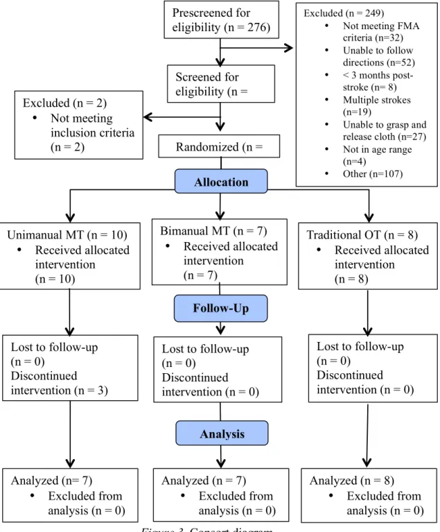
1 Mirror therapy set-up ... 5 2 UMT and BMT therapy ... 6 3 Consort diagram ... 45
I - INTRODUCTION
This dissertation is a report of a quantitative randomized controlled study designed to compare the efficacy of two home-based mirror therapy protocols as an adjunct to outpatient occupational therapy for upper limb recovery post-stroke. This study was carried out with adult subacute/chronic stroke patients in an urban outpatient
occupational therapy setting. The first chapter of the dissertation presents the background, literature review, research aims, and hypotheses of the study.
Background
Stroke is the leading cause of adult disability in the United States, with over 7 million survivors. Each year, an estimated 795,000 people have a new stroke or recurrent stroke (Mozzafarian et al., 2016). The majority of stroke survivors have persistent hemiparesis, with over 85% experiencing upper limb dysfunction, which is a significant barrier to recovery of function and participation in life (Nakayama et al., 1994). There are several rehabilitation interventions for upper limb recovery post-stroke, all with a common premise that individualized and goal-directed tasks that promote repetition of movement are essential for improvement in motor function and daily activities (Kwakkel, Veerbeek, van Wegen, & Wolf, 2015; Nilsen et al., 2015). The premise of repetition and practice is fundamental in stroke rehabilitation and is a common theme through many disciplines, including occupational therapy, physical therapy, speech therapy, and psychology, across the continuum of care.
The most common clinical pathway of stroke rehabilitation begins at the acute stage, whereby the patient becomes medically stable, the weakened arm is mobilized, and basic activities of daily living (BADL) need to be relearned. This is followed by inpatient rehabilitation, where the patient receives at least 3 hours of intensive rehabilitation, including but not limited to upper limb retraining, BADL, safety, and family education. Home health follows, whereby the patient receives rehabilitation in the home setting, under the supervision of the therapist, that may include upper limb training, safety, functional mobility, self-care, and relearning instrumental activities of daily living (IADLs). The final step is the outpatient setting, where the patient receives intervention 2-3 times per week with a focus on maximizing independence with ADL and IADL through task practice and repetition of movement, elicited through a comprehensive home exercise/task-based program (Duncan et al., 2005; Wolf & Baum, 2016).
Despite the continuum of care in stroke rehabilitation, it has been shown that the amount of upper limb movement training during traditional stroke rehabilitation is small, compared to animal models (Lang et al., 2009). Lang et al. (2009) conducted an
observational study examining the amount of movement practice that occurred in stroke rehabilitation in the inpatient and outpatient setting. For the upper extremity, which included active and passive exercises, sensory therapy, and functional tasks, there were a total of 132 repetitions per session. More specifically in the outpatient setting, Lang, MacDonald, and Gnip (2007) showed that upper limb movement practice was an average of 85 repetitions per session. In comparison, in animal stroke models, monkeys who performed 600 repetitions of a pellet retrieval task per day had improved hand and arm function as well as neuroplastic changes in the brain (Nudo, Wise, SiFuentes, & Milliken,
1996). In human stroke research, specialized interventions with the same premise of intensive practice and repetition have also been shown to be beneficial for upper limb recovery post-stroke, such as constraint-induced movement therapy (CIMT). CIMT is an intervention whereby a mitt is placed on the unaffected limb, forcing the patient to use the affected limb during therapy and at home, thus increasing practice time and use of the limb (Kwakkel et al., 2015). Furthermore, research has also shown that larger amounts of therapy and movement practice result in better outcomes in motor relearning 2 to 3 months after the stroke, regardless of setting (outpatient or inpatient) or the target of rehabilitation (upper limb recovery or mobility) (Lang, Lohse, & Birkenmeir, 2015).
In addition, Schneider, Lannin, Ada, and Schmidt (2016) performed a systematic review with a meta-analysis of randomized controlled studies regarding dosage of
traditional rehabilitation to reduce activity limitation after stroke. In this analysis, studies were included only if the additional training was traditional therapy. In order to compare the additional training between studies, the percentage increase per week was calculated. The results showed that increasing traditional therapy by at least an extra 240% improved activity performance. In contrast, Lang et al. (2016) reported that greater amounts of therapy post-stroke did not result in better outcomes; however, in this research, the group with the lowest dosage of 3,200 repetitions (100 repetitions per session) was still greater than the amount of upper limb repetition and practice typically reported in stroke
outpatient rehabilitation. In this study, the participants received one hour of task specific upper limb training, 4 days a week for 8 weeks. Therefore, it may be possible that the movement dosage for the paretic limb in traditional OT is too low for motor recovery and improved activity.
Post-stroke upper limb interventions, such as robotic training (Péter, Fazekas, Zsiga, & Denes, 2011), constraint-induced movement therapy (CIMT) or modified constraint induced movement therapy (mCIMT) (Kwakkel et al., 2015), and functional electrical stimulation coupled with task training (Meadmore et al., 2014), have been used as an adjunct to traditional therapy. These interventions have been shown to promote motor recovery; however, they are costly, labor-intensive, and limited to specific groups of stroke individuals. For example, because upper extremity robotic devices are
expensive, there are few devices in clinics or the home. Qualified personnel are required for set-up and assistance—thus the need for supervision. Furthermore, many robotic devices are not suitable for stroke individuals with limited passive range of motion of the arm/hand because the paretic limb cannot be placed in the device (Maciejasz, Eschweiler, Gerlach-Hahn, Jansen-Troy, & Leonhardt, 2014). There are similar issues with CIMT. Although CIMT has been shown to be effective for upper limb recovery, there is a minimal requirement of both voluntary wrist and finger extension of the paretic arm, thus excluding people who have minimal to no paretic hand movement (Kwakkel et al., 2015). Furthermore, many facilities do not offer CIMT because of the labor-intensive
requirements and need for trained individuals—thus less access to the greater population. Mirror therapy (MT) may be a suitable adjunct to occupational therapy (see Appendix A for definitions of terms), as it has been shown to improve upper limb
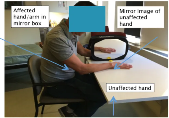
recovery post-stroke (Thieme et al., 2012) and facilitate neuroplastic changes in the brain (Deconinck et al., 2014). Mirror therapy is an intervention in which a mirror box is placed in approximately the mid-sagittal plane to the seated participant and the affected hand is placed in the mirror box, while the unaffected hand is placed outside of the box
facing the mirror (see Figures 1 and 2). During the intervention, the person moves the unaffected hand while watching the mirror reflection, which is superimposed on the affected limb, giving the visual illusion that the affected limb is moving. Thus, the mirror is providing augmented feedback, a motor learning principle, which is a term to describe information external from the person that can be used to facilitate learning or relearning a motor skill (Magill, 2011). Furthermore, MT also requires minimal one-on-one therapy, is simple to implement in the clinic and home environment, is low-cost (Michielsen et al., 2011a), and can be performed with stroke individuals with minimal to no upper limb movement (Thieme et al., 2012).
Figure 1. Mirror therapy set-up
Mirror therapy was first introduced to relieve phantom limb pain after amputation in 1995. It was speculated that the decrease in phantom pain was due to the mirror
reflection acting as a visual illusion and tricking the person into the idea that the amputated limb was intact, thus causing cortical reorganization and decrease in pain (Ramachandran & Hirstein, 1998; Ramachandran, Rogers-Ramachandran, & Cobb, 1995). Mirror therapy was later introduced to treat hemiparesis post-stroke (Altschuler et al., 1999). Altschuler et al. (1999) examined the effectiveness of MT in nine chronic
stroke participants, using a randomized crossover design. Participants were randomly assigned to the MT group or the control group (transparent plastic replacing the mirror) and were instructed to move the affected limb as best as possible to match the unaffected limb (bilateral movements). In other words, the MT group viewed the mirror reflection of the unaffected limb, while the control group had direct view of the affected limb through the plastic. After 4 weeks, all participants crossed over to the other group. Results
showed that substantially more participants in the MT group improved in movement ability, as compared to the control. In addition, all participants reported favoring the mirror intervention, as compared to the use of plastic (Altschuler et al., 1999).
Since the introduction of MT, two different protocols have been used: unimanual mirror therapy (UMT) and bimanual mirror therapy (BMT). During UMT, the affected hand is placed in the mirror box and is static, while the patient views the mirror reflection of the unaffected hand performing various activities. During BMT, the affected hand is also placed in the mirror box; however, the patient attempts to move the affected hand as best as possible to duplicate the movements of the unaffected hand, while viewing the mirror reflection of the unaffected hand (see Figure 2).
Figure 2. UMT and BMT therapy (a)UMT; hand in mirror box not moving. (b) BMT; bilateral movements with cup in both hands.
Mirror therapy research studies have shown improvements in upper limb recovery with both MT protocols (Peréz-Cruzado, Merchán-Baeza, González-Sánchez, & Cuesta-Vargas, 2017; Thieme et al., 2012); however, the results have been inconsistent across the spectrum of functioning and disability. According to the International Classification of Functioning, Disability and Health (ICF), there are three levels of human functioning, which include the body parts (body structure and function), the whole person (activity), and the whole person in the context of society (participation). Body function refers to the physiological functions of the body, while body structure refers to the anatomical parts of the body; impairment refers to the loss of body functions or structures. Activity refers to executing tasks, and activity limitations are difficulties a person has with performing the task. Participation refers to experiencing different life situations, and participation restrictions are problems one may encounter in the life situation (World Health Organization [WHO], 2001.
The three levels of human functioning (body structure and function, activity and participation) interact with one another, although not necessarily in a linear fashion, and are influenced by the health condition and contextual factors. For example, after a person has a stroke (health condition), he/she may have considerable impairments, such as loss of range of motion or sensation in the arm (body functions), which may negatively affect his/her performance with dressing and bathing (activity), thus causing activity limitations. The person’s inability to dress may therefore impact his/her ability and desire to go to a movie (participation), thus resulting in participation restrictions (WHO, 2001). Mirror
therapy has been shown to be effective in improving impairments, such as range of motion and motor function, and activities, such as self-care (Thieme et al., 2012), in people post-stroke. However, it is less effective in improving the person’s ability to participate in life scenarios (Michielsen et al., 2011a; Thieme et al., 2013).
Literature Review
The challenges in upper extremity post-stroke rehabilitation entail inaccessibility to upper limb recovery technologies (Maciejasz et al., 2014) and low dosage of upper limb practice (Lang et al., 2009). Thus, there is an urgent need to develop innovative rehabilitation interventions targeting upper limb recovery that can be self-directed and adhered to, and that are accessible to chronic stroke survivors, with a broad range of impairments, who are living in the community. Mirror therapy requires minimal one-on-one therapy, is simple to implement in the clinic and home environment (Michielsen et al., 2011a), and has been shown to be beneficial for upper limb recovery post-stroke (Thieme et al., 2012). In addition, mirror therapy combined with task practice, as an adjunct to traditional therapy, has been shown to be more beneficial than mirror therapy or task practice alone (Khandare, Singaravelan, & Khatri, 2013), thus suggesting the importance of not only task practice but also augmented visual feedback. Furthermore, mirror therapy has been shown to facilitate neuroplastic changes in the brain (Deconinck et al., 2014).
Mirror Therapy
As indicated earlier, mirror therapy was first introduced to treat hemiparesis post-stroke in 1999 (Altschuler et al., 1999). Since its introduction, both UMT and BMT have
been used in therapy with positive benefits in upper limb recovery. Thieme et al. (2012) performed a systematic review and meta-analysis of the mirror therapy literature from 1999 to 2011, which yielded 12 randomized controlled studies and 2 crossover studies. The studies examined the effectiveness of mirror therapy for improving impairments, such as decreased motor function, pain, and neglect, as well as activity limitations, such as decreased performance of activities of daily living (ADL). Time post-stroke varied for each study and consisted of individuals who were either in the acute and subacute stage (within 3 months post-stroke) or the chronic stage (>3 months post-stroke). For all studies, regardless of the mirror protocol, the mirror box was placed in the mid-sagittal plane to the participant. The MT intervention durations varied between studies and included intensities that ranged from 10 to 60 minutes per session, frequencies of 1, 2, 5, or 7 days per week, and intervention time periods ranging from 2 to 6 weeks. In addition, the control groups varied from no additional intervention other than traditional
rehabilitation, to sham MT and direct view of the affected hand.
The results of this review provided evidence in support of MT compared to control groups with respect to motor function, pain, neglect, and activities of daily living. For example, 11 studies were pooled regarding motor function post-intervention, which included 234 participants in the mirror group and 247 in the control group. Mirror therapy had significant effects on motor function post-stroke, as compared to all other types of interventions (SMD 0.61; 95% CI, 0.22 to 1.0; p = 0.002). Furthermore, motor function data at 6-month follow-up were pooled from four studies, which included 78 patients in the MT group and 79 in the control group. At 6 months, the MT group had a significant lasting effect on motor function, as compared to the control interventions
(SMD 1.09; 95% CI, 0.30 to 1.87; p = 0.007). For activities of daily living, four studies were pooled with 90 participants in the MT group and 98 in the control group. Results showed significant effect of MT on ADLs, as compared to the control group (SMD 0.33; 95% CI, 0.05 to 0.60; p = 0.02). One study not only examined the effect of MT on motor function and activities of daily living, but also on participation; however, the participation results were not included in the review (Michielsen et al., 2011a). The results of the Michielsen et al. (2011a) study showed no main effect of time or between-group interactions for the participation outcome measure. Furthermore, the researchers of the review reported that six studies used the UMT protocol, while five used the BMT; however, there was no further analysis comparing the two protocols (Thieme et al., 2012). Thus, conclusions could not be drawn about the benefit differences of the two mirror therapy protocols.
A literature review was conducted for randomized controlled MT studies after 2012 to examine the effectiveness of MT on upper limb recovery in stroke (>2 weeks stroke) individuals. Studies with participants who were 2 weeks to 6 months post-stroke were defined as subacute, while studies with participants who were greater than 6 months post-stroke were defined as chronic. Studies that included participants whose range post-stroke was greater than 2 weeks until 2 years were defined as
subacute/chronic. Studies were excluded when MT was combined with other modalities such as neuromuscular stimulation, brain stimulation, action observation, mental practice, sensory treatment, and robotics, or consisted of group MT. In addition, MT studies were excluded when examining the effectiveness of MT on lower limb recovery post-stroke, complex regional pain syndrome, or phantom pain (Table 1).
As seen in Table 1, between the years of 2012-2017, 15 randomized controlled studies were published that examined the effectiveness of MT on upper limb recovery. Table 1
Comparison of Recent UMT and BMT Efficacy Studies Recovery
Stage
Intervention Outcome Measures Impairment Activity UMT studies Arya et al. (2015) Chronic CG: TT EG: TT + UMT Brunnstrom FMA* Colomer et al. (2016) Chronic CG: TT + passive mobilization FMA NSA* WMFT Gurbuz et al.
(2016) Subacute CG: TT + sham UMT EG: TT + UMT Brunnstrom FMA* FIM Invernizzi et al.
(2013) Subacute CG: TT + sham UMT EG: TT + UMT Motricity Index* ARAT* FIM* Khandare et al. (2013) Subacute/ Chronic CG: TSE EG 1: UMT EG 2: UMT + TSE* FMA* ARAT* Kim et al.
(2016) Chronic CG: TOT EG: UMT FMA* ARAT* BBT*
FIM* Park et al.
(2015) Subacute/ Chronic CG: Sham UMT EG: UMT MFT* FIM* BMT studies
Cristina et al.
(2015) Subacute CG: TT EG: TT + BMT Brunnstrom FMA Ashworth Bhakta Test* Lee et al.
(2012) Subacute CG: TT EG: TT + BMT Brunnstrom* FMA* MFT* Lim et al. (2016) Subacute CG: TT + sham BMT EG: TT + BMT Brunnstrom FMA* MBI* Rajappan et al.
(2015) Subacute/ Chronic CG: TT + sham BMT EG: TT + BMT FMA* UEFI* Rodrigues et al.
(2016) Chronic CG: sham BMT EG: BMT TEMPA
Samuelkamal-eshkumar et al. (2014)
Subacute CG: TT
EG: TT + BMT FMA* Brunnstrom* MAS
BBT*
Wu et al.
(2013) Chronic CG: TT EG: TT + BMT FMA* Kinematics* rNSA* MAL ABILHAND UMT & BMT Hajializade et al. (2017) Chronic CG: TT EG: TT + UMT&BMT MMDT* Jebsen Taylor BI* BBT* *Denotes significant difference in favor of mirror therapy
Abbreviations: CG, control group; EG, experimental group; UMT, unimanual mirror therapy; BMT, bimanual mirror therapy; TT, traditional therapy; ARAT, Action Research Arm Test; BBT, Box and Blocks Test; BI, Barthel Index; FMA, Fugl-Meyer Assessment; FIM, Functional Independence Measure; MAL, Motor Activity
Log; MAS, Modified Ashworth Scale; MBI, Modified Barthel Index; MFT, Motor function test; MMDT, Minnesota manual dexterity test; NSA, Nottingham Sensory Assessment; rNSA, Revised Nottingham Sensory Assessment; TSE, task specific exercises, UEFI, Upper Extremity Functional Index; TEMPA, Test
d’Evaluation des Supérieurs de Personnes Agées; WMFT, Wolf Motor Function Test
Seven of the studies reported using the unimanual protocol (Arya, Pandian, Kumar, & Puri, 2015; Colomer, Noé, & Llorens, 2016; Gurbuz, Afsar, Ayaş, & Cosar, 2016; Invernizzi et al., 2013; Khandare et al., 2013; Kim, Lee, Kim, Lee, & Kim, 2016; Park, Chang, Kim, & An, 2015); seven studies reported using the bimanual protocol (Cristina, Matei, Ignat, & Popescu, 2015; Lee, Cho, & Song, 2012; Lim, Lee, Yoo, Yun, & Hwang, 2016; Rajappan, Abudaheer, Selvaganapathy, & Gokanadason, 2015;
Rodrigues, Farias, Gomes, & Michaelsen, 2016; Samuelkamaleshkumar et al., 2014; Wu, Huang, Chen, Lin, & Yang, 2013); and one study used a combination of UMT and BMT as the intervention protocol (Hajializade et al., 2017).
All but one study (Arya et al., 2015) that utilized the unimanual protocol included at least one impairment level outcome measure and one activity level outcome measure. With respect to the impairment level outcome, all of the studies (100%) reported positive findings in favor of UMT on at least one of the included outcome measures. With regard to the activity level measures, 67% of the studies reported positive findings in favor of UMT on at least one of the included outcome measures. Furthermore, no studies examined the impact of UMT on participation restrictions. Thus, there is evidence in support of UMT’s positive effects at the impairment level; however, the findings are inconsistent with regard to improvements in the activity level domain, and no studies have investigated the effects on participation outcomes.
There were similar findings in seven randomized controlled studies examining the efficacy of the BMT protocol in stroke participants. As seen in Table 1, four studies (Lim et al., 2016; Rajappan et al., 2015; Samuelkamaleshkumar et al., 2014; Wu et al., 2013)
included at least one impairment and one activity level outcome measure; two studies (Cristina et al., 2015; Lee et al., 2012) included only impairment level outcome measures, while one study (Rodrigues et al., 2016) included only an activity level outcome measure. With respect to the impairment level outcome, all of the studies (100%) reported positive findings in favor of BMT on at least one of the included outcome measures, while 60% of the studies reported positive findings in favor of BMT on at least one of the activity level outcome measures. Similar to the UMT findings, there is evidence in support of BMT’s positive effects at the impairment level; however, the findings are inconsistent with regard to improvements in activity level domains. In addition, no studies in this review (2012-2017) examined the impact of BMT on participation restrictions.
One study examined the effectiveness of a combined BMT and UMT protocol (Hajializade et al., 2017). The intervention consisted of 5 minutes of exercise with the UMT protocol, followed by 10 minutes of exercise with the BMT protocol, and ending with 15 functional tasks with the UMT protocol. The results showed gains at both the impairment and activity levels in favor of MT. Possibly, a combination of both UMT and BMT protocols would be most beneficial for upper limb improvements post-stroke. However, there are no intervention studies comparing UMT to BMT or comparing either protocol to the combined protocol.
It is apparent that both mirror therapy protocols are beneficial for upper limb recovery in subacute and chronic stroke patients; however, no studies to date have directly compared the two intervention protocols for clinical application. Selles et al. (2014) compared UMT and BMT during a short-term (1 session) motor learning study with chronic stroke patients during a simple reaching task. In this study, 93 stroke
subjects at least 6 months post-stroke were randomly allocated to one of five
experimental groups: (a) direct view of the affected hand with no mirror, (b) direct view of the unaffected hand with no mirror, (c) UMT, (d) BMT, and (e) BMT sham (mirror was covered preventing view of the affected arm). The session consisted of 70 reaching trials as per group allocation, while kinematic data were collected pre- and post- session with movement time being the primary outcome measure. The results showed that the direct view of the paretic limb group (no mirror) improved the most with respect to movement time and improved significantly more than the BMT group. In addition, the UMT group was not significantly different from the direct view group (Selles et al., 2014).
In the aforementioned study, the researchers argued that BMT might not be the optimal protocol because the effect of the visual feedback decreases when the paretic hand movement (behind the mirror) is incongruent with the visual mirror image of the unaffected hand. In other words, the visual feedback from the mirror image of the intact hand is incongruent with the proprioceptive feedback from the paretic hand in the mirror box. Research on healthy adults has supported this argument. Holmes, Crozier, and Spence (2004) examined the impact of visual and proprioceptive conflict during a reaching task with a mirror. Participants were seated with a mirror placed mid-sagittal plane on the table, with their left hand placed 12 cm facing the mirror and the right hand behind the mirror. During the reaching trials, the right hand was placed at four different positions behind the mirror. When the right hand was placed 12 cm behind the mirror, thus both hands were equidistant from the mirror, participants perceived the mirror reflection (visual feedback) as the same as the actual position of the right hand
(proprioceptive feedback). When the right hand was placed at any other distance behind the mirror, the perceived mirror reflection was different than the actual position of the right hand—thus the visual/proprioceptive conflict. Results showed that reaching error was significantly greater when there was visual-proprioceptive conflict.
In summary, both BMT and UMT have been shown to be effective in improving upper limb recovery in post-stroke individuals (Thieme et al., 2012). However,
preliminary evidence has suggested that UMT may be more beneficial than BMT for impairment level gains (Selles et al., 2014). Therefore, comparing these two mirror protocols as an intervention study is important for clinical application and best practice.
Feasibility and Effectiveness of Home-Based Mirror Therapy
Home-based MT and MT in the clinic are identical, except for the amount of direct supervision of the therapist. Home-based MT is self-directed and entails limited supervision, while MT in the clinic entails more continuous feedback and instruction. There is limited research on the effectiveness and feasibility of home-based mirror
therapy programs on upper limb recovery post-stroke. Michielsen et al. (2011a) examined the efficacy of a home-based BMT program in chronic stroke survivors across the
disability spectrum (impairment, activity, and participation levels) as well as cortical reorganization. Participants were randomized to the BMT group or the control group (direct view) and were instructed to perform all tasks bilaterally. All subjects participated in a 6-week program, which included a home program of 1 hour a day five times per week, plus one session per week under the supervision of a therapist. Subjects were provided with home practice material as well as regular phone calls to assure compliance of the home program. The results showed significantly greater improvement
post-intervention in the FMA (impairment level domain) in favor of the MT group, but not at the six-month follow-up. In addition, there was no transfer from impairment gains to activity or participation level domains. On the other hand, fMRI results showed a shift in activation toward the affected hemisphere in the primary motor cortex of MT group participants, suggesting cortical reorganization. In addition, there were no differences between the groups in total home-based practice time, which averaged a total of 30 hours per participant. This study showed the feasibility and adherence of a home MT program as well as impairment level changes and cortical reorganization.
Amasyali and Yaliman (2016) examined the effects of home-based UMT and electromyography-trigged neuromuscular stimulation (ES) on hand function in post-stroke individuals. Participants were randomized into a control group, UMT group, or ES group for a 3-week intervention. All participants received conventional therapy; however, the experimental groups (UMT and ES) received an additional 7.5 hours of treatment according to group allocation. In addition, the MT group participants were educated to practice their MT at home after each supervised session and were questioned with regard to properly performing the home MT program; however, there was no mention of the adherence or practice time at home. Results showed that all groups improved pre-post intervention; however, the MT group improved significantly more than the control group on motor performance (FMA), manual dexterity (BBT), wrist extension AROM and grip strength.
Hajializade et al. (2017) examined the effectiveness of a combined clinic and home-based mirror therapy program for upper limb recovery in chronic stroke
control group for a 4-week intervention. Both groups received conventional rehabilitation, while the mirror group also received a combination UMT and BMT protocol. The clinic mirror intervention consisted of a 1- hour session, 3 days a week, while the home-based program consisted of a 1-hour session, 4 days a week. Participants were provided with a training video clip of the home-based MT exercises as well as a timetable to track the home sessions. Results showed that both groups improved on all outcome measures; however, the MT group significantly improved on the impairment level outcome measures (BBT, Minnesota manual dexterity test) and the activity level measures (Jebsen Taylor Test and Barthel Index), as compared to the control group.
In summary, it is feasible to administer a home-based mirror therapy program with minimal supervision as long as the home program is structured and includes handouts, photos, logs, or a video clip of the home program. Furthermore, there is evidence that UMT, BMT, or a combination of both protocols are feasible and effective for upper limb recovery in chronic post-stroke individuals.
Stroke Mechanism and Recovery
In healthy individuals, the left hemisphere of the brain controls the right side of the body, while the right hemisphere controls the left side of the body. More specifically, the primary motor cortex is an important area for execution of movement; however, other areas of the brain contribute to movement coordination and control. For instance, the posterior parietal cortex is responsible for movement planning, the premotor cortex for movement observation, the parietal lobe for somatosensory function, the parietal-occipital lobes for visuomotor processing, and the cerebellum for motor control and coordination.
A stroke occurs when there is lack of blood—thus oxygen—to an area of the brain, which leads to brain cell death (Arya, 2016; Bartels, Duffy, & Beland, 2016). In global terms, a stroke to the right hemisphere of the brain affects the left side of the body and vice versa. Furthermore, the location of the stroke dictates the deficits of the person. For example, a stroke in the occipital cortex will affect vision, while a stroke in the motor cortex will affect movement. As a result of a stroke, many individuals may have an array of impairments, such as decreased strength, range of motion, and sensation in the body parts, which negatively affect their ability to perform activities such as dressing, bathing, cooking, and walking.
Research has suggested a relationship between motor deficits and an imbalance in the hemispheres, presenting as decreased activation in the ipsilesional hemisphere and excessive activation in the contralesional hemisphere (Calautti et al., 2006; Zhang et al., 2016) and/or interhemispheric disruptions (Murase, Duque, Mazzocchio, & Cohen, 2004). Caluatti et al. (2006) examined the relationship between motor deficits and
hemisphere activation in 19 right-handed first-time unilateral stroke participants, using an index thumb-tapping task of the affected hand during fMRI. Results showed that the greater the hemispheric shift toward the contralesional side in the primary motor and sensory areas, and therefore greater imbalance between the two hemispheres, the worse the performance of the index thumb-tapping task. Thus, the degree of recovery may be linked to the activation balance of the hemispheres. Zhang et al. (2016) examined the structural and functional connectivity between the bilateral primary motor cortex in 24 unilateral subcortical stroke participants and 25 health controls with multimodal magnetic resonance imaging. Results showed significantly decreased connectivity between the
primary motor areas in stroke participants, compared to controls. In addition, there was higher activation in the contralesional hemisphere in stroke subjects, suggesting the imbalance of the two hemispheres.
Similarly, Murase et al. (2004) examined the influence of interhemispheric connectivity with regard to motor function in chronic stroke participants. Nine stroke participants and eight age- and sex-matched healthy controls performed index finger movements with their affected or right hand, respectively, to examine the
interhemispheric inhibition (IHI) from the intact hemisphere to the lesioned hemispheres with TMS. Results showed that IHI in controls decreased progressively by the voluntary index finger movement, while stroke participants showed no changes in the IHI prior to voluntary movement. This suggested high interhemispheric inhibition from the intact to lesioned hemispheres. This increased inhibition may adversely affect motor recovery of the hand post-stroke. Thus, interventions that re-establish or promote a normal balance between the motor cortices may optimize upper limb recovery post-stroke.
It is well known that the affected hemisphere after a stroke is able to reorganize itself structurally or functionally as a result of repetition and practice of sensory-motor tasks and exercises, which is known as neuroplasticity (Arya, 2016). Warraich and Kleim (2010) defined neuroplasticity as “any change in the neuron structure or function that is observed either directly from measures of individual neurons or inferred from measures taken across populations of neurons” (p. S209), and is not limited to an area of the central nervous system (CNS). Despite the complexity of the brain, there is evidence that new learning and persistent behavior changes suggest neural circuitry changes and
important role in supporting functional reorganization following disease or injury. The interconnectedness creates redundancy, which contributes to the ability of the brain to adapt and change after injury, thus possibly re-establishing a normal balance between the motor cortices after stroke.
There are three neural strategies with regard to motor improvement that take advantage of the redundancy in the brain: restoration, recruitment, and retraining (Warraich & Kleim, 2010). The concept of restoration involves the encouragement of “normal” movement during rehabilitation that can re-engage neglected neural networks, thus improving movement and functional outcomes. Recruitment refers to engaging motor areas that have the capacity to perform a motor movement, but were not originally designed for that movement prior to injury. Finally, retraining involves training intact brain areas to take on additional functions to improve movement and function. These strategies play a role in cortical reorganization and functional improvement post-stroke (Warraich & Kleim, 2010).
Studies have shown that repetition of movement and practice can affect cortical reorganization after stroke and improve behavioral outcomes. Liepert, Bauder, Miltner, Taub, and Weiller (2000) examined the effect of a 12-day constraint-induced movement therapy intervention (intensive practice and repetition intervention) in 13 chronic stroke patients regarding function and cortical organization. Results showed significant
improvements in function and significantly larger motor output in the affected hemisphere, while at 6 months, the cortical areas in both hemispheres were almost identical, suggesting a balance of activation between the two hemispheres. Veldema, Bösl, and Nowak (2017) examined the relationship between motor recovery of the
affected hand post-stroke and cortical hand motor representation in 17 first-time
unilateral subacute stroke participants with hemiplegia. Results showed that participants with poor motor improvement of the affected hand showed an increase in the motor map area (MMA) size and volume in the contralesional primary motor area, while motor improvement in the affected hand was associated with a decrease in MMA size and volume in the contralesional primary motor area.
In summary, many individuals post-stroke present with upper limb dysfunction, which impedes their ability to care for themselves and participate in the community. Research has shown that after a stroke, the affected hemisphere has the ability to reorganize itself structurally or functionally as a result of repetition and practice of sensory-motor tasks and exercises (Liepert et al., 2000; Veldema et al., 2017). More specifically, it has been shown that the activation balance between the two hemispheres is an important element for better recovery post-stroke (Calautti et al., 2006; Zhang et al., 2016). Possibly, mirror therapy can promote preferable patterns of reorganization of the brain post-stroke for optimal upper limb recovery.
Mirror Therapy Underlying Mechanisms
The use of MT has been shown to be effective in upper limb recovery post-stroke; however, the underlying mechanisms of MT have been disputed. Brain imaging studies, predominantly in healthy subjects, have shown that MT can lead to neuroplastic changes, thus leading to improved arm/hand function post-stroke. Deconinck et al. (2014)
performed a systematic review to identify the underlying mechanisms of mirror visual feedback on the brain. An extensive literature review with regard to the underlying mechanisms of both mirror therapy protocols was performed by Deconinck et al. (2014)
from 1972 to January 2014, which was limited to: (a) experimental studies or clinical trials, (b) healthy and/or motor-impaired subjects, and (c) use of imaging techniques. Studies that were excluded focused only on pain and tactile perception with MT, not on sensorimotor control. Thirty-three studies were deemed eligible by two independent researchers, with the majority of the studies examining healthy adults and eight on stroke patients. Of the eight stroke studies, five examined the immediate modulatory effects of MT, while three investigated the neuroplastic changes after a period of training.
Furthermore, the researchers suggested that BMT may be a special case of bilateral training and would therefore have similar underlying mechanisms; however, no analysis compared possible differences in the underlying mechanisms of UMT and BMT.
According to Deconinck et al. (2014), there are three possible hypotheses for the underlying mechanisms of MT: (a) perceptual motor control process, whereby there is activation of attention and spatial areas in the brain; (b) direct facilitation of the motor network by means of facilitation of the affected primary motor cortex or unmasking of dormant ipsilateral pathways that are normally inhibited; and (c) activation of the mirror neuron system (MNS) that is associated not only with movement but also action
observation. While these are three distinct hypotheses, it is possible that it could be a combination of all three; however, there is still no clear understanding of the underlying mechanism of MT.
The results of the systematic review suggested that MT increases activation of attention and cognitive control areas of the brain, such as the dorsolateral prefrontal cortex, the superior posterior parietal cortex and its medial extension, and the posterior aspect of the parietal and cingulate cortex (Deconinck et al., 2014), thus supporting the
first hypothesis. One study from the review examined the neural correlates of MT in stroke participants with the use of a functional MRI (Michielsen et al., 2011b). Twenty-two participants, who were eligible to be scanned, were randomized into the experimental group (mirror) or control group. In the first experiment, participants either moved their unaffected limb while looking directly at this limb (unimanual no mirror condition) or with the use of a mirror, viewing the mirror image (UMT) while in the scanner. The second experiment was exactly the same as the unimanual trial; however, participants were asked to move both hands with the mirror (BMT) and without a mirror (bimanual no mirror condition). Results showed increased activity at the precuneus and posterior cingulate cortex, areas associated with spatial attention and awareness, with the
participants in the BMT condition, but not the UMT condition. The researchers proposed that this occurred because the mismatch between the movement of the affected hand and the superimposed mirror image, which occurs during BMT, causes greater attention and awareness to the affected limb rather than the mirror image alone, which occurs in UMT. However, this appears to contradict Selles et al. (2014), who compared UMT to BMT in a single-session motor learning study. The results showed that the UMT group improved significantly more than the BMT group in movement time during the simple reaching task. The researchers suggested that the mismatch between movement of the affected arm and the visual illusion as a result of BMT was detrimental, causing a decrease in the positive effects of the mirror image and thus less improvement.
Regarding the second hypothesis, Deconinck et al. (2014) suggested that MT decreases the motor threshold by way of reduction in interhemispheric and/or
primary motor cortex in stroke patients; thus, MT directly affects the motor network. One study in the review examined cortical reorganization after MT in chronic stroke patients (Michielsen et al., 2011a), which supports this hypothesis. Forty participants were randomized to the BMT group or the control, while only nine experimental and seven control group participants underwent both baseline and post fMRI testing. All
participants underwent a 6-week home-based program according to group allocation. Results showed a shift in activation within the primary motor cortex toward the affected hemisphere, thus balancing the two hemispheres, only in the mirror groups. Another study in the review investigated the fMRI changes in 20 chronic stroke patients after 8 weeks of computer-based bimanual MT, compared to 10 healthy controls (Bhasin, Srivastava, Kumaran, Bhatia, & Mohanty, 2012). The authors reported significant
changes in the FMA (impairment level domain) and Barthel index (activity level domain) following BMT, but also an increase in the laterality index of the ipsilesional primary and premotor cortex, thus supporting the second hypothesis.
The third hypothesis relates to activation of the “mirror neurons system” (MNS), which is divided into the parietal and frontal MNS. The frontal MNS consists of the pars opercularis of the inferior frontal gyrus and ventral premotor cortex, while the parietal MNS consists of the inferior parietal lobule of the brain (Liew, Garrison, Werner, & Aziz-Zadeh, 2012). The MNS is associated with both execution and observation of movement, which are supported by neurophysiological, behavioral, and brain imaging studies (di Pellengrino, Fadiga, Fogassi, Gallese, & Rizzolatti, 1992; Rizzolatti & Craighero, 2004; Small, Buccino, & Solodkin, 2012). For instance, “mirror neurons” vigorously fire when either an object is manipulated by an individual or when the
individual observes an object being manipulated (action observation). Thus, MT provides the visual illusion of the affected limb moving, a form of action observation, which activates parts of the motor system and has been hypothesized to induce motor learning and skill acquisition (Buccino, Solodkin, & Small, 2006). However, according to Deconinck et al. (2014), the MNS plays a minimal role in MT as it only activates the superior temporal gyrus and the premotor cortex and no other part of the MNS.
Since the review, Rossiter, Borrelli, Borchert, Bradbury, and Ward (2014) examined the mechanism of MT with the use of magnetoencephalography (MEG) to measure cortical activity, more specifically movement-related Beta desynchronization (MRBD), during mirror training post-stroke. Stroke and healthy subjects’ MBRD were measured during bimanual movements in both mirror and no mirror conditions. The results showed that in controls, MRBD was the same in both hemispheres and unchanged by the mirror; however, for stroke patients, the imbalance in MRBD between the
hemispheres in the no mirror condition was made more symmetrical with the mirror. Thus, the presence of the mirror was balancing the primary motor cortex activity in ipsilesional and contralesional hemispheres, thus supporting the second hypothesis as proposed by Deconinck et al. (2014).
Summary
In summary, both UMT and BMT have been shown to be effective in upper limb recovery post-stroke (Thieme et al., 2012). However, there are inconsistencies in both UMT and BMT literature as to the areas of improvement, with some studies showing improvement only at the impairments level (Colomer et al., 2016; Gurbuz et al., 2016;
Wu et al., 2013) and others showing improvement at both impairment and activity levels (Invernizzi et al., 2013; Khandare et al., 2013; Kim et al., 2016; Lim et al., 2016; Park et al., 2015; Rajappan et al., 2016; Samuelkamlaeshkumar et al., 2014). One motor learning study suggested that UMT may be more beneficial than BMT; however, this was not an intervention study for the purpose of MT application (Selles et al., 2014). The researchers proposed that the mismatch of information of the affected hand and the visual image during BMT decreased the positive effects of the mirror illusion; thus, there was less benefit of BMT compared to UMT for upper limb movement post-stroke.
Home-based MT programs using both protocols as well as a combination of both protocols have been shown to be feasible and effective for upper limb recovery post-stroke (Amasyali & Yaliman, 2016; Hajializade et al., 2017; Michielsen et al., 2011a). These studies supported the notion that home-based MT programs can be self-directed and adhered to with minimal supervision and structured supports, such as home pamphlets and instructions.
Research has shown that after a stroke, the affected hemisphere has the ability to reorganize itself structurally or functionally as a result of repetition and practice of sensory-motor tasks and exercises (Liepert et al., 2000; Veldema et al., 2017). More specifically, it has been shown that the activation balance between the two hemispheres is an important element for better recovery post-stroke (Calautti et al., 2006; Zhang et al., 2016). While there is evidence that MT has an effect on cortical reorganization post-stroke, there is still no clear consensus on the underlying mechanism. According to Deconinck et al. (2014), there are three possible hypotheses: (a) MT activates attention and spatial areas in the brain; (b) MT has direct facilitation of the motor network; and
(c) MT activates the mirror neuron system, which is associated not only with movement but also with action observation (Deconinck et al., 2014).
While there is evidence of the effectiveness of both MT protocols in the clinic (Thieme et al., 2012) and in the home (Amasyali & Yaliman, 2016; Hajializade et al., 2017; Michielsen et al., 2011a), no intervention research studies have compared the two protocols for MT application. Therefore, it is imperative to compare the two home-based mirror therapy protocols as an intervention study to determine if one is more beneficial than the other for upper limb recovery in chronic stroke patients. This information could be used to guide clinical decision making about the use of mirror therapy for patients with stroke.
Research Aims and Hypotheses
The American Occupational Therapy Association’s (2007) centennial vision states, “We envision that occupational therapy is a powerful, widely recognized, science-driven, and evidence-based profession…meeting society’s occupational needs.” The OT profession has focused on the importance of science-driven, evidence-based research to meet the needs of clients to provide them with the most beneficial treatments that can improve their daily living skills and overall quality of life. Since mirror therapy was introduced as an intervention to improve upper limb recovery post-stroke, research has shown the benefits of mirror therapy; however, information on the optimal mode of delivery is limited. Thus, in accordance with the AOTA centennial vision, it is essential to determine the optimal mode of mirror therapy delivery for stroke recovery best practice. This study was designed to address the following research aims:
Aim 1. To determine whether one home-based MT protocol (i.e., UMT and BMT) is more efficacious than the other for upper limb recovery post-stroke. It is hypothesized that:
a. The UMT group would demonstrate better performance on the primary outcome measure (ARAT) as compared to the BMT group.
b. The UMT group would demonstrate better performance on the secondary outcome measures (FMA, ABILHAND, grip strength, and SIS) as compared to the BMT group.
Aim 2. To determine whether home-based MT programs (UMT or BMT) are more efficacious for upper limb recovery post-stroke, as compared to the control group receiving a traditional home-based occupational therapy program.
It is hypothesized that:
a. Both MT groups would demonstrate better performance on the primary outcome measure (ARAT) as compared to the control group. b. Both MT groups would demonstrate better performance on the
secondary outcome measures (FMA, ABILHAND, grip strength, and SIS), as compared to the control group.
II - METHODS
Participants
Participants were recruited from the outpatient occupational therapy department at New York University (NYU) Langone Medical Center.
Inclusion Criteria
The following inclusion criteria were used to determine participant selection: 1. age 19-85;
2. first unilateral stroke at least 3 months prior to recruitment; 3. ability to follow directions and consent to participate in the study;
4. Fugl-Meyer Assessment (FMA) score between 10-50, indicating moderate to severe upper limb impairment (Woodbury, Velozo, Richards, & Duncan, 2013); and
5. ability to grasp and release a small washcloth with any grasp.
Exclusion Criteria
The following exclusion criteria were used to determine participant selection: 1. complex medical problems, history of pre-existing neurological or psychiatric
diseases, orthopedic conditions of the upper limb, or peripheral nerve injuries; 2. hearing and/or visual impairments that may impede participation in the home
3. perceptual deficits such as apraxia, neglect, or agnosias as per clinical evaluation;
4. botox injection in affected arm/hand within 3 months; and
5. global aphasia that may interfere with understanding instructions for testing or home exercise program.
All patients with a diagnosis of cerebral vascular accident (CVA) or stroke were prescreened by non-study occupational therapists within the first three OT outpatient sessions. The prescreening consisted of the following inclusion criteria: age 19 to 85, first unilateral stroke at least 3 months prior to recruitment, ability to follow commands, and ability to pick up and release a washcloth with any grasp. In addition, a prescreen FMA was performed on potential participants, and those who were at most 3 points below the minimum (FMA score of 7) or 3 points above the maximum (FMA score of 53) were referred to the research team and provided with informed consent prior to formal screening for the study. Participants were consented by the research OT and provided with a copy of the consent form. This was followed by screening of all of the inclusion and exclusion criteria, except for the FMA. If the participants met these criteria, they were scheduled for the baseline assessments with one of two senior OTs, who were the research assessors and trained on all outcome measures. During the baseline assessment, the senior OT administered the FMA prior to the other assessments as the final inclusion criteria screening. Thus, those who met the FMA inclusion criteria completed the
baseline assessment, and those who did not were dropped from the study and continued with their regularly scheduled OT. Eligible participants were randomized into one of
three home program groups: UMT, BMT, or Traditional OT (TOT). A non-research study OT performed randomization by a sealed envelope method.
Sample Size Calculation
To calculate the sample size, G*power, an online tool (available at
https://www.macupdate.com/app/mac/24037/g-power) was used with the statistical test ANOVA: with repeated measures, between factor. The power analysis was computed given α set at 0.05, power (1-β) set at 0.80, effect size set at 0.5, for 3 groups, 2 measurements times, and 5 outcome measures. The sample calculation was N = 27.
Study Design
This was a single-blinded, randomized controlled design. One of two senior OTs in the study, blind to group allocation, administered the pretest and posttest outcome measures. The baseline measures were administered during the first OT outpatient
session after the participants provided consent. The post-assessments, administered by the same senior OT (except for one instance), was completed after the 12th OT session in the clinic. The primary therapist (one of five therapists) who provided the conventional OT twice a week in the clinic were also blind to the participants’ group allocation.
Data Collection
All outcome measures were obtained by the senior OT and recorded on standardized case report forms. Dynamometer grip strength was collected from the primary therapist’s evaluation and follow-up re-evaluations for the pilot study to decrease the burden on the senior OT. However, the primary therapists did not consistently take
grip strength measures on the re-evaluation, therefore, the post-assessment grip strength data were not consistently assessed at the 6-week mark. Hence, the dynamometer grip strength assessment was assigned to the senior OT after the pilot study for consistency. Demographic data were collected from participants’ health records by the research OT. All data were entered by the research OT into the Research Electronic Data Capture (RED Cap), an online database management tool. The logs were collected at the end of the intervention by the research OT and data were entered into Excel for analysis.
Outcome Measures
The outcome measures were chosen to assess the participants’ recovery across the full spectrum of disability, including impairment, activity and participation level
domains. Outcome measures were performed by one of two senior occupational therapists, blind to group allocation, at baseline and post-intervention, and recorded on standardized report forms. Except for one occasion, due to logistical issues, the same senior OT performed the baseline and post-intervention assessment for each participant. Both assessors were trained on all of the outcome measures by means of lectures and videos, followed by administration of the FMA on two stroke patients with supervision and feedback. Outcome measures were completed (total time approximately 45 minutes) in the following order: Fugl-Meyer Assessment (secondary measure), Action Research Arm Test (primary measure), Stroke Impact Scale (secondary measure), and then ABILHAND (secondary measure) and grip strength (secondary measure). On the post-assessment, a Likert scale questionnaire, which evaluated the acceptability of the home
program, was administered to the participants after completing the primary and secondary outcome measures.
Primary Measure
The Action Research Arm Test (ARAT) is a standardized objective assessment used to evaluate arm and hand function at the activity level domain (Bushnell et al., 2015). This instrument contains 19 items and is divided into four subtests, including five-finger grasp, cylindrical grasp, pincer grip, and gross arm movements. All items are scored on a 4-point ordinal scale (0 to 3), with a maximum score of 57, with higher scores reflecting greater hand and arm recovery. Both interrater and intrarater reliability are excellent (ICC > .93) for the ARAT in chronic stroke subjects for all subscale scores and totals. The ARAT has been shown to have construct validity with the upper extremity FMA, (r = .94, P < .01), which is the gold standard for assessment of upper extremity motor function in individuals with hemiplegia (Yozbatran, Der-Yeghiaian, & Cramer, 2008). The minimal clinical important difference (MCID) was established to be 5.7 points for the ARAT in chronic stroke individuals (Van der Lee et al., 2001). The ARAT was chosen as the primary outcome because it measures activity performance, which was deemed more important than impairment level gains in stroke individuals (Duncan, Jorgensen, & Wade, 2000) and it has been used in previous MT studies (Invernizzi et al., 2013; Kandare et al., 2013; Kim et al., 2016).
Secondary Measures
The Fugl-Meyer Assessment (FMA) measures recovery in patients with
changes in stroke research (Fugl-Meyer, Jääskö, Leyman, Olsson, & Steglind, 1975). The upper extremity motor function section of the FMA measures performance at the
impairment/body function domain (Bushnell et al., 2015) and is divided into four sections, including upper extremity, wrist, hand, and coordination. All items are scored on a 3-point ordinal scale (0 to 2) with a maximum score of 66, with higher scores indicating greater level of motor function recovery. The FMA has excellent test-retest reliability (ICC = 0.97) and interrater reliability (ICC = 0.99) for the motor score (Platz et al., 2005). The MCID was established to be 5.25 points for the upper extremity FMA in chronic stroke individuals (Page, Fulk, & Boyne, 2012).
Grip strength, an impairment level measurement, was evaluated with a
dynamometer, which entails a standard method consisting of the participant seated with the shoulder at 0 degrees, elbow at 90 degrees, forearm neutral. The final score was taken from the average of three measurements, with higher scores indicating greater grip strength. Dynamometer grip strength demonstrates good reliability in both chronic stroke and healthy subjects (ICC > 0.86), and is significantly correlated (p < 0 .01) with four upper extremity tests, including the FMA (Boissy, Bourbonnais, Carlotti, Gravel, & Arsenault, 1999).
ABILHAND, an activity level measurement, is a valid and reliable interview-based tool, which measures participants’ perceived difficulty with the use of their arms and hands with 23 bimanual hand activities, such as filing one’s nails, taking the cap off a bottle, and opening mail. For each item, participants were asked to rate their perceived ability to perform the task by checking one of the following boxes: impossible, difficult, or easy. Participants who had not performed the task within the past 3 months were asked
to check the question mark box, which was included in the analysis. The ABILHAND was first developed to measure patients’ perceived ability to perform both bimanual and unimanual tasks; however, it was later calibrated for chronic stroke patients, resulting in a decline of the original 56 items to only 23 tasks, which were only bimanual tasks, as per Rasch analysis. For chronic stroke patients, the maximum logit score on the
ABILHAND is 6.0 with higher scores indicating higher level of perceived manual ability during bilateral upper extremity tasks (Penta, Tesio, Arnould, Zancan, & Thonnard, 2001). Simone, Rota, Tesio, and Perucca (2011) examined the reliability and validity of the ABILHAND in 83 chronic stroke patients. The results demonstrated high reliability (item reliability index = 0.94; Cronbach’s α = 0.99) and moderate correlations with grip strength, box and blocks test, and the Purdue pegboard. The MCID of the ABILHAND was established to be 0.26 to 0.35 logits in chronic stroke individuals (Wang et al., 2011).
The Stroke Impact Scale (SIS), version 3.0, is a subjective standardized 59-item eight-domain questionnaire assessing health status post-stroke. The following domains of the SIS were used to measure changes in impairment, activity, and participation levels: strength domain, activities of daily living and hand use domains, and participation domain, respectively (Bushnell et al., 2015). Each item is rated on a 5-point Likert scale (1 to 5) regarding one’s perceived difficulty (Duncan et al., 2005), with higher scores indicating less difficulty. Vellone et al. (2015) examined the psychometric properties of the SIS, version 3.0, in 392 acute/subacute stroke individuals. Results showed internal consistency and test-retest reliability between 0.79 and 0.98. The participation domain of the SIS was the only measure assessing participation in this study. The questions in this domain focus on the individual’s ability to integrate into the community and revolve
around work, social and spiritual activities, recreation, roles, the ability to control one’s life and to help others. For this domain, Cronbach’s α was 0.87, thus showing that this domain is sensitive and useful in assessing participation (Vellone et al., 2015).
Usability Questionnaire
The Likert Scale Questionnaire (Appendix B) was created by the research OT to evaluate acceptability of the home program, which was divided into three themes: usability, perceived improvement of the affected limb, and continuation of the program. Usability, a quality attribute of the ease of using a tool such as the home program, was assessed with seven questions, such as requiring assistance to perform the program or ease of set-up. Perceived improvement of the affected limb was assessed with five
questions, such as perceived benefits during the MT protocol and perceived benefits after the 6-week intervention. The final question was the likelihood of continuing the home program after the research study was completed. There were a total of 13 questions with a rating of 1 to 5 for each question (1 = strongly disagree, 2 = disagree, 3 = neutral, 4 = agree, and 5 = strongly agree). The questions were created such that the higher scores indicated increased usability and perceived improvement and more likelihood of continuing the home program.
Log Data
A log data sheet (Appendix C) was created by the research OT in order to track the frequency of MT performance during the week and time spent during each of the different tasks in the home program in minutes. If the participant adhered to the home program, the frequency would be 5 days a week for a total of 900 minutes over the