ESTUDO CLÍNICO DO CONTEÚDO INFLAMATÓRIO E INFECCIOSO
DE DENTES COM INSUCESSO DO TRATAMENTO ENDODÔNTICO
CLINICAL STUDY OF THE INFLAMMATORY AND INFECTIOUS
CONTENT OF TEETH WITH FAILURE OF ENDODONTIC
TREATMENT
Piracicaba 2015
UNIVERSIDADE ESTADUAL DE CAMPINAS FACULDADE DE ODONTOLOGIA DE PIRACICABA
ESTUDO CLÍNICO DO CONTEÚDO INFLAMATÓRIO E INFECCIOSO
DE DENTES COM INSUCESSO DO TRATAMENTO ENDODÔNTICO
CLINICAL STUDY OF THE INFLAMMATORY AND INFECTIOUS
CONTENT OF TEETH WITH FAILURE OF ENDODONTIC
TREATMENT
Orientadora: Profa. Dra. Brenda Paula Figueiredo de Almeida Gomes
ESTE EXEMPLAR CORRESPONDE À
VERSÃO DA TESE DEFENDIDA PELO ALUNO MARLOS BARBOSA RIBEIRO, E ORIENTADO PELA PROFA. DRA. BRENDA PAULA FIGUEIREDO DE ALMEIDA GOMES. ______________________________________
Piracicaba 2015
MARLOS BARBOSA RIBEIRO
Tese apresentada à Faculdade de Odontologia de Piracicaba da Universidade Estadual de Campinas como parte dos requisitos exigidos para a obtenção do título de Doutor em Clínica Odontológica, na Área de Endodontia.
Thesis presented to the Piracicaba Dental School of the University of Campinas in partial fulfillment of the requirements for the degree of Doctor, in the area of Endodontics.
A Deus, por conceder tantas graças em minha vida. O caminho do bem nem sempre é o fácil, mas Ele está sempre comigo, guiando-me e protegendo-me. - Obrigado Senhor por Tua bondade e amor!
Aos meus pais e irmãos, pelo respeito a todas as minhas decisões e pelo amor com que cuidamos uns dos outros. Minha trajetória de vida é pautada em todos os valores positivos que obtive neste núcleo. Por isso, agradeço pelo apoio incondicional. Vocês são a razão pela qual sigo tentando tornar-me um ser humano melhor a cada dia.
À minha orientadora, profa. Brenda Paula Figueiredo de Almeida Gomes. Seu empenho em realizar todas as coisas com capricho e dedicação é um exemplo a ser seguido. Agradeço-lhe pela oportunidade de compartilhar de seu vasto conhecimento, por todas as oportunidades que contribuíram para meu crescimento profissional e pessoal, mas, sobretudo, por acreditar e apoiar os meus sonhos.
trataram. Agradeço por compartilharem dos meus sonhos.
Aos professores da banca examinadora de defesa de tese, Profa. Dra. Brenda Paula Figueiredo de Almeida Gomes, Prof. Dr. Marco Antônio Húngaro Duarte, Prof. Dr. Luciano Tavares Ângelo Cintra, Prof. Dr. Rafael Nóbrega Stipp, Profa. Dra. Adriana de Jesus Soares, Prof. Dr. João Eduardo Gomes Filho, Profa. Dra. Daniela Cristina Miyagaki e Prof. Dr. José Flávio Affonso de Almeida, que de maneira gentil e cordial se dispuseram a contribuir para a melhoria da minha tese de doutorado. Muito obrigado.
Aos professores da banca de qualificação de tese, Prof. Dr. Alexandre Augusto Zaia, Profa. Dra. Fernanda Graziela Corrêa Signoretti, Profa. Dra. Erika Nikitza Shiauha Harth Chu e Prof. Dr. Caio Cezar Randi Ferraz, pelas pertinentes considerações.
Aos amigos de Piracicaba, Aline Cristine Gomes, Andréa Cardoso Pereira, Ana Carolina Correia Laurindo Cerqueira Neto, Ana Carolina Pimental Corrêa, Aniele Carvalho Lacerda, Augusto Rodrigues Lima, Daniela Cristina Miyagaki, Diogo Henrique da Silva, Felipe Nogueira Anacleto, Jaqueline Mafra Lazzari, Maicon Ricardo Zieberg Passini, Pauline Magalhães Cardoso, Priscila Amanda Francisco e Rodrigo Arruda Vasconcelos. Agradeço pelo cuidado, paciência e carinho de todos.
A Rodrigo Arruda Vasconcelos, pela amizade desde minha chegada em Piracicaba. Obrigado pela parceria!
À direção da Faculdade de Odontologia de Piracicaba, da Universidade Estadual de Piracicaba, na pessoa do seu diretor, o Prof. Dr. Guilherme Elias Pessanha Henriques.
À Coordenação de Aperfeiçoamento de Pessoal de Nível Superior (CAPES) pela concessão da bolsa de doutorado.
À Profa. Dra. Cínthia Pereira Machado Tabchoury, coordenadora do Programa de Pós-Graduação da FOP/UNICAMP e à Profa. Karina Gonzales Silvério Ruiz, coordenadora do curso de Pós-Graduação em Clínica Odontológica.
Aos professores da Área de Endodontia da FOP/UNICAMP, Profa. Adriana de Jesus Soares, Prof. Dr. Alexandre Augusto Zaia, Profa. Dra. Brenda Paula Figueiredo de Almeida Gomes, Prof. Dr. Caio Cezar Randi Ferraz e Prof. Dr. José Flávio Affonso de Almeida.
Aos professores da FOP/UNICAMP, Altair Antoninha Del Bel Cury, Jacks Jorge Júnior, José Ricardo de Albergaria Barbosa, Luciana Asprino, Marcio de Moraes, Rafael Nóbrega Stipp e Ricardo Della Coletta pelo convívio durante este período.
Aos funcionários da FOP/UNICAMP, Adriano Luis Martins, Ana Cristina Godoy, Elisângela Barbosa Vendemiatti, Maria Helídia Neves Pereira, Leny Cecília Faro Pereira e Maicon Ricardo Zieberg Passini.
Às secretárias da Área de Endodontia da FOP/UNICAMP, Jéssica, Lorena e Tifanny.
Santos, equipe técnica da secretaria de Pós-Graduação da FOP/UNICAMP.
Aos colegas de mestrado, Augusto Rodrigues Lima, Bruna Alves Taveira Ueno, Bruna Milaré Angelieri, Carlos Henrique Meloni, Diogo Henrique da Silva, Eloá Cristina Bícego Pereira, Flávia Medeiros Saavedra de Paula, Humberto Ramah Menezes de Matos, Jaqueline Mafra Lazzari, Priscila Amanda Francisco e Rafaela Casadei Chapola.
Aos colegas de doutorado, Aline Cristine Gomes, Ana Carolina Correia Laurindo Cerqueira Neto, Ana Carolina Pimental Corrêa, Andréa Cardoso Pereira, Aniele Carvalho Lacerda, Ariane Cássia Salustiano Marinho, Cimara Barroso Braga Brum, Cláudia Leal Sampaio Suzuki, Elilton Cavalcante Pinheiro Júnior, Érika Manuela Asteria Clavijo, Fabrício Rutz da Silva, Felipe Nogueira Anacleto, Frederico Campos Manhães, Júlio Vargas Neto, Marcelle Louise Sposito Bourreau, Maria Cristina Coelho de Carvalho, Mário Luis Zuolo, Mateus Silveira Martins Hartmann, Thaís Mageste Duque, Tiago Pereira da Rosa e Volmir João Fornari.
Aos alunos da iniciação científica, Bárbara Di Santi, Fábio Lourenço Fabretti, Renata Campos Pelegrini e Rodrigo Arruda Vasconcelos. Obrigado pela confiança!
Aos colegas de Piracicaba, Carlos Augusto de Morais Souto Pantoja, Emmanuel João Nogueira Leal da Silva, Fernanda Graziela Corrêa Signoretti, Giselle Priscilla Cruz Abi Rached, Jefferson José de Carvalho Marion, Juliana Yuri Nagata, Maria Rachel Figueiredo Penalva Monteiro, Marcos Sérgio Endo.
A todos que participaram direta ou indiretamente, contribuindo para realização deste trabalho.
“A sabedoria é um paradoxo. O homem que mais sabe é aquele que mais reconhece a vastidão da sua ignorância”
RESUMO
Introdução: O conteúdo infeccioso dos canais radiculares, incluindo bactérias e seus bio-produtos como o LTA (ácido lipoteicóico), e produtos derivados de genes de virulência, podem induzir a liberação de citocinas pró-inflamatórias (CPI) e metaloproteinases de matriz (MMPs) causando injuria aos tecidos periapicais. Os objetivos deste estudo foram: (Capítulo I) a) caracterizar os microrganismos Gram-positivos e estabelecer a prevalência de Enterococcus faecalis nas diferentes fases da terapia endodôntica; b) investigar a presença de LTA; (Capítulo II) c) monitorar in vivo o efeito do preparo químico-mecânico (PQM) e da medicação intracanal (MIC) na redução de bactérias cultiváveis (BC), LTA, CPI (TNFα e IL1β) e MMPs (2, 3, -8, -9 e -13) em canais radiculares de dentes com periodontite apical pós-tratamento endodôntico; (Capítulo III) d) investigar a suscetibilidade antimicrobiana de E. faecalis isolados e a prevalência dos seus fatores de virulência.
Metodologia: Vinte canais radiculares infectados de dentes unirradiculares foram divididos aleatoriamente em dois grupos de acordo com a substância química auxiliar (SQA) utilizada durante o PQM (n = 10 por grupo): G1 - clorexidina (CLX) 2% gel e G2 - hipoclorito de sódio (NaOCl) 6%. As amostras do conteúdo infeccioso e inflamatório dos canais radiculares foram obtidas usando cones de papel estéreis antes (C1) e após (C2) o PQM e após 30 dias com MIC (Ca[OH]2 + clorexidina 2%
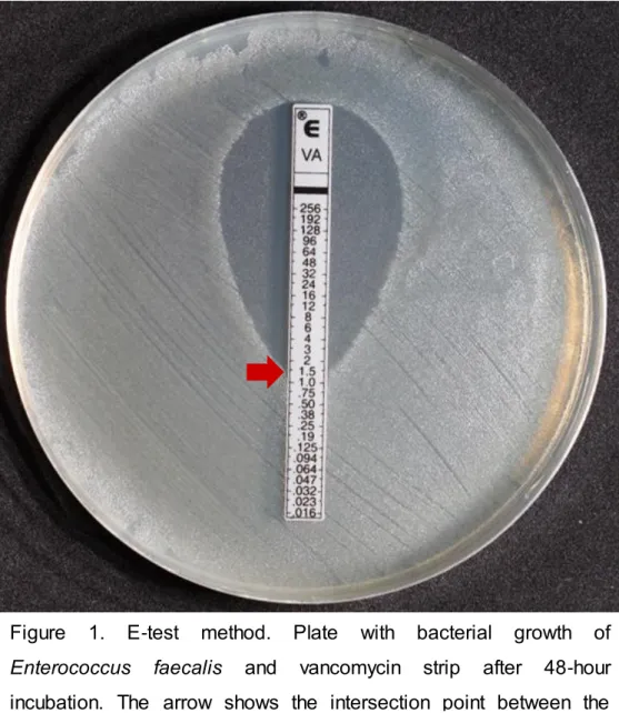
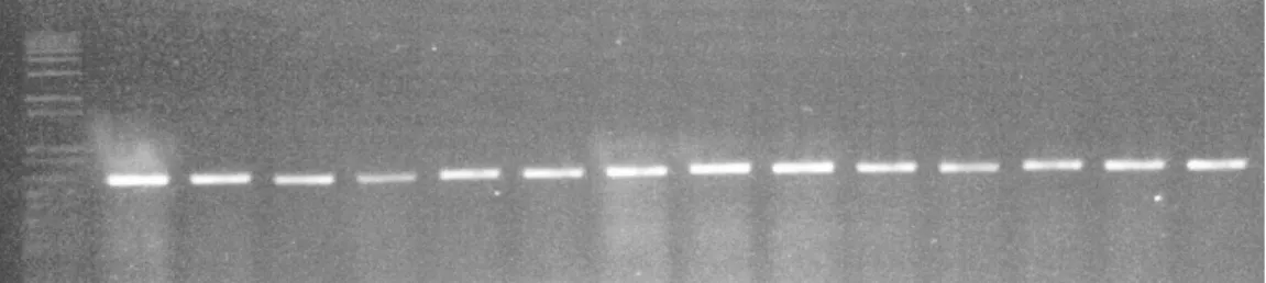
gel) (C3). (Capítulo I) A identificação microbiana foi realizada por testes bioquímicos; BC foram determinadas por contagem das unidades formadoras de colônia (UFC/mL); os níveis de LTA foram mensurados usando o ensaio imunoenzimático (ELISA) (pg/mL); (Capítulo II) CPIs e MMPs foram também mensuradas usando o ensaio imunoenzimático (ELISA) (pg/mL); (Capítulo III) a suscetibilidade antimicrobiana de diferentes antibióticos foi determinada pela concentração inibidora mínima (CIM) utilizando o método do E-test. Os genes de virulência (ace, asa, asa373, cylA, efaA, esp e gelE) dos E. faecalis isolados foram detectados através da técnica de PCR.
Resultados: (Capítulo I) 82 espécies bacterianas Gram-positivas, de um total de 102 bactérias isoladas, foram encontradas nos canais radiculares (62 na C1, 4 na C2 e 16 na C3). Os gêneros mais prevalentes foram Actinomyces spp, Aerococcus spp, Enterococcus spp, Gemella spp and Staphylococcus spp. E. faecalis foi a bacteria mais prevalente. BC (101,2±79,2), LTA (94,1±61,7), (Capítulo II) CPI
(TNF-α: 8,8±4,7 e IL1-β: 1,2±0,4) e MMPs (MMP-2: 803,7±96,4; MMP-3: 453,9±229,3; MMP-8: 245,9±122,4; MMP-9: 129,4±29,6 e MMP-13: 70,8±12,8) estavam presentes em todas as amostras (C1). Foi encontrada diminuição dos níveis globais de BC, LTA, CPI e MMPs, em todos os grupos (P < 0,05) na C2. (Capítulos I e II) O percentual de redução na C2 foi o seguinte: BC (94,4%), LTA (60,8%), CPI (TNF-α: 89,7% e IL1-β: 91,6%) e MMPs (2: 7,8%; 3: 30,3%; 8: 6,9%; MMP-9: 6% e MMP-13: 13,9%) (P < 0,05). Não houve diferença estatística entre as SQA testadas na redução do conteúdo infeccioso e inflamatório na C2 (P > 0,05). O percentual de redução na C3 foi o seguinte: BC (16,7%) (P > 0,05), LTA (39%) (P < 0,05) e MMP-8 (2,4%) (P > 0,05). Por outro lado, observou-se aumento de TNF-α, IL1-β, MMP-2 (P < 0,05), MMP-3, MMP-9 e MMP-13 (P > 0,05) na C3. A MIC foi efetiva na redução de LTA no G1; e BC, MMP-3 e MMP-8 no G2 (P < 0,05). No entanto, houve aumento de todas as CPIs e MMPs (3, 8, 9 e 13) no G1 e (2 e -13) no G2 (P < 0,05). (Capítulo III) Amoxicilina + ácido clavulânico foi efetivo contra todas as cepas. Resistência intermediária de algumas cepas foi observada para Amoxicilina (5%), Azitromicina (20%), Benzilpenicilina (5%), Ciprofloxacina (15%), Doxiciclina (5%), Eritromicina (75%), Tetraciclina (10%) e Vancomicina (15%). Total resistência a Clindamicina (60%), Clorafenicol (5%), Gentamicina (65%), Metronidazol (95%), Moxifloxacina (5%) e Rifampicina (10%) foi observada para algumas cepas. Em relação aos fatores de virulência dos E. faecalis isolados, houve detecção para ace (100%), asa (60%), asa373 (15%), esp (70%) e gelE (75%). Em contrapartida, os genes cylA e efaA não foram detectados.
Conclusão: (Capítulos I e II) Independente da utilização de CLX ou NaOCl, o PQM é efetivo na redução do conteúdo infeccioso/inflamatório de canais radiculares de dentes com insucesso do tratamento endodôntico. A MIC elevou os níveis de TNF-α, IL-1β e MMP-2. (Capítulo III) Amoxicilina + Ác. Clavulânico mostrou atividade antimicrobiana para todas as cepas de E faecalis testadas, enquanto que alguns isolados apresentaram resistência a Clindamicina, Clorafenicol, Gentamicina, Metronidazol, Moxifloxacina e Rifampicina. Todas as cepas de E. faecalis apresentaram os genes ace, asa, asa373, esp e gelE.
Palavras-chave: Bactérias. Clorexidina. Hipoclorito de sódio. Enterococcus faecalis. Testes de sensibilidade microbiana. Fatores de virulência. Citocinas. Metaloproteinases da matriz.
ABSTRACT
Introduction: The infectious contents of root canals, including bacteria and their by-products such as LTA (lipoteichoic acid) and derived by-products of virulence genes can induce the release of proinflammatory cytokines (PIC) and matrix metalloproteinases (MMP), causing injuries to the periapical tissues. The aims of this study were: (Chapter I) a) to characterize the Gram-positive microorganisms and to establish the prevalence of Enterococcus faecalis in the different phases of the endodontic therapy; b) to investigate the presence of LTA, (Chapter II) PIC and MMP, and also to monitor in vivo the effect of chemomechanical preparation (CMP) and intracanal medication (ICM) on the reduction of cultivable bacteria (CB), LTA, PIC (TNF-α and IL1-β) and MMPs (-2, -3, -8, -9 and -13) in the root canals of teeth with post-treatment apical periodontitis; and (Chapter III) c) to investigate the prevalence of virulence factors and the antimicrobial susceptibility of E. faecalis isolates.
Methods: Twenty infected root canals of single-rooted teeth were randomly assigned into two groups according to the irrigant used during CMP (n = 10 per group): G1 - 2% chlorhexidine (CHX) gel and G2 - 6% sodium hypochlorite (NaOCl). Root canal contents were taken by using paper points before (S1) and after CMP (S2) and after 30 days of ICM (Ca[OH]2 + 2% CHX gel) (S3). (Chapter I) The microbial
identification was performed using biochemical tests; CB was determined by counting the colony-forming unit (CFU/mL); LTA levels were measured using enzyme-linked immunosorbent assay (pg/mL);, (Chapter II) PICs and MMPs levels were measured by using enzyme-linked immunosorbent assay (pg/mL); (Chapter III) the antimicrobial susceptibility of different antibiotics was determined by the minimum inhibitory concentration (MIC) by using the E-test method. E. faecalis virulence factors (ace, asa, asa373, cylA, efaA, esp and gelE) were detected by PCR assay.
Results: (Chapter I) A total of 82 Gram-positive bacteria species, out of 102 bacteria isolated, were found in the root canals (62 in S1, 4 in S2 and 16 in S3). The most prevalent genera were Actinomyces spp, Aerococcus spp, Enterococcus spp, Gemella spp and Staphylococcus spp. E. faecalis was the most frequent bacteria isolated. CB (101.2±79.2), LTA (94.1±61.7), (Chapter II) PICs (TNF-α: 8.8±4.7 and
IL1-β: 1.2±0.4) and MMPs (MMP-2: 803.7±96.4; MMP-3: 453.9±229.3; MMP-8: 245.9±122.4; MMP-9: 129.4±29.6 and MMP-13: 70.8±12.8) were present in all S1 samples. Decrease of the overall levels was found in all groups (P < 0.05) in S2
samples. (Chapters I and II) Reduction percentage in S2 was the following: CB (94.4%), LTA (60.8%), PIC (TNF-α: 89.7% and IL1-β: 91.6%) and MMPs (MMP-2: 7.8%; MMP-3: 30.3%; MMP-8: 6.9%; MMP-9: 6% and MMP-13: 13.9%) (P < 0.05). There was no difference between the chemical substances tested on the infectious content reduction in S2 (P > 0.05). Reduction percentage in S3 was the following: CB (16.7%) (P > 0.05), LTA (39%) (P < .05), and MMP-8: (2.4%) (P > 0.05). On the other hand, there was an increase of TNF-α, IL1-β, MMP-2 (P < 0.05), MMP-3, MMP-9 and MMP-13 (P > 0.05) levels in S3. ICM was effective in reducing LTA in G1; and CB, MMP-3 and MM-8 in G2 (P < 0.05). However, there was an increase of all PCI and MMPs in S3 for G1 (-3, -8, -9 and -13) and G2 (-2 and -13) (P < 0.05). (Chapter III) Amoxicilin + clavulanate was effective against the all strains. Intermediate resistance of some strains was observed for Amoxicillin (5%), Azithromycin (20%), Benzylpenicillin (5%), Ciprofloxacin (15%), Doxycycline (5%), Erythromycin (75%), Tetracycline (10%) and Vancomycin (15%). A total resistance to Clindamycin (60%), Chloramphenicol (5%), Gentamicin (65%), Metronidazole (95%), Moxifloxacin (5%) and Rifampicin (10%) was observed for some strains. In relation to the virulence factors of E. faecalis isolates, there was detection of ace (100%), asa (60%), asa373 (15%), esp (70%) and gelE (75%) genes. However, cylA and efaA genes were not detected.
Conclusion: (Chapters I and II) Regardless the use of NaOCl or CHX, CMP is effective in reducing the infectious/inflammatory content of the root canals of teeth with endodontic treatment failure. The MIC increased the levels of TNF-α, IL-1β and MMP-2. (Chapter III) Amoxicillin + Clavulanate showed antimicrobial activity against all strains of E faecalis tested, while some isolates showed resistance to Clindamycin, Chloramphenicol, Gentamicin, Metronidazole, Moxifloxacin and Rifampicin. All strains of E. faecalis had the genes ace, asa, asa373, esp and gelE.
Keywords: Bacteria. Chlorhexidine. Sodium hypochlorite. Enterococcus faecalis. Cytokines. Matrix metalloproteinases. Microbial sensitivity tests. Virulence factor.
SUMÁRIO INTRODUÇÃO...17 PROPOSIÇÃO...23 CAPÍTULO I...24 CAPÍTULO II...42 CAPÍTULO III...62 DISCUSSÃO...84 CONCLUSÃO...88 REFERÊNCIAS...90 APÊNDICE 1...100 APÊNDICE 2...102 APÊNDICE 3...103 APÊNDICE 4...104 ANEXO 1...105 ANEXO 2...106
INTRODUÇÃO
As elevadas taxas de sucesso do tratamento endodôntico se devem ao avanço das técnicas e materiais utilizados, como também ao aumento do número de profissionais especializados no mercado. No entanto, diversos fatores intrínsecos e/ou extrísecos podem influenciar de maneira negativa no tratamento e conduzi-lo ao fracasso (Occhi et al., 2011; Margarit et al., 2012; Estrela et al., 2014; Wong et al, 2014). Por isso é importante destacar que mesmo que o percentual de insucessos seja baixo (Estrela et al., 2014; Wong et al., 2014), os tratamentos estão sujeitos a falhas.
O principal fator de insucesso endodôntico é a infecção intrarradicular (Nair et al., 1990; Siqueira Jr, 2001; Schirrmeister et al., 2009; Endo et al., 2012, 2013; Rahimi et al., 2014). Microrganismos podem se organizar na forma de biofilme e serem capazes de resistir aos procedimentos intracanais (Nair et al., 1999; Distel et al., 2002; Wang et al., 2012), levando à falha da terapia. As áreas não atingidas durante o preparo químico-mecânico (PQM) são favoráveis à manutenção deste biofilme, contribuindo para a persistência ou reinfecção intraradicular, seja devido ao dano tecidual direto causado pelos microrganismos, ou pela ativação de uma rede de mediadores químicos capazes de perpetuar um processo inflamatório crônico na região periapical, caracterizado como periodontite apical (Lin et al., 1991; Chugal et al., 2003; Peters et al., 2004; Occhi et al., 2011; Endo et al., 2013; Siqueira Jr et al., 2014).
Os microrganismos são os principais agentes etiológicos da periodontite apical (Kakehashi et al., 1965; Gomes et al., 1996; Rocas et al., 2010; Siqueira Jr et al., 2011; Rahimi et al, 2014) e possuem diferentes mecanismos de resistência e virulência capazes induzir ou perpetuar um processo inflamatório na região periapical após obturação de canal (Siqueira Jr & Rocas, 2007; Dos Santos et al, 2014.; Zhao et al., 2014). Dentre eles, destaca a resistência a antibióticos utilizados na prática endodôntica (Costa, Souza-Filho & Barbosa, 2003), formação de biofilme (Siqueira Jr & Rocas, 2007; Baik et al., 2008; Lee & Baek, 2012), expressão de genes de virulência (Baik et al., 2008; Lee & Baek, 2012), liberação de fatores de virulência (Ginsburg, 2002; Hermann et al., 2002; Hahn e Liewehr, 2007; Baik et al., 2008; Ryu et al., 2009), e características de patogenicidade (Kayaoglu e & Ørstavik, 2004).
A microbiota de um dente com insucesso endodôntico é diferente daquela relacionada a infecção primária, com predomínio de microrganismos anaeróbios facultativos, como os gênero Actinomyces, Candida e Enterococos (Pinheiro et al, 2003a,b; Endo et al., 2012, 2013). Enterococcus faecalis é bactéria mais freqüentemente detectada e é capaz de suportar grande variação de pH, temperatura e tensão de O2 no interior do canal radicular (Baik et al., 2008; Gomes
et al., 2008; Lee & Baek, 2012; Endo et al, 2013; Gomes et al., 2013).
A patogenicidade dos E. faecalis depende não somente do seu número e diversidade de espécies presentes na infecção, mas também do potencial antigênico de seus mecanismos de virulência, que são capazes de perpetuar um processo inflamatório na região apical mesmo após a obturação do canal radicular. (Souza-Filho & Barbosa, 2003; Hahn & Liewehr, 2007; Ryu et al., 2009; Ozeki et al., 2015) Fatores de virulência são mecanismos que os microrganismos possuem para facilitar sua aderência, colonização, resistência, patogenicidade e evasão da resposta imune do hospedeiro (Medeiros et al., 2014). O papel dos fatores de virulência dos Enterococcus ainda não foi completamente elucidado e tem despertado interesse devido a sua habilidade em potencializar a infecção e de gerar respostas exacerbadas. Estas cepas, por estarem presas num ambiente nutricional restrito e seletivo podem possuir mecanismos de virulência diversificados e favorecer trocas genéticas entre elas (Wang et al., 2011; Camargo et al., 2014). A presença destes fatores de virulência no biofilme endodôntico pode ativar ou exacerbar respostas teciduais distintas na região periapical, por isso, torna-se imperioso entender o papel específico de cada um deles na patogenicidade do conteúdo infeccioso dos canais radiculares.
O ácido lipoteicóico (LTA) é um importante fator de virulência das bactérias Gram-positivas, dentre elas, E. faecalis e tem propriedades patogênicas similares ao lipopolissacárideo (LPS) de bactérias Gram-negativas (Ginsburg, 2002; Hermann et al., 2002; Han et al., 2003; Wang et al., 2003; Hahn e Liewehr, 2007; Siqueira Jr e Rocas, 2007; Baik et al., 2008; Ryu et al., 2009). O número de unidades de repetição e o teor de D-alanina (porção glicolipídica) parecem estar estreitamente relacionados com o seu potencial pró-inflamatório (Wang et al., 2003; Baik et al., 2008). No entanto, a estrutura química que dá ao LTA purificado a sua atividade biológica não foi completamnte elucidada. Esta molécula pode iniciar uma série de reações por ligação específica (CD14 e receptores toll-like 2) ou não específica (membrana de
fosfolípido; ativação do sistema do complemento; e liberação de citocinas pró-inflamatórias TNF-α, IL-1, IL-6, IL-8 e PGE2) (Card, Jasuja e Gustafson, 1994;
Ginsburg, 2002; Hermann et al., 2002; Costa, Souza-Filho e Barbosa, 2003; Han et al., 2003; Telles et al., 2003; Wang et al., 2003; Kayaoglu e Ørstavik, 2004; Hahn e Liewehr, 2007; Siqueira Jr e Rocas, 2007; Seo et al., 2008; Ryu et al., 2009). Sua difusão para os tecidos periapicais pode induzir o dano tecidual, tanto direta ou indiretamente (via sistema imune). Contudo, pouco se conhece a respeito da ação do LTA no desenvolvimento de sintomatologia dolorosa e reabsorção óssea.
Os fatores de virulência mais frequentemente relacionados com os Enterococcus são: ace (proteína de ligação ao colágeno), asa e asa373 (substância de agregação), cylA (ativador de hemolisina), efaA (antígeno da endocardite), esp (proteína de superfície) e gelE (gelatinase). A expressão destes genes no biofilme endodôntico pode ativar ou exacerbar respostas teciduais distintas na região periapical, por isso, torna-se imperioso entender o papel específico de cada um deles na patogenicidade do conteúdo infeccioso dos canais radiculares (Sedgley et al., 2005; Wang et al., 2011; Endo et al., 2013).
Mais recentemente, estudos têm revelado um aumento surpreendente de cepas de Enterococcus spp resistentes a uma variedade de antibióticos comumente utilizados na terapêutica medicamentosa, principalmente pelo seu uso indiscriminado (Murray, 1990; Morrison et al., 1997; Poeschl et al., 2010; Skucaite et al., 2010; Endo et al., 2012, 2014). Enterococcus spp. têm adquirido determinantes genéticos que conferem resistência a várias classes de antibióticos, incluindo eritromicina, tetraciclina, cloranfenicol, e, mais recentemente, vancomicina (Murray, 1990; Morrison et al., 1997; Mundy et al., 2000; Embora a emergência de cepas resistentes seja mais pronunciada em infecções hospitalares ou sistêmicas (Poeschl et al., 2010), estudos de Enterococcus spp. possibilitam o monitoramento constante da sua resistência aos agentes antimicrobianos, permitindo a adoção de um tratamento mais eficaz (Skucaite et al., 2010).
A periodontite apical é um processo inflamatório dinâmico localizado na região periapical, com atuação de diferentes mediadores químicos produzidos por células do sistema imune em resposta ao estímulo causado pela ação dos microrganismos e seus bio-produtos. A exacerbação deste processo inflamatório, diretamente relacionada ao aumento da concentração desses mediadores, pode resultar na morte de tecidos e modular o aparecimento dos sinais e sintomas clínicos
e/ou radiográficos (Martinho et al., 2011; Endo et al., 2013, Zhao et al., 2013). A resposta do hospedeiro frente a agressões é caracterizada por um padrão no qual uma rede de mediadores químicos é acionada no intuito de debelar o processo inflamatório instalado na região periapical. LTA é capaz de estimular a liberação de citocinas inflamatórias, tais como IL1-α, IL1-, IL6, IL10, TNF- e PGE2 por diferentes linhagens celulares (Ryu et al., 2009), dentre as quais estão os macrófagos presentes em maior população, sendo considerados a principal fonte de produção de citocinas pró-inflamatórias (Martinho et al, 2010).
As citocinas atuam como mensageiros químicos no sistema imunológico, realizando comunicação com células de outros sistemas. A interpretação das mensagens fica a cargo da via metabólica a ser ativada naquele tipo de célula (Consolaro, 2009), podendo ser autócrina (atuando na mesma célula que a secreta), parácrina (em células diferentes) ou endócrina (agindo sistemicamente) (Tayal e Kalra, 1999). Suas principais funções são: mediar e regular respostas imunitárias, inflamação, hematopoiese e controle da proliferação e diferenciação celular. Durante o processo inflamatório há o recrutamento celular que é fator essencial para controlar a infecção estabelecida na região periapical. Isso é resultado da expressão de moléculas de adesão na superfície vascular de células endoteliais, induzidas por citocinas IL1-α, IL1-, TNF- e quimiocinas.
TNF-α é um dos principais mediadores da resposta inflamatória a bactérias Gram-positivas e outros microrganismos (Safavi et al., 1994; Ataoglu et al., 2002; Hong et al., 2004). Suas principais funções biológicas são o recrutamento de neutrófilos e monócitos para o local da infecção e a ativação destas células para erradicar os microrganismos. Já a IL1-β é capaz de induzir à síntese e à liberação de mediadores como a IL-6 e IL-8 e levar ao aparecimento de hipotensão, taquicardia, acidose láctica, neutrofilia, contudo, seu efeito biológico mais consistente é o aumento da síntese de prostaglandina E2 (PGE2), que possui a capacidade de
sensibilizar nociceptores (Van Deuren et al., 1992). O sinergismo existente entre o TNF-α e IL1-β é um fenômeno comumente descrito na literatura (Dinarello et al., 1986; Martinho et al., 2012). Claramente, ambas as citocinas são produzidas no mesmo sítio de inflamação e, desta forma, a interação de seus efeitos deve ser considerada exacerbada quando avaliada a severidade da doença.
Metaloproteinases da matriz (MMP) são enzimas são capazes de degradar todos os componentes da matriz extracellular (MEC) e proteínas da membrana
basal, atuando ativamente na remodelação óssea, seja em situações fisiológicas ou patológicas (Paula-Silva, da Silva e Kapila, 2010; Silva et al., 2012; Gomes et al., 2013; Sambandam e Neelakantan, 2014; Ozeki et al., 2015). Elas podem estar ancoradas à superfície celular ou serem secretadas por uma variedade de células, dentre elas leucócitos polimorfonucleares, monócitos, macrófagos, fibroblastos e osteoblastos (Ahmed et al., 2013).
Sua ativação está condicionada a uma regulação imunológica, que em condições desfavoráveis como a periodontite apical, fica na dependência de uma via de sinalização tecidual iniciada diretamente pelos microrganismos oriundos do canal radicular ou através dos seus fatores de virulência que ativam uma série de mediadores químicos, que podem promover a expressão das MMPs (Sambandam e Neelakantan, 2014).
Inibidores teciduais de MMPs (ITMMPs), citocinas, fatores de crescimento e indutor de metaloproteinases da matriz (EMMPRIN) são algumas moléculas regulatórias da expressão de MMPs (Hannas et al., 2007; Ozeki et al., 2015). Citocinas pró-inflamatórias como TNF-α e IL-1β que possuem maiores taxas de expressão em processos inflamatórios da região periapical podem modular a ação das MMPs, e consequentemente agir na destruição óssea ou até mesmo perpetuar uma inflamação periapical que não possibilite o reparo (Ozeki et al., 2014, 2015).
O papel dos microrganismos, seus bio-produtos e mediadores químicos da inflamação nas infecções endodônticas primárias já está bem elucidado na literatura científica (Martinho et al., 2010, 2012; Marinho et al., 2014, 2015). No entanto, nos casos onde houve insucesso do tratamento endodôntico, existe uma escassez de estudos que relacionam in vivo o real efeito do PQM e medicação intracanal (MIC) no controle da infecção e inflamação. A redução do tempo de contato da substância química auxiliar (SQA) e MIC e a simplificação das técnicas de instrumentação do canal radicular podem ser um dos fatores que influenciam na diminuição das taxas de sucesso. Por isso, torna-se importante o conhecimento e monitoramento dos fatores causais e mediadores químicos envolvidos neste processo, a fim de estabelecer estratégias de controle do conteúdo infeccioso dos canais radiculares (Baugh e Wallace, 2014; Gade et al., 2013, Miranda et al., 2013; Koçak et al., 2014; KungWani et al., 2014). Desta forma, este trabalho visa através do monitoramento microbiológico, dos níveis de LTA, citocinas pró-inflamatórias e MMPs, investigar in
vivo o efeito do PQM e da MIC na redução e/ou eliminação desse conteúdo em canais radiculares de dentes com periodontite apical pós-tratamento endodôntico.
PROPOSIÇÃO
Capítulo I
Caracterizar a microbiota Gram-positiva presente nas diferentes etapas da terapia endodôntica, e monitorar in vivo o efeito do PQM e da MIC na redução dos níveis de LTA e de bactérias cultiváveis em dentes com insucesso do tratamento endodôntico.
Capítulo II
Monitorar in vivo o efeito do PQM e da MIC na redução dos níveis das citocinas pró-inflamatórias (TNF-α e IL1-β) e das MMPs (-2, -3, -8, -9 e -13) em dentes com insucesso do tratamento endodôntico.
Capítulo III
Analisar a suscetibilidade antimicrobiana aos antibióticos freqüentemente prescritos na endodontia e determinar a prevalência dos fatores de virulência de cepas de E. faecalis isolados de dentes com insucesso do tratamento endodôntico.
CAPÍTULO I
Quantification of Lipoteichoic Acid Contents and Cultivable
Bacteria at the Different Phases of Endodontic Therapy of Teeth
with Post-Treatment Apical Periodontitis
Abstract
Aim: To quantify the levels of both LTA and cultivable bacteria at the different phases of the endodontic retreatment (ER) of teeth with post-treatment apical periodontitis. It also aimed to investigate the presence of Gram-positive microorganisms before and after chemomechanical preparation (CMP) and intracanal medication (ICM).
Methods: Twenty infected root canals of single-rooted teeth were randomly assigned into two groups according to the irrigant used for CMP (n=10 per group): G1 - 2% chlorhexidine gel and G2 - 6% sodium hypochlorite. Root canal samples were taken using paper points before (S1) and after CMP (S2) and after 30 days of ICM with Ca[OH]2 + 2% chlorhexidine gel (S3). Microorganisms were identified by culture
technique using biochemical tests. Cultivable bacteria were determined by counting the colony-forming unit (CFU/mL). LTA levels were measured using the enzyme-linked immunosorbent assay (pg/mL). Results: A total of 70 Gram-positive bacteria, out of 102 bacteria isolated, were found in the root canals (54 in S1, 4 in S2 and 12 in S3). Enterococcus faecalis was the most frequent isolated bacteria in all phases of the ER. LTA (94.1±61.7) and cultivable bacteria (101.2±79.2) were present in all S1 samples. CMP decreased the overall levels of cultivable bacteria by 99.4% and LTA by 60.8% (P< .05), whereas the total overall reduction level of ICM on viable bacteria was 99.5% and on LTA was 76% (P < 0.05). CMP with 2% CHX gel (G1, 99.3%) was more effective (P < 0.05) than 6% NaOCl (G2, 92.1%). On the other hand, ICM showed 100% reduction in the CHX-group (G1) and 98.5% in NaOCl-group (G2). Regarding the reduction of LTA, CMP with 2% CHX gel (G1, 55.6%) was less effective (P < 0.05) than 6% NaOCl (G2, 67.5%). On the other hand, ICM showed 74.4% reduction in the CHX-group (G1) and 78.2% in NaOCl-group (G2) (P > 0.05).
Conclusion: Regardless the CMP and ICM the reduction rates of bacteria were higher compared to LTA. Gram-positive microorganisms were present in all phases of the endodontic retreatment.
Keywords: Bacteria. Chlorhexidine. Sodium hypochlorite. Enterococcus faecalis. Virulence factor.
Introduction
The role of microorganisms in the pathogenesis of apical periodontitis has been nicely elucidated by the literature (1-4) as they can perpetuate an infection after root canal filling or induce new inflammation in the periapical region (3,5-6).
The microbiota of teeth with failure of the endodontic treatment is predominantly composed of facultatively anaerobic microorganisms such as Actinomyces, Candida and Enterococcus species (7-8). Enterococcus faecalis, a facultative Gram-positive bacterium is the most detected microorganism using culture-dependent or independent techniques, being able to endure severe conditions of survival in the root canal with large variation of pH, temperature and O2
tension (9-12). When exposed to environmental stress, some strains can adopt a viable existing state and resuscitate when normal conditions are re-established (13). Even after bacterial death, some components of the Gram-positive bacteria cell wall (e. g. LTA) persist in the root canal for long periods of time, which can cause chronic inflammation (6,11,14-16).
The antigenic complexity of the root canal content is influenced by the diversity and the number of microbial species, including synergistic and antagonistic relationships and presence of bacterial virulence factors (e.g. LTA), modulating the toxicity of this infectious content (17-18).
Lipoteichoic acid (LTA) is an important virulence factor of the Gram-positive bacteria (9,19-22) which is released during bacterial multiplication, mainly after bacteriolysis by lysozyme, bactericidal cationic peptides, phospholipase A, cathepsins or beta-lactam antibiotics (19). It has pathogenic properties similar to the lipopolysaccharides (LPS) of Gram-negative bacteria (3,16,19,21-23), resulting in well-known injuries to dental pulp and periapical tissues. However, LTA differs from LPS in term of structure and gene transcription (22).
LTA can begin a series of reactions by binding to specific receptors (CD14 and toll-like 2 receptors) or by non-specific events (phospholipid membrane; complement system activation; release of pro-inflammatory cytokines TNF-α, IL-1β, IL-6 IL-8 and PGE2; angiogenesis regulation; release of hydrolases, proteases, prostaglandins and
reactive oxygen species from neutrophils and macrophages; and regulation, recruitment and activation of neutrophils) (3,15-16,19-24).
Thus, the present study aimed to investigate the levels of LTA and the cultivable Gram-positive microorganisms and evaluate the effect of chemomechanical preparation (CMP) and intracanal medication (ICM) on the reduction of LTA and bacteria of teeth with post-treatment apical periodontitis.
Materials and Methods
Patient Selection
Twenty patients were selected from those who attended the Piracicaba Dental School, State University of Campinas- UNICAMP, Piracicaba, SP, Brazil, with a need for nonsurgical endodontic retreatment.
The Human Volunteers Research and Ethics Committee of the Piracicaba Dental School approved a protocol (# 018/2014), describing the specimen collection for this investigation, and all patients signed an informed consent for their participation in this research. The age of the patients ranged from 30 to 60 years. All selected teeth (n=20) had been previously single root-filled and showed radiographic evidence of apical periodontitis.
Failure of root canal treatment was determined based on clinical and radiographic examinations. Presence of persistent periapical radiolucent lesion, voids in or around the root canal filling, persistent symptoms such as pain of palpation, discomfort to percussion, persistent sinus were considered reasons for retreatment (7).
Exclusion criteria were as follows. Subjects who had received antibiotic treatment within the preceding three months; reported systemic disease starting with ASA 3 (American Society of Anestesiology); teeth that could not be isolated with rubber dam, teeth with absence of coronary sealing, and teeth with periodontal pockets deeper than 3 mm were excluded.
Endodontic sample collection and clinical procedures
The teeth were isolated with rubber dam. The crown and surrounding structures were disinfected with 30% hydrogen peroxide (volume/volume for 30 seconds) followed by 2.5% sodium hypochlorite (NaOCl) for the same period of time
and then inactivated with 5% sodium thiosulfate (25). Disinfection of the external surfaces of the crown was checked by taking a swab sample from the crown surface and streaking it onto blood agar plates, which were then incubated aerobically and anaerobically.
The sampling procedures were performed according to Martinho and Gomes (25). Under anesthesia (2% lidocaine with 1:100,000 epinephrine), a two-stage access preparation was performed. The access cavity was made without the use of water spray but under manual irrigation with sterile saline and by using sterile high-speed diamond bur. This first stage was performed to promote a major removal of contaminants. In the second stage, before entering the pulp chamber, the access cavity was disinfected according to the decontamination protocol described above. Disinfection of the internal surface of the access cavity was checked as previously described, and all procedures were performed aseptically. Root-filling materials were removed by using Reciproc R25 files (VDW, Munich, Germany) in the working length obtained by preoperatory radiography and used according to the manufacturer's instructions in a crown-down technique, with no chemical solvent.
Before the first sample (S1) of the root canal, a K-file #20 (Dentsply Maillefer, Ballaigues, Switzerland) was used to confirm the working length (previously estimated by radiographs), with an apex locator (Novapex; Forum Technologies, Rishon le-Zion, Israel). A sterile paper point (Dentsply-Maillefer, Ballaigues, Switzerland) was then introduced into the full length of the canal and retained in position during 60 seconds for LTA sampling. Next, this paper point was placed in a sterile tube for enzyme-linked immunosorbent assay (ELISA). Other three paper points were pooled in a sterile tube containing 1 mL of VMGA III transport medium (26) for microbial sampling. The samples were transported within 15 minutes to an anaerobic workstation (Don Whitley Scientific, Bradford, UK) for bacterial culture analysis. The LTA samples were frozen at −80 °C for further analysis.
Root canals were then prepared by using Reciproc R40 files (VDW, Munich, Germany) according to the manufacturer's instructions in a reciprocating working motion generated by the motor. The instrument was used in an in-an-out pecking motion of about 3 mm in amplitude with apical pressure. After three pecking motions, the instrument was removed from the canal and cleaned. Next, a K-file #20 was taken to the working length (WL) to check whether the canal was patent. These
procedures were repeated until the Reciproc instrument reached the WL (zero point displayed on the apex locator).
Retreatment was deemed complete when the Reciproc R40 file reached the working length, with no filling material covering the instrument and canal walls being smooth and free of visible debris. Furthermore, a close inspection under high magnification with dental operating microscope (DF Vasconcelos S.A., São Paulo, SP, Brazil) showed complete removal of gutta-percha.
The twenty infected root canals of single-rooted teeth with post-treatment apical periodontitis were divided randomly into two groups according to the chemical substances used.
• Group 1 (n = 10): 2% chlorhexidine gel (CHX) • Group 2 (n = 10): 6% sodium hypochlorite (NaOCl)
Calcium hydroxide + 2% cholorhexidine gel was used as ICM in all cases for 30 days.
EndoVac System (Discus Dental, Culver, CA, USA) was used to irrigate both groups, using saline in group 1 and NaOCl in group 2. In group 1, during instrumentation the root canals were filled with 1 mL of 2% CHX gel (Endogel; Itapetininga, SP, Brazil) using a syringe (27-gauge needle) before the use of each instrument and immediately rinsed afterwards with 5 mL of saline solution using the EndoVac System. In the end of the instrumentation, CHX was inactivated with 5 mL of 5% Tween-80 and 0.07% (w/v) lecithin solution during 1-minute period, which was removed with 5 mL of saline solution
In group 2 during instrumentation the root canals were filled with 1 mL of 6% NaOCl (Drogal; Piracicaba, SP, Brazil), using a syringe (27-gauge needle) before the use of each instrument and immediately rinsed afterwards with 5 mL of 6% NaOCl using the EndoVac System. In the end of the instrumentation, NaOCl was inactivated with 5 mL of a solution of 5% sodium thiosulfate (Drogal, Piracicaba, SP, Brazil) for 60 seconds, which was also removed with 5 mL saline solution.
Before the second sampling procedure (S2), a rinse with 5 mL of 17 % EDTA was applied continuously for 3 minutes under stirring with ultrasound (Advanced SE, Microdont, São Paulo, SP, Brazil) with tip ET40 (Satelec / Acteon, Mount Laurel, NJ, USA), for 60 seconds alternately followed by a final rinse with 5 mL of sterile saline
solution. Next, a second LTA and microbiological samples were taken (S2) as previously described.
The canal was dried with paper points. A calcium hydroxide [Ca(OH)2] paste
was placed over the entire length of the prepared canal by using Lentulo spiral fillers. The access cavity was then temporarily sealed with a temporary cement (Coltosol, Coltène/Whaledent, Cuyahoga Falls, OH, USA) at a thickness of at least 2 mm, and a second layer of composite material (Filtek Z250; 3M ESPE, St. Paul, MN, USA) was applied in combination with a single bond adhesive (3M ESPE). After 30 days, the canal was aseptically accessed and the medication removed with 5 mL saline solution and the master apical file (# 40). Ca(OH)2 activity was neutralized with 0.5%
citric acid during 1-minute period, which was then removed with 5 mL of saline solution. Next, the third sample (S3) was immediately taken.
A final irrigation with 3 ml of 17% EDTA with ultrasound as described previously, followed by the irrigation with 5 ml of sterile saline. Finally, as all canals were asymptomatic and dried, the teeth were filled with a single Reciproc gutta-percha cone and Endométhasone sealer (Septodont, Saint-Maur-des-Fossés, France). Access cavities were restored with Coltosol (Coltène/Whaledent) at a thickness of at least 2 mm and a second layer of composite material (Filtek Z250; 3M ESPE) was applied in combination with single bond adhesive.
Culture Procedure and Microbial Identification
The method for counting the colony-forming unit (CFU) has been previously published by Martinho and Gomes (25). Briefly, the tubes containing root canal samples were shaken thoroughly for 60 seconds (Vortex; Marconi, Piracicaba, São Paulo, Brazil) and then serial 10-fold dilutions were made to 10−4 in tubes containing Fastidious Anaerobe Broth (FAB; Lab M, Bury, UK). By using sterile plastic spreaders, 50μL of each serial dilution were plated onto 5% defibrinated sheep blood Fastidious Anaerobe Agar (FAA; LAB M, Bury, UK) to obtain non-selectively obligate anaerobes and facultative anaerobes. The plates were incubated at 37°C in anaerobic atmosphere for up to 14 days. After this period, colony-forming units (CFUs) were visually quantified for each plate.
The microbial identification was performed from each bacterial plate, where representative colonies of each morphologic type were cultured. Pure cultures were initially characterized according to their gaseous requirements, Gram-stain
characteristic, and ability to produce catalysis. They were then biochemically identified using specific kits (API 20 A, API 20 Strep, API 20 Staph, API NH, BioMérieux, Marcy-l’Etoile, France).
Measurement of LTA Levels
Levels of LTA in the different phases of endodontic retreatment were measured by using the human lipoteichoic acid ELISA Kit (My BioSource; San Diego, CA, USA). Standard, control, and sample solutions were added to an ELISA well plate, which had been precoated with the specific monoclonal antibody for LTA supplied by the manufacturer. Anti-LTA antibodies labeled with biotin were added to unite with streptavidin-HRP, forming an immune complex. The plate was incubated for 60 minutes at 37°C and then washed for removal of unbound enzymes. A substrate solution containing hydrogen peroxidase and chromogen was added and allowed to react. Shades of solution and the concentration of LTA were positively correlated. The levels of LTA were assessed by an ELISA reader at 450 nm and normalized for negative control values. Each densitometric value, expressed as mean and standard deviation, was obtained from two independent experiments.
Statistical Analysis
Data collected for CFUs and LTA concentrations were statistically analyzed by using SAS for Windows (SAS Inc, Cary, NC, USA). The normality of the data was verified by the Shapiro-Wilk test, and those presenting normal distribution were analyzed with one-way analysis of variance and post hoc Tukey-Kramer method for inter-group analysis. Paired t-test and repeated measure analysis of variance were also applied for intra-group analysis at the different phases of endodontic therapy. All tests were performed at significance of 5%.
Results
The samples obtained from the external surfaces of the selected teeth before and after pulp chamber penetration presented no microbial growth, thus showing that the whole procedure was performed under sterile conditions.
A total of 70 Gram-positive bacteria, out of 102 bacteria isolated, were found in the root canals (54 in S1, 4 in S2 and 12 in S3). The most prevalent Gram-positive
genera found in the initial samples (S1) were Actinomyces, Aerococcus, Enterococcus, Gemella and Staphylococcus. After CMP, the most frequently-detected genera were Enterococcus and Micrococcus. After ICM the genera most frequently detected were Enterococcus and Staphylococcus (Figure 1). Enterococcus faecalis was the most frequently found bacteria in all phases of the endodontic retreatment (Figure 1).
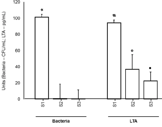
Cultivable bacteria (101.2±79.2) and LTA (94.1±61.7) were present in all S1 samples (20/20). CMP decreased the overall levels of cultivable bacteria by 99.4% (P < 0.05) and of LTA by 60.8% (P < 0.05), whereas the total overall reduction level of ICM on viable bacteria was 99.5% and on LTA was 76% (P < 0.05). Figure 2 shows the reduction of cultivable bacteria and lipoteichoic acid at the different phases of endodontic retreatment.
Regarding the reduction of cultivable bacteria, CMP with 2% CHX gel (G1, 99.3%) was more effective (P < 0.05) than 6% NaOCl (G2, 92.1%). On the other hand, ICM showed 100% reduction in the CHX-group (G1) and 98.5% in NaOCl-group (G2).
Regarding the reduction of LTA, CMP with 2% CHX gel (G1, 55.6%) was less effective (P < 0.05) than 6% NaOCl (G2, 67.5%). On the other hand, ICM showed 74.4% reduction in the CHX-group (G1) and 78.2% in NaOCl-group (G2), without statistical difference between the groups after the use of ICM (P > 0.05).
It is important to stress that the reduction of cultivable bacteria was greater than the reduction of LTA in all phases of ER (Table 1).
Figure 1. Gram-positive found in microbial culture at the different phases of root canal therapy with pos-treatment apical periodontitis.
0 2 4 6 8 10 12 Aerococcus spp Enterococcus faecalis Staphylococcus spp Actinomyces naeslundii Bifidobacterium spp Eggerthella lenta Enterococcus spp Gemella spp Actinomyces israelii Actinomyces viscosus Clostidium histolyticum Gemella morbillorum Lactococcus lactis spp Micrococcus spp Staphylococcus capitis Staphylococcus xylosus Staphylococcus warneri Streptococcus salivarius Streptococcus uberis Enterococcus faecalis Micrococcus spp Bifidobacterium spp Enterococcus faecalis Enterococcus spp Gemella morbillorum Granulicatella spp Micrococcus spp Staphylococcus spp S1 S2 S3
Figure 2. Reduction of cultivable bacteria and lipoteichoic acid at the different phases of endodontic therapy of teeth with post-treatment apical periodontitis.
Table 1 – Cultivable bacterial (x102 CFU/mL), LTA concentration (pg/mL), standard deviation and reduction (%) found in the 20
cases of post-treatment apical periodontitis.
Treatment phase Overall
values Overall reduction (%) Chemical substance 2% Chlorhexidine gel Reduction (%) 6% sodium hypochlorite Reduction (%) Bacterial S1 101.2±79.2 a - 129.10±83.4 a - 73.2±67.4 a - S2 0.6±1.4 b 99.4 0.90±2.8 b 99.3 5.8±6.1 c 92.1 S3 0.5±0.8 b 99.5 0.0±0.0 b 100 1.1±1.9 b 98.5 LTA S1 94.1±61.7 a - 107.2±77.7 a - 80.9±45.3 a - S2 36.9±28.8 b 60.8 47.6±33.6 b 55.6 26.3±21.4 c 67.5 S3 22.5±14.5 c 76 27.4±16.9 c 74.4 17.6±11.3 c 78.2
Note: CFU, colony-forming units; CMP, chemomechanical preparation; ICM, intracanal medication; LTA, lipotecichoic acid. Columns and row with different letters indicate statistical differences (P< 0.05).
Discussion
Our results confirm the strong presence of Gram-positive bacteria in infections related to the failure of endodontic treatment as 70/102 microorganisms were found in the initial samples of the root canals investigated, agreeing with the findings of the literature (4,7,12). Moreover, Enterococcus faecalis was the most prevalent bacteria involved in this clinical situation (4,8). This bacterium has been associated with the ability to remain inside the root canal even after the endodontic procedures (27).
The literature shows that elimination of bacteria from root canals after CMP ranges between 80 and 95% (8, 28-31). In the present study, CMP with either CHX or NaOCl was able to reduce the bacterial levels in 99.3% and 92.1% respectively. The group irrigated with CHX achieved a greater reduction on the bacterial levels after CMP, probably because CHX has the ability to act on both Gram-positive bacteria and Gram-negative bacteria (12). The total overall reduction of ICM on the bacterial levels was 99.5%. ICM was more effective in reducing the bacterial levels in root canals irrigated with CHX (100%).
Regarding the LTA reduction, after CMP, the group irrigated with NaOCl achieved a greater reduction on the LTA levels (67.5%) compared to the CHX group (55.6%). The use of intracanal medication was able to reduce even more the LTA levels (G1: 74.4%; G2: 78.2%).
It is important to mention that the greatest reduction rate of microorganisms and LTA was achieved after CMP. The intracanal medication actually reduced 16.7% (microorganisms) and 39% (LTA). However, considering the cumulative effect of all phases it came to 99.5% and 76%, respectively.
Our findings that CMP and ICM are more effective in reducing microbial levels than LTA levels are similar to the LPS reduction profile either in primary endodontic infection (17,34-35) or in secondary endodontic infection (8). In primary infections the LPS reduction percentual after CMP is between 59%-98.06% (35, 36) and in secondary infections in 60.6% (8), which is in agreement with the present study.
High levels of bacteria and LTA may be responsible for the maintenance of an inflammatory process in the periapical region and consequent failure of endodontic treatment (9,22). Further studies are needed to test the acceptable clinical levels of residual LTA and also to confirm the results found in the present work, as there is a
lack in the literature regarding clinical studies investigating the presence of LTA in infected root canals.
It was concluded that regardless the CM and ICM the reduction rates of bacteria were higher compared to LTA. Gram-positive microorganisms were present in all phases of the endodontic retreatment.
Acknowledgments
This study was supported by grants from Research Support Foundation of the State of São Paulo (FAPESP) (protocol number 2012/23697-4), National Scientific and Technological Development Council (CNPq) (protocol number 308162/2014-5) and Coordination for Improvement of Higher Education Personnel (CAPES), and Brazilian governmental institutions. We would like to thank Mr Maicon R Z Passini and Priscila Amanda Francisco for their technical support.
The authors deny any conflicts of interest related to this study.
References
1. Kakehashi S, Stanley HR, Fitzgerald RJ. The effects of surgical exposures of dental pulps in germ-free and conventional laboratory rats. Oral Surg Oral Med Oral Pathol. 1965; 20: 340-9.
2. Gomes BP, Lilley JD, Drucker DB. Associations of endodontic symptoms and signs with particular combinations of specific bacteria. Int Endod J. 1996; 29(2): 69-75.
3. Siqueira JF Jr, Rôças IN. Bacterial pathogenesis and mediators in apical periodontitis. Braz Dent J. 2007; 18(4): 267-80.
4. Rahimi S, Janani M, Lotfi M, Shahi S, Aghbali A, Vahid Pakdel M, Salem Milani A, Ghasemi N. A review of antibacterial agents in endodontic treatment. Iran Endod J. 2014; 9(3): 161-8.
5. dos Santos LG, Felippe WT, Teixeira CS, Bortoluzzi EA, Felippe MC. Endodontic re-instrumentation enhances hydroxyl ion diffusion through radicular dentine. Int Endod J. 2014; 47(8): 776-83.
6. Zhao L, Chen J, Cheng L, Wang X, Du J, Wang F, Peng Z. Effects of Enterococcus faecalis lipoteichoic acid on receptor activator of nuclear factor-κB ligand and osteoprotegerin expression in periodontal ligament fibroblasts. Int Endod J. 2013; 47(2): 163-72.
7. Pinheiro ET, Gomes BP, Ferraz CC, Sousa EL, Teixeira FB, Souza-Filho FJ.Microorganisms from canals of root-filled teeth with periapical lesions. Int Endod J. 2003; 36(1): 1–11.
8. Endo MS, Martinho FC, Zaia AA, Ferraz CC, Almeida JF, Gomes BP. Quantification of cultivable bacteria and endotoxin in post-treatment apical periodontitis before and after chemo-mechanical preparation. Eur J Clin Microbiol Infect Dis. 2012; 31(10): 2575-83.
9. Baik JE, Jang KS, Kang SS, Yun CH, Lee K, Kim BG, Kum KY, Han SH. Calcium hydroxide inactivates lipoteichoic acid from Enterococcus faecalis through deacylation of the lipid moiety. J Endod. 2011; 37(2): 191-6.
10. Gomes BPFA, Pinheiro ET, Jacinto RC, Zaia AA, Ferraz CC, Souza-Filho FJ. Microbial analysis of canals of root-filled teeth with periapical lesions using polymerase chain reaction. J Endod.2008; 34(5): 537-40.
11. Lee SH, Baek DH. Antibacterial and neutralizing effect of human β-defensins on Enterococcus faecalis and Enterococcus faecalis lipoteichoic acid. J Endod. 2012; 38(3):351-6.
12. Gomes BP, Vianna ME, Zaia AA, Almeida JF, Souza-Filho FJ, Ferraz CC. Chlorhexidine in endodontics. Braz Dent J. 2013; 24(2): 89-102.
13. Lleò MM, Bonato B, Tafi MC, Signoretto C, Boaretti M, Canepari P. Resuscitation rate in different enterococcal species in the viable but non-cultivable state. J Appl Microbiol. 2001; 91(6): 1095-102.
14. Seltzer S, Farber PA. Microbiologic factors in endodontology. Oral Surg Oral Med Oral Pathol. 1994; 78(5): 634-45.
15. Costa ED, de Souza-Filho FJ, Barbosa SV. Tissue reactions to a component of root canal system bacteria: lipoteichoic acid. Braz Dent J. 2003; 14(2): 95-8.
16. Wang JE, Dahle MK, McDonald M, Foster SJ, Aasen AO, Thiemermann C. Peptidoglycan and lipoteichoic acid in gram-positive bacterial sepsis: receptors, signal transduction, biological effects, and synergism. Shock. 2003; 20(5): 402-14.
17. Martinho FC, Chiesa WM, Leite FR, Cirelli JA, Gomes BP. Antigenic activity of bacterial endodontic contents from primary root canal infection with periapical lesions against macrophage in the release of interleukin-1beta and tumor necrosis factor alpha. J Endod. 2010; 36(9): 1467-74.
18. Martinho FC, Chiesa WM, Leite FR, Cirelli JA, Gomes BP. Correlation between clinical/radiographic features and inflammatory cytokine networks produced by macrophages stimulated with endodontic content. J Endod. 2012; 38(6): 740-5.
19. Ginsburg I. Role of lipoteichoic acid in infection and inflammation. Lancet Infect Dis. 2002; 2(3): 171-9.
20. Hermann C, Spreitzer I, Schröder NW, Morath S, Lehner MD, Fischer W, Schütt C, Schumann RR, Hartung T. Cytokine induction by purified lipoteichoic acids from various bacterial species--role of LBP, sCD14, CD14 and failure to induce IL-12 and subsequent IFN-gamma release. Eur J Immunol. 2002; 32(2):541-51.
21. Hahn CL, Liewehr FR. Relationships between caries bacteria, host responses, and clinical signs and symptoms of pulpitis. J Endod. 2007; 33(3): 213-9.
22. Ryu YH, Baik JE, Yang JS, Kang SS, Im J, Yun CH, Kim DW, Lee K, Chung DK, Ju HR, Han SH. Differential immunostimulatory effects of Gram-positive bacteria due to their lipoteichoic acids. Int Immunopharmacol. 2009; 9(1): 127-33.
23. Han SH, Kim JH, Martin M, Michalek SM, Nahm MH. Pneumococcal lipoteichoic acid (LTA) is not as potent as staphylococcal LTA in stimulating Toll-like receptor 2. Infect Immun. 2003; 71(10): 5541-8.
24. Kayaoglu G, Ørstavik D. Virulence factors of Enterococcus faecalis: relationship to endodontic disease. Crit Rev Oral Biol Med. 2004; 15(5): 308-20.
25. Martinho FC, Gomes BP. Quantification of endotoxins and cultivable bacteria in root canal infection before and after chemomechanical preparation with 2.5% sodium hypochlorite. J Endod. 2008; 34(3): 268-72.
26. Möller AJ. Microbiological examination of root canals and periapical tissues of human teeth. Methodological studies. Odontol Tidskr. 1966; 74(5): Suppl 1-380.
27. Gomes BP, Drucker DB, Lilley JD. Positive and negative associations between bacterial species in dental root canals.Microbios. 1994; 80(325):231-43.
28. Gomes BP, Martinho FC, Vianna ME. Comparison of 2.5% sodium hypochlorite and 2% chlorhexidine gel on oral bacterial lipopolysaccharide reduction from primarily infected root canals. J Endod. 2009; 35(10): 1350-3.
29. Dornelles-Morgental R, Guerreiro-Tanomaru JM, de Faria-Júnior NB, Hungaro-Duarte MA, Kuga MC, Tanomaru-Filho M. Antibacterial efficacy of endodontic irrigating solutions and their combinations in root canals contaminated with Enterococcus faecalis. Oral Surg Oral Med Oral Pathol Oral Radiol Endod. 2011; 112(3): 396-400.
30. Rôças IN, Siqueira JF Jr. Comparison of the in vivo antimicrobial effectiveness of sodium hypochlorite and chlorhexidine used as root canal irrigants: a molecular microbiology study. J Endod. 2011; 37(2): 143-50.
31. Kaushik N, Rehani U, Agarwal A, Kaushik M, Adlakha V. Antimicrobial Efficacy of Endodontic Irrigants against Enterococcus faecalis and Escherichia Coli: An in vitro study. Int J Clin Pediatr Dent. 2013; 6(3): 178-82.
32. Mohammadi Z, Abbott PV. Antimicrobial substantivity of root canal irrigants and medicaments: a review. Aust Endod J. 2009; 35(3): 131-9.
33. Mozayeni MA, Haeri A, Dianat O, Jafari AR. Antimicrobial effects of four intracanal medicaments on Enterococcus faecalis: an in vitro study. Iran Endod J. 2014; Summer; 9(3): 195-8.
34. Marinho AC, Martinho FC, Zaia AA, Ferraz CC, Gomes BP. Monitoring the effectiveness of root canal procedures on endotoxin levels found in teeth with chronic apical periodontitis. J Appl Oral Sci. 2014; 22(6): 490-5.
35. Martinho FC, Gomes AP, Fernandes AM, Ferreira NS, Endo MS, Freitas LF, Camões IC. Clinical comparison of the effectiveness of single-file reciprocating systems and rotary systems for removal of endotoxins and cultivable bacteria from primarily infected root canals. J Endod. 2014; 40(5): 625-9.
36. Vianna ME, Hortz HP, Conrads G, Zaia AA, Souza-Filho FJ, Gomes BPFA. Effect of root canal procedures on endotoxins and endodontic pathogens. Oral Microbiol Immunol 2007; 22(6):411-8.
CAPÍTULO II
Effectiveness of root canal procedures on the reduction
proinflammatory cytokines and matrix metalloproteinases in cases
of post-treatment apical periodontitis
Abstract
Aim: To monitor in vivo the effect of chemomechanical preparation (CMP) and intracanal medication (ICM) on the reduction of proinflammatory cytokines (PIC) (TNF-α and IL1-β) and matrix metalloproteinases (MMPs) (-2, -3, -8, -9 and -13) in root canals of teeth with post-treatment apical periodontitis. Methodology: Twenty infected root canals of teeth single-rooted were randomly assigned into two groups according to the irrigant used for CMP (n = 10 per group): G1 - 2% chlorhexidine gel and G2 - 6% sodium hypochlorite. Root canal contents were taken by using paper points before (S1) and after CMP (S2) and after 30 days of ICM (Ca[OH]2 + 2%
chlorhexidine gel) (S3). PIC and MMP (pg/mL) were measured using enzyme-linked immunosorbent assay. Results: PIC and MMP were present in all S1 samples. Lower initial values were found for PIC (TNF-α: 8.8±4.7; IL1-β: 1.2±0.4) compared to the levels of MMP-2 (803.7±96.4), followed by -3 (453.9±229.3), -8 (245.9±122.4), -9 (129.4±29.6) and -13 (70.8±12.8). Decreased levels of PIC and MMP were found for all groups (P < 0.05) in all S2 samples. On the other hand, PIC, MMP-2 and MMP-13 increased after the ICM (P < 0.05). There was no difference between the chemical substances tested on PIC reduction in S2 (P > 0.05). Regarding MMP, 2% CHX gel reduced the levels of all of them in S2 (P < 0.05), whereas 6% NaOCl reduced only MMP-2, -3 and -13 (P < 0.05). TNF-α and IL1-β increased in S3 for both G1 and G2 (P < 0.05). The ICM reduced MMP-3 and -8 only in G2 (P < 0.05). However, there was an increase of MMP in S3 (P < 0.05) for G1 (3, 8, 9 and 13) and G2 (2 and -13). Conclusion: regardless of the chemical substance tested, CMP is effective in reducing PIC and MMP. While the ICM increases the levels of TNF-α, IL-1β, MMP-2 e MMP-13.
Keywords: Bacteria. Chlorhexidine. Sodium hypochlorite. Enterococcus faecalis. Cytokines. Matrix metalloproteinases.