FABRICATION AND APPLICATIONS OF DOPAMINE SENSITIVE ELECTRODES
Andre Hermans
A dissertation submitted to the faculty of the University of North Carolina at Chapel Hill in partial fulfillment for the degree of Doctor of Philosophy in the Department of
Chemistry
Chapel Hill 2007
Approved by:
Advisor: R. Mark Wightman
Reader: Mark H. Schoenfish
ABSTRACT
ANDRE HERMANS: Fabrication and Applications of Dopamine Sensitive Electrodes (Under the direction of Dr. R. Mark Wightman)
The neurotransmitter dopamine has shown to be of central importance to
difference brain functions, such as movement, reward, and addiction. A biosensor for the detection of dopamine in the brain should have a fast time response to monitor concentration changes which happen on a subsecond time scale.
Furthermore, the sensor should have a high sensitivity to dopamine, because the physiological concentrations of dopamine were found to be in the range form nanomolar to lower micromolar. High selectivity is also necessary to distinguish the desired signal from electrochemical interferences in the brain such as ascorbic acid. Fast scan cyclic voltammetry at glass-encased carbon fiber microelectrodes has been shown to fulfill these requirements and is therefore often used for
measurements of easily oxidizable neurotransmitters like dopamine. In this dissertation, some drawbacks of the technique and the sensor are addressed and improved.
Chapter 1 contains an overview of electrochemical methods that have been used to detect various neurotransmitters in the brain. Chapter 2 explains a method to increase the sensitivity and selectivity for dopamine of carbon fiber
electrode surface. A method utilizing tungsten microwires as substrate for the construction of flexible gold, platinum, and carbon microelectrodes is described in Chapter 3 and 4. Carbon-coated tungsten microwires have then been examined for use as in vivo dopamine sensor. The microwires showed the same
electrochemical properties as conventional glass-encased carbon fiber
ACKNOWLEDEGMENTS
First, I would like to thank my academic advisor Dr. R. Mark Wightman for his support throughout the last 5 years. He has been a great mentor who gave me the opportunity to gain a lot of experience in the field of academic research and science.
I would like to thank my family and friends, especially Cherie Lanyi, for support and help outside the lab environment.
Furthermore, I would like to acknowledge everyone who worked with me over the last five years and help me pursuing my research goals. I thank Andrew Seipel, Justin Kita and Dr. Leslie Sombers for help with in vivo applications, Charlie Miller for slice experiments, Richard Keithley for his help with data analysis, the UNC electronics facility, and Dr. Michael Heien.
The experiments presented in chapter 7 were performed in the laboratories of Wolfram Schultz in Cambridge, England. I would like to thank Dr. Schultz and Dr. Istvan Hernardi for this wonderful collaboration.
TABLE OF CONTENTS
LIST OF TABLES ... xiii
LIST OF FIGURES ... xiv
LIST OF ABBREVIATIONS ... xiv
Chapter I. ELECTROCHEMICAL DETECTION IN THE BRAIN ... 1
Introduction ... 1
Direct electrochemical detection of neurotransmitters in group 1 ... 2
Electrode materials ... 9
Electrochemical techniques ... 12
Modified electrodes ... 17
Reference and auxiliary electrodes ... 19
Detection of changes in pH in vivo ... 19
Enzyme electrodes for detection of molecules in group 2 ... 21
Detection of molecules in group 3 ... 27
References ... 29
Introduction ... 41
Experimental section ... 44
Chemicals ... 44
Synthesis and characterization of 4-sulfobenzenediazonium tetrafluoroborate (4-SBD) ... 44
Electrode preparation ... 45
Chemical surface modification ... 46
XPS analysis ... 47
Electrochemical measurements ... 47
Measurement in brain slices ... 48
Results and discussion ... 48
Growth of sulfobenzene layers ... 48
Fast-scan cyclic voltammetry at electrodes grafted with 4-sulfobenzene ... 52
Dopamine adsorption at electrodes grafted with 4-sulfobenzene ... 54
Voltammetric response to other compounds ... 56
Use in brain slices ... 60
Summary ... 62
References ... 63
III. CONICAL TUNSGTEN TIPS AS SUBSTRATES FOR THE PREPARATION OF ULTRAMICROELECTRODES ... 67
Introduction ... 67
Experimental section ... 69
Electrodeposition of Platinum and Gold ... 70
Carbon Deposition ... 71
Glass encased microelectrodes ... 73
Voltammetric characterization ... 74
Chemicals ... 75
Results and Discussion ... 75
Fabrication Considerations ... 75
Electrochemical behavior of platinum plated electrodes ... 77
Electrochemical behavior of gold plated electrodes ... 79
Electrochemical behavior of pyrolyzed photoresist film (PPF) electrodes ... 81
Surface area of conical electrodes ... 84
Summary ... 85
References ... 87
IV. CARBON COATED TUNGSTEN MICROELECTRODES FOR DOPAMINE DETECTION IN VITRO AND IN VIVO ... 90
Introduction ... 90
Experimental section ... 93
Chemicals ... 93
PPF microelectrodes ... 93
Construction of multiwire arrays ... 97
Glass encased carbon fiber microelectrodes ... 98
Data acquisition and analysis ... 99
Electrically evoked dopamine release in anesthetized rats ... 99
Single unit recording in freely moving rats ... 100
Results and discussion ... 101
Electrochemical detection of dopamine in vitro ... 101
Electrochemical detection of other compounds in vitro ... 103
Electrochemical and electrophysiological measurements in vivo ... 105
Construction of PPF-microelectrode arrays ... 107
Summary ... 109
References ... 111
V. MONITORING BRAIN DOPAMINE FLUCTUATIONS WITH FAST-SCAN CYCLIC VOLTAMMETRY FOR MULTIPLE MINUTES .... 115
Introduction ... 115
Experimental Section ... 118
Chemicals ... 118
Data acquisition and background correction ... 118
Flow-injection analysis ... 121
Electrode preparation ... 121
Noise analysis ... 122
In vivo measurements in anesthetized rats ... 122
In vivo measurements in freely moving rats ... 123
Data analysis ... 124
Quantitative comparison between components ... 125
Results and discussion ... 128
Noise reduction ... 128
Effect of drift of the background ... 130
Monitoring dopamine concentrations in vivo ... 133
Investigation of chemical fluctuations ... 139
Summary ... 142
References ... 144
VI. CHANGES IN BACKGROUND SIGNAL AT CARBON MICRO ELECTRODES DURING FAST SCAN CYCLIC VOLTAMMETRY ... 148
Introduction ... 148
Experimental Section ... 150
Chemicals ... 150
Data acquisition and electrochemical pretreatment ... 151
Flow-injection analysis ... 151
Electrode preparation ... 152
In vivo measurements in anesthetized rats ... 153
Results and discussion ... 153
Electrochemical activation of pyrolyzed photoresist films ... 153
Background changes at carbon-fiber microelectrodes in vitro ... 156
Background changes at carbon fiber microelectrodes in-vivo ... 160
Polymerization of dopamine ... 166
Summary ... 168
References ... 169
VII. ELECTROCHEMICAL MEASUREMENT OF PH SHIFTS AND DOAPMINE RELEASE IN PRIMATES DURING REWARD DELIVERY ... 173
Introduction ... 173
Experimental section ... 175
Chemicals ... 175
Electrode preparation ... 175
Flow-injection apparatus ... 178
Data acquisition and analysis ... 178
In vivo recordings ... 179
Presentation of free reward ... 180
Presentation of predictable reward ... 180
Results ... 182
Responses to pH changes at carbon fiber microelectrodes ... 182
pH changes in vivo during delivery of free reward ... 182
Correlation between pH changes and behavior during delivery of free reward ... 184
pH changes during presentation of predicted reward ... 186
Correlation between pH changes and behavior during delivery of predicted reward ... 190
Dopamine release during delivery of predicted reward ... 192
Discussion ... 193
Summary ... 199
LIST OF TABLES
Table
LIST OF FIGURES
Figure
2.1. Modification of carbon electrodes with 4-SBD ... 50
2.2. Characterization of 4-SBD modified electrodes in pH 7.4 ... 53
2.3. Adsorption characteristics of dopamine at P-55 elliptical electrodes ... 55
2.4. Background-subtracted cyclic voltammograms for various compounds ... 57
2.5. Dopamine detection in mouse brain slices with carbon fiber electrodes ... 61
3.1. SEM images of conical microelectrodes ... 72
3.2. Cyclic voltammograms at platinum microelectrodes ... 78
3.3. Cyclic voltammograms at gold microelectrodes ... 80
3.4. Cyclic voltammograms at carbon microelectrodes ... 82
4.1. Fabrication process for PPF microelectrodes ... 94
4.2. SEM micrograph of PPF microelectrodes ... 95
4.3. Response to dopamine at carbon fibers and PPF electrodes ... 102
4.4. Background subtracted cyclic voltammograms for various compounds ... 104
4.5. Electrochemical and electrophysiological measurements in-vivo . 106 4.6. Construction of multiwire array ... 108
5.1. Electronic setup for digital background subtraction ... 120
5.2. Background change ... 127
5.4. Dopamine injections in vitro ... 132
5.5. Dopamine stimulations in vivo ... 134
5.6. Response to iv cocaine and saline injections ... 136
5.7. Construction of pure component color plots and RMS traces ... 140
5.8. Standard-deviation of RMS-current traces ... 141
6.1. Electrochemical activation of PPF microelectrodes ... 154
6.2. Changes in background signal at carbon-fiber microelectrodes in vitro ... 157
6.3. Changes in background signal at carbon fiber microelectrodes in vivo (cortex) ... 161
6.4. Background changes in caudate/putamen ... 162
6.5. Dopamine induced background changes in vitro ... 164
7.1. SEM image of electrodes for primate recordings ... 176
7.2. Timing diagram of the behavioral tasks performed by monkeys during predicted reward delivery ... 181
7.3. Cyclic voltammetric response to a basic pH change in vitro ... 183
7.4. Delivery of free reward ... 185
7.5. Delivery of predicted reward ... 187
7.6. Delivery and non-delivery of predicted reward with 50% probability ... 189
LIST OF ABBREVIATIONS
3-MT 3–Methoxytyramine 4-SB 4-Sulfobenzene
4-SBD 4-Sulbebenzene diazonuim
AC Alternating current
AChE Acetylcholine esterase
ADP Adenosine diphosphate
AP Anterior / posterior
ChO Choline oxidase
CLS Classical least squares CNS Central nervous system
COMT catechol-o-methyltransferase
DC Direct current
DOPA 3,4 dihydroxyphenylalanine DOPAC 3,4 dihydroxyphenylacetic acid DPV Differential pulse voltammetry
DV Dorsal / ventral
E1/2 Half-wave potential
F Faraday’s constant
FAD Flavine adenine dinucleotide
GABA Gamma-aminobutyric acid
GluOx Glucose oxidase
H Aspect ratio of a cone
HPLC High performance liquid chromatography
HRP Horseradish peroxidase
HVA Homovanilic acid
L-Dopa Levo-dihydroxyphenylalanine
LOx Lactate oxidase
MAO Monoamine oxidase
MHPG 3-Methoxy-4-Hydroxy-phenylethyleneglycol
ML Medial / lateral
n Number of electrons
NADH Nicotinamide adenine dinucleotide PAN Polyacrylonitrile PCA Principal component analysis
PPF Pyrolyzed photoresist film
Q Charge
RMS Root-mean-square
SEM Standard error of the mean, scanning electron microscopy SSDH Succinic semialdehyde dehydrogenase
UA Uric acid
VMA vanilylmandeleic acid
CHAPTER 1
ELECTROCHEMICAL DETECTION IN THE BRAIN
Introduction
With respect to electrochemical detection, neurotransmitters can be
separated into three different categories. The first group of neurotransmitters is the
electrochemically active compounds such as the tyrosine derivatives dopamine,
norepinephrine, and epinephrine. Many of their metabolites are also electroactive
such as 3,4 dihydroxyphenylacetic acid (DOPAC), homovanilic acid (HVA),
3-methoxytyramine, or l-dopa. The neuroactive tryptophan derivatives are also
electroactive and include 5-hydroxytryptamine (serotonin) precursors or metabolites
like 5-hydroxyindolacetic acid, 5-hydroxyindoletryptophan or melatonin. Other
electroactive neurotransmitters are histamine and adenosine. All of these
compounds can be directly detected by electrochemical oxidation of the molecule.
Furthermore, other electroactive substances in the brain like ascorbic acid, uric acid,
nitric oxide, oxygen or hydrogen peroxide are also readily detectable by
electrochemical methods.
The second group of neurotransmitters is not inherently electroactive, and
thus these compounds cannot be detected by traditional electroanalytical methods.
However, those that can be oxidized by an enzymatic reaction can be measured
Some of the neurotransmitters in this category are amino acid transmitters
like glutamate and γ-aminobutyric acid (GABA), but also acetylcholine and its
precursor choline have been detected with this approach. Glucose and lactate,
compounds important in energy production in the brain, can also be detected with
such indirect electrochemical approach.
Neuroactive peptides cannot be detected directly in the brain with
electrochemical biosensors. However, neuropeptides with inherently
electrochemically active group can be detected off-line electrochemically. Inate
redox-active functionalities include tyrosine, tryptophan, methionine and cysteine
residues. Neuropetides and some amino acid neurotransmitters form the third group
of neurotransmitters.
Direct electrochemical detection of neurotransmitters in group 1
Catecholamine neurotransmitters are well suited for electrochemical detection
because the potential required for their oxidation is well within normal scan ranges
for carbon and metal electrodes in physiological buffer (Adams and Marsden, 1982).
Catecholamines are derived in the biosynthetic pathway from tyrosine (Cooper et al.,
2003). The rate limiting step in the synthesis is the hydroxylation of tyrosine to
3,4-dihydroxyphenylalanine (DOPA) with tyrosine hydroxylase. Dopamine is formed by
the decarboxylation of l-DOPA. Norepinephrine is formed after transfer of a hydroxyl
group onto the β-position of the side chain via dopamine-β-hydroxylase. The amine
of norepinephrine can be methylated by phenylethanolamine-N-methyltransferase to
Molecule Redox-Reaction Approximate oxidation potential in vivo(Adams and Marsden, 1982) (vs Ag/AgCl) Tyrosine derivatives O H NH3 +
COO- +0.7 V
L-DOPA O H O H NH3 + COO-O O NH3 + COO- H + -2e
+2 +0.4 V
Dopamine O H O H NH3 + O O
NH3+ H+ -2e
+2 +0.2 V
Norepinephrine
O H
O H
NH3+ OH
O
O
NH3+ OH
H+ -2e
+2 +0.2 V
Epinephrine
O H
O H
NH2+ OH
O
O
NH2+ OH
H+ -2e
+2 +0.2 V
DOPAC O H O H COO- O O COO-H+ -2e
+2 +0.2 V
Homovanilic Acid O H O COO-C H3 O O COO-C
H3 OH
H+ -2e
+
+ +0.5 V
3-Methoxytyramine O H O C H3
NH3+ O
O
NH3+
C
H3 OH
H+
-2e +
+ +0.5 V
Tryptophan
derivatives N
NH3 +
COO- +0.8 V
Serotonin N NH3 + COO-O H N NH3 + COO-O H+ -2e
+ 2 +0.35 V
5-Hydroxyindolacetic acid N O COO-H+ COO-N O H -2e
+2 +0.35 V
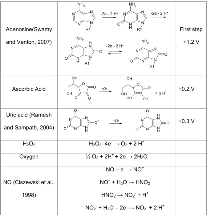
Adenosine(Swamy
and Venton, 2007)
N N N N R1 NH2 N N H N N R1 O NH2 N N H N H N R1 O NH2 O N N N N R1 O NH2 O -2e - 2 H+ -2e - 2 H+
-2e - 2 H+
First step +1.2 V Ascorbic Acid O O O O OH OH O O OH O H OH
OH H+
-2e
+2 +0.2 V
Uric acid (Ramesh
and Sampath, 2004)
N N O O N H N O N N O O N H N H O
-2e +0.3 V
H2O2 H2O2 -4e-→ O2 + 2 H+
Oxygen ½ O2 + 2H+ + 2e-→ 2H2O
NO (Ciszewski et al.,
1998)
NO – e-→ NO+
NO+ + H2O → HNO2
HNO2→ NO2- + H+
NO2- + H2O – 2e-→ NO3- + 2 H+
monoamine oxidase (MAO) and catechol-O-methyltransferase(COMT). These
metabolites are dihydroxyphenylacetic acid (DOPAC), homovanilic acid and
3-methoxytyramine (3-MT). The action of MAO on norepinephrine mainly produces
vanilylmandeleic acid (VMA) and 3-methoxy-4-hydroxy-phenlethyleneglycol
(MHPG). As can be seen in table 1.1a all of these compounds show similar 2
electron oxidation reactions with similar oxidation potentials. For this reason it is
difficult to distinguish different catecholamines and their metabolites with
electrochemical methods. However, most of these compounds undergo secondary
reactions. The methoxylated derivatives are oxidized at more positive potentials
than the catecholamines, however, the oxidation product loses methanol and forms
the o-quinone of the corresponding catecholamine. In the case of 3-MT oxidation,
dopamine-o-quinone is formed while HVA leads to the formation of the oxidized form
of DOPAC.
Another example for a secondary reaction intracyclization reaction of the
oxidized hydroquinone form of catecholamines (Hawley et al., 1967; Blank et al.,
1976; Zhang and Dryhurst, 1993).
O
O
NH3+
N H O
H
O H
(1)
N H O
H
O
H N
H O
O
H+
+
2e+
2The 5-6 dihydroxyindoline product can undergo redox-reactions itself to an
aminochrome (reaction (2)). However, In the case of dopamine the rate constant for
the formation reaction is very slow, on the order of k = 0.1 s-1 at physiological pH.
The rate of cyclization is faster for epinephrine than for norepinephrine. This has
been used to distinguish these compounds electrochemically (Ciolkowski et al.,
1992). By using fast scan methods or measuring quickly after a potential step the
effect of these reactions can be diminished. The formation of the aminochrome
product can be used to distinguish norepinephrine and epinephrine
electrochemically since the rate of cyclization is faster for epinephrine (Ciolkowski et
al., 1992). In addition to this, the secondary-amine side chain of epinephrine can be
oxidized at very positive potentials whereas the primary-amine side chain of
norepinephrine cannot (Pihel et al., 1994).
The other important group of electrochemically detectable neurotransmitter
and metabolites are the tryptophan derivatives including serotonin. During
biosynthesis tryptophan is converted by tryptophan hydroxylase to
5-hydroxytryptophan which then forms serotonin after decarboxylation. Metabolism of
serotonin is primarily by MAO followed by an aldehyde dehydrogenase that leads to
5-hydroxyindole acetic acid. A more minor pathway is metabolism of serotonin to
form 5-hydroxytryptophenol. Tryptophan derivatives can also be oxidized and
electrochemically detected in a two electron process with similar oxidation potential
as catecholamines. After oxidation many different secondary reactions have been
and involves the dimerization of serotonin, as well as, after addition of water,
formation of tryptamine 4-5 dione.
Histamine and adenosine are other oxidizable neurotransmitters but neither
has been directly detected with electrochemical methods in the brain yet. Histamine
secretion from isolated mast cells has been detected electrochemically(Pihel et al.,
1995). Histamine is synthesized from histidine via decarboxylation and has multiple
metabolic products such as imidazoleacetaldehyde and imidazolacetic acid. Recent
research has shown that adenosine can be detected by fast scan cyclic voltammetry
(Abou El-Nour and Brajter-Toth, 2000; Brajter-Toth et al., 2000; Abou El-Nour and
Brajter-Toth, 2003; Swamy and Venton, 2007). Adenosine seems to undergo a 3
step oxidation of which the first two steps lead to a distinct voltammogram which can
be differentiated from other interferences (see table 1.2).
The main electroactive interference for electrochemical measurements in the
extracellular fluid of the brain is ascorbic acid. The ascorbic acid concentrations in
the extracellular fluid of the brain are 0.5 mM which is 104-106 times higher than the
concentrations of catecholamines (Mefford et al., 1981; Nagy et al., 1982). Early
amperometric measurements showed that the oxidation of ascorbic acid was the
main signal recorded in the brain (Kissinger et al., 1973; Mueller, 1986). However,
the electron transfer kinetics for the oxidation of ascorbic acid at carbon are very
sensitive to the surface conditions. Electrooxidation of carbon at very high potentials
(~3.0 V) causes an acceleration of electron transfer rates so that the ascorbate
oxidation peak occurs near its thermodynamically anticipated value. Using such
concentration following pharmacological treatments expected to affect
cathecholamines (Gonon et al., 1980a; Gonon et al., 1981). This established that
brain sensors for dopamine had to be able to distinguish the two molecules. This is
accomplished in fast scan methods because the oxidation of ascorbate shifts to
more positive potentials as a result of slow electron-transfer kinetics, distinguishing
the ascorbate signal from other compounds (Marsden et al., 1988; Millar et al.,
1992). Alternatively, electrode modification with selective membranes can reject
ascorbate from the electrode surface (Nagy et al., 1982; Baur et al., 1988;
Wiedemann et al., 1990).
Beside direct electrochemical interference ascorbic acid can contribute
indirectly to the measured signal. Ascorbate functions as the major antioxidant in
the body. Thus, even when it is rendered inactive at the electrode surface, it can
play this role during the electrochemical detection. For example, after the oxidation
of dopamine, dopamine-o-quinone is reduced by ascorbate, regenerating dopamine.
This catalytic reaction by ascorbic acid provides more dopamine for electrooxidation
with the net result that the electrochemical current is proportional to both dopamine
and ascorbate (Sternson et al., 1973).
Recently it has been shown that hydrogen peroxide plays an important role in
brain signaling and neurotransmitter regulation(Chen et al., 2001; Avshalumov et al.,
2007). Hydrogen peroxide can be detected directly by electrochemical methods
such as amperometry (Kulagina and Michael, 2003). Oxygen levels in the brain are
also often determined with microelectrodes (Zimmerman and Wightman, 1991;
determination of oxygenation of brain tissue, electrochemical methods are an
alternative approach to BOLD-MRI measurements which monitors the oxygen
dissociation from hemoglobin (Ogawa et al., 1990). Although electrochemical
oxygen measurements have a much better spatial and temporal resolution than fMRI
techniques for many studies use fMRI techniques because of the non-invasive
nature. Furthermore, amperometric detection of hydrogen peroxide or oxygen is the
basis for enzyme based electrodes that are discussed further later.
Nitric oxide (NO) has been shown to act in the central nervous system as
secondary messenger and as mediator in cardiovascular system (Schuman and
Madison, 1994; Dawson and Dawson, 1996; Cooper et al., 2003). Nitric oxide is
synthesized from argenine. When measuring nitric oxide electrochemically, normally
electrodes with coatings or membranes that are specific to NO are used. A
description of these coating can be found later in this review.
Electrode materials
The developments of microelectrodes several decades ago formed the basis
for in vivo applications of electrochemical detection of neurotransmitters. Early
microelectrodes for voltammetric recordings in the CNS were constructed from
carbon paste (Adams, 1958; Kissinger et al., 1973; O'Neill, 2005). The carbon paste
was prepared by mixing carbon powder with either Nujol or silicone oil (O'Neill et al.,
1982) and then packed into the end of Teflon insulated metal microwires resulting in
disk electrodes with diameters between 100 and 300 µm. Glass capillaries can also
curing resulting in a more rigid electrode (Conti et al., 1978; Huff and Adams, 1980).
Carbon paste electrodes have been reported to be stable over several months for in
vivo applications. However, the relatively large size of carbon-paste electrodes limits
the applications to larger brain regions. Because of the larger size more brain
damage occurs in comparison to smaller sized electrodes.
Smaller carbon electrodes can be made from carbon fibers (Gonon et al.,
1978; Ponchon et al., 1979). Carbon fibers range from a few micrometers in
diameter to 40 µm but the majority of fibers range between 5-15 µm (McCreery,
1996). Carbon fibers are prepared from the pyrolysis of either petroleum pitch or
polyacrylonitrile (PAN) and undergo thermal processing similar to glassy carbon. To
make an electrode the carbon fiber is inserted into a glass capillary, pulled with a
pipette puller, and cut to obtain a cylindrical electrode or polished for disk electrodes.
To form a well insulated seal between the carbon fiber and the insulating glass
sheath, epoxy resin is allowed to creep between the fiber and the glass and then
cured (Kawagoe et al., 1993). Carbon-fiber microelectrodes constructed in this way
cause minimal damage to the surrounding tissue (Peters et al., 2004) when inserted
into the brain because the dimensions of the whole electrode including the
surrounding insulation are in the lower micrometer range.
The electrochemical properties of carbon electrodes depend on the oxidation
state of carbon-containing functional groups of the carbon surface. It has been
proposed that surface carbonyl- and hydroxyl-groups can catalyze electron transfer
for inner-sphere reactions at the electrode surface (Chen and McCreery, 1996).
increase the sensitivity to positively charged analytes (Heien et al., 2003). This is
probably due to increased adsorption of cations to the carbon surface. An increase
in sensitivity of 5 to 7 has been observed in vitro for dopamine when using fast scan
cyclic voltammetry at overoxidized carbon fibers. However, overoxidized electrodes
show a slower time response and lower selectivity than electrode which have not
been exposed to high oxidative potentials. In early studies overoxidation was
accomplished by repetitive excursions + 3 V vs. Ag/AgCl at 70 Hz (Gonon et al.,
1980b; Gonon et al., 1981). Electrodes treated this way showed higher sensitivity
for catecholamines. However, in addition to increasing the amount of surface
oxides, this surface treatment seems to drastically increase the surface area of the
electrode (Swain and Kuwana, 1991).
Carbon microelectrodes are well suited for direct electrochemical detection of
neurotransmitters of group 1 for various reasons. Beside the relatively easy and
cost-effective construction, carbon electrodes show very little bio-fouling during
in-vivo applications in contrast to metal electrodes which have to be further coated.
Fouling of the electrode surface has a huge impact on electron transfer kinetics on
which especially voltammetry techniques depend on. Furthermore, a larger potential
range can be applied to carbon electrodes than to metal electrodes which readily
undergo electro-oxidation.
Gold and platinum microelectrodes have been used to directly detect
catecholamines in vitro (Matos et al., 2000; Vandaveer et al., 2003; Yan et al., 2003;
Etienne et al., 2005). However, because of the relatively small potential window
commonly not used for direct electrochemical detection of neurotransmitters of
group one. However, platinum and gold electrodes are commonly used for enzyme
electrodes to detect molecules of group two in vivo. Because of the possibility to
micro pattern gold and platinum onto substrates metal microelectrodes are often
fabricated in arrays with multiple electrode sites (Burmeister, 2000; Burmeister and
Gerhardt, 2001, 2003).
Electrochemical techniques
Different electrochemical techniques are used to directly detect the molecules
of group 1. The most common techniques are constant-potential amperometry,
chonoamperometry, differential pulse voltammetry and fast-scan cyclic voltammetry.
In constant-potential amperometry the working electrode is held at a DC
potential sufficient to oxidize or reduce the compound of interest at the electrode
surface. The presence of an electroactive species that undergoes electron transfer
at this potential will lead to a current. The integral of the current with respect to time
(charge, Q) is directly proportional to the amount (m) of the species electrolyzed at
the electrode surface by Faraday’s law (Q = nFm where n is the number of electrons
in the redox step, and F is Faraday’s constant). With constant-potential
amperometry very high time resolution can be achieved. With a sampling rate in the
kHz range constant-potential amperometry can resolve signals on the
sub-millisecond time scale. Adsorption of reactants at electrodes is not a concern with
constant-potential amperometry because species are electrolyzed immediately when
not slow down the response to concentration changes of the analyte as occurs with
voltammetric techniques. Collectively, these properties make constant-potential
amperometry a useful technique to measure vesicular neurotransmitter release from
single cells (Leszczyszyn et al., 1991; Wightman et al., 1991; Cahill and Wightman,
1995; Zhou and Misler, 1995). Studies using amperometry have allowed for the
detection of attomole to zeptomole amounts secreted from single cells (Chen et al.,
1994; Jaffe et al., 1998; Pothos et al., 1998; Hochstetler et al., 2000).
Although amperometry can be used to determine amounts secreted, it is not
particularly useful for determining concentrations. This is because the dimensions
and shape of the diffusion layer must be designed to be identical during calibration
and at single cells. Despite this, amperometry has also been successfully used to
study catecholamine concentrations in the brain and in brain slices (Falkenburger et
al., 2001; Troyer et al., 2002). It has been shown that the addition of ascorbic acid
provides a way to obtain accurate in calibrations for catecholamine detection
(Venton et al., 2002). Recall that the catalytic reaction of ascorbate with
dopamine-o-quinone regenerates dopamine. If the reaction occurs at a similar rate in the
calibration solution and in the brain preparation, the diffusion layer dimensions will
be the same in both environments, and the calibration factor obtained in vitroo will
be valid in vivo. Despite these advantageous features, constant potential
amperometry is inherently non-selective. All electroactive compounds that oxidize or
reduce at the holding potential will produce a faradaic current detected at the
electrode. Therefore it is important to use independent measures to identify the
In chronoamperometry the applied potential is instantaneously stepped from
an initial potential at which no electrochemical reaction is occurring to a potential
sufficient to oxidize or reduce the molecule of interest. After that the potential is then
stepped back to the initial holding potential in a rectangular fashion. The current
observed during the initial potential step is proportional to the concentration of the
electroactive species present, and it decays with the inverse of the square root of
time if the current is governed by diffusion. Traditionally the current is measured at a
fixed time into the potential step and this is used to calculate the concentration of the
analyte. On the potential step back to the initial potential the species that were
originally oxidized will be reduced. From the ratio of the currents measured on the
reverse and forward potential step, information about the stability of the oxidized
species can be made. This feature provides chronoamperometry with somewhat
greater selectivity than constant-potential amperometry. Chronoamperometry has
often been used to measure neurotransmitter concentrations especially serotonin
(Daws et al., 2005; Perez and Andrews, 2005) and dopamine (Hoffman and
Gerhardt, 1998; Miller et al., 2005; Unger et al., 2006) in the extracellular fluid of the
brain in real time. However, because of its limited chemical selectivity, it is most
often used to measure neurotransmitter dynamics following injection of the authentic
substance or an agent that immediately causes release.
An electrochemical method used in many early reports of in vivo
neurotransmitter measurements is differential-pulse voltammetry (DPV). DPV is a
combination of linear sweep voltammetry with square wave techniques. The applied
superimposed on a slow linear potential ramp. The current is measured both shortly
before each square wave is applied and again shortly before each pulse ends. The
difference between these currents is potted versus the potential of the linear sweep.
The differential currents give a symmetrical voltammetric peak whose amplitude is
proportional to the concentration of the analyte. In contrast to amperometric
methods, it is possible with DPV to measure simultaneously different analytes as
long as the oxidation potentials of these compounds are separated by more than
100 mV(Adams and Marsden, 1982). DPV has been used in the past for in-vivo
neurotransmitter detection of catecholamines (Gonon et al., 1980b; Gonon et al.,
1984) and serotonin (Crespi et al., 1984) as well as oxygen (Bolger and Lowry,
2005) measurements. However, DPV shows a relatively poor time resolution since
one scan takes longer than 30s while neurotransmitter fluctuations occur on a
subsecond time scale.
Fast-scan cyclic voltammetry (FSCV) is an electrochemical technique that
provides a much higher temporal resolution than DPV but still shows high selectivity
(Millar et al., 1985). In an FSCV experiment the potential applied to the electrode is
ramped at scan rates larger than 100 V/s in a triangular fashion. The voltage limits
are chosen so that the reduction and oxidation of the analyte of interest lies within
this potential window.. At high scan rates the majority of the current detected at the
working electrode is a background current due to charging of the double layer and,
depending on the material of the electrode used, redox processes at the electrode
surface (Chen and McCreery, 1996; Hsueh et al., 1997). Typically this large
faradaic currents due to redox processes of electroactive species can be monitored.
Because the background current is only stable for a brief time, FSCV is typically only
used to observe concentration changes over the time course of a minute.
Furthermore, because of the differential nature of the technique it is not possible to
measure basal level concentrations of electroactive species. FSCV has been shown
to be very useful for the detection of catecholamines in vivo because of the high
sensitivity and selectivity (Cahill et al., 1996). Typically with FSCV a time resolution
of 100 ms is achieved by applying the waveform for 10 ms and repeating it at 100
ms intervals. Because it involves a potential sweep, FSCV provides the possibility to
distinguish analytes with different oxidation potentials by their peak positions for the
oxidation and the reduction as well as by their peak shape (Heien et al., 2004).
In the intervals between each scan, where the electrode is typically held at a
negative potential the catecholamines have been shown to adsorb to the electrode
surface (Bath et al., 2000). This adsorption causes a preconcentration of the
catecholamine at the surface before each voltammetric scan. Anionic compounds
do not show this preconcentration process, which increases selectivity for the
cations. A further increase in selectivity is achieved by the application of high scan
rates itself. Compounds with slower electron transfer rates like ascorbate can be
distinguished very easily form biogenic amines at high scan rates because the
Modified electrodes
Different surface coatings have been applied to electrode surfaces to
overcome some of the limitations of the electrochemical detection schemes.
Surface coatings are widely employed to increase sensitivity and selectivity for
certain analytes as well as way to prevent surface fouling. Nafion, a perfluorinated
cation-exchange polymer, is a commonly used electrode coating(Baur et al., 1988)
for catecholamine detection because the biogenic amines are positively charged at
physiological pH. Nafion can be applied by dip coating the electrode in a
suspension of 2.5% Nafion in isopropanol and allowing the solvent to evaporate
leaving a Nafion film. The considerable hydrophobicity of the Nafion layer minimizes
surface fouling while sulfonate groups within the Nafion network promote
accumulation of cations while rejecting anions. An alternate approach is the
deposition of an overoxidized polypyrrole-film on the electrode surface (Witkowski
and Brajtertoth, 1992; Hsueh and Brajtertoth, 1994; Pihel et al., 1996; Wang et al.,
1997). These films exhibit similar properties to those described for Nafion films.
However, both coatings are noncovalently attached layers that have finite thickness.
The thickness means that there is a finite time required for molecules to diffuse
through the coating. This increases the response time of the electrode. To remove
this component, deconvolution methods have to be used to extract real time
response curve at electrodes modified with this way(Kawagoe and Wightman, 1994).
Carbon paste electrodes have been modified by mixing stearic acid into the carbon
paste (Lyne and Oneill, 1989; Lane and Blaha, 1990; Blaha and Phillips, 1996).
dopamine over other electrochemical interferences present in the brain like ascorbic
acid and DOPAC. More recently carbon-fiber microelectrodes have been modified
by covalent attachment of molecules via diazonuim salt reduction. Most commonly
the electrodes are modified with anionic functional groups like carboxyphenyl (Bath
et al., 2001), phenylacetate (Downard et al., 1995), or sulfobenzene (Hermans et al.,
2006). Electrodes modified this way show a higher sensitivity and selectivity to
cationic analytes such as catecolamines over anionic analytes like ascorbic acid
without slowing down the time response of the detection.
To create an electrode that is sensitive to nitric oxide different surface
coatings have been used to minimize interferences from oxygen and other derivates
of nitric oxide such as the main interference NO2-. Elimination of these interferences
is the biggest challenge in creating a nitric oxide sensor. Early nitric oxide sensing
electrodes used a Clark-type electrode with chloroprene rubber as NO selective
membrane (Shibuki, 1990). Carbon fiber microelectrodes coated with
o-phenylenediamine and Nafion (Friedemann et al., 1996) showed also a very high
selectivity for nitric oxide. Platinum-iridium electrodes coated with a
nitrocellulose/pyroxylin layer were used to measure NO concentrations in various
biological applications (Ichimori et al., 1994). Recently NO concentrations have
been electrochemically detected in tumor-bearing mice with a a
Nafion/o-phenyldiamine coating on a platinum-iridium microelectrode (Griveau et al., 2007).
Other approaches include electrodes modified with layers of Nafion and Ni
with silicone based xerogels which are doped with methoxysilanes (Shin et al.,
2005). However, these approaches have not been tested in vivo yet.
Reference and auxiliary electrodes
When using a 3-electrode setup for in-vivo electrochemical measurements
the auxiliary electrode is most commonly a stainless steel electrode which is brought
in contact with the cortex at a convenient location. When using microelectrodes
often a 2-electrode setup is used consisting just of a working electrode and a
reference electrode. Two electrode systems are preferred because the currents are
sufficiently small that the reference electrode does not get polarized during the
course of the experiment. Also, with an extracellular NaCl concentration of ~
150mM the electrolyte concentration is high enough to minimize the solution
resistance. Reference electrodes are normally a small-diameter silver wire, which
has been anodized in hydrochloric acid to form a silver chloride layer on the surface
of the wire (Phillips et al., 2003). The wire is directly implanted into the brain tissue.
Detection of changes in pH in vivo
pH changes have been shown to follow electrical stimulation of dopamine
neurons and seem to be an indirect measure of blood vessel dilation and
oxygenation of the tissue (Venton et al., 2003a). Microelectrodes to measure pH in
the brain have been developed with different approaches. One approach for
electrodes for in vivo pH-measurements have been construction from double
Chesler et al., 1994). The pH sensitive barrel is filled with a hydrogen ionophore
cocktail to provide a liquid pH sensitive junction. The second barrel is traditionally
the reference barrel that is filled with a buffer solution at physiological pH. The
electrodes are used in a potentiometric mode with the voltage difference between
the two electrodes measured. Electrodes produced in this fashion have shown to be
able to resolve at least 0.001 pH units in a millisecond time scale (Chesler and
Chan, 1988). A different approach uses metal microelectrodes as micro-pH
sensors. Most commonly iridium-oxide has been used electrode as electrode
material. Iridium oxide electrodes show a linear sensitivity over a pH range of pH 2 -
pH 10 and have low susceptibility to other cationic interferences and seem to
function under in-vivo conditions (Marzouk et al., 1998; Bezbaruah and Zhang,
2002). Recent research showed that iridium-oxide pH sensors can be patterned in a
microelectrode array to allow simultaneous recording at multiple sites (Johnson et
al., 2007). Anhydrous iridium oxides have Nernstian responses to pH changes that
originate from the reactions between the +III and the +IV states of iridium:
IrO·OH →IrO2 + H+ + e- (3)
Ir2O3 + H2O → 2 IrO2 + 2H+ + e- (4)
Commonly the measurement is done in a potentiometric measurement versus a
silver-silver chloride reference electrode. In-vivo pH changes can also be measured
with background subtracted fast-scan cyclic voltammetry at carbon electrodes
(Runnels et al., 1999; Venton et al., 2003a; Cheer et al., 2006). Carbon-fiber
electrodes respond to pH changes because one of the contributions to the
electrolysis is pH dependent, and a differential signal is formed when the
background is subtracted. The amplitude of this change is directly proportional to
the amplitude of the pH change.
Enzyme electrodes for detection of molecules in group 2
Considerable research has been conducted over the last couple of decades
to design electrochemical sensors for molecules that are not electroactive. These
sensors rely on the principle that during the oxidation of an analyte by an enzymatic
reaction an electroactive species is formed that can be detected at the electrode
surface. With this technique electrochemical sensors that measure glucose and
lactate concentrations in the brain have been constructed to study brain metabolism.
The design features of those electrodes have been adapted to detect glutamate,
choline, acetylcholine, GABA and adenosine (Dale et al., 2000; Llaudet et al.,
2003a). Detection with enzyme electrodes is normally used with amperometric
detection. This means that is of great importance to construct electrodes with
selective membranes to minimize interfering signals (Wilson and Gifford, 2005).
An important driving force for the development of enzyme electrodes was the
need for a reliable glucose sensor for blood measurements for diabetic patients.
The first enzyme-based glucose sensing electrodes have been developed in the 60’s
(Clark and Lyons, 1962; Updike and Hicks, 1967) by immobilizing glucose oxidase
embedded in a gel-matrix on Clark-type oxygen electrode. The enzymatic reaction
turns glucose into gluconic acid with consumption of oxygen.
The configuration of the sensor depends on whether reaction (6) is monitored by
measuring H2O2 or oxygen. When oxygen is measured a low reducing potential is
required for amperometric detection which eliminates interferences because only a
few endogenous electroactive species undergo electron transfer in this potential
region. However, often hydrogen peroxide detection is preferred because of easier
construction although the relatively high potential (0.6 V vs. Ag/AgCl) that is applied
to the electrode significantly increases the number of interfering species. An
alternative approach uses redox mediators which enable the use of lower potentials
applied (0V vs Ag/AgCl). These mediators are commonly horseradish peroxidase
coupled to an osmium complex or polypyrrole (Gregg and Heller, 1990, 1991;
Georganopoulou et al., 2000). An example of a redox-mediated enzyme reaction is
shown the reaction schemes 10a to 12a.
Miniaturized glucose sensors based on hydrogen peroxide sensing have
been developed for subcutaneous monitoring (Bindra et al., 1991) as well as direct
measurements in the brain (Silver and Erecinska, 1994; Hu and Wilson, 1997b) to
study brain metabolism (Hu and Wilson, 1997a). Lactate oxidase (Wang and Heller,
1993; Shram et al., 1998; Shram et al., 2002) can also be used as reactive enzyme
to obtain further information about brain metabolism
lactate + O2 LOx pyruvate H2O2 (7)
Often electrochemical glucose and lactate detectors have also been used with micro
dialysis probes that allow removal of extracellular fluid for on-line detection outside
The developments in the field of glucose sensors were exploited to develop
electrochemical detectors for other neurotransmitters in the brain. Glutamic acid, or
glutamate, is the major excitatory neurotransmitter in the central nervous system. It
is synthesized from glutamine in glial cells and then converted by glutaminase into
glutamate or synthesized from glucose via the Krebs cycle. Most electrochemical
glutamate sensors have been constructed from metal electrodes, most commonly
platinum, coated with a thin layer of electropolymerized o-phenylenediamine
(Alvarez-Crespo et al., 1997) or Nafion. These surface coated polymers minimize
interferences, especially anionic compounds. Glutamate oxidase can be embedded
in a cross-linked redox polymer with horseradish peroxidase (Kulagina et al., 1999),
or, more commonly in a layer of BSA and glutaraldehyde on top of the Nafion layer
(Pan and Arnold, 1996; Burmeister and Gerhardt, 2001; Huettl et al., 2002;
Burmeister and Gerhardt, 2003). In a recent research study glucose oxidase was
embedded in a hydrogel matrix (Oldenziel et al., 2006). Horseradish peroxidase,
and ascorbate oxidase were wired via poly(ethylene glycol) diglycidyl ether to an
osmium-containing redox polymer and integrated into the hydrogel to provide high
selectivity to glutamate.
All these electrodes follow the basic electrochemical detection scheme for
glutamate (8 to 10) as described by Kusakabe (Kusakabe et al., 1983):
O O
NH3+
O O
O O
O
O O
+H2O+GluOx/FAD + NH
3 + GluOx/FADH2
(8)
GluOx/FADH2 + O2→ GluOx/FAD + H2O2 (9)
H2O2 + HRPred → O2 + 2 H+ + HRPox (10a)
HRPox + Os(II) → HRPred + Os (III) (11a)
Os (III) + 1 e-→Os (II) (12a)
The first 2 steps of this reaction scheme occur in the outer enzyme containing layer.
The hydrogen peroxide that is generated in reaction (0) diffuses then through the
selective membrane to the electrode surface where it gets oxidized in a 2 electron
process (reaction (9)). An alternative detection with an osmium-containing
redox-layer is shown in the reaction (10a to 12a). In this case the hydrogen peroxide will
be reduced by horseradish peroxidase (HRP) which then will be oxidized itself by
Osmium (II) to Osmium (III). At the electrode the reduction of Osmium (III) will then
be detected. Glutamate electrodes currently show a limit of detection of around 1
µM and a linear range up to 200 µM for calibrations in vitro (Burmeister and
Gerhardt, 2001).
The neurotransmitter acetylcholine is synthesized in vivo from choline by
choline acetyltransferase. Electrodes designed for the detection of choline are
similar to those described for glutamate detection(Garguilo and Michael, 1993,
1994). Instead of immobilization of glutamate oxidase, choline oxidase is used. The
first reaction step is shown in the following reaction scheme:
N+
OH N
+
OH O
+
2 O2+
H2O ChO+
2 H2O2(13)
Hydrogen peroxide can then either be detected directly at the electrode(Guerrieri et
(10a) to (12a). To monitor acetylcholine concentrations acetylcholine esterase is
added to the electrode surface to convert acetylcholine to choline:
N+
O O
N+
OH
+
H2O AChE+
AceticAcid
(14)
Choline is then oxidized according to reaction (13) which then will lead to the
reaction (10) or (10a) to (12a). Because choline is present in the brain as well as
being one of the products in the reaction cascade, it is important to have an
independent measure of choline. This can be done in a differential manner to
subtract out the signal from a choline sensor from the overall signal recorded at the
actetylcholine sensitive electrode (Garguilo and Michael, 1996; Guerrieri et al.,
2006).
Another neurotransmitter that can be detected electrochemically after an
enzymatic reaction is the inhibitory amino acid transmitter γ-aminobutyric acid
(GABA). Glutamate is the biochemical precursor for GABA. GABA is formed after
α-decarboxylation of glutamate. The bioenzymatic system used for GABA detection
is comprised of the enzymes GABA-α-oxoglutarate transaminase (GABA-T) and
succinic semialdehyde dehydrogenase (SSDH)(Mazzei et al., 1996):
O O
NH3+
O O O O O O O
NH3+
O
O O
O
O
+
GABA T+
(15) O O O O O O O H++
NADP+ SSDH+
NHDHP+
2NADPH + O2 + 2H+ HRP 2NADP+ + 2H2O (17)
The reaction steps for the electrochemical detection of GABA three step process.
The product of the first two enzymatic reactions (15+16) is NADHP, which is formed
from NADH+ embedded in the enzyme layer. NADHP is then re-oxidized to NADP+
after reacting with horseradish peroxidase. The amperometric detection is based on
oxygen consumption in reaction (17). However, GABA electrodes are not commonly
used today for direct in-vivo applications. Mostly GABA detection is conducted with
HPLC detection after microdialysis.
Beside direct electrochemical detection(Swamy and Venton, 2007)
purines such as ATP (Llaudet et al., 2005), ADP, and adenosine (Llaudet et al.,
2003b) have been detected in vivo with enzyme electrodes (Dale et al., 2005). ATP
sensors have been constructed by combining glucose oxidase and hexokinase. The
signal recorded for changes in ATP is due to the reduction in the glucose signal via
phosphorylation of glucose to glucose 6-phosphate (Compagnone and Guilbault,
1997; Kueng et al., 2004). A different detection scheme relies on the
phosphorylation of glycerol(Murphy and Galley, 1994; Llaudet et al., 2005).
O H OH OH O H O OH P O O O
ATP
+
Glycerolkinase ADP+
(18) O H O OH P O O O O H O O P O O O
+
O2Glycerol-3 phosphate
oxidase
+
H2O2(19)
This detection scheme enables again the amperometric detection of hydrogen
To further eliminate interferences in the amperometric signal
self-referencing electrodes have been developed (Burmeister and Gerhardt, 2001,
2003). With this electrode setup two electrodes are placed within a very close
proximity of each other. This can be achieved by the construction of microelectrode
via photolithographic etching. Only one of the electrodes sites will be coated with
the enzyme containing coating, while the other site will be coated in the same
manner just without addition of the enzyme. The non-enzyme containing electrode
serves as a reference electrode that can sense all electrochemical signals
originating from interfering species. This signal is then subtracted from the signal
measured at the enzyme-containing electrode to obtain a signal that is purely due to
the analyte of interest. This method can be applied to all enzyme electrode-types
described here. However, the prerequisite for this technique is that the chemical
environment is identical at each of the two electrode sites.
Detection of molecules in group 3
Some neurotranmitters and neuromodulators cannot be detected directly in
the brain with electrochemical biosensors at this time. The most prominent
members of this group are some of the amino acid transmitters like gycine as well as
the large group of neuroactive peptides. For the detection of these molecules
typically microdialysis probes are used. In microdialysis a small probe containing a
dialysis membrane is inserted into the target region. A perfusion fluid, the dialysate,
is pumped through the probe and the molecules of interest can diffuse though the
different independent analytical methods (Watson et al., 2006). Typically HPLC is
used to analyze the composition and concentrations of the dialysate. For analysis of
neuropeptides, commonly isolated or cultured neurons or whole neuronal tissue is
homogenized and then separated with chromatographic techniques. Detection can
be done with variety of methods such as UV absorbance, florescence,
electrochemical detection or radioactive detection (Sandberg and Weber, 2003).
Soft ionization techniques in mass spectrometry are also employed in discovering
REFERENCES
Abou El-Nour K, Brajter-Toth A (2000) Sensitivity of electrochemically
nanostructured carbon fiber ultramicroelectrodes in the determination of adenosine. Electroanalysis 12:805-810.
Abou El-Nour K, Brajter-Toth A (2003) Development of adenosine sensor: effect of physiological buffers on activity and sensitivity in adenosine determinations by fast scan voltammetry. Analyst 128:1056-1061.
Adams RN (1958) Carbon Paste Electrodes. Analytical Chemistry 30:1576-1576.
Adams RN, Marsden CA (1982) Electrochemical detection methods for monoamine measurements in vitro and in vivo. Handb Psychopharmacol 15:1-74.
Alvarez-Crespo SL, Lobo-Castanon MJ, Miranda-Ordieres AJ, Tunon-Blanco P (1997) Amperometric glutamate biosensor based on
poly(o-phenylenediamine) film electrogenerated onto modified carbon paste electrodes. Biosensors & Bioelectronics 12:739-747.
Avshalumov MV, Bao L, Patel JC, Rice ME (2007) H2O2 signaling in the
nigrostriatal dopamine pathway via ATP-sensitive potassium channels: Issues and answers. Antioxidants & Redox Signaling 9:219-231.
Bath BD, Martin HB, Wightman RM, Anderson MR (2001) Dopamine adsorption at surface modified carbon-fiber electrodes. Langmuir 17:7032-7039.
Bath BD, Michael DJ, Trafton BJ, Joseph JD, Runnels PL, Wightman RM (2000) Subsecond adsorption and desorption of dopamine at carbon-fiber
microelectrodes. Anal Chem 72:5994-6002.
Baur JE, Kristensen EW, May LJ, Wiedemann DJ, Wightman RM (1988) Fast-scan voltammetry of biogenic amines. Analytical Chemistry 60:1268-1272.
Bezbaruah AN, Zhang TC (2002) Fabrication of anodically electrodeposited iridium oxide film pH microelectrodes for microenvironmental studies. Analytical Chemistry 74:5726-5733.
Bindra DS, Zhang YN, Wilson GS, Sternberg R, Thevenot DR, Moatti D, Reach G (1991) Design and Invitro Studies of a Needle-Type Glucose Sensor for Subcutaneous Monitoring. Analytical Chemistry 63:1692-1696.
Blank CL, Mccreery RL, Wightman RM, Chey W, Adams RN, Reid JR, Smissman EE (1976) Intracyclization Rates of Hydroxydopamine and
6-Aminodopamine Analogs under Physiological Conditions. Journal of Medicinal Chemistry 19:178-180.
Bolger FB, Lowry JP (2005) Brain tissue oxygen: In vivo monitoring with carbon paste electrodes. Sensors 5:473-487.
Brajter-Toth A, Abou El-Nour K, Cavalheiro ET, Bravo R (2000) Nanostructured carbon fiber disk electrodes for sensitive determinations of adenosine and uric acid. Analytical Chemistry 72:1576-1584.
Burmeister JJ, Gerhardt GA (2001) Self referencing ceramic based multisite microelectrodes for the detection and elimination of interferences from the measurement of L-glutamate and other analytes. Analytical Chemistry 73:1037-1042.
Burmeister JJ, Gerhardt GA (2003) Ceramic-based multisite microelectrode arrays for in vivo electrochemical recordings of glutamate and other neurochemicals. Trac-Trends in Analytical Chemistry 22:498-502.
Burmeister JJ, Moxon, Karen, Gerhardt, Greg A. (2000) Ceramic-Based Multisite Microelectrodes for Electrochemical Recordings. Anal Chem 72:187-192.
Cahill PS, Wightman RM (1995) Simultaneous Amperometric Measurement of Ascorbate and Catecholamine Secretion from Individual Bovine Adrenal-Medullary Cells. Analytical Chemistry 67:2599-2605.
Cahill PS, Walker QD, Finnegan JM, Mickelson GE, Travis ER, Wightman RM (1996) Microelectrodes for the measurement of catecholamines in biological systems. Analytical Chemistry 68:3180-3186.
Cheer JF, Wassum KM, Wightman RM (2006) Cannabinoid modulation of
electrically evoked pH and oxygen transients in the nucleus accumbens of awake rats. Journal of Neurochemistry 97:1145-1154.
Chen BT, Avshalumov MV, Rice ME (2001) H2O2 is a novel, endogenous modulator of synaptic dopamine release. Journal of Neurophysiology 85:2468-2476.
Chen JCT, Chesler M (1992) Ph Transients Evoked by Excitatory Synaptic Transmission Are Increased by Inhibition of Extracellular
Carbonic-Anhydrase. Proceedings of the National Academy of Sciences of the United States of America 89:7786-7790.
Chen TK, Luo G, Ewing AG (1994) Amperometric monitoring of stimulated catecholamine release from rat pheochromocytoma (PC12) cells at the zeptomole level. Anal Chem 66:3031-3035.
Chesler M, Chan CY (1988) Stimulus-Induced Extracellular Ph Transients in the Invitro Turtle Cerebellum. Neuroscience 27:941-948.
Chesler M, Chen JCT, Kraig RP (1994) Determination of Extracellular Bicarbonate and Carbon-Dioxide Concentrations in Brain-Slices Using Carbonate and Ph-Selective Microelectrodes. Journal of Neuroscience Methods 53:129-136.
Ciolkowski EL, Cooper BR, Jankowski JA, Jorgenson JW, Wightman RM (1992) Direct Observation of Epinephrine and Norepinephrine Cosecretion from Individual Adrenal-Medullary Chromaffin Cells. Journal of the American Chemical Society 114:2815-2821.
Ciszewski A, Milczarek G, Kubaszewski E, Lozynski M (1998) Oxidation of nitric oxide at a porphyrinic-based sensor new results from rotating disk
experiments. Electroanalysis 10:628-632.
Clark LC, Lyons C (1962) Electrode Systems for Continuous Monitoring in Cardiovascular Surgery. Annals of the New York Academy of Sciences 102:29-&.
Compagnone D, Guilbault GG (1997) Glucose oxidase/hexokinase electrode for the determination of ATP. Analytica Chimica Acta 340:109-113.
Conti JC, Strope E, Adams RN, Marsden CA (1978) Voltammetry in Brain-Tissue - Chronic Recording of Stimulated Dopamine and 5-Hydroxytryptamine Release. Life Sciences 23:2705-2715.
Cooper JR, Bloom FE, Roth RH, Editors (2003) The Biochemical Basis of Neuropharmacology, Eighth Edition.
Crespi F, Paret J, Keane PE, Morre M (1984) An Improved Differential Pulse Voltammetry Technique Allows the Simultaneous Analysis of Dopaminergic and Serotonergic Activities Invivo with a Single Carbon-Fiber Electrode. Neuroscience Letters 52:159-164.
Dale N, Pearson T, Frenguelli BG (2000) Direct measurement of adenosine release during hypoxia in the CA1 region of the rat hippocampal slice. Journal of Physiology-London 526:143-155.
Daws LC, Montanez S, Owens WA, Gould GG, Frazer A, Toney GM, Gerhardt GA (2005) Transport mechanisms governing serotonin clearance in vivo revealed by high-speed chronoamperometry. J Neurosci Methods 143:49-62.
Dawson VL, Dawson TM (1996) Nitric oxide actions in neurochemistry. Neurochemistry International 29:97-110.
Downard AJ, Roddick AD, Bond AM (1995) Covalent modification of carbon electrodes for voltammetric differentiation of dopamine and ascorbic acid. Analytica Chimica Acta 317:303-310.
Etienne M, Oni J, Schulte A, Hartwich G, Schuhmann W (2005) Solvent-free electrodeposition of polypyrrole as a base for the preparation of carbonised platinum microelectrodes. Electrochimica Acta 50:5001-5008.
Falkenburger BH, Barstow KL, Mintz IM (2001) Dendrodendritic inhibition through reversal of dopamine transport. Science 293:2465-2470.
Friedemann MN, Robinson SW, Gerhardt GA (1996) o-phenylenediamine-modified carbon fiber electrodes for the detection of nitric oxide. Analytical Chemistry 68:2621-2628.
Garguilo MG, Michael AC (1993) An Enzyme-Modified Microelectrode That Detects Choline Injected Locally into Brain-Tissue. Journal of the American Chemical Society 115:12218-12219.
Garguilo MG, Michael AC (1994) Quantitation of Choline in the Extracellular Fluid of Brain-Tissue with Amperometric Microsensors. Analytical Chemistry 66:2621-2629.
Garguilo MG, Michael AC (1996) Amperometric microsensors for monitoring choline in the extracellular fluid of brain. Journal of Neuroscience Methods 70:73-82.
Georganopoulou DG, Carley R, Jones DA, Boutelle MG (2000) Development and comparison of biosensors for in-vivo applications. Faraday Discussions:291-303.
Gonon F, Buda M, Cespuglio R, Jouvet M, Pujol JF (1980a) In vivo electrochemical detection of catechols in the neostriatum of anesthetized rats: Dopamine or DOPAC? Nature 286:902-904.
Gonon F, Buda M, Cespuglio R, Jouvet M, Pujol JF (1980b) Invivo Electrochemical Detection of Catechols in the Neostriatum of Anesthetized Rats - Dopamine or Dopac. Nature 286:902-904.
Gonon FG, Navarre F, Buda MJ (1984) Invivo Monitoring of Dopamine Release in the Rat-Brain with Differential Normal Pulse Voltammetry. Analytical
Chemistry 56:573-575.
Gonon FG, Fombarlet CM, Buda MJ, Pujol JF (1981) Electrochemical Treatment of Pyrolytic Carbon-Fiber Electrodes. Analytical Chemistry 53:1386-1389.
Gregg BA, Heller A (1990) Cross-Linked Redox Gels Containing Glucose-Oxidase for Amperometric Biosensor Applications. Analytical Chemistry 62:258-263.
Gregg BA, Heller A (1991) Redox Polymer-Films Containing Enzymes .2. Glucose-Oxidase Containing Enzyme Electrodes. Journal of Physical Chemistry 95:5976-5980.
Griveau S, Dumezy C, Seguin J, Chabot GG, Scherman D, Bedioui F (2007) In vivo electrochemical detection of nitric oxide in tumor-bearing mice. Analytical Chemistry 79:1030-1033.
Guerrieri A, Lattanzio V, Palmisano F, Zambonin PG (2006) Electrosynthesized poly(pyrrole)/poly(2-naphthol) bilayer membrane as an effective anti-interference layer for simultaneous determination of acethylcholine and choline by a dual electrode amperometric biosensor. Biosensors & Bioelectronics 21:1710-1718.
Hawley MD, Tatawawa.Sv, Piekarsk.S, Adams RN (1967) Electrochemical Studies of Oxidation Pathways of Catecholamines. Journal of the American Chemical Society 89:447-&.
Heien ML, Phillips PE, Stuber GD, Seipel AT, Wightman RM (2003) Overoxidation of carbon-fiber microelectrodes enhances dopamine adsorption and increases sensitivity. Analyst 128:1413-1419.
Heien MLAV, Johnson MA, Wightman RM (2004) Resolving neurotransmitters detected by fast-scan cyclic voltammetry. Analytical Chemistry 76:5697-5704.
Hermans A, Seipel AT, Miller CE, Wightman RM (2006) Carbon-fiber
microelectrodes modified with 4-sulfobenzene have increased sensitivity and selectivity for catecholamines. Langmuir 22:1964-1969.
Hochstetler SE, Puopolo M, Gustincich S, Raviola E, Wightman RM (2000) Real-time amperometric measurements of zeptomole quantities of dopamine released from neurons. Analytical Chemistry 72:489-496.
Howell JO, Kuhr WG, Ensman RE, Wightman RM (1986) Background Subtraction for Rapid Scan Voltammetry. Journal of Electroanalytical Chemistry 209:77-90.
Hsueh C, Bravo R, Jaramillo AJ, BrajterToth A (1997) Surface and kinetic enhancement of selectivity and sensitivity in analysis with fast scan
voltammetry at scan rates above 1000 V/s. Analytica Chimica Acta 349:67-76.
Hsueh CC, Brajtertoth A (1994) Electrochemical Preparation and Analytical
Applications of Ultrathin Overoxidized Polypyrrole Films. Analytical Chemistry 66:2458-2464.
Hu YB, Wilson GS (1997a) A temporary local energy pool coupled to neuronal activity: Fluctuations of extracellular lactate levels in rat brain monitored with rapid-response enzyme-based sensor. Journal of Neurochemistry 69:1484-1490.
Hu YB, Wilson GS (1997b) Rapid changes in local extracellular rat brain glucose observed with an in vivo glucose sensor. Journal of Neurochemistry 68:1745-1752.
Huettl P, French K, Pomerleau FP, Palmer MR, Burmeister JJ, Granholm AC, Gerhardt GA (2002) Dynamics of glutamate release and uptake in the rat CNS. Experimental Neurology 175:439-439.
Huff RM, Adams RN (1980) Dopamine Release in N Accumbens and Striatum by Clozapine - Simultaneous Monitoring by Invivo Electrochemistry.
Neuropharmacology 19:587-590.
Hummon AB, Sweedler JV, Corbin RW (2003) Discovering new neuropeptides using single-cell mass spectrometry. Trac-Trends in Analytical Chemistry 22:515-521.
Hummon AB, Amare A, Sweedler JV (2006) Discovering new invertebrate
neuropeptides using mass spectrometry. Mass Spectrometry Reviews 25:77-98.
Ichimori K, Ishida H, Fukahori M, Nakazawa H, Murakami E (1994) Practical Nitric-Oxide Measurement Employing a Nitric Nitric-Oxide-Selective Electrode. Review of Scientific Instruments 65:2714-2718.
Isik S, Castillo J, Blochl A, Csoregi E, Schuhmann W (2007) Simultaneous detection of L-glutamate and nitric oxide from adherently growing cells at known