GESTATIONAL WEIGHT GAIN AND PRE-CONCEPTIONAL CARDIOVASCULAR HEALTH IN PREGNANT WOMEN WITH SYSTEMIC LUPUS ERYTHEMATOSUS
Amanda Marie Eudy
A dissertation submitted to the faculty at the University of North Carolina at Chapel Hill in partial fulfillment of the requirements for the degree of Doctor of Philosophy in the Department of Epidemiology in the
Gillings School of Global Public Health.
Chapel Hill 2016
Approved by:
ABSTRACT
Amanda Marie Eudy: Gestational Weight Gain and Pre-Conceptional Cardiovascular Health in Pregnant Women with Systemic Lupus Erythematosus
(Under the direction of Anna Maria Siega-Riz)
Systemic lupus erythematosus (SLE) is an autoimmune disease largely affecting women of childbearing age. Compared to the general population, SLE patients have a higher risk of poor pregnancy outcomes. We investigated three aspects of pregnancy in SLE patients in the Hopkins Lupus Pregnancy Cohort: a) weight gain during pregnancy, b) preconceptional cardiovascular health as a risk factor for pregnancy outcomes, and c) the effect of pregnancy on disease activity.
For the analysis of gestational weight gain (GWG), of the 211 pregnancies with available data, 34%, 24%, and 42% had inadequate, adequate, and excessive GWG, respectively, based on pre-pregnancy BMI, according to Institute of Medicine (IOM) guidelines. In exploratory analyses, differences in IOM adherence were observed by pre-pregnancy BMI, race, elevated creatinine, and pre-pregnancy blood pressure. Odds of inadequate and excessive GWG increased 12% with each 1 kg/m2 increase in pre-pregnancy BMI. Lower maternal education was associated with increased odds of inadequate and excessive GWG.
Next, we analyzed 308 births with available preconceptional cardiovascular data, of which 56% had ideal BMI (<25 kg/m2), 86% ideal total cholesterol (<200 mg/dL untreated), and 51% ideal blood pressure (<120/<80 mm Hg untreated). In adjusted models, overweight was associated with decreased odds of small for gestational age (OR: 0.26; 95% CI: 0.11, 0.63) compared to ideal weight,
intermediate/poor total cholesterol was associated with increased odds of preterm birth (OR: 1.91; 95% CI: 0.96, 3.79), and intermediate/poor blood pressure was associated with decreased gestational age at birth (β: -0.96; 95% CI: -1.62, -0.29).
1.96) and SELENA SLEDAI flare (HR: 1.57; 95% CI: 1.25, 1.92). The HR of flares during pregnancy compared to unexposed periods was modified by hydroxychloroquine use.
ACKNOWLEDGEMENTS
Writing a dissertation is a long and tedious labor of love and a task I certainly could have not accomplished without the help and support of so many individuals in my life. First, I am forever grateful for my dissertation committee, Drs. Anna Maria Siega-Riz, Stephanie Engel, Nora Franceschini, Annie Green Howard, and Megan Clowse, for their expertise, kindness, and guidance. Thank you for agreeing to delve into the world of lupus pregnancies with me and for making me a better epidemiologist along the way. I also thank Dr. Michelle Petri for allowing me to use the data she has spent more than 25 years collecting.
I would be remiss to not acknowledge my friends, who cheered me on over the past six years, never complained when I canceled plans to handle a SAS crisis, and supplied care packages in times of need. Katie Holliday, Hannah Scherr, Tyler Craft, Lauren Kinney, Kara Yates, and Peter Romanov – thanks for always being there. To my friend and mentor, Deanna Hill, I’m thankful for the years we worked together and the knowledge I gained while being your research assistant (both epidemiology- and cookware-related).
TABLE OF CONTENTS
LIST OF TABLES ... xi
LIST OF FIGURES ... xiii
LIST OF ABBREVIATIONS... xiv
CHAPTER 1: INTRODUCTION ... 1
CHAPTER 2: LITERATURE REVIEW ... 3
Systemic Lupus Erythematosus ... 3
Pregnancy Outcomes in SLE & Risk Factors for Poor Pregnancy Outcomes ... 4
Fetal Loss ... 4
Preterm Birth ... 6
Low Birth Weight for Gestational Age ... 7
Gestational Weight Gain ... 8
Cardiovascular Health and Pregnancy ... 10
SLE Disease Activity during Pregnancy ... 11
Research Gaps ... 13
CHAPTER 3: METHODS ... 16
Study Population ... 16
Data Collection and Measurement ... 16
Exposure Classification ... 17
Outcome Classification ... 18
Covariates ... 19
Study Analysis Plan ... 20
Methods... 25
Results ... 29
Discussion ... 30
Conclusion... 32
CHAPTER 5: PRE-CONCEPTIONAL CARDIOVASCULAR HEALTH AND PREGNANCY OUTCOMES IN WOMEN WITH SYSTEMIC LUPUS ERYTHEMATOSUS ... 40
Introduction... 40
Methods... 41
Results ... 45
Discussion ... 46
Conclusions ... 49
CHAPTER 6: EFFECT OF PREGNANCY ON DISEASE FLARES IN PATIENTS WITH SYSTEMIC LUPUS ERYTHEMATOSUS ... 57
Introduction... 57
Methods... 58
Results ... 61
Discussion ... 65
Conclusions ... 68
CHAPTER 7: CONCLUSIONS ... 81
Summary of Findings ... 81
Study Limitations ... 83
Study Strengths ... 85
Public Health Implications ... 86
Direction of Future Research ... 87
APPENDIX 1. 1997 UPDATE OF THE 1982 AMERICAN COLLEGE OF RHEUMATOLOGY (ACR) CRITERIA FOR CLASSIFICATION OF SLE (12, 13) ... 89
APPENDIX 2. SYSTEMIC LUPUS ERYTHEMATOSUS DISEASE ACTIVITY INDEX (SLEDAI) SELENA MODIFICATION ... 90
APPENDIX 4. SUPPLEMENTAL TABLES FOR CHAPTER IV: GESTATIONAL WEIGHT GAIN IN PREGNANT WOMEN WITH SYSTEMIC LUPUS
ERYTHEMATOSUS ... 93 APPENDIX 4.1. UNIVARIATE LOGISTIC REGRESSION MODELS FOR
CLINICAL AND DEMOGRAPHICS FACTORS ASSOCIATED WITH GESTATIONAL WEIGHT GAIN IN WOMEN WITH SLE IN THE HOPKINS
LUPUS PREGNANCY COHORT (N=211). ... 93 APPENDIX 4.2. MEAN PREDICTED CHANGE IN WEIGHT DURING
PREGNANCY FROM MIXED EFFECTS MODELS, STRATIFIED BY
PRE-PREGNANCY BMI (EXCLUDING UNDERWEIGHT WOMEN). ... 95 APPENDIX 5. SUPPLEMENTAL TABLES FOR CHAPTER V:
PRE-CONCEPTIONAL CARDIOVASCULAR HEALTH AND PREGNANCY
OUTCOMES IN WOMEN WITH SYSTEMIC LUPUS ERYTHEMATOSUS ... 96 APPENDIX 5.1. DEMOGRAPHICS STRATIFIED BY PRE-PREGNANCY OR
1ST TRIMESTER VISIT (N=309) ... 96 APPENDIX 5.2. LIVE BIRTH OUTCOMES STRATIFIED BY
PRE-PREGNANCY OR 1ST TRIMESTER VISIT (N=309) ... 97 APPENDIX 5.3. PRE-CONCEPTIONAL CARDIOVASCULAR HEALTH
ACCORDING TO AHA CRITERIA STRATIFIED BY PRE-PREGNANCY OR
1ST TRIMESTER VISIT ... 98 APPENDIX 5.4. DISTRIBUTION OF PRETERM BIRTH, SGA AND LGA BY
PRE-CONCEPTIONAL CARDIOVASCULAR HEALTH AMONG PATIENTS
WITH PRE-PREGNANCY VISITS ONLY (N=195) ... 99 APPENDIX 5.5. MEAN GESTATIONAL AGE AND BIRTH WEIGHT
Z-SCORES BY PRE-CONCEPTIONAL CARDIOVASCULAR HEALTH, WITH ANOVA TESTS FOR DIFFERENCES IN MEANS, AMONG PATIENTS WITH
PRE-PREGNANCY VISITS ONLY (N=195) ... 100 APPENDIX 5.6. MULTIVARIABLE LOGISTIC REGRESSION MODELS FOR
ASSOCIATION OF PRE-CONCEPTIONAL CARDIOVASCULAR HEALTH AND PREGNANCY OUTCOMES IN SLE, AMONG PATIENTS WITH
PRE-PREGNANCY VISITS ONLY (N=195). ... 101 APPENDIX 5.7. MULTIVARIABLE LINEAR REGRESSION MODELS FOR
ASSOCIATION OF PRE-CONCEPTIONAL CARDIOVASCULAR HEALTH AND PREGNANCY OUTCOMES IN SLE, AMONG PATIENTS WITH
PRE-PREGNANCY VISITS ONLY (N=195). ... 102 APPENDIX 5.8. PREVALENCE OF PRETERM BIRTH, SGA AND LGA
AMONG LIVE BIRTHS BY PRE-CONCEPTIONAL CARDIOVASCULAR
HEALTH IN THE HOPKINS LUPUS PREGNANCY COHORT (N=309) ... 103 APPENDIX 6. SUPPLEMENTAL TABLES FOR CHAPTER VI: EFFECT OF
PREGNANCY ON DISEASE FLARES IN PATIENTS WITH SYSTEMIC LUPUS
APPENDIX 6.1. MODIFICATION BY PREDNISONE OF HAZARD RATIOS OF FLARES BASED ON PGAA DURING PREGNANCY AND 1-YEAR
POSTPARTUM PERIOD COMPARED TO UNEXPOSED PERIODS FOR WOMEN WITH SLE IN THE HOPKINS LUPUS COHORT, 1987-2015
(N=1349). ... 104 APPENDIX 6.2. MODIFICATION BY PREDNISONE OF HAZARD RATIOS OF
FLARES BASED ON SELENA SLEDAIA DURING PREGNANCY AND 1-YEAR POSTPARTUM PERIOD COMPARED TO UNEXPOSED PERIODS FOR WOMEN WITH SLE IN THE HOPKINS LUPUS COHORT, 1987-2015
(N=1349). ... 105 APPENDIX 6.3. NUMBER AND CRUDE INCIDENCE OF FLARES DURING
PREGNANCY, 1-YEAR POSTPARTUM PERIOD, AND UNEXPOSED PERIODS OF TIME FOR WOMEN WITH SLE IN THE HOPKINS LUPUS
COHORT, 2000-2015 (N=1073). ... 106 APPENDIX 6.4. NUMBER AND CRUDE INCIDENCE OF PGAA FLARES
DURING PREGNANCY, 1-YEAR POSTPARTUM PERIOD, AND UNEXPOSED PERIODS OF TIME FOR WOMEN WITH SLE IN THE HOPKINS LUPUS COHORT STRATIFIED BY HYDROXYCHLOROQUINE
USE, 1987-2015 (N=1349). ... 107 APPENDIX 6.5. NUMBER AND CRUDE INCIDENCE OF SELENA-SLEDAIA
FLARES DURING PREGNANCY, 1-YEAR POSTPARTUM PERIOD, AND UNEXPOSED PERIODS OF TIME FOR WOMEN WITH SLE IN THE HOPKINS LUPUS COHORT STRATIFIED BY HYDROXYCHLOROQUINE
USE, 1987-2015 (N=1349). ... 108 APPENDIX 6.6. NUMBER AND CRUDE INCIDENCE OF FLARES DURING
PREGNANCY, 1-YEAR POSTPARTUM PERIOD, AND UNEXPOSED PERIODS OF TIME FOR WOMEN WITH SLE IN THE HOPKINS LUPUS
COHORT, 1987-2015 (N=1426). ... 109 APPENDIX 6.7 HAZARD RATIOS OF FLARES BASED ON PGAA DURING
PREGNANCY AND 1-YEAR POSTPARTUM PERIOD COMPARED TO UNEXPOSED PERIODS FOR WOMEN WITH SLE IN THE HOPKINS LUPUS
COHORT, 1987-2015 (N=1426). ... 110 APPENDIX 6.8. HAZARD RATIOS OF FLARES BASED ON SELENA
SLEDAIA DURING PREGNANCY AND 1-YEAR POSTPARTUM PERIOD COMPARED TO UNEXPOSED PERIODS FOR WOMEN WITH SLE IN THE
LIST OF TABLES
Table 1. Frequency of spontaneous abortions, stillbirths and fetal loss in SLE
pregnancy... 5 Table 2. Frequency of preterm birth in SLE pregnancy. ... 7 Table 3. 2009 IOM Recommendations for Gestational Weight Gain in the
General Population (8) ... 9 Table 4. American Health Association Definitions of Poor, Intermediate, and Ideal
Cardiovascular Health (95) ... 10 Table 5. Incidence of flares in SLE pregnancy per person-month ... 12 Table 6. Differentiating pregnancy-related changes from SLE flares during
pregnancy (37). ... 13 Table 7. Demographic and clinical factors associated with estimated total weight
gain during pregnancy for women with SLE in the Hopkins Lupus Pregnancy
Cohort (n=211) ... 34 Table 8. Predictors of adherence to 2009 IOM guidelines for gestational weight
gaina for women with SLE in the Hopkins Lupus Pregnancy Cohort (n=211). ... 36 Table 9. Population characteristics in the Hopkins Lupus Pregnancy Cohort
(n=309) ... 50 Table 10. Live birth outcomes in the Hopkins Lupus Pregnancy Cohort (n=309) ... 51 Table 11. Mean gestational age and birth weight z-scores by pre-conceptional
cardiovascular health, with ANOVA tests for differences in means in the Hopkins
Lupus Pregnancy Cohort (n=309) ... 52 Table 12. Multivariable logistic regression models for association of
pre-conceptional cardiovascular health and pregnancy outcomes in SLE in the
Hopkins Lupus Pregnancy Cohort. ... 53 Table 13. Multivariable linear regression models for association of
pre-conceptional cardiovascular health and pregnancy outcomes in SLE in the
Hopkins Lupus Pregnancy Cohort. ... 54 Table 14. Demographics for SLE patients at baseline and pregnant women at
time of first pregnancy in cohort in the Hopkins Lupus Cohort, 1987-2015. ... 69 Table 15. Pregnancy outcomes observed in the Hopkins Lupus Pregnancy
Cohort, 1987-2015 (n=398 pregnancies in n=304 patients) ... 70 Table 16. Number and crude incidence of flares during pregnancy, 1-year
postpartum period, and unexposed periods of time for women with SLE in the
Table 18. Hazard ratios of flares based on SELENA SLEDAIA during pregnancy and 1-year postpartum period compared to unexposed periods for women with
SLE in the Hopkins Lupus Cohort, 1987-2015 (n=1349). ... 73 Table 19. Modification by hydroxychloroquine of hazard ratios of flares based on
PGAA during pregnancy and 1-year postpartum period compared to unexposed
periods for women with SLE in the Hopkins Lupus Cohort, 1987-2015 (n=1349). ... 74 Table 20. Modification by hydroxychloroquine of hazard ratios of flares based on
SELENA SLEDAIA during pregnancy and 1-year postpartum period compared to unexposed periods for women with SLE in the Hopkins Lupus Cohort, 1987-2015
(n=1349). ... 75 Table 21. Hazard ratios of flares based on PGAA during pregnancy, 1-year
postpartum period, and unexposed periods of time for women with SLE in the
Hopkins Lupus Cohort, 2000-2015 (n=1073). ... 76 Table 22. Hazard ratios of flares based on SELENA SLEDAIA during pregnancy,
1-year postpartum period, and unexposed periods of time for women with SLE in
the Hopkins Lupus Cohort, 2000-2015 (n=1073). ... 77 Table 23. Modification by hydroxychloroquine of hazard ratios of flares based on
PGAA during pregnancy and 1-year postpartum period compared to unexposed
periods for women with SLE in the Hopkins Lupus Cohort, 2000-2015 (n=1073). ... 78 Table 24. Modification by hydroxychloroquine of hazard ratios of flares based on
SELENA SLEDAIA during pregnancy and 1-year postpartum period compared to unexposed periods for women with SLE in the Hopkins Lupus Cohort, 2000-2015
(n=1073). ... 79 Table 25. Correlation of flares defined by PGAA and SELENA-SLEDAIB for
LIST OF FIGURES
Figure 1. Study population for the Hopkins Lupus Pregnancy Cohort, 1987 to
February 2015. ... 37 Figure 2. Proportion of pregnancies with SLE meeting IOM recommendations for
gestational weight gain based on maternal pre-pregnancy body mass indexA in
the Hopkins Lupus Pregnancy Cohort (n=211). ... 38 Figure 3. Mean predicted change in weight during pregnancy from mixed effects
models with a random effect for individualsA, stratified by pre-pregnancy BMIB for
women with SLE in the Hopkins Lupus Pregnancy Cohort (n=211). ... 39 Figure 4. Aim 2 Study population for the Hopkins Lupus Pregnancy Cohort, 1987
to February 2015. ... 55 Figure 5. Pre-conceptional cardiovascular health according to American Heart
LIST OF ABBREVIATIONS aCL anti-cardiolipin
ACR American College of Rheumatology AHA American Heart Association
ANOVA analysis of variance
BILAG British Isles Lupus Assessment Group
BMI body mass index
C3 complement 3
C4 complement 4
CARDIA Coronary Artery Risk Development in Young Adults Study CDC Centers for Disease Control and Prevention
CI confidence interval DAG directed acyclical graph
dL deciliter
DNA deoxyribonucleic acid dsDNA double-stranded DNA
ECLAM European Consensus Lupus Activity Measurement GWG gestational weight gain
HCQ anti-malarial
HELLP hemolysis, elevated liver enzymes, and low platelets
Hg mercury
HR hazard ratio
IOM Institute of Medicine IQR interquartile range
IR incidence rate
IRR incidence rate ratio
kg kilogram
LGA large for gestational age LMP last menstrual period
m meter
mg milligram
mm millimeter
NDAIDs non-steroidal anti-inflammatory drugs
NHANES National Health and Nutrition Examination Survey
OR odds ratio
PEA physician’s estimate of lupus activity
PEPP Pregnancy Exposures and Preeclampsia Prevention PGA Physician’s Global Assessment of disease activity PRAMS Pregnancy Risk Assessment Monitoring System
PY person-years
SD standard deviation
SDI Systemic Lupus International Collaborating Clinics/American College of Rheumatology (SLICC/ACR) Damage Index
SGA small for gestational age
SLAM Systemic Lupus Activity Measure SLE systemic lupus erythematosus
SLEDAI Systemic Lupus Erythematosus Disease Activity Index SLICC Systemic Lupus International Collaborating Clinics
US United States
UK United Kingdom
CHAPTER 1: INTRODUCTION
Systemic lupus erythematosus (SLE) is an autoimmune disease which largely affects women, with disease onset typically occurring between the childbearing ages of 15 and 44 (1). In the United States, the most recent analyses of population-based registries in Georgia and Michigan estimate the overall age-adjusted prevalence to be 73 per 100,000 persons (~128 per 100,000 women) (2, 3).
Pregnancy remains contraindicated in SLE patients with severe end-organ manifestations of SLE (e.g., kidney, heart, brain) or in patients who have experienced a severe disease flare within the previous six months (4). Complications during pregnancy in women with SLE are quite common, with up to 76% of women experiencing complications, including disease flares, worsening or new onset of kidney failure, hypertension, preeclampsia, or pulmonary embolism (5).
Compared to women in the general population, women with SLE have a higher risk of poor pregnancy outcomes, including four times the risk of preterm deliveries and three times the risk of fetal loss (6, 7). In SLE patients, the presence of thyroid disease, kidney disease, and certain autoantibodies are associated with an increased risk of poor pregnancy outcomes. There are, however, a number of risk factors associated with poor pregnancy outcomes that have not been examined in the population of women with SLE.
Gestational weight gain (GWG) has been shown to be associated with preterm birth, small for gestational age (SGA) and large for gestational age (LGA) in the general population, with pre-pregnancy weight being an important modifier (8). Previous research has found that among women who are
This dissertation investigated three interrelated aspects related to pregnancy in women with SLE not previously examined: a) weight gain during pregnancy, b) pre-conceptional cardiovascular health as a risk factor for pregnancy outcomes, and c) the effect of pregnancy on disease activity. The knowledge obtained from this research will provide a basis for understanding the effects of SLE on pregnancy, as well as the effect of pregnancy on SLE. We used data from the Hopkins Lupus Pregnancy Cohort of 515 pregnancies that occurred over a period of almost 30 years.
Specifically, we investigated the following aims:
Specific Aim 1: To estimate the proportion of pregnant women with systemic lupus erythematosus (SLE) who meet the Institute of Medicine (IOM) guidelines for gestational weight gain (GWG) and to determine factors associated with adherence to IOM guidelines for GWG.
Sub-Aim 1a: To estimate gestational weight gain trajectories for women with SLE.
Specific Aim 2: To estimate the effect of preconceptional cardiovascular health, as measured by blood pressure, total cholesterol and body mass index, on preterm birth and fetal growth (birth weight for gestational age z-score) in women with SLE.
Specific Aim 3: To estimate the effect of pregnancy on disease activity (i.e., disease flares) in SLE using a Cox proportional hazards model.
Sub-Aim 3a: To compare traditional methods for estimating the incidence of disease flares to the estimates from counting process and stratified Cox proportional hazards models.
CHAPTER 2: LITERATURE REVIEW
Systemic Lupus Erythematosus
Systemic lupus erythematosus (SLE) is an autoimmune rheumatic disease that affects a wide range of organ systems, including the skin, kidney, heart, lungs, central nervous system and
musculoskeletal system. SLE is diagnosed according to the American College of Rheumatology (ACR) criteria (Appendix 1), a list of 11 measures, of which a patient must meet at least four of the criteria in order to be diagnosed with SLE (12, 13).
In the United States, the annual incidence of SLE is approximately 5 cases per 1,000 persons (2, 3). Women have a higher prevalence of SLE than men and are typically diagnosed at a younger age than men, most often during the childbearing years (ages 15 – 44 years) (14, 15). A racial/ethnic discrepancy is apparent in SLE, with African Americans, Asians, and Hispanics having a higher prevalence and incidence of SLE compared to Caucasians (16-35).
SLE is typically treated with a multi-drug combination of corticosteroids, anti-malarials,
immunosuppressants, and non-steroidal anti-inflammatory drugs (NDAIDs) (36). During pregnancy, some of these medications that are toxic to the fetus need to be stopped or have the dose reduced in order to protect the fetus. Due to concerns surrounding the treatment effects on the fetus, a challenge of treating SLE during pregnancy is that changes in medications may lead to an increase in SLE disease activity, which could also lead to a poor pregnancy outcome (37).
Hydroxychloroquine (HCQ), an anti-malarial, is recommended for continued use during
pregnancy (38). HCQ use during pregnancy appears to be beneficial to the mother, with a study of SLE patients reporting higher disease activity and flares in women who stopped taking HCQ compared to women who continued to take HCQ throughout pregnancy, thus providing evidence that HCQ use should not be discontinued during pregnancy (39).
restrict fetal growth (40-43). Azathioprine, an immunosuppressant, can be used during pregnancy if limited to a maximum daily dose of 2 mg/kg/day. Higher doses during pregnancy can increase the risk of fetal blood cell reduction (cytopenia; e.g. anemia, thrombocytopenia, leucopenia, and pancytopenia), and immune suppression, which will increase the susceptibility to infections (37, 38). Other
immunosuppressants, such as cyclophosphamide, methotrexate, and mycophenolate, should be avoided during pregnancy, as first trimester exposure can lead to birth defects (36, 37).
Pregnancy Outcomes in SLE & Risk Factors for Poor Pregnancy Outcomes
It was previously recommended that women with SLE avoid pregnancy, as the risk for poor fetal and maternal outcomes was high. Over the past several decades as the treatment and management of SLE has improved, so have fetal and maternal outcomes in pregnancies to women with SLE (44). It is currently advised that women with SLE consult with their rheumatologist prior to becoming pregnant for preconception counseling to determine factors that could increase the risk of pregnancy complications, such as severe organ system damage, high disease activity, antiphospholipid syndrome, Ro or anti-La antibodies, or medications that may harm the fetus (4).
Standard obstetrical care dictates close monitoring of women throughout pregnancy for changes in disease activity and kidney function, with regular monitoring of blood to assess cell counts,
inflammatory changes and renal function, urine for protein and cells that would signify lupus nephritis, and frequent measurements of blood pressure to detect preeclampsia or imminent kidney flares (45). For women who are anti-Ro or anti-La antibody positive, which are found in up to 40% of women with SLE, repeated ultrasounds of the fetus’s heart between 18 and 28 weeks of gestation should be conducted to detect congenital heart block (4).
Fetal Loss
Fetal loss is defined by the Centers for Disease Control and Prevention (CDC) as the “spontaneous intrauterine death of a fetus at any time during pregnancy” and can be divided into
to survive outside of the uterus, whereas stillbirths are intrauterine fetal deaths at a gestational age when a fetus would be able to survive outside of the uterus (47). The gestational age cut-point used to
distinguish between spontaneous miscarriages and stillbirths varies by state, country and study, ranging from 20 to 28 weeks of gestation (46, 47).
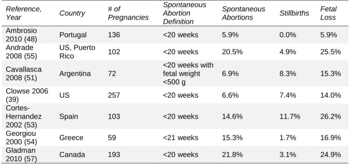
Presently, the majority of SLE pregnancies, 66 to 95% depending on the study population, result in a live birth (6, 7, 39, 48-54); however, previous research has shown that women with SLE have poorer pregnancy outcomes compared to women in the general population. In women with SLE, the frequency of spontaneous miscarriage (loss of pregnancy prior to 20 weeks gestation) ranges from 6 to 22%, and the frequency of stillbirth (loss of pregnancy after 20 weeks gestation) ranges from 0 to 12% (Table 1) (7, 39, 48-57). Comparatively, the CDC estimates that 17% of pregnancies to women of all ages in the US end in a fetal loss (spontaneous miscarriage or stillbirth) (58). The majority of the data for SLE pregnancies comes from prospective cohorts, which can under-count early pregnancy loss if a woman suffers the loss before she visits her physician to be enrolled in the study.
Table 1. Frequency of spontaneous abortions, stillbirths and fetal loss in SLE pregnancy
Reference,
Year Country
# of Pregnancies Spontaneous Abortion Definition Spontaneous
Abortions Stillbirths
Fetal Loss
Ambrosio
2010 (48) Portugal 136 <20 weeks 5.9% 0.0% 5.9%
Andrade 2008 (55)
US, Puerto
Rico 102 <20 weeks 20.5% 4.9% 25.5%
Cavallasca
2008 (51) Argentina 72
<20 weeks with fetal weight <500 g
6.9% 8.3% 15.3%
Clowse 2006
(39) US 257 <20 weeks 6.6% 7.4% 14.0%
Cortes-Hernandez 2002 (53)
Spain 103 <20 weeks 14.6% 11.7% 26.2%
Georgiou
2000 (54) Greece 59 <21 weeks 15.3% 1.7% 16.9%
Gladman
2010 (57) Canada 193 <20 weeks 21.8% 3.1% 24.9%
General risk factors for spontaneous miscarriage, in women with or without SLE, include
disease activity and co-morbidities such as kidney disease, hypertension, antiphospholipid syndrome, heart failure, and pulmonary disease are associated with an increased risk for fetal loss (4, 7, 53, 55, 59).
Preterm Birth
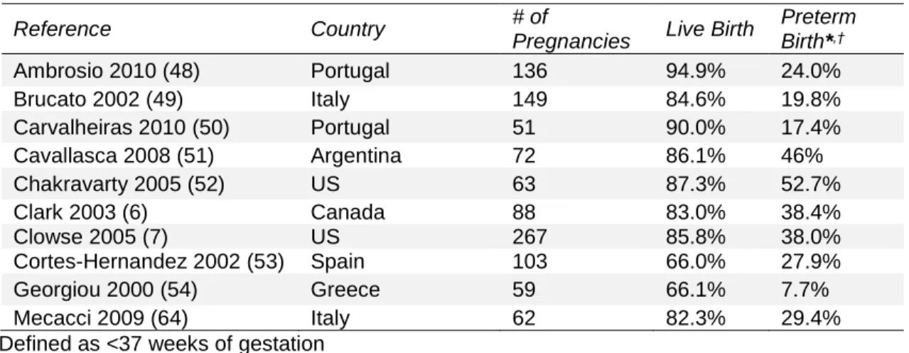
Preterm birth is defined as delivery prior to 37 completed weeks of gestation. In the general population, approximately 12% of births in the United States are preterm (60). The rate of preterm births in the US increased by more than 20% from 1990 to 2006, although more recent reports indicate that the rate is slowly declining after its peak in 2006 (61). A racial disparity is apparent in the rate of preterm birth, with 17.5% of births to non-Hispanic black mothers in 2008 being preterm, compared to 11.1% and 12.1% in non-Hispanic white and Hispanic mothers, respectively (61). Although the majority of preterm infants survive, the risks of mortality and morbidity (such as neurological, respiratory, gastrointestinal, and kidney systems development) are higher in preterm infants compared to infants born at term (62).
Within the overarching outcome of preterm birth, there are subtypes: spontaneous preterm labor, premature rupture of membranes, and medically indicated preterm birth. In the general population, the largest proportion of preterm births is due to spontaneous labor (45%), and the pathways that lead to each type of preterm delivery vary (62), though some overlap. Medically indicated preterm births occur when a baby is intentionally delivered before 37 weeks due to medical complications in the mother or the fetus. In addition to the high burden of morbidity due to preterm birth, the costs associated with a preterm delivery increase substantially for each week prior to term that an infant is delivered (63). Once study reported that for medical costs during the first 5 years of life, infants born at <28 weeks gestation accrued almost $23,000 more in hospital utilization costs compared to term infants, and infants born at 28 to 31 weeks gestation had costs almost $19,000 greater than term infants (63).
preterm birth include hypertension, increased disease activity during pregnancy, the presence of anti-cardiolipin (aCL) antibodies, and prednisone use either prior to or during pregnancy (6, 7, 51-54). Although data are limited, one study reported that 75% of preterm births in women with SLE were medically indicated (50). Reasons for a medically indicated preterm delivery in SLE, either by a
caesarean section or induction of labor, include maternal high blood pressure, pre-eclampsia, proteinuria, decreased amniotic fluid volume, intrauterine growth restriction, and HELLP syndrome (hemolysis, elevated liver enzymes, and low platelets), a complication of eclampsia (6).
Table 2. Frequency of preterm birth in SLE pregnancy.
Reference Country # of
Pregnancies Live Birth
Preterm Birth*,†
Ambrosio 2010 (48) Portugal 136 94.9% 24.0%
Brucato 2002 (49) Italy 149 84.6% 19.8%
Carvalheiras 2010 (50) Portugal 51 90.0% 17.4%
Cavallasca 2008 (51) Argentina 72 86.1% 46%
Chakravarty 2005 (52) US 63 87.3% 52.7%
Clark 2003 (6) Canada 88 83.0% 38.4%
Clowse 2005 (7) US 267 85.8% 38.0%
Cortes-Hernandez 2002 (53) Spain 103 66.0% 27.9%
Georgiou 2000 (54) Greece 59 66.1% 7.7%
Mecacci 2009 (64) Italy 62 82.3% 29.4%
*Defined as <37 weeks of gestation †Frequency among live birth infants
Low Birth Weight for Gestational Age
Low birth weight is defined as a birth weight of <2500 g, and the CDC estimates that 8% of births in the US general population in 2012 were low birth weight (66). Low birth weight infants are at increased risk for infant mortality (death within 1-year after birth) than heavier infants (53.05 deaths per 1,000 births vs. 2.21 deaths per 1,000 births, respectively) (67) and are at increased risk for complications such as respiratory distress syndrome, intraventricular hemorrhage, and necrotizing enterocolitis (68). It has been estimated that 5 to 39% of infants born to mothers with SLE are classified as low birth weight (48, 51, 53, 54). Hypertension and aCL antibodies are risk factors for delivering a low birth weight infant in SLE (53).
A methodological issue of low birth weight is that it is largely dependent on the duration of
gestation, as approximately half of infants who are classified as low birth weight are born preterm (47). An alternative measure of fetal growth that takes gestational age into consideration is classifying infants as
based on their gestational age and sex. Among infants born to mothers with SLE, 16 to 23% are SGA (39, 57). Women with SLE have an increased risk of delivering a SGA infant, with one study finding the odds of delivering a SGA infant to be 2.5 times that of women in the general population (OR=2.54; 95% CI: 1.42, 4.55), when adjusted for instrumentation to assist in the delivery and caesarean section (65). The prevalence of SGA is higher among women with active kidney disease during pregnancy (defined as the presence of hematuria, pyuria, casts, and proteinuria), compared to women without active kidney disease (57).
Gestational Weight Gain
Gestational weight gain (GWG) is the amount of weight a mother gains throughout her pregnancy and is composed of maternal and fetal factors. For fetal factors, the average weight gained is 4.8
kilograms, with the fetus itself comprising an average of 3.3 kilograms (kg) of the weight gained. The remaining fetal weight gained is from the placenta (~0.7 kg) and amniotic fluid (~0.8 kg). For maternal factors, the average weight gained is 7 kilograms, largely due to increase in fat (~4.0 kg), blood volume (~1.2 kg) and extracellular fluid (~1.2 kg) (69).
The greatest increase in weight in nonfat tissues is due to water, with an increase of 6 to 7 liters of water (70). Fat is used to meet a mother’s metabolic requirements and as an energy source throughout pregnancy in times of deprivation as well as for lactation postpartum. The largest amount of maternal fat is accrued during the second trimester, but fat begins to be deposited early in pregnancy (69). The increase in body fat during pregnancy has been found to vary with pre-pregnancy BMI, with one study noting that women classified as overweight and obese had a minimal increase in total body fat during pregnancy. The study also found that women who met the 1990 IOM guidelines for gestational weight gain did not amass excessive amounts of fat (71).
The pattern of weight gain during pregnancy varies greatly between women, and is most variable for obese women. One study found that the average weekly weight gain for the second and third
In 1990, the Institute of Medicine (IOM) published guidelines on the ideal weight gain during pregnancy based on pre-pregnancy BMI, and these guidelines were updated in 2009 due to the concern of rising obesity rates in the population (Table 3) (8).
Table 3. 2009 IOM Recommendations for Gestational Weight Gain in the General Population (8)
Pre-Pregnancy BMI (kg/m2)
Total Weight Gain (kg)
2nd and 3rd Trimester Rates of Weight Gain, mean kg/wk (range)
Underweight (<18.5) 12.5-18 0.51 (0.44-0.58) Normal weight (18.5-24.9) 11.5-16 0.42 (0.35-0.50) Overweight (25.0-29.9) 7-11.5 0.28 (0.23-0.33)
Obese (≥30.0) 5-9 0.22 (0.17-0.27)
The vast majority of women do not meet these guidelines for weight gain, with one study finding 53% of mothers gain more than the recommended weight, 17% gain less than the recommended weight, and only 31% gain the ideal amount of weight (11). In this analysis, women who were classified as overweight or obese were at increased risk of gaining more than the recommended amount of weight during pregnancy, compared to women with normal BMI. The proportion of women who are not meeting the guidelines for GWG is increasing. In 1997, 39.6% of women who had a pre-pregnancy BMI in the normal range exceeded the IOM’s recommendations for GWG, which increased to 46.3% in 2007. The increase in the proportion of women exceeding the IOM weight gain recommendations has also been seen in women classified as being overweight or obese prior to pregnancy (8).
Cardiovascular Health and Pregnancy
The American Heart Association (AHA)’s 2020 Impact Goals included the development of the concept of “ideal cardiovascular health,” which focuses on primordial prevention and is composed of seven modifiable cardiovascular metrics: health factors (glucose, cholesterol, and blood pressure) and health behaviors (body mass index, physical activity, diet, and cigarette smoking; Table 4) (95). Meeting these metrics for ideal cardiovascular health is associated with a lower risk of cardiovascular disease, lower cardiovascular mortality rates, and lower all-cause mortality.
Table 4. American Health Association Definitions of Poor, Intermediate, and Ideal Cardiovascular Health (95)
Goal/Metric Poor Health Intermediate Health Ideal Health Current smoking Yes Former ≤12 months Never or quit >12
months Body mass index ≥30 kg/m2 25-29.9 kg/m2 <25 kg/m2 Physical activity None 1–149 minutes/week moderate
intensity
or 1–74 minutes/week vigorous intensity
or 1–149 minutes/week moderate + vigorous
≥150 min/week moderate intensity or ≥75 minutes/week vigorous intensity or≥150 minutes /week Healthy diet score 0–1 Components 2–3 Components 4-5 Components Total cholesterol ≥240 mg/dL 200–239 mg/dL or treated to
goal
<200 mg/dL Blood pressure Systolic ≥140
Or
Diastolic ≥90 mm Hg
Systolic 120–139 or Diastolic
80–89 mm Hg or treated to goal <120/<80 mm Hg
Fasting plasma glucose
≥126 mg/dL 100–125 mg/dL or treated to goal
<100 mg/dL
The guidelines for ideal total cholesterol, blood pressure, and fasting glucose are in agreement with the definitions used by the Third Adult Treatment Panel of the National Cholesterol Education Program (96), the Seventh Joint National Committee of the National Blood Pressure Education Program (97), and the American Diabetes Association (98), respectively.
proinflammatory proteins, and obese women are more likely to enter pregnancy in a state of subclinical inflammation than non-obese women (102-104). Maternal obesity increases the risk of delivering an infant who is macrosomic (>4000 g) or large for gestational age (105-107).
Studies have shown that hypertension is a risk factor for preterm birth (108, 109), even in studies where pregnancies affected by preeclampsia were removed from the study population, with one study noting the risk of preterm birth increased 29% for each 10 mm Hg increase in diastolic blood pressure (110). Additionally, chronic hypertension is associated with fetal growth restriction and low birth weight (108, 109, 111), with the risk of preterm small for gestational age births being 5.5 times greater than in woman without hypertension and the risk of term small for gestational age births being 1.5-1.7 times greater than in women without hypertension (109).
Previous research, although limited, has demonstrated that increased total cholesterol during the first trimester is associated with preterm birth, and it has also been suggested that the association of cholesterol and preterm birth may be modified by maternal inflammation (110, 112, 113). One study reported the risk of preterm birth at <34 weeks to be 2.8 times greater among women with
hypercholesterolemia than women with normal cholesterol (112), and another study estimated a 24% increase in the risk of preterm birth for each 40 mg/dL increase in cholesterol (110).
It has been theorized that maternal risk factors for cardiovascular disease may also be risk factors for fetal growth restriction and fetal programming (114). As SLE is a chronic inflammatory disease, it will be important to study the way these cardiovascular health factors affect preterm birth and fetal growth during SLE pregnancies. Additionally, understanding how SLE specific components of the disease, such as disease activity and autoantibodies, may modify the association is important in improving the outcomes of infants born to mothers with SLE.
SLE Disease Activity during Pregnancy
(BILAG) index (119, 120), Systemic Lupus Activity Measure (SLAM) Index (121, 122) and European Consensus Lupus Activity Measurement (ECLAM) index (123-125).
The effect of pregnancy on disease activity and flares in SLE has long been debated. Previous research has found that between 19 and 68% of women with SLE experience a flare during pregnancy (7, 39, 50-53, 57, 126-129). Risk factors for flare during pregnancy include active disease at conception, prednisone use, kidney disease and previous flares (52, 53, 57).
When compared to SLE patients who are not pregnant, there are conflicting results about the effect pregnancy has on disease activity (Table 6). Some studies report an increased rate of flares during pregnancy, while others report no difference in disease activity during pregnancy or post-partum. The rate of flares per person-months in pregnancy ranges from 0.06 – 0.14, compared to 0.04 – 0.05 in non-pregnant SLE patients (126, 127, 130, 131). A study by Lockshin et al. (132) analyzed flare
characteristics of pregnant and non-pregnant SLE patients, including laboratory values (urine protein, anti-dsDNA, complement, hemoglobin, etc.) and symptoms (rash, fever, serositis, arthritis, neurologic events, etc.), and did not find a difference between women who were pregnant and women who were not (Table 5).
Table 5. Incidence of flares in SLE pregnancy per person-month
Reference Country Pregnancies (n) Rate of Flares per Person-Month Garsenstein 1962 (130) US 33 32 weeks pre-pregnancy: 0.04
0-20 weeks of pregnancy: 0.13 21-40 weeks of pregnancy: 0.07 0-8 weeks postpartum: 0.27 9-40 weeks postpartum: 0.04 Mintz 1986 (130) Mexico 102 Pregnant patients: 0.06
Non-pregnant patients: 0.04
Petri 1991 (126) US 40 During pregnancy: 0.14
Post-partum: 0.05
Non-pregnant patients: 0.05 Ruiz-Irastorza 1996 (127) UK 78 Pregnant patients (any time): 0.08
1st trimester: 0.008 2nd trimester: 0.15 3rd trimester: 0.07 1-year postpartum: 0.15 Non-pregnant patients: 0.04 Wong 1991 (131) China 29 Pregnant patients: 0.08
Non-pregnant patients: 0.04
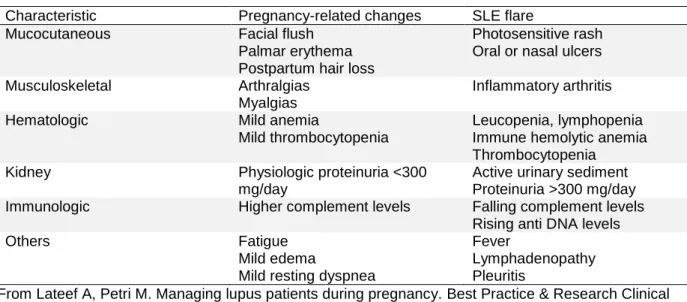
studies. Many previous studies were also limited by a small sample size, which reduced power to determine differences in the rate of flares between pregnant and non-pregnant patients. Additionally, it can be challenging to differentiate between pregnancy related changes and changes that are related to SLE flares. Lateef et al. (37) constructed a comparison of pregnancy-related changes and flare
characteristics to assist in distinguishing between the signs and symptoms of these two conditions (Table 6).
Table 6. Differentiating pregnancy-related changes from SLE flares during pregnancy (37). Characteristic Pregnancy-related changes SLE flare
Mucocutaneous Facial flush
Palmar erythema Postpartum hair loss
Photosensitive rash Oral or nasal ulcers
Musculoskeletal Arthralgias
Myalgias
Inflammatory arthritis
Hematologic Mild anemia
Mild thrombocytopenia
Leucopenia, lymphopenia Immune hemolytic anemia Thrombocytopenia
Kidney Physiologic proteinuria <300
mg/day
Active urinary sediment Proteinuria >300 mg/day Immunologic Higher complement levels Falling complement levels
Rising anti DNA levels
Others Fatigue
Mild edema
Mild resting dyspnea
Fever
Lymphadenopathy Pleuritis
From Lateef A, Petri M. Managing lupus patients during pregnancy. Best Practice & Research Clinical Rheumatology 2013;27(3):435-47 (37).
Research Gaps
Although pregnancy outcomes to women with SLE have improved in recent years, the prevalence of preterm birth and infants born small for gestational age remain two- to six-times greater in women with SLE, as compared to women in the general population (65). Many well-researched aspects of pregnancy in the general, “healthy” population remain unstudied in the population of women with SLE, and there are gaps in the literature relating to risk factors for poor pregnancy outcomes in SLE and the effect on pregnancy on the SLE disease course.
One aspect of pregnancy that has yet to be studied in a population of women with SLE is
recommendations to be specific to a woman’s pre-pregnancy body mass index (BMI) and co-morbidities. Although the 2009 committee was not intended to develop GWG guidelines for specific diseases or conditions, a noticeable gap in the literature was the availability of data on the weight gain patterns in patients with autoimmune diseases, namely SLE. It is not presently known if women with SLE are gaining the appropriate amount of weight and what factors may affect weight gain in these women. Additionally, it has yet to be determined if the IOM guidelines for weight gain in the general population are appropriate for women with SLE.
The impact preconceptional cardiovascular health has on the occurrence of poor pregnancy outcomes in women with SLE is presently unknown. The American Heart Association (AHA)’s 2020 Impact Goals included the development of the concept of “ideal cardiovascular health,” which focuses on primordial prevention and is composed of seven modifiable cardiovascular metrics: health factors
(glucose, cholesterol, and blood pressure) and health behaviors (body mass index, physical activity, diet, and cigarette smoking) (95). Longitudinal cohort studies have reported that hypertension, dyslipidemia, and obesity are common in SLE, afflicting 30-60% of patients (99-101). Maternal cardiovascular health at conception and during the beginning of pregnancy has implications for the in utero environment, with obesity, hypertension and increased total cholesterol being associated with an increased risk of preterm birth and small for gestational age infants (108-113). As SLE is a chronic inflammatory disease, it will be important to study the way these cardiovascular health factors affect preterm birth and fetal growth during SLE pregnancies. Additionally, understanding how SLE specific components of the disease, such as disease activity and autoantibodies, may modify the association is important in improving the outcomes of infants born to mothers with SLE.
This dissertation addressed several of the gaps in the literature relating to pregnancy in women with SLE and determined:
1. if women with SLE are meeting the recommended Institute of Medicine guidelines for gestational weight gain,
2. factors associated with not meeting or exceeding gestational weight gain guidelines in SLE, 3. the proportion of women with SLE meeting the American Heart Association’s classification of
cardiovascular health in a cohort of pregnant SLE patients,
4. the effects of poor and intermediate cardiovascular health on pregnancy outcomes in SLE, and
CHAPTER 3: METHODS Study Population
The Hopkins Lupus Pregnancy Cohort is a subset of the Hopkins Lupus Cohort, which has prospectively followed patients with SLE since 1987, with data available through February 6, 2015. Patients meeting the ACR or SLICC criteria for SLE (12, 13, 133) were eligible for enrollment in the cohort following informed consent. Patients enrolled in the Hopkins Lupus Cohort and SLE patients seen in the Hopkins Obstetrics Clinics were automatically referred to the Hopkins Lupus Pregnancy Cohort. Outside of Johns Hopkins Hospital, local patients were referred by their local rheumatologists, the Maryland Lupus Foundation, and self-referral (126). Patients who were not pregnant were seen on a quarterly basis at the Lupus Center in Baltimore, Maryland by a single rheumatologist.
Pregnant women were seen every on average every 4-6 weeks throughout their pregnancy. At the first clinic visit, patients were given a full medical examination and self-reported their obstetrical history, including previous abortions (spontaneous and elective) and previous deliveries. During each subsequent visit, a patient’s weight was recorded, lupus disease activity was measured, medications were updated and laboratory tests were conducted. Laboratory tests included complete blood count (complement levels, autoantibodies, cholesterol and glucose) and urinalysis. Pregnancy outcome data were collected from women at the first postpartum visit to the Lupus Clinic or by telephone or email if a woman did not continue her medical care at the Lupus Clinic. Multiple pregnancies per patient were allowed in the analysis.
Data Collection and Measurement
morbidities (hypertension, diabetes, proteinuria, lupus nephritis), laboratory tests, patient demographics, SLE disease history, SLE disease activity, and treatment history were collected in the larger Hopkins Lupus Cohort.
Exposure Classification
Specific Aim 1 was an exploratory analysis of adherence to gestational weight gain and correlates of adherence. There were no specific exposures of interest.
In Specific Aim 2, the exposure of interest was pre-conceptional cardiovascular health defined according to three of the American Heart Association (AHA)’s metrics, body mass index (BMI), total cholesterol, and blood pressure, using the following criteria: BMI: (1) poor health (obese): ≥30 kg/m2; (2) intermediate health (overweight): 25-29.9 kg/m2; (3) ideal health (underweight/normal weight): <25 kg/m2;
total cholesterol: (1) poor health: ≥240 mg/dL; (2) intermediate health: 200–239 mg/dL or treated to goal; (3) ideal health: <200 mg/dL without treatment; blood pressure: (1) poor health: systolic ≥140 or diastolic
≥90 mm Hg; (2) intermediate health: systolic 120–139 or diastolic 80–89 mm Hg or treated to goal; (3) ideal health: <120/<80 mm Hg without treatment. Each metric was coded as a categorical variable, with “ideal health” as the referent group. Due to small sample size, poor health and intermediate health were collapsed into one exposure category for total cholesterol and blood pressure, with ideal health remaining the referent group. Each metric was also analyzed as a continuous variable. BMI, total cholesterol, and blood pressure at the most recent clinic visit in the one-year prior to conception were used to classify patients’ cardiovascular health. If a clinic visit prior to conception was unavailable, the first measurement taken during the first trimester served as a surrogate for preconception health.
Outcome Classification
In Specific Aim 1, the outcome of interest was the proportion of women with SLE who met the 2009 IOM guidelines for GWG based on pre-pregnancy BMI. Pre-pregnancy weight was defined as the most recent weight recorded at a visit within 12 months prior to pregnancy or, if not available, in the first trimester. The final pregnancy weight was the weight recorded closest to birth in the third trimester. Observed weight gain was calculated as the difference in the first and final weight measurement. The estimated total weight gain was calculated to account for variations in the timing of the first and final weight: (observed weight gain / weeks of gestation between weight measurements) x 40 weeks. Estimated total weight gain was classified according to IOM guidelines based on a woman’s pre-pregnancy BMI: underweight (<18.5 kg/m2), normal weight (18.5-24.9 kg/m2), overweight (25.0-29.9 kg/m2), obese (≥30 kg/m2). The guidelines recommend the following total weight gain during pregnancy (8):
underweight: 12.5-18 kg
normal weight: 11.5-16 kg
overweight: 7-11.5 kg
obese 5-9 kg.
Total weight gain below the recommendations was considered inadequate weight gain, and total weight above the recommendations was considered excessive weight gain.
percentile of birth weight for gestational age: <10th percentile (small for gestational age; SGA) and >90th percentile (large for gestational age; LGA).
In Specific Aim 3, the outcome was time to disease flare (allowing for multiple flares), with a flare classified according to two disease activity indices. PGA is a disease activity index ranging from 0 to 3, with 0 being no activity and 3 being severe disease activity (135). SELENA SLEDAI is a weighted disease activity index for activity related to SLE present within the previous 10 days, with a score range of 0 to 105 (118). Flares during follow-up were classified as:
1. Change in PGA ≥1 from previous visit
2. Change in SELENA SLEDAI ≥4 from the previous visit.
Covariates
Maternal age: Maternal age at the time of conception was analyzed as a continuous variable and categorized as ≤30 and >30 years in descriptive analyses in Aim 1.
Race: Patient race was classified as black and non-black.
Maternal education: Maternal education was based on self-reported years of education and categorized as ≤12 years, 13-16 years, and >16 years.
Duration of SLE: Duration of SLE at the time of conception was analyzed as a continuous variable and categorized as ≤5 and >5 years.
Infant Delivery Date: In Aims 1 and 2, infant delivery date (categorized as prior to January 1999 or between January 1999-February 2015) was considered a variable of interest due to changes in SLE prescribing patterns and general population shifts in BMI over time.
Medication Use during Pregnancy: Medication use during pregnancy was classified as (1) yes or (0) no for the following medications: anti-malarial, immunosuppressants, and prednisone. High-dose prednisone use was further classified as prednisone ≥15 mg/day during pregnancy. In Aim 3, prednisone and anti-malarial (hydroxychloroquine) use were analyzed as time-varying covariates.
Organ System Damage: Organ system damage at conception was classified according to the Systemic Lupus International Collaborating Clinics/American College of Rheumatology (SLICC/ACR) Damage Index (SDI), with a score of ≥1 indicating any organ system damage.
Renal Involvement: Renal involvement during pregnancy was defined as renal Lupus Activity Index score >1 at any time during pregnancy.
Autoantibodies during Pregnancy: Autoantibodies during pregnancy included the presence of any of the following: low complement 3 (C3), low complement 4 (C4), and anti-double stranded DNA (anti-dsDNA; ever positive).
Elevated Serum Creatinine: Elevated serum creatinine during pregnancy was defined as serum creatinine ever >1 mg/dl.
Study Analysis Plan
Specific Aim 1: To estimate the proportion of pregnant women with systemic lupus erythematosus (SLE) who meet the Institute of Medicine (IOM) guidelines for gestational weight gain (GWG) and to determine factors associated with adherence to IOM guidelines for GWG.
Sub-Aim 1A: To estimate gestational weight gain trajectories for women with SLE.
fixed effects included a linear effect for time, quadratic effect for time, BMI group, and interaction for BMI group and time.
Specific Aim 2: To estimate the effect of preconceptional cardiovascular health, as measured by blood pressure, total cholesterol and body mass index, on preterm birth and fetal growth (birth weight for gestational age z-score) in women with SLE.
Unadjusted differences in the prevalence of preterm birth, SGA, and LGA among live births by pre-conceptional cardiovascular health were analyzed descriptively by Fischer’s exact test. Differences in mean gestational age and mean birth weight for gestational age z-score by pre-conceptional
cardiovascular health were analyzed by ANOVA. Multivariable logistic regression models estimated odds ratios (OR) and 95% confidence intervals for the association of each maternal cardiovascular health factor and categorical pregnancy outcomes of interest (preterm birth, SGA, and LGA). Multivariable linear regression models estimated associations of each maternal cardiovascular health factor with continuous outcome measures (gestational age at birth and birth weight for gestational age z-score). To account for the correlation between outcomes that would occur from patients contributing more than one pregnancy to this analysis, generalized estimating equations (GEE) with an exchangeable correlation structure were used (136). Confounders were assessed based on combined directed acyclic graph (DAG) minimally sufficient set that was reduced based on a 10% change in beta (β) estimates. Models with BMI as the exposure were adjusted for prednisone use during pregnancy and patient race, and blood pressure models were adjusted for renal involvement during pregnancy and patient race. For the exposure of total cholesterol, three adjusted models were estimated: 1) adjusted for patient race and prednisone use during pregnancy; 2) adjusted for patient race and anti-malarial use during pregnancy; and 3) adjusted for patient race, prednisone use during pregnancy, and anti-malarial use during pregnancy.
Specific Aim 3: To estimate the effect of pregnancy on disease activity (i.e., disease flares) in SLE using a Cox proportional hazards model.
Sub-Aim 3a: To compare traditional methods for estimating the incidence of disease flares
Sub-Aim 3b: To perform a sensitivity analysis excluding women without a pregnancy from the study population.
All women with SLE in the Hopkins Lupus Cohort between the ages of 15 and 45 were included in the analysis for Specific Aim 3, regardless of pregnancy status. Women with only one measurement of disease activity were excluded as time to event could not be determined for these women. The time of entry into the Hopkins Lupus Cohort was considered the initial measurement for all women. Patients were right censored and removed from the risk set at age 45 (end of reproductive years), menopause (if prior to age 45 years), death, loss to follow-up, or February 6, 2015, the end of follow-up. If patients had a gap of more than one year in study visits, patients were considered lost to follow-up, but were allowed to re-enter the cohort when study visits resumed. The time between when a patient exited and re-re-entered the cohort did not contribute to person-time at risk.
Crude incidence rates were calculated as the observed number of flares / total person-time for each exposure period. Incidence rate ratios and corresponding 95% confidence intervals were calculated for pregnancy vs. unexposed periods and postpartum vs. unexposed periods. The analysis used two separate variations of Cox models to estimate the hazard rate ratio of flares in pregnancy and postpartum periods compared to unexposed periods: the standard counting process Cox proportional hazards model and the stratified Cox model. The counting process Cox proportional hazards model accounted for repeated measures, but did not take into account the order in which events occur. In the stratified Cox model, a stratum for the time interval number was included in the model so a patient was not at risk for a second flare without having experienced a previous flare.
If a woman had more than one pregnancy, all pregnancies (as well as postpartum periods) were included in the analysis. Due to repeated events of flares being counted in the same patient and patients being allowed to exit and re-enter the analytic cohort, 95% confidence intervals were estimated with 1,000 bootstrap replications sampled with replacement (138). Using the same model, hazard ratios were
CHAPTER 4: GESTATIONAL WEIGHT GAIN IN PREGNANT WOMEN WITH SYSTEMIC LUPUS ERYTHEMATOSUS
Background
In 1990, the Institute of Medicine (IOM) published guidelines for ideal weight gain during pregnancy based on pre-pregnancy BMI. These guidelines were updated in 2009 due in part, to the concern of rising obesity rates in the population (8). Although the 2009 committee was not intended to develop gestational weight gain (GWG) guidelines for specific diseases or conditions, a noticeable gap in the literature was the availability of data on the weight gain patterns in patients with autoimmune
diseases. Of particular interest was systemic lupus erythematosus (SLE), a disorder that largely affects women between the ages of 15 and 44 (1). It is not presently known if women with SLE are gaining the appropriate amount of weight and what factors may affect weight gain in these women.
Gestational weight gain is the amount of weight a mother gains throughout her pregnancy and is composed of maternal and fetal products of conception. The average weight gain attributable to fetal components is 4.8 kilograms, comprised of the fetus (~3.3 kilograms), the placenta (~0.7 kilograms) and amniotic fluid (~0.8 kilograms). For maternal components, the average weight gained is 7 kilograms, largely due to increase in fat (~4.0 kilograms), blood volume (~1.2 kilograms) and extracellular fluid (~1.2 kilograms) (69). The pattern of weight gain during pregnancy varies greatly among women. One study found the average weekly weight gain for the second and third trimesters was higher for underweight and normal weight women, compared to overweight and obese women. Additionally, in this study, all women except obese women had higher weekly rates of weight gain in the second trimester than in the third trimester (72).
with modification by pre-pregnancy BMI (10, 91-94). Among women who are underweight according to their pre-pregnancy BMI, insufficient GWG is associated with an increased risk of preterm birth, and this association weakens as pre-pregnancy BMI increases. Excessive GWG may be associated with preterm birth in women of all pre-pregnancy BMI categories (9, 10). Gestational weight gain also has implications throughout childhood, with excessive weight gain being associated with childhood obesity (94, 139, 140). The vast majority of women in the general population do not meet the IOM guidelines for weight gain, with one study finding that 17% of mothers had inadequate, 31% had adequate, and 53% had excessive weight gain (11). Women classified as overweight or obese are at increased risk of gaining more than the recommended amount of weight during pregnancy, compared to women with normal BMI. Unfortunately, the proportion of women who are exceeding the guidelines for GWG is increasing (11), which is why the IOM committee has called for a paradigm shift in how preconception and prenatal advice concerning weight gain is being delivered to women of childbearing ages. The objectives of this study were to estimate the proportion of women with SLE who meet the IOM guidelines for GWG and to determine correlates of adherence to IOM guidelines for GWG.
Methods
Study population
The Hopkins Lupus Pregnancy Cohort is a subset of the Hopkins Lupus Cohort, which has prospectively followed patients with SLE since 1987, with data available through February 6, 2015 (n=515). Patients meeting the American College of Rheumatology (ACR) or Systemic Lupus International Collaborating Clinics (SLICC) criteria for SLE (12, 13, 133) were eligible for enrollment in the cohort following informed consent. Patients enrolled in the Hopkins Lupus Cohort and SLE patients seen in the Hopkins Obstetrics Clinics were automatically referred to the Hopkins Lupus Pregnancy Cohort. Outside of Johns Hopkins Hospital, local patients were referred by their local rheumatologists, the Maryland Lupus Foundation and self-referral (126). Pregnant women were seen every 4-6 weeks throughout their
conducted. Laboratory tests included complete blood count, complement levels, autoantibodies and urinalysis.
Gestational weight gain
The outcome of interest was the proportion of women with SLE who met the 2009 IOM guidelines for GWG based on pre-pregnancy BMI. Pre-pregnancy weight was defined as the most recent weight recorded at a visit within 12 months prior to pregnancy (average weeks prior to pregnancy: 8.4 weeks, SD: 1.9) or, if not available in the first trimester (n=64, average gestational age: 8.4 weeks, SD: 3.2). The final pregnancy weight was the weight recorded closest to birth in the third trimester (average gestational age: 34.8 weeks, SD: 2.9). Observed weight gain was calculated as the difference in the first and final weight measurement. The estimated total weight gain was calculated to account for variations in the timing of the first and final weight: (observed weight gain / weeks of gestation between weight measurements) x 40 weeks.
Estimated total weight gain was classified according to IOM guidelines based on a woman’s pre-pregnancy BMI: underweight (<18.5 kg/m2), normal weight (18.5-24.9 kg/m2), overweight (25.0-29.9 kg/m2), obese (≥30 kg/m2). The guidelines recommend the following total weight gain during pregnancy (8):
underweight: 12.5-18 kg
normal weight: 11.5-16 kg
overweight: 7-11.5 kg
obese 5-9 kg.
Total weight gain below the recommendations was considered inadequate weight gain, and total weight above the recommendations was considered excessive weight gain.
Covariates
prescribing patterns and general population shifts in BMI over time. Information on medication SLE treatment used during pregnancy included: anti-malarial, immunosuppressants, prednisone, and
prednisone ≥15 mg/day. Clinical characteristics and biomarkers of SLE recorded as ever occurring during pregnancy were: renal involvement (renal Lupus Activity Index >1), elevated serum creatinine (>1 mg/dl), high Physician Global Assessment (PGA ≥2), low complement (C3 and C4), and anti-dsDNA (ever positive). Maternal cumulative organ system damage at conception was measured by the
SLICC/American College of Rheumatology Index (SDI), with a score of ≥1 representing the presence of any organ system damage. Pre-pregnancy blood pressure on the study visit closest to conception in the one-year prior to pregnancy or 1st trimester was classified according to American Heart Association (AHA) criteria for cardiovascular health: poor/intermediate blood pressure: systolic ≥120 or diastolic ≥80 mm Hg or treated to goal; ideal health: <120 and <80 mm Hg without treatment (95). Pre-pregnancy cholesterol on the study visit closest to conception in the one-year prior to pregnancy or 1st trimester was classified according to AHA criteria for cardiovascular health: poor/intermediate cholesterol: ≥200 mg/dL or treated to goal; ideal health: <200mg/dL without treatment (95).
Pregnancy outcomes of interest included gestational age at birth and birth weight for gestational age z-score. Gestational age at birth was based on maternally reported last menstrual period date and date of delivery and categorized as preterm (<37 weeks) and term (≥37 weeks), as well as analyzed as a continuous variable. Birth weight for gestational age z-score was based on US population reference percentiles of birth weight for singleton infants, stratified by infant sex (134). Z-scores in Oken et al. 2003 were calculated based on the distribution of birth weights for all live births born 22 to 44 weeks gestation in the US, 1999-2000, with a potential range of -2.58 to 2.58. Birth weight for gestational age z-score was analyzed as a continuous variable, as well as categorized based on the percentile of birth weight for gestational age: <10th percentile (small for gestational age; SGA) and >90th percentile (large for gestational age; LGA).
Subject selection
Pregnancies without a weight measurement in the one year prior to the last menstrual period pre-pregnancy or during the first trimester, and/or without a weight measurement in the third trimester, were excluded. Of the 421 singleton live births, 291 pregnancies had a weight measurement during the one year prior to pregnancy or during the first trimester, and of these, 211 pregnancies had an additional weight measurement during the third trimester. Live births excluded from the analysis (210 of 421 singleton live births) were more frequently to mothers with a high school education and a pregnancy outcome date prior to 1999. Additionally, excluded births were to mothers with a lower frequency of anti-malarial use during pregnancy and shorter disease duration.
Analysis
Adherence to the IOM recommendations was classified as a categorical variable (inadequate, adequate, or excessive weight gain) based on pre-pregnancy BMI. The percent of women who had inadequate, adequate, or excessive weight gain, based on their pre-pregnancy BMI group, was
estimated, and the mean estimated total weight gain was calculated. An exploratory analysis determined factors associated with not meeting IOM guidelines by Fisher’s exact test of differences in proportions and ANOVA compared differences in means. A generalized logit model analysis with stepwise selection determined predictors of inadequate and excessive weight gain, both compared to adequate weight gain. Generalized estimating equation methods were used to account for the potential correlation of multiple pregnancies per patient being included in the analysis (136). Potential variables were entered into the model if α was <0.2 and remained in the model if α was <0.05. Covariates included in model were race, education, infant delivery year, age at conception, duration of SLE, medication use ever during pregnancy (anti-malarial, immunosuppressants, prednisone, and prednisone ≥15 mg/day), SDI at conception and clinical characteristics ever occurring during pregnancy (renal involvement, elevated serum creatinine, high PGA, low complement, and anti-dsDNA).
Weight trajectories for gestational weight gain were estimated using mixed models. Mixed models include fixed and random effects and are ideal for repeated measures with varying number of
effect for time, BMI group, and interaction for BMI group and time. All analyses were conducted with SAS 9.3 (Cary, North Carolina).
Results
There were 211 pregnancies among 182 women included in the analysis. The majority of pregnancies were to women who were white (59%), with a median age at pregnancy of 30 years and median disease duration of 5 years. Overall, 34% of pregnancies had inadequate weight gain, 24% had adequate weight gain, and 42% had excessive weight gain (Figure 2). Differences were observed by pre-pregnancy BMI. Among underweight women, 67% of pregnancies had inadequate GWG, and 33% had adequate GWG. Among normal weight women, pregnancies were fairly evenly divided, with 30%, 32%, and 38% having inadequate, adequate, and excessive weight gain, respectively. On the other hand, among overweight and obese women, few had inadequate GWG, 51% of both groups had excessive GWG, and only 19% and 7% of overweight and obese women, respectively, gained within the
recommended guidelines. There were nine pregnancies in which the mother lost weight, ranging from 1.5 kg to 16.0 kg; all had BMI in the range of overweight or obese. The mean (SD) estimated total weight gain was 10.9 (3.4) kg for underweight women, 14.7 (6.4) for normal weight women, 12.9 (8.8) for overweight women, and 8.3 (12.4) for obese women.
In exploratory analyses, there were observed differences in adherence to IOM guidelines by race, elevated creatinine during pregnancy, pre-pregnancy blood pressure, and pre-pregnancy BMI (Table 7). The mean pre-pregnancy BMI for patients with inadequate, adequate, and excessive weight gain was 26.9 kg, 23.4 kg, and 26.6 kg, respectively (p=0.004). Of interest, there were no differences in weight gain adherence to IOM guidelines by SLE medication use during pregnancy, and adherence to GWG
guidelines did not appear to correlate with pregnancy outcomes.
In logistic regression models, stepwise selection determined continuous pre-pregnancy BMI and maternal education level were predictors of inadequate and excessive weight gain (Table 8). With each 1 kg/m2 increase in pre-pregnancy BMI, the odds of inadequate weight gain and excessive weight gain both increased 12%. Compared to patients with a greater than college education, patients with a high school education had approximately three times the odds of inadequate weight gain and twice the odds of excessive weight gain.
Figure 3 illustrates the mean predicted change in maternal weight, stratified by pre-pregnancy BMI category (underweight/normal weight, overweight, and obese). Normal weight and underweight women were pooled into one category due to the small number of underweight women. The weight gain trajectory did not change in a sensitivity analysis removing underweight women from the analytic cohort. The weight gain trajectories in normal weight/underweight women and overweight women appear to be similar, with weight increasing steadily throughout pregnancy. The trajectories for obese women, however, were different from normal weight/underweight and overweight women, with a decrease in weight observed at the beginning of pregnancy.
Discussion
In this study of pregnant women with SLE, 34% of pregnancies had inadequate weight gain, 24% of pregnancies had adequate weight gain, and 42% had excessive weight gain, rates similar to those observed in the general population of pregnant women in the United States (141). In a recent analysis of the Pregnancy Risk Assessment Monitoring System (PRAMS) 2010-2011, 21%, 32%, and 47% of women reported having inadequate weight gain, adequate weight gain, and excessive weight gain during
pregnancy, respectively (141). In PRAMS, underweight women and normal weight women had decreased odds of excessive weight gain, while overweight and obese women had increased odds of excessive weight gain (141, 142). Similar patterns were observed in our cohort of SLE women, with the frequency of excessive weight gain lower in normal weight and underweight women than in overweight and obese women.