DESIGN OF β-HAIPRINS AND β-SHEETS FOR MOLECULAR RECOGNITION
Jessica Hyui-Su Park
A dissertation submitted to the faculty of the University of North Carolina at Chapel Hill in partial fulfillment of the requirements for the degree of Doctor of Philosophy in the
Department of Chemistry.
Chapel Hill 2012
Approved by Marcey Waters Matthew Redinbo
Dorothy Erie Kevin Weeks
ii ©2012
iii ABSTRACT
JESSICA HYUI-SU PARK: Design of β-hairpins and β-sheets for Molecular Recognition (Under the direction of Marcey L. Waters)
Protein-nucleic acid interactions are essential in a multitude of biological processes.
Protein interactions with single-stranded DNA are particularly important in DNA replication, repair, and telomere regulation. Previously, our laboratory had designed a β-hairpin dimer, (WKWK)2 which binds ssDNA with a Kd of 3 µM and dsDNA with a Kd of 5 µM. These
results later led to the redesign of a β-sheet peptide from a native protein which displayed 10-fold selectivity for dsDNA but overall lower affinity for ssDNA at a Kd of 20 µM. In this
work, with the insight gained from these studies, a third de novo β-sheet was designed, S123. This new system was found to bind ssDNA with a dissociation constant of 170 nM. Several derivatives were investigated to determine the origins of the marked improvement in binding
affinity. It was found that high β-sheet structure was necessary to achieve the observed nanomolar affinity of S123 to ssDNA.
In another study, the use of the copper(I)-assisted azide-alkyne cycloaddition as a method of β-hairpin stabilization was investigated at several different positions. It was
determined that the CuAAC reaction was a suitable method for locking in β-hairpin structure in peptides possessing the type I’ turn, VNGO and the type II’ turn, VpGO. All cyclic
variants exhibited improved thermal stability and resistance to proteolysis as compared to the
iv
v
ACKNOWLEDGEMENTS
The past five years would not have been possible without the constant love and support of my family and friends. This journey was one I never would have started without
the guidance of my late father who always encouraged and helped me to try my best in all things. Dad, you have sacrificed so much for me and without your example of hard work and
passion, I know that I would not be where I am today.
To my mom, thank you for never ceasing to pray for me every day of my life. You have always displayed strength, patience, and wisdom and for that I can never be thankful
enough.
Brother, “obba”, where do I even start. You are the absolute best friend and sibling
anyone could have ever asked for. Thanks for always impressing on me that the grass can be greener on my side of the fence.
I must thank Professor Marcey Waters for both being a friend and an amazing mentor. Thank you for encouraging me to branch out and learn new things. I greatly appreciate all the opportunities I have come across under your guidance. I could not have
hoped for a better advisor.
To Dr.’s Alexander Riemen, Amanda Stewart, and Dale Wilger – thank you for
vi
Kaiulani Houston, I am not sure how I lived my life without you. There are not many people that know me better than you. Thank you for being such a wonderful friend – for
caring enough to call me out on my faults, for being crazy and silly with me, for listening to my unending complaints, and most importantly for being you.
To the rest of the Waters’ lab, past and present, you have been such an amazing group
of people. I will certainly miss the daily jabs at each other, the attempts at making me laugh (mostly successful), and just the general jovial atmosphere. Thank you for making graduate
school a pleasant experience. I hope I have been a blessing to you all as you have been to me.
Lastly, Rob Lindsey, there is not a man like you in existence anywhere in the world. To have found someone that displays so much patience, love, and understanding towards me is truly a blessing. Thank you for continually encouraging me to be creative, to try new
vii
TABLE OF CONTENTS
LIST OF TABLES ...xii
LIST OF FIGURES ... xvi
LIST OF ABBREVIATIONS ...xxiii
Chapter I. INTRODUCTION ... 1
A. Model systems: design principles of β-hairpins and β-sheets ... 1
B. Targeting ssDNA ... 4
i. Proteins and nucleotides: the importance of protein-ssDNA interactions ... 4
ii. Mimicking the OB-fold ... 9
C. Stabilizing secondary structures for targeting biomolecules ... 10
i. Covalent modifications in peptide chemistry to stabilize secondary structures... 10
ii. A click reaction: the copper(I)-assisted azide-alkyne cycloaddition (CuAAC) in peptidomimetics ... 12
D. Structural characterization ... 20
E. Conclusions ... 23
II. DE NOVO DESIGN OF A THREE-STRANDED β-SHEET PEPTIDE FOR RECOGNITION OF ssDNA ... 29
A. Background and significance ... 29
B. System design ... 31
viii
D. Characterization of the recognition of ssDNA by S123 ... 36
E. Conclusions ... 39
F. Experimental section ... 39
i. Peptide synthesis and purification ... 39
ii. Cyclization of cysteine containing peptides ... 40
iii. DNA ... 40
iv. Isothermal titration calorimetr ... 40
v. Circular dichroism ... 41
vi. NMR characterization ... 41
vii. Determination of fraction folded ... 42
III.MUTANTS OF S123 TO UNDERSTAND NECESSARY MOTIFS FOR ssDNA RECOGNITION ... 52
A. Introduction ... 52
B. Mutant designs ... 52
C. Characterization of mutant peptide-ssDNA interactions ... 55
i. Importance of β-hairpin cyclizations... 55
ii. Importance of Trp19... 59
iii. Importance of residue location ... 61
D. Conclusions ... 64
E. Experimental section ... 65
i. Peptide synthesis and purification ... 65
ii. Cyclization of cysteine containing peptides ... 66
iii. DNA ... 66
ix
v. Circular dichroism ... 69
vi. NMR characterization ... 69
vii. Determination of fraction folded ... 70
IV.DEVELOPMENT OF β-HAIRPIN PEPTIDE CYCLIZATION VIA THE CuAAC REACTION ... 77
A. Background and significance ... 77
i. Disruption of protein-protein interactions ... 77
ii. Previous studies of a well folded β-hairpin peptide, WKWK... 81
B. Development of CuAAC for cyclization of β-hairpins ... 83
C. Conclusions ... 89
D. Experimental section ... 89
i. Peptide synthesis and purification ... 89
ii. Synthesis of tris-(triazolylmethyl)amine ligand ... 90
iii. Optimized reaction conditions for CuAAC ... 91
iv. NMR characterization ... 92
V. INCLUSION OF AN AROMATIC AZIDE FOR CuAAC MEDIATED β-HAIRPIN CYCLIZATION ... 95
A. System design ... 95
B. Structural insights of singly clicked β-hairpins using azidophenylalanine ... 98
C. Prelimary work on double click reactions ... 102
D. Conclusions ... 108
E. Experimental section ... 109
i. Peptide synthesis and purification ... 109
x
iii. Synthesis of diynes ... 111
iv. General CuAAC reaction for peptide cyclization ... 113
v. General double CuAAC reaction for peptide cyclization ... 113
vi. Circular dichroism ... 114
vii. NMR characterization ... 114
viii. Determination of fraction folded... 115
VI.INCORPORATION OF FLEXIBLE AZIDES FOR CuAAC MEDIATED β-HAIRPIN CYCLIZATION ... 129
A. Introduction ... 129
B. System design ... 129
C. Positional impact on structure and stability of CuAAC in WKWK derived peptides with a type II’ turn... 132
i. Influence on β-hairpin structure of the CuAAC mediated cyclization in the hydrogen bonded position at residues 3 and 10 ... 132
ii. Influence on β-hairpin structure of the CuAAC mediated cyclization in the non-hydrogen bonded position as residues 2 and 11... 134
iii. Influence on β-hairpin structure of the CuAAC mediated cyclization in the terminal position at residues -1 and 13 ... 136
iv. Thermal and proteolytic stability of type II’ turn CuAAC cyclized peptides ... 138
D. Positional impact on structure and stability of CuAAC in WKWK derived peptides with a type I’ turn ... 140
xi
ii. Influence on β-hairpin structure of the CuAAC
mediated cyclization in the non-hydrogen bonded position ... 143
iii. Influence on β-hairpin structure of the CuAAC mediated cyclization in the terminal position ... 146
iv. Thermal stability of type I’ turn CuAAC cyclized peptides ... 148
v. Recognition of ATP by CuAAC cyclized peptide ... 149
E. Preliminary results of varying the chain length of azidolysine ... 150
F. Preliminary results of the CuAAC cyclization with WKFK-NG ... 154
G. Conclusions ... 156
H. Experimental section ... 157
i. Peptide synthesis and purification ... 157
ii. Cyclization of peptides using a disulfide bridge ... 158
iii. Synthesis of AzK, AzO, AzB, and AzP ... 158
iv. General CuAAC reaction for peptide cyclization ... 159
v. Circular dichroism ... 159
vi. NMR characterization ... 162
vii. Determination of fraction folded ... 162
viii. Peptidase concentration ... 208
ix. Peptidase degradation reactions ... 208
x. Fluorescence quenching experiments with ATP ... 213
xii
LIST OF TABLES
Table
2.1 Fraction folded for β-hairpin controls and S123……….…..36
2.2 Proton chemical shift assignments for Strand 1………..43
2.3 Proton chemical shift assignments for Strand 2……….…...44
2.4 Proton chemical shift assignments for Strand 3………..45
2.5 Proton chemical shift assignments for S12C………....…....46
2.6 Proton chemical shift assignments for S12………..………...…..47
2.7 Proton chemical shift assignments for S22C………...…..…….……..48
2.8 Proton chemical shift assignments for S23………...………..……..49
2.9 Proton chemical shift assignments for S1………..………….……..50
3.1 Sequences of mutants of S123………..………..……….…….……55
3.2 Parameters characterizing the mutant interactions with ssDNA………...…56
3.3 Fraction folded for β-sheet mutants………..………..………...…….59
3.4 Parameters characterizing peptide interactions with oligo1…………...………..66
3.5 Proton chemical shift assignments for Parent………..………..……..……73
3.6 Proton chemical shift assignments for S12C………..………..………74
3.7 Proton chemical shift assignments for S23C………..………..………75
4.1 CuAAC conditions on NHB-PheN3………...….………..84
5.1 Sequences of β-hairpin peptides using PheN3 and Pra……….96
5.2 Results of aFaF double click reaction……….………...102
5.3 Sequences of aFaF β-hairpin peptides………...…..…..105
xiii
5.5 Proton chemical shift assignments for HB-aF-U………...116
5.6 Proton chemical shift assignments for HB-aF-C………...……117
5.7 Proton chemical shift assignments for NHB-aF-U………118
5.8 Proton chemical shift assignments for NHB-aF-C………...….…119
5.9 Proton chemical shift assignments for Term-aF-U………120
5.10 Proton chemical shift assignments for aF RC………..…121
5.11 Proton chemical shift assignments for Pra RC…………...……….……122
5.12 Proton chemical shift assignments for aFaF………..………..123
5.13 Proton chemical shift assignments for aFaF-Cys………....…124
5.14 Proton chemical shift assignments for aFaF-hex……….……125
5.15 Proton chemical shift assignments for aFaF-hept…………...………126
5.16 Proton chemical shift assignments for aFaF-oct………..……127
6.1 Sequences of WKWK modified peptides for the CuAAC cyclization………..130
6.2 Sequences of WKFK modified peptides for the CuAAC cyclization………....131
6.3 Half-lives of type II’ turn peptides treated with Pronase E………139
6.4 Fraction folded of HB peptides………...……..………..141
6.5 Fraction folded of NHB peptides………..………..…144
6.6 Fraction folded of Term peptides………...…..…..…147
6.7 Binding constants of Term and HB peptides for ATP………...149
6.8 Fraction folded of TFK-AzK-NG peptides………156
6.9 Proton chemical shift assignments for AzK RC……….……163
6.10 Proton chemical shift assignments for WKWK-pG………164
xiv
6.12 Proton chemical shift assignments for HB-pG-U……….166
6.13 Proton chemical shift assignments for HB-pG-C………167
6.14 Proton chemical shift assignments for NHB-pG-U………..……168
6.15 Proton chemical shift assignments for NHB-pG-C………..…169
6.16 Proton chemical shift assignments for Term-pG-U……….……170
6.17 Proton chemical shift assignments for Term-pG-C……….……171
6.18 Proton chemical shift assignments for HB-U………...………172
6.19 Proton chemical shift assignments for HB-C………...………173
6.20 Proton chemical shift assignments for HB-rev-U………....…………174
6.21 Proton chemical shift assignments for HB-rev-C………...…….………175
6.22 Proton chemical shift assignments for NHB-U……….…...………176
6.23 Proton chemical shift assignments for NHB-C………177
6.24 Proton chemical shift assignments for NHB-rev-U……….…178
6.25 Proton chemical shift assignments for NHB-rev-C……….…179
6.26 Proton chemical shift assignments for Term-U………...…180
6.27 Proton chemical shift assignments for Term-C………...181
6.28 Proton chemical shift assignments for Term-rev-U………...….182
6.29 Proton chemical shift assignments for Term-rev-C………183
6.30 Proton chemical shift assignments for Term-pG-AzO-C………184
6.31 Proton chemical shift assignments for Term-pG-AzB-C………185
6.32 Proton chemical shift assignments for Term-pG-AzP-C...………….………186
6.33 Proton chemical shift assignments for FK RC……….………187
xv
6.35 Proton chemical shift assignments for WKFK-Cys……….…………189
6.36 Proton chemical shift assignments for TFK-AzK-U………190
6.37 Proton chemical shift assignments for TFK-AzK-C………191
6.38 Proton chemical shift assignments for TFK-AzO-U………192
6.39 Proton chemical shift assignments for TFK-AzO-C………193
6.40 Proton chemical shift assignments for TFK-AzB-U………194
6.41 Proton chemical shift assignments for TFK-AzB-C………195
6.42 Proton chemical shift assignments for TFK-AzP-U………196
6.43 Proton chemical shift assignments for TFK-AzP-C………197
6.44 Proton chemical shift assignments for HBFK-AzK-U………198
6.45 Proton chemical shift assignments for HBFK-AzK-C………199
6.46 Proton chemical shift assignments for HBFK-AzO-U………200
6.47 Proton chemical shift assignments for HBFK-AzO-C………201
6.48 Proton chemical shift assignments for HBFK-AzB-U……….…202
6.49 Proton chemical shift assignments for HBFK-AzB-C……….…203
6.50 Proton chemical shift assignments for HBFK-AzP-U……….…204
6.51 Proton chemical shift assignments for HBFK-AzP-C……….205
6.52 Proton chemical shift assignments for TFK-AzK-NG-U………206
xvi
LIST OF FIGURES
Figure
1.1 Representation of a β-hairpin………...………...….…...3
1.2 The OB-old………..….…..5
1.3 POT1 bound to ssDNA……….…..6
1.4 Schematic for the hnRPA1 mediated ATR kinase deactivation……….…7
1.5 RPA and TPP1……….….………..8
1.6 Dimer of WKWK………..10
1.7 A minimalistic mimic for dnMAML1………..12
1.8 Scheme for 1,3-dipolar cycloaddition……….………..13
1.9 Amide bond versus 1,4-triazole………..…..15
1.10 Triazole substitution for amprenavir……….……..15
1.11 Triazolamer structures……….………….……..17
1.12 Tachyleptsin I 1,4-triazole substitution……….…..19
1.13 Representation of NMR control peptides……….…..21
1.14 Representation of NMR data for β-hairpins……….………..22
2.1 Structure of WW1 domain and Mut1………30
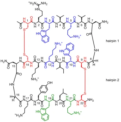
2.2 Structure of S123………...…….…….….……32
2.3 Structure of S123 control peptides………..…..……33
2.4 CD and NMR data for S123 and control peptides………...……….………34
2.5 ITC binding isotherm of S123 with ssDNA……….………37
2.6 Unambiguous NOEs for S123………...………...………42
xvii
2.8 1H NMR of Strand 2………44
2.9 1H NMR of Strand 3………45
2.10 1H NMR of S12C………46
2.11 1H NMR of S12………...………47
2.12 1H NMR of S23C………48
2.13 1H NMR of S23……….………...……...…49
2.14 1H NMR of S123……….…50
3.1 Cartoon representation of mutants used of S123………..…54
3.2 CD and NMR data for mutants of S123………....…………58
3.3 CD of W19L……….………60
3.4 Example ITC curve for W19L titrated with ssDNA………61
3.5 Cartoon representation of YKswitch conformations………62
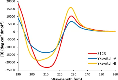
3.6 CD of YKswitch-A and YKswitch-B……….……….………63
3.7 Example ITC curves for YKswitch peptides titrated with ssDNA…………..…64
3.8 Example ITC curves for Parent peptides titrated with ssDNA………69
3.9 Example ITC curves for S12C peptides titrated with ssDNA………..70
3.10 Example ITC curves for S23C peptides titrated with ssDNA………...….71
3.11 1H NMR of Parent………..………...……73
3.12 1H NMR of S12C………..……….…….……74
3.13 1H NMR of S23C………..…...………...75
4.1 Targeting hotspots or allosteric sites……….………....78
4.2 Small molecules developed to target PPIs………79
xviii
4.4 WKWK peptide structure………...……….….81
4.5 Computation model of WKWK………..………..82
4.6 CuAAC catalytic cycle……….83
4.7 Scheme of CuAAC on a β-hairpin and the NMR results………..…85
4.8 Synthesis of β-hairpin peptides and the tris-(triazolylmethyl)amine ligand….…88 4.9 1H NMR of tris-(triazolylmethyl)amine ligand………..….…..91
5.1 WKWK peptide with a type II’ turn……….……..…..96
5.2 aFaF peptide for the double CuAAC……….…………..97
5.3 CD and NMR data for HB-aF peptides………..…..99
5.4 CD and NMR data for NHB-aF peptides………...100
5.5 CD data for Term-aF peptides………...……101
5.6 Cartoon of singly and doubly CuAAC peptides……….101
5.7 Cartoon of configurations for double CuAAC peptides……….103
5.8 CD and NMR data for aFaF peptides……….106
5.9 Lys side-chain chemical shifts for aFaF peptides………..…………108
5.10 1H NMR of 6………..……...…..…………..112
5.11 1H NMR of 7………..….……..112
5.12 1H NMR of 8………..…….…..113
5.13 1H NMR of HB-aF-U……….………..116
5.14 1H NMR of HB-aF-C………..………...117
5.15 1H NMR of NHB-aF-U………..…………..118
5.16 1H NMR of NHB-aF-C………..…………..119
xix
5.18 1H NMR of aF RC………..…………..121
5.19 1H NMR of Pra RC………..……122
5.20 1H NMR of aFaF………..…………123
5.21 1H NMR of aFaF-Cys………..…………124
5.22 1H NMR of aFaF-hex………..………….125
5.23 1H NMR of aFaF-hept………..……...……126
5.24 1H NMR of aFaF-oct………127
6.1 Modifications of WKWK for CuAAC cyclization……….130
6.2 CD and NMR data for HB-pG peptides……….…………132
6.3 Chemical shift differences of Lys side-chain for HB-pG-C………..…………133
6.4 NOEs for HB-pG peptides……….…134
6.5 CD and NMR data for NHB-pG peptides………..135
6.6 NOEs for NHB-pG peptides………..………136
6.7 CD and NMR data for Term-pG peptides………..……...137
6.8 NOEs for Term-pG peptides………..………137
6.9 Thermal stability of HB-pG, NHB-pG, and Term-pG peptides………...138
6.10 CD and NMR data for HB peptides………..…………141
6.11 NOEs for HB peptides………..…142
6.12 CD and NMR data for NHB peptides………...…143
6.13 NOEs for NHB peptides………...…....………145
6.14 CD and NMR data for Term peptides………..146
6.15 NOEs for Term peptides………..……148
xx
6.17 CD and NMR data for Term-pG-AzX peptides………...…...150
6.18 CD and NMR data for TFK-AzX peptides………..152
6.19 CD and NMR data for HBFK-AzX peptides………..……….153
6.20 CD and NMR data for HBFK-NG and TFK-NG peptides……….155
6.21 Thermal denaturation by CD of WKWK-pG modified peptides………160
6.22 Thermal denaturation by CD of WKWK modified peptides…………...……161
6.23 1H NMR of AzK RC………...……….………163
6.24 1H NMR of WKWK-pG………...…………164
6.25 1H NMR of WKWK-pG-Cys………..…165
6.26 1H NMR of HB-pG-U……….….…………166
6.27 1H NMR of HB-pG-C………...……...…167
6.28 1H NMR of NHB-pG-U………...168
6.29 1H NMR of NHB-pG-C………...…169
6.30 1H NMR of Term-pG-U………..…170
6.31 1H NMR of Term-pG-C………...………...171
6.32 1H NMR of HB-U……….…172
6.33 1H NMR of HB-C……….………173
6.34 1H NMR of HB-rev-U………...…...174
6.35 1H NMR of HB-rev-C………..175
6.36 1H NMR of NHB-U………..176
6.37 1H NMR of NHB-C………..…177
6.38 1H NMR of NHB-rev-U………...…178
xxi
6.40 1H NMR of Term-U……….…180
6.41 1H NMR of Term-C………..…...181
6.42 1H NMR of Term-rev-U………..182
6.43 1H NMR of Term-rev-C………..183
6.44 1H NMR of Term-pG-AzO-C……….184
6.45 1H NMR of Term-pG-AzB-C………..…185
6.46 1H NMR of Term-pG-AzP-C………...……186
6.47 1H NMR of FK RC………...……187
6.48 1H NMR of WKFK………...………...……188
6.49 1H NMR of WKFK-Cys………..189
6.50 1H NMR of TFK-AzK-U……….190
6.51 1H NMR of TFK-AzK-C………...……191
6.52 1H NMR of TFK-AzO-U……….…192
6.53 1H NMR of TFK-AzO-C………...…193
6.54 1H NMR of TFK-AzB-U………...……194
6.55 1H NMR of TFK-AzB-C………..…195
6.56 1H NMR of TFK-AzP-U………..…196
6.57 1H NMR of TFK-AzP-C………..197
6.58 1H NMR of HBFK-AzK-U………...…...198
6.59 1H NMR of HBFK-AzK-C………..…199
6.60 1H NMR of HBFK-AzO-U………..200
6.61 1H NMR of HBFK-AzO-C………...…………...…201
xxii
6.63 1H NMR of HBFK-AzB-C………...………203 6.64 1H NMR of HBFK-AzP-U………...204
6.65 1H NMR of HBFK-AzP-C………...…205 6.66 1H NMR of TFK-AzK-NG-U………...……206
6.67 1H NMR of TFK-AzK-NG-C………...…...207 6.68 HPLC traces monitoring Pronase E degradation of Scramble……….209
6.69 HPLC traces monitoring Pronase E degradation of HB-pG-U…...……….…209 6.70 HPLC traces monitoring Pronase E degradation of HB-pG-C………210 6.71 HPLC traces monitoring Pronase E degradation of NHB-pG-U……….210
6.72 HPLC traces monitoring Pronase E degradation of NHB-pG-C……….……211 6.73 HPLC traces monitoring Pronase E degradation of Term-pG-U…...….……211
6.74 HPLC traces monitoring Pronase E degradation of Term-pG-C ………...…212 6.75 HPLC traces monitoring Trypsin degradation of NHB-pG-U……….212 6.76 HPLC traces monitoring Trypsin degradation of NHB-pG-C……….213
xxiii
LIST OF ABBREVIATIONS
A Adenine
Ac Acetyl cap
ACN Acetonitrile
ADP Adenosine diphosphate
Ala, A Alanine
AMP Adenosine monophosphate
Arg, R Arginine Asn, N Asparagine
Asp, D Aspartic acid
ATP Adenosine triphosphate
ATR Ataxia telangiectasia and Rad3-related checkpoint kinase
Azk Azidolysine
Boc t-Butoxycarbonyl
BRCA2 Breast cancer 2 susceptibility protein
C Cytosine
CD Circular dichroism
csDNA Complementary strand DNA CspA Cold shock protein A
CuAAC Copper(I) assisted azide-alkyne cycloaddition
Cu(I); (II) Copper(I); copper(II)
Cu(CH3CN)4PF6 Tetrakis(acetonitrile)copper(I) hexafluorophosphate
xxiv Cys, C Cysteine
Dab Diaminobutyric acid Dab(N3) Azidohomoalanine
Dap Diaminopropionic acid
Dap(N3) Azidoalanine
DBU 1,8-diazobicyclo[5.4.0]undec-7-ene
DCM Dichloromethane
DFT Density functional theory
DIPEA Diisopropylethylamine DMF Dimethylformamide
DMSO Dimethylsulfoxide
DNA Deoxyribonucleic acid
dnMAML1 Dominant-negative fragment of MAML1
DOS Diversity oriented synthesis
dPro, p d-Proline
dsDNA Double stranded DNA
DSS 3-(trimethylsilyl)-1-propanesulfonic acid sodium salt
DTT Dithiothreitol
EDT Ethanedithiol
ESI-MS Electrospray ionization – mass spectrometry
FAM 5-(and -6)-carboxyfluorescein
Fmoc N-9-fluorenylmethoxycarbonyl
xxv Gdn∙HCl Guanidine hydrochloride
Glu, E Glutamic acid Gly, G Glycine
HB Hydrogen bonded
HBTU O-benzotriazole-N,N,N',N'-tetramethyluronium hexafluorophosphate HIF-1α Hypoxia inducible factor 1
H2O Water
HOBt N-hydroxybenzotriazole
HPLC High pressure liquid chromatography HTS High throughput screening
Ile, I Isoleucine
ITC Isothermal calorimetry Leu, L Leucine
Lys, K Lysine
Lys(N3), K(N3) Azidolysine
MeOH Methanol
NaAsc Sodium ascorbate
NH2 C-terminal amide
NHB Non-hydrogen bonded NMP N-methyl-2-pyrrolidinone
NMR Nuclear magnetic resonance
NOE Nuclear Overhauser effect
xxvi NTP Nucleotide triphosphate
OB-fold Oligonucleotide/oligosaccharide binding fold Orn, O Ornithine
Pbf 2,2,4,6,7-Pentamethyl-dihydrobenzofurane-5-sulfonyl
Phe, F Phenylalanine
POT1 Protection of telomeres 1
PPI Protein-protein interaction
Pra Propargylglycine
Pro, P Proline
Ptch1 Patched protein
RCM Ring closing metathesis
RNA Ribonucleic acid RPA Replication protein A
ShhN Sonic hedgehog protein cleavage product
ssDNA Single stranded DNA
T Thymine
TAMRA 5-(and -6)-carboxytetramethylrhodamine
tBu t-Butyl
TFA Trifluoroacetic acide TIPS Triisopropyl silane
TOCSY Total correlation spectroscopy
xxvii Trt Trityl
Tyr, Y Tyrosine
UV-vis Ultraviolet-visible spectroscopy
CHAPTER I INTRODUCTION
A. Model systems: design principles of β-hairpins and β-sheets
Chemists and biologists alike admire the complexity of even the simplest mechanisms
in living systems. Understanding the complex function of biomolecules, in concert or alone, remains an elusive challenge. Often when studying a single interaction or event in vivo a
host of other downstream effects convoluting the results are created leading to more questions to answer. To simplify these challenges, researchers have devised model systems to study complex systems. Model systems are advantageous because they allow for the study
of a single interaction under a controlled environment. Examples of methods for the design of model systems include: recreation of the system outside of the living organism using
native parts, creation of an artificial system, and chemical synthesis of a portion of the system based on design principles as well as a variety of other approaches.1
For many years model systems have been utilized to study protein structure and
function.2 Secondary structure stabilization of α-helices, β-sheets, and β-turns has been a sought after goal to understand native protein folding and function. In addition such insights
will aide in the design of minimalistic functional peptides and novel enzymes for catalysis of new reactions. Design rules for forming stable monomeric and multimeric α-helices have long been established.3,2a,2e However, insights into β-hairpin and β-sheet design have lagged
2
Despite these limitations, over the past two decades, a significant amount of work has been accomplished in the investigation of peptides that form β-hairpins. The first de novo
β-hairpin was reported in 1993 and was based on residues 15-23 of Tendamistat, a protein which inhibits α-amylases.5
Shortly after, the same group reported on the isolation of a
second hairpin from Streptococcal protein G which was found to be 40% folded, native-like β-hairpin structure, via NMR studies.6
These early discoveries and characterizations of isolable β-hairpins/sheets defined structural factors that contributed to numerous reports of stable β-sheets in the late 90’s and early 2000’s.7
β-Hairpins, the smallest secondary structural motif, are composed of two β-strands
oriented in an antiparallel fashion connected by a turn sequence. When designing a stable monomeric hairpin there are several aspects to consider: the sequence of the turn residues, side-chain–side-chain interactions, and the β-sheet propensities of amino acids.4,8 The
residues that nucleate the turn are of particular importance as they can impart proper strand alignment and stability. The common β-turn motifs are the Type I and Type II turns as well
as their mirror images, Type I’ and Type II’ turns, which are very strong turn promoters of structure in β-hairpins.9
These turn types also furnish a right-handed twist which occurs in native antiparallel β-sheets.4
Thus turn sequence known to adopt either Type I’ or Type II’ conformations were used in the β-hairpins and β-sheets in the work presented in this thesis
3
A B
Figure 1.1 Representation of a β-hairpin. (A) Cartoon 3D representation showing the two faces, hydrogen bonded (HB, red) and non-hydrogen bonded (NHB, blue) faces, of a β-hairpin. This figure depicts the hydrogen bonding (red dashes) and possible interactions between cross-strand NHB residues (blue dashes). (B) Example structure of a typical β-hairpin showing the turn nucleating sequence (blue box) and again the hydrogen bonds (red dashes) present within the peptide backbone. Shown here is a type II’ turn sequence, dPro-Gly.
Side-chain–sidechain interactions are also a major contributing factor to hairpin
stability. The residues in the HB sites are known to be packed together due to the inherent twist in a β-hairpin. Thus, the HB residues are typically quite hydrophobic and less flexible
to substitution.10 The residues in the NHB sites tend to have the greatest interaction with
each other as cross-strand residues are pointed towards each other in an antiparallel configuration.11 It is for this reason that β-hairpins have been used to elucidate a variety of
interactions such as hydrogen bonding12, salt bridge formation13, π-π14,13a and cation-π interactions15, interactions between post-translational modifications16, and proton/electron transfer17. Using a small peptide scaffold to understand the driving forces of stability
4
More recently, β-hairpins and β-sheets have been designed for functional purposes.
Robinson and co-workers have made great strides in developing cyclic β-hairpin mimics of α-helices18
and portions of antibodies19 that work to inhibit protein-protein18a and protein-nucleic acid interactions.20 The Schneider lab has taken a different approach using design principles to synthesize β-hairpins that self assemble under environmentally controlled
conditions allowing for biological applications such as drug delivery.21 Our lab has had an interest in targeting nucleotides, in particular the selective recognition of single-stranded
DNA (ssDNA) over duplex DNA.22 Successful developments of minimalistic model systems for this goal will be discussed in further detail in Chapter II.
B. Targeting ssDNA
i. Proteins and nucleotides: the importance of protein-ssDNA interactions
Proteins and nucleotides are essential in all aspects of the central dogma of biology.
From single nucleotide triphosphates to individual strands of ribonucleic acid (RNA) and deoxyribonucleic acid (DNA), nucleotides are involved in far more complex functions than
previously hypothesized. Several of these processes require highly specific interactions with regulatory protein complexes. A portion of the work presented here aims to define the nature of these protein-nucleotide interactions via the design of β-sheet model systems containing a
nucleotide recognition motif. A major theme in the first part of this dissertation will be the understanding of the nature of the specific interactions necessary for targeting ssDNA.
The interactions between proteins and ssDNA are critical in a number of biological processes such as recombination, DNA replication repair23, the cold shock response24, and telomere maintenance.25 Understanding the specifics of why and how these interactions bind
5
physical and chemical properties of ssDNA. The repeating unit of ssDNA, the nucleotide, is composed of a nucleoside base, a sugar, and a negatively charged phosphate tail. All of these
features are exploited by single-stranded binding proteins and the need to manipulate DNA in single stranded form has given rise to a specialized set of proteins.
Nearly all proteins that recognize and interact with ssDNA share a common structural motif called the OB-fold (oligosaccharide/oligonucleotide binding fold) that enables them to bind ssDNA.26, 23c, 23d Structurally, most OB-fold domains consist of a closed β-barrel
consisting of five β-strands with an α-helix cap lying between the third and fourth β-strands (Figure 1.2A). These domains can range from 70 to 150 amino acids and of the known
OB-fold family members there is no strong sequence homology.27 These are, however, easily recognized within proteins due to the distinct structure of these domains. The recognition of
A B
6
ssDNA by OB-fold domains is mediated by aromatic stacking and edge-to-face interactions between prevalent aromatic residues on the solvent exposed face of the protein and the
aromatic bases of the ssDNA.28 The OB-fold motif and aromatic residues are clearly identified in the crystal structure of Cold shock protein A (CspA, Figure 1.2B) and the
co-crystal structure of Protection of telomeres 1 (POT1, Figure 1.3). The negatively charged phosphate backbone of ssDNA provides a handle for electrostatic interactions with OB-fold proteins which contain a number of positively charged basic residues.29 The sugar moiety is
often less utilized by ssDNA binding proteins as it is not readily accessible. However, as presence of the 2’ hydroxyl group on the sugar in RNA is the only chemical difference in
RNA and DNA, proteins that bind ssRNA can be make use of this to increase affinity for ssRNA versus ssDNA.30 These binding sites that are highly selective for ssRNA are typically found in DNA or RNA polymerases.
7
To highlight the importance and complexity of the protein-ssDNA interactions in biological systems, we can turn to Replication protein A (RPA) and downstream protein
players (Figure 1.4). RPA (Figure 1.5A), a classic example of a ssDNA binding protein in eukaryotes, is a heterotrimer consisting of six OB-folds, four of which are used in the binding
event.23,26,31 As the name suggests, RPA is important in replication as it prevents ssDNA from forming secondary structures. RPA is also implicated in telomere maintenance as is POT1 and tripeptidylpeptidase 1 (TPP1, Figure 1.5B).32 RPA and POT1 work in conjunction
to activate or suppress the ATR checkpoint kinase of telomeres, respectively. Activation of the ATR checkpoint halts the cell cycle progression and promotes DNA repair. This process
is mediated by yet another OB-fold containing protein, hnRNPA1, which is bound to telomeric repeat-containing RNA (TERRA) in the early S phase of the cell cycle. However, in late S phase, TERRA levels decline allowing hnRNPA1 to displace RPA. After S phase, it
is presumed that TERRA reaccumulates thereby removing hnRNPA1 and allowing POT1/TPP1 to bind the telomeric ssDNA, which inhibits ATR kinase activation allowing the
cell cycle to continue.33
8
A B
Figure 1.5 RPA and TPP1. Crystal structures of (A) Replication protein A (RPA) which has six OB-folds, PDB: 1L1O, and (B) Tripeptidylpeptidase 1 (TPP1), PDB: 2I46.
RPA also functions to bind ssDNA during the initial phase of homologous recombination, an important process in DNA repair. Again, RPA prevents ssDNA secondary formation thus allowing Rad51, the central protein of recombination34, and its cofactors to
bind. This process has been found to be encouraged by BRCA2, a tumor suppressor protein associated with breast cancer susceptibility.35 This recognition allows error-free repair of
DNA double strand breaks. Dysregulation of the BRCA2 protein or mutations in the gene leads to nonfunctional protein, inhibiting DNA repair and potentially causing uncontrolled cellular replication thereby forming a tumor.36
Other examples of β-hairpin or β-sheet containing proteins involved in DNA repair
include UvrB37, Chlorella virus DNA ligase38, RecA39, and Rad4.40 All of the
aforementioned proteins exemplify the variety of roles that OB-fold domains play in DNA recognition. Dysregulation of any of these proteins can lead to a variety of different disease states, most implicated in cancer. Understanding the interactions between the OB-fold
9
Thus it is important to study and fully understand the critical contacts necessary for ssDNA recognition.
ii. Mimicking the OB-fold
The significance of nucleotides in biology has led our laboratory to explore the use of designed β-hairpins and β-sheets to recognize ATP, ssDNA as well as RNA structures. One
of the earliest works included the design of a 12-residue β-hairpin peptide, WKWK, to bind ATP preferentially over ADP and AMP due to electrostatic interactions.41 The details of
WKWK binding to ATP are covered in Chapter IV. This minimalistic model laid the ground work for understanding how OB-fold containing proteins can interact with ssDNA. It was
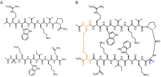
later found that dimerization of the WKWK peptide via a disulfide linkage between N-terminal Cys residues, (WKWK)2 (Figure 1.6), could bind ssDNA sequences with slight selectivity over dsDNA.22a Structural studies of (WKWK)2 were conducted to determine the
contributing factors for ssDNA recognition and were insightful towards the possible modes of dsDNA recognition. It was proposed that dsDNA recognition by the WKWK dimer was
not only mediated by electrostatic interactions between the positively charged basic residues and the negatively charged phosphate backbone but could also be achieved through DNA groove binding.22b In order to test this theory, the WKWK motif was grafted on to a WW
domain which is composed of a three stranded β-sheet.22c By addition of the third strand, the peptide achieved much higher selectivity for ssDNA than (WKWK)2 as it was now too large
10
Figure 1.6 Dimer of WKWK. Structure of WKWK dimerized via N-terminal Cys residues (orange). Trp and Lys residues (blue) are involved in the recognition of ssDNA. Trp residues participate in stacking interaction with the nucleotide bases while the Lys provide electrostatic interactions with the phosphates of ssDNA.
C. Stabilizing secondary structures for targeting biomolecules
i. Covalent modifications in peptide chemistry to stabilize secondary structures The development of orthogonality in peptide synthesis for biological applications dates back to the mid 1950’s when chemists realized the need for methods to functionalize
peptides post or during synthesis. Early and contemporary bioorthogonal methods in peptide synthesis involved heavy use of various protecting group strategies. While these methods are
11
structure and bioconjugation of either a reactive handle or probe. For the purpose of this research only examples of secondary structure stabilization will be mentioned.
The most notable examples of secondary structural stabilization are those of Verdine42 and Arora43, where each worked to stabilize biologically important α-helices
through inclusion of orthogonal chemical functionalities. Verdine and coworkers successfully employed ring closing metathesis (RCM) with Grubbs-I catalyst on an α-helix of the dnMAML1 protein, to give what has been termed a “stapled” peptide (Figure 1.7).42c
dnMAML1 is known to bind the Notch1:Lag1 complex which has been linked to several different cancers, in particular leukemia.44 The RCM between two unnatural side chains
containing terminal alkenes at i and i+4 positions gave a highly structured α-helix and
studies showed that without the preorganized α-helical structure the peptide was unable to efficiently antagonize Notch signaling.
Arora and coworkers used RCM between reactive alkenes at the backbone carbonyl and amide at i and i+4 positions to afford a hydrogen bond surrogate (HBS).43c They aimed
to stabilize a portion of the HIF-1α protein which mediates the cellular response in low tissue oxygen concentrations. The recognition of p300 by HIF-1α is accomplished via helical domains.45 Thus by stabilizing an α-helical portion of the HIF-1α via a HBS, Arora and
coworkers effectively inhibited the interaction. These two cases exemplify the need for robust methods of secondary stabilization. The copper(I)-assisted azide-alkyne cycloaddition
12
A B
Figure 1.7 A minimalistic mimic for dnMAML1. (A) Co-crystal structure of Notch1 (cyan):Lag1 (green):dnMAML1 (magenta) complex bound to DNA, PDB: 2F8X. (B) Scheme of the Grubbs I catalyzed RCM of a fragment of dnMAML1 to yield a structured α-helix that can act like a dnMAML1 mimic.42c
ii. A click reaction: the copper(I)-assisted azide-alkyne cycloaddition (CuAAC) in peptidomimetics
Click chemistry is often misconstrued as the single reaction between an azide and
alkyne but it should be understood that it defines a class of reactions. A click reaction is one in which several requirements are met46: the reaction should 1) be modular, 2) be wide in scope, 3) give very high chemical yields, 4) generate only inoffensive byproducts, 5) be
stereospecific, 6) be physiologically stable, 7) exhibit a large thermodynamic driving force, (8) have high atom economy. Additionally the process should 1) have simple reaction
non-13
chromatographic methods. This chapter serves as a report on the discovery and utilization of one such click reaction, the copper(I)-assisted azide-alkyne cycloaddition (CuAAC), as it
pertains to peptidomimetics.
The inception of the CuAAC began in the early 1900s with Otto Dimroth’s discovery
of the 1,3-dipolar cycloaddition between an organic azide and an acetylene to give a 1,2,3-triazole47 (Figure 1.8). Its mechanism and scope of use was expanded upon some sixty years later by Rolf Huisgen for whom the reaction was named, the Huisgen cycloaddition. Initial
mechanistic studies led to the belief that the reaction proceeded via a diradical intermediate; however, this was met with controversy and was largely disproved by Huisgen who showed
that it was indeed a concerted process.48 Huisgen was also the first to capitalize on the usefulness of this reaction as a clean method to produce a large number of 5-membered heterocycles. This transformation has the advantage of high chemoselectivity since few
functional groups react with azides or alkynes in the absence of other reagents. However, the reaction conditions of the Huisgen cycloaddition require high heat and as such degradation
and safety concerns arose. In addition, this reaction exhibits poor regioselectivity as both the 1,4- and 1,5-adducts are observed. Despite these limitations, the Huisgen cycloaddition has found use in bioorganic and organic chemistry.
14
The utility of the azide-alkyne Huisgen cycloaddition led to the development of the copper(I)-assisted azide-alkyne cycloaddition (CuAAC), commonly referred to as the “click”
reaction. This reaction was concurrently discovered and developed by Sharpless and Meldal in 2002.49 It was found that the use of a copper(I) catalyst dramatically accelerated the
reaction and made it completely regioselective for the 1,4-adduct. The mechanism will be briefly discussed in subsequent chapters. Following its discovery, this conjugation method rapidly became a work-horse reaction finding application in drug discovery, polymer and
materials synthesis, bioconjugation, carbohydrate chemistry, and peptidomimetics. For the purpose of this thesis its applicability in peptidomimetics will be discussed.
The triazole functionality resulting from the CuAAC reaction is of unique interest to bioorganic chemists as the triazole moiety bears strong physicochemical resemblance to amide bonds due to their relative planarity and strong dipole moment, ~5 D (Figure 1.9). In
addition biologically active molecules typically suffer enzymatic degradation, hydrolysis, and oxidation whereas traizoles are robust in these conditions. Consequently, the CuAAC
reaction has been utilized as a means to incorporate the 1,4-triazole as an amide bond surrogate in small molecules and peptides. For example, the CuAAC reaction has been used to ligate two small molecules generating a mimic, AB2, of the HIV-1 protease inhibitor
amprenavir50 (Figure 1.10). Simulations and crystallographic analyses showed AB2 was able to bind to the HIV-1 protease active site with the triazole moiety localized to the position that
15
A B
Figure 1.9 Amide bond versus 1,4 triazole. Comparison of (A) a typical amide bond to (B) a 1,4-triazole.
A B
Figure 1.10 Triazole substitution for amprenavir. (A) Structure of amprenavir, the HIV-1 protease inhibitor and AB2 which has a triazole substituted for an amide bond (yellow). (B) X-ray crystal overlay of amprenavir (colored) with AB2 (grey) to show 3D structural similarities.
Other research with small molecule ligations have shown that the 1,4-substituted
triazole serves as a more appropriate surrogate for the amide functionality over the 1,5-substitution. This can be exemplified by the research of Di Marzo and coworkers where the
16
made by the CuAAC reaction and ruthenium(II) catalyzed Huisgen cycloaddition (RuAAC), respectively.51 These derivatives were tested for biological activity and the 1,4-triazole
containing capsaicinoid exhibited comparable efficacy at ~4-fold less activity as compared to capsaicin. However, the 1,5-substituted triazole was found to be 10 to 20-fold less active
compared to capsaicin. The results demonstrated that 1) the 1,4-triazole serves as a better amide bond surrogate in this system and 2) the CuAAC reaction can be used to generate potent biologically active lead compounds. Thus it was found that the 1,4-triazole is a better
substitute for trans-peptide bonds, whereas 1,5-triazoles promote cis-peptide bonds.
The above examples highlight the use of the CuAAC reaction to first ligate small
molecules as well as replace single amides susceptible to enzymatic degradation. Longer chain sequences have been generated with the CuAAC reaction where the triazole units replace some or all amide bonds in a biomimetic oligomeric backbone. Several groups have
reported the synthesis of peptidotriazoles where peptide bonds and triazole units were alternated through the backbone in an (AB)n pattern52 as well as completely triazole
backbone substituted polypeptide mimic53, termed triazolamers (Figure 1.11A). Solution NMR analysis of these triazolamers, trimers and tetramers, suggests that they adopt an anti
conformation, much like a beta-strand, preferentially over syn conformations (Figure 1.11B).
In addition dipole-dipole interactions between neighboring triazole rings are postulated to stabilize these conformations. Because proteases are known to have substrates in a β-strand
17
Figure 1.11 Triazolamer structures. (A) Structure of a typical triazolamer. (B) Possible anti
and syn conformations for a peptide triazolamer. Anti conformations are approximately 4 kcal/mol more stable than syn conformers.
Single insertions of triazole motifs into structured peptides, including alpha-helices and beta-sheets, have been accomplished by several researchers. Perhaps the most noted
example of the 1,4-triazole moiety in alpha helices is the modification of GCN4, a well-characterized α-helical peptide known to form coiled-coil bundles. In this work Ghadiri and
coworkers replaced a dipeptide in various positions along the backbone of the helix with a unit containing the triazole to determine the influence on helix formation.54 Circular dichroism studies showed that the peptides retained substantial α-helical character in the
presence of the substitution when they were placed near the termini rather than in the core of the helix. In some of the cases, interestingly an unusual right-handed interhelical crossover
18
the triazole moiety with an amide NH group as well as the triazole proton with backbone carbonyl groups was observed. Recently, Klaveness and Bong have also used the CuAAC
reaction to stabilize 3,10-helices55 and α-helices.56
The CuAAC reaction in peptidomimetics has been widely applied to
macrocyclizations and turn mimetics. However, it has often been reported that the major product observed from these reactions was a macrocyclic dimer with little to no macrocyclic monomer.57 This was initially met with disappointment however, researchers have used this
to their advantage and have been able to synthesize cyclic ditriazole peptides to be used for organized nanotube structures58 and generate large libraries of ditriazole and monotriazole
containing peptides.59 Studies using γ-peptides or peptoids conclude that a critical density of hydrogen bond networks between two adjacent peptide chains is responsible for positioning the reactive groups in an antiparallel fashion thus facilitating the cyclodimerization.60
Presumably the cyclodimerization can be overcome by incorporating turn promoting sequences such that the intrastrand azide and alkyne are proximal to each other than with
those on another strand. The CuAAC has additionally been used as a turn mimetic by reacting two strands each with either a terminal azide or alkyne, as seen in the research by Guan and coworkers.61 In this work, intermolecular CuAAC produced a β-turn confirmed by
NOEs between strands.
Most recently, Holland-Nell and Meldal have successfully replaced two native
disulfide bridges in tachylepsin-I (TP-I), a 17-residue bicyclic peptide possessing antimicrobial activity, with triazoles (Figure 1.12).62 The first two cysteines, Cys3 and Cys7, were replaced with propargylglycine, and Cys12 and Cys16 were replaced with either
19
Figure 1.12 Tachylepsin I triazole substitution. Scheme for the substitution of 1,4-triazole for disulfide linkages in the antimicrobial peptide, tachylepsin-I. The CuAAC reaction yields both misfolded and properly folded analogues. Propargylglycine was used as the alkyne and either the diaminopropionioc and diaminobutyric acid derived as the terminal azides.
to two different structures: a misfolded and properly folded β-hairpin, as seen in the native TP-I, peptide. The properly folded bicyclic analogues exhibited comparable biological
activity to the native TP-I peptide while the misfolded and linear peptides did not possess antimicrobial activity. Solution phase NMR structures confirmed the active peptides were a close mimic of TP-I. This work demonstrates that triazoles can be approximate mimics of
disulfide bridges in peptides. All the aforementioned examples validate that the 1,4-triazole furnished by the CuAAC reaction is a viable method for introducing orthogonality to peptide
20 D. Structural characterization
The two main techniques used to characterize the structure of the peptides in this
work were circular dichroism (CD) and 1D and 2D NMR. Since CD and NMR are used extensively throughout this thesis a brief explanation of these methods is warranted
(additional details are provided in the experimental sections). Circular dichroism measures difference in the absorption of left-handed versus right-handed polarized light and is used to study optically active chiral molecules. Thus CD can be used to ascertain information about
protein/peptide secondary structure as the building blocks, the amino acids, are chiral. In CD, the β-sheet structure is commonly characterized by a global minimum between 210 nm
and 215 nm with a corresponding maximum at 195 nm. A random coil peptide is expected to have a global minimum at 195 nm. While CD is a good method for the approximation of global peptide secondary structure, additional details gained by NMR are required for full
characterization.
The 1H NMR spectra of β-sheet/β-hairpin peptides are typically well dispersed
making analysis by conventional 1D and 2D NMR techniques possible. A number of NMR methods are used to confirm β-hairpin structure and quantify β-hairpin stability. The Hα
chemical shifts of amino acids relative to random coil values obtained through 2D TOCSY
NMR is a common method to confirm β-hairpin structure (Figure 1.13A). Relative downfield shifts of greater than 0.1 ppm are consistent with β-hairpin formation (Figure
21
A B
Figure 1.13 Representation of NMR control peptides. (A) Example of unstructured peptides used to obtain random coil chemical shifts. (B) Example of a fully folded control β-hairpin cyclized via disulfide bond between terminal Cys residues (orange). The diastereotpic glycine protons (blue) are also used to calculate fraction folded.
The chemical shift of the glycine protons in the turn sequence is also indicative of
β-hairpin formation at the residues near the turn.64 The Hα’s at this glycine are diastereotopic thus their chemical shifts differ significantly in β-hairpin structures (Figure 1.14B). The more well-folded a β-hairpin is the greater the splitting between the two glycine protons. In order to determine fraction folded, the glycine splitting of a β-hairpin is compared to a
disulfide cyclized control peptide (Figure 1.13B) which is considered to be fully folded.65 Folding has been found to be two state,66 thus fraction folded defines the relative population of β-hairpin versus the unfolded state. This is quantifiable since folding is fast on the NMR
timescale so the Hα’s are an average of the two populations.66 Fraction folded can also be calculated on a per residue basis where the Hα chemical shifts are compared to the fully
22
accurate way to calculate overall fraction folded as they have minimal contacts with cross-strand residues which can influence the chemical shift.65
A
B
Figure 1.14 Representation of NMR data for β-hairpin peptides. (A) Example of Hα shifts a 12 residue β-hairpin peptide with a Type II’ dPro-Gly turn. Hydrogen bonded residues are highlighted with red squares. (C) Example of Gly splitting observed from a Type I’ Asn-Gly turn.
Proximity between residues on opposite strands can be determined from the 2D
through space NMR technique, NOESY. Additionally, data from NOESY spectra will also
-0.40 -0.20 0.00 0.20 0.40 0.60 0.80
R W V K V dP G* O W I K Q
Δδ
H
α
,
p
p
m
23
confirm the folded state as well as correct strand registry. One must be careful when interpreting NOE spectra as either a lack of NOEs represents a poorly folded β-hairpin or a
well-folded but highly dynamic hairpin structure. Most well folded β-hairpins will display at the least critical NOEs that are present between HB residues which confirm correct
cross-strand registries, but not always show an overabundance in aqueous solution. NMR is an established method to analyze formation of β-hairpin structure and can provide minute
details.
E. Conclusions
The purpose of the work presented here is two-fold: to present 1) the de novo design of a preorganized three stranded β-sheet as a minimalistic model system of an OB-fold for
the recognition of ssDNA and 2) a method for β-hairpin cyclization to improve stability yet maintain functionality with the use of the CuAAC reaction. β-sheet and β-hairpin structures
are being found to be crucial in the recognition of oligonucleotides and at the interface of protein-protein interactions. A more thorough understanding of how these interactions occur
in minimal models will shed light on the specific function of the domains participating in the recognition motif. This work also aims to improve the understanding of the drive forces behind β-sheet and β-hairpin structure stabilization, which will be useful in the development
24 References
1. Zimmerman, S.C.; Hamilton, A.D. Curr Opin Chem Biol 1999, 3, 711-713.
2. (a) Hill, R. B.; Raleigh, D. P.; Lombardi, A.; Degrado, W. F. Acc Chem Res 2000, 33, 745-754. (b) Ali, M. H.; Taylor, C. M.; Grigoryan, G.; Allen, K. N.; Imperiali, B.; Keating, A. E. Structure 2005, 13, 225-234. (c) Dahiyat, B. I.; Mayo, S. L. Science 1997, 278, 82-87. (d) Dai, Q. H.; Tommos, C.; Fuentes, E. J.; Blomberg, M. R. A.; Dutton, P. L.; Wand, A. J. J Am Chem Soc 2002, 124, 10952-10953. (e) DeGrado, W. F. Science 1997, 278, 80-81. (f) Imperiali, B.; Ottesen, J. J. J Pept Res 1999, 54, 177-184. (g) Kraemer-Pecore, C. M.; Lecomte, J. T. J.; Desjarlais, J. R. Protein Sci 2003, 12, 2194-2205. (h) Lim, A.; Saderholm, M. J.; Makhov, A. M.; Kroll, M.; Yan, Y. B.; Perera, L.; Griffith, J. D.; Erickson, B. W.
Protein Sci 1998, 7, 1545-1554.
3. Micklatcher, C.; Chmielewski, J. Curr Opin Chem Biol 1999, 3, 724-729. 4. Gellman, S.H. Curr Opin Chem Biol 1998, 2, 717-725.
5. Blanco, F.J.; Jiménez, M.A.; Herranz, J.; Rico, M.; Santoro, J.; Nieto, J.L.. J Am Chem Soc 1993, 115, 5887-5888.
6. Blanco, F.J.; Rivas, G.; Serrano, L. Nat Struct Biol 1994, 1, 584-590.
7. (a) Searle, M.S. Biopolymers 2004, 76, 185-195. (b) Smith, C.K.; Regan, L. Acc Chem Res 1997, 30, 153-161. (c) Searle, M.S. J Chem Soc Perkin Trans 2 2001, 7, 1011-1020. (d) Waters, M.L. Biopolymers 2004, 76, 435-445.
8. (a) Stotz, C. E.; Borchardt, R. T.; Middaugh, C. R.; Siahaan, T. J.; Vander Velde, D.; Topp, E. M. J Pept Res 2004, 63, 371-382. (b) Ramirez-Alvarado, M.; Kortemme, T.; Blanco, F. J.; Serrano, L. Bioorg Med Chem 1999, 7, 93-103.
9. Cochran, A.G.; Tong, R.T.; Starovasnik, M.A.; Park, E.J.; McDowell, R.S.; Theaker, J.E.; Skelton, N.J. J Am Chem Soc 2001, 123, 625-632.
10. Russell, S.J.; Blandl, T.; Skelton, N.J.; Cochrran, A.G. J Am Chem Soc 2003, 125, 388-395.
11. Syud, F. A.; Stanger, H. E.; Gellman, S. H. J Am Chem Soc 2001, 123, 8667-8677. 12. Sharman, G. J.; Searle, M. S. J Am Chem Soc 1998, 120, 5291-5300.
13. (a) Kiehna, S. E.; Waters, M. L. Protein Sci 2003, 12, 2657-2667. (b) Searle, M. S.; Griffiths-Jones, S. R.; Skinner-Smith, H. J Am Chem Soc 1999, 121, 11615-11620.
25
15. (a) Tatko, C. D.; Waters, M. L. Protein Sci 2003, 12, 2443-2452. (b) Hughes, R. M.; Waters, M. L. J Am Chem Soc 2005, 127, 6518-9.
16. (a) Riemen, A.J.; Waters, M.L. J Am Chem Soc 2010, 132, 9007-90013. (b) Riemen, A.J.; Waters, M.L. J Am Chem Soc 2009, 39, 14081-14087.
17. Sibert, R.S.; Josowicz, M.; Barry, B.A. ACS Chem Biol 2010, 5, 1157-1168.
18. (a) Fasan, R.; Dias, R.L.A; Moehle, K.; Zerbe, O.; Obrecht, D.; Mittl, P.R.E.; Grütter, M.G.; Robinson, J.A. ChemBioChem 2006, 7, 515-526. (b) Moehle, K.; Athanassiou, Z.; Patora, K.; Davidson, A.; Varani, G.; Robinson, J.A. Angew Chem Int Ed 2007, 46, 9101-9018.
19. Favre, M.; Moehle, K.; Jiang, L.; Bfeiffer, B.; Robinson, J. A.J Am Chem Soc 1999, 121, 2679– 2685.
20. (a) Athanassiou, Z.; Dias, R.L.A.; Moehle, K.; Dobson, N.; Varani, G.; Robinson, J.A. J Am Chem Soc 2004, 126, 6906-6913. (b) Leeper, T.C.; Athanassiou, Z.; Dias, R.L.A.; Robinson, J.A.; Varani, G. Biochemistry 2005, 44, 12362-12372. (c) Athanassiou, Z.; Patora, K.; Dias, R.L.A.l Moehle, K.; Robinson, J.A.; Varani, G. Biochemistry 2007, 46, 741-751.
21. (a) Altunbas, A.; Lee, S.J.; Rajasekaran, S.A.; Schneider, J.P.; Pochan, D.J. Biomaterials
2011, 32, 5906-5914. (b) Branco, M.C.; Sigano, D.M.; Schneider, J.P. Curr Opin Chem Biol
2011, 15, 427-434. (c) Sinthuvanich, C.; Veiga, A.S.; Gupta, K.; Gaspar, D.; Blumenthal, R.; Schneider, J.P. J Am Chem Soc 2012, ASAP.
22. (a) Butterfield, S.M.; Cooper, W.J.; Waters, M.L. J Am Chem Soc 2005, 127, 24-25. (b) Stewart, A.L.; Waters, M.L. ChemBioChem 2009, 10, 539-544. (c) Stewart, A.L.; Park, J.H.; Waters, M.L. Biochemistry 2011, 50, 2575-2584.
23. (a) Bochkarev, A.; Pfuetzner, R.A.; Edwards, A.M.; Frappier, L. Nature 1997, 385, 176-181. (b) Wold, M.S. Annu Rev Biochem 1997, 66, 61-92. (c) Theobald, D.L.; Mitton-Fry, R.M.; Wuttke, D.S. Annu Rev Biophys Biomol Struct 2003, 32, 115-133. (d) Bochkarev, A.; Bochkareve, E. Curr Opin Struct Biol 2004, 14, 36-42.
24. (a) Max, K.E.A.; Zeeb, M.; Binert, R.; Balbach, J.; Heinemann, U. FEBS J 2007, 274, 1265-1279. (b) Max, K.E.A.; Zeeb, M.; Binert, R.; Balbach, J.; Heinemann, U. J Mol Biol
2006, 360, 702-714. (c) Hillier, B. J.; Rodriguez, H. M.; Gregoret, L.M. Folding Des 1998,
3, 87-93. (d) Newkirk, K.; Feng, W.; Jiang, W.; Tejero, R.; Emerson, S.D.; Inouye, M.; Montelione, G.T. Proc Natl Acad Sci USA 1994, 91, 5114-5118. (e) Schindelin, H.; Marahiel, M. A.; Heinemann, U. Nature 1993, 364, 164-168. (f) Schnuchel, A., Wiltscheck, R., Czisch, M.; Herrier, M.; Willimsky, G.; Graumann, P.; Marahiel, M. A.; Holak, T. A.
26
25. (a) Anderson, E.M.; Halsey, W.A.; Wuttke, D.S. Biochemistry 2003, 42, 3751-3758. (b) Mitton-Fry, R.M.; Anderson, E.M.; Hugues, T.R.; Lundbald, V.; Wuttke, D.S. Science 2002,
296, 145-147.
26. Murzin, A. G. EMBO J 1993, 12, 861-867.
27. Arcus, V. Curr Opin Struct Biol 2002, 12, 794-801.
28. (a) Kloks, C.P.; Spronk, C.A.; Lasonder, E.; Hoffmann, A.; Vuister, G.W.; Grzesiek, S.; Hilbers, C.W. J Mol Biol 2002, 316, 317-326. (b) Schroder, K.; Graumann, P.; Schnuchel, A.; Holak, T.A.; Marahiel, M.A. Mol Microbiol 1995, 16, 699-708.
29. Shamoo, Y. Single-stranded DNA Binding Proteins. Encyclopedia of Life Sciences 2000. 30. Messias, A.C.; Sattler, M. Acc Chem Res 2004, 37, 279-287.
31. (a) Wold, M.S. Annu Rev Biochem 1997, 66, 61-92. (b) Bochkarev, A.; Bochkareva, E.; Frappier, L.; Edwards, A.M. EMBO J 1999, 18, 4498-4504.
32. (a) Guo, X.; Deng, Y.; Lin, Y.; Cosme-Blanco, W.; Chane, S.; He, H.; Yuan, G.; Brown, E.J.; Chang, S. EMBO J 2007, 26, 4709-4719. (b) Denchi, E.L.; deLange, T. Nature 2007,
448, 1068-1071.
33. (a) Porro, A.; Feurerhahn, S.; Reichenbach, P.; Lingner, J. Mol Cell Biol 2010, 30, 4808-4817. (b) Flynn, R.L.; Centore, R.C.; O’Sullivan, R.J.; Rai, R.; Tse, A.; Songyan, Z.; Chang, S.; Karlseder, J.; Zou, L. Nature 2011, 471, 532-538.
34. Conway, A.B.; Lynch, T.W.; Zhang, Y.; Fortin, G.S.; Fung, C.W.; Symington, L.S.; Rice, P.A. Nat Struct Mol Biol 2004, 11, 791-796.
35. (a) Wooster, R. et al. Nature 1995, 378, 789-792. (b) Moynahan, M.E.; Pierce, A.J.; Jasin, M. Mol Cell 2001, 7, 263-272.
36. Liu, J.; Doty, T.; Gibson, B.; Heyer, W. Nat Struct Mol Biol 2010, 17, 1260-1262.
37. (a) Malta, E.; Moolenaar, G.F.; Goosen, N.J. J Biol Chem 2006, 281, 2184-2194. (b) Truglio, J.J.; Karakas, E.; Rhau, B.; Wang, H.; DellaVecchia, M.J.; Van Houten, B.; Kisker, C. Nat Struct Mol Biol 2006, 13, 360364.
38. Nair, P.A.; Nandakumar, J.; Smith, P.; Odell, M.; Lima, C.D.; Shuman, S. Nat Struct Biol 2007, 14, 770-778.
39. (a) Story, R.M.; Weber, I.T.; Steitz, T.A. Nature 1992, 355, 318-325. (b) Cox, M.M.
Crit Rev Biochem Mol Biol 2007, 42, 41-63.