INSURANCE STATUS AND DIAGNOSTIC FOLLOW-UP DISPARITIES IN BREAST CANCER SCREENING
Danielle D. Durham
A dissertation submitted to the faculty at the University of North Carolina at Chapel Hill in partial fulfillment of the requirements for the degree of Doctor of Philosophy in the
Department of Epidemiology in the Gillings School of Global Public Health.
Chapel Hill 2016
ABSTRACT
Danielle D. Durham: Insurance Status and Diagnostic Follow-Up Disparities in Breast Cancer Screening
(Under the direction of Whitney R. Robinson)
Screening mammography’s effectiveness may be reduced if women do not receive timely diagnostic follow-up after a positive mammogram. Lack of insurance may pose challenges to adequate follow-up. The objectives of this study were 1) to describe clinical follow-up after a positive screening mammogram, 2) to assess the association between insurance status and time to initial diagnostic follow-up after a positive screening
ACKNOWLEDGEMENTS
This dissertation work would not have been possible without the support and
guidance from the following. I would like to express my sincere gratitude to my dissertation committee members who have guided me through the dissertation process. I would like to thank my dissertation chair and academic advisor, Dr. Whitney R. Robinson, for her support and encouragement throughout the academic and dissertation process. I would like to thank my mentor Dr. Louise Henderson for her support, knowledge and friendship through my doctoral career. I would also like to thank Dr. Mike Bowling, Dr. Sheila Lee, Dr. Andy Olshan, Dr. Katie Reeder-Hayes, and Dr. Stephanie Wheeler, for their generous feedback and support which greatly improved the quality of this work and positively influenced my
educational experience at UNC Chapel Hill. I express sincere appreciation to the Carolina Mammography Registry staff for answering my questions and providing thoughtful insight as I learned about and used the data.
Funding for my doctoral degree was provided by graduate research assistant support from the following entities: F31CA180755, UNC Lineberger Comprehensive Cancer Center, Carolina Mammography Registry 5U01CA070040-15 and P01CA154292.
TABLE OF CONTENTS
LIST OF TABLES ... xi
LIST OF FIGURES ... xiii
CHAPTER 1: REVIEW OF THE LITERATURE ... 1
Breast Cancer Incidence, Mortality, and Burden ... 1
Mammography ... 2
Mammography for Breast Cancer Screening in the U.S. ... 3
Guidelines for Mammography Screening in the US ... 3
Breast Imaging Reporting and Data System (BI-RADS) ... 4
Diagnostic Mammography ... 5
Diagnostic Follow-up and Resolution of Screening Mammography ... 5
The importance of diagnostic follow-up ... 5
Defining Follow-up and Diagnostic Resolution ... 5
Current Recommendations for Follow-up after a Positive Screening Mammogram ... 6
Breast imaging and reporting data system ... 6
The National Comprehensive Cancer Network ... 7
Other recommendations for follow-up after positive screening mammography ... 8
Delays in Follow-up after a Positive Mammogram ... 13
Diagnostic Follow-up Pathways ... 13
Factors Contributing to Time to Diagnostic Follow-up after a
Positive Screening Mammogram ... 15
Conceptual Model to Inform Follow-up after a Positive Screening Mammogram ... 15
Disparities in Diagnostic Follow-up after Positive Screening Mammogram ... 16
Insurance ... 16
Medicare ... 17
Medicaid ... 18
National Breast and Cervical Cancer Early Detection Program (NBCCEDP) ... 19
Private insurance ... 19
Other Factors Thought to Influence Follow-up after a Positive Screening Mammogram ... 20
Breast density ... 20
Race ... 22
Socioeconomic status ... 23
Family history and previous breast biopsy ... 23
Facility-level factors ... 24
Imaging modality ... 24
Patient residence ... 25
Summary of the Literature ... 26
REFERENCES ... 29
CHAPTER 2: SPECIFIC AIMS ... 36
CHAPTER 3: METHODS ... 38
Overview ... 38
Data Acquisition and Dataset Creation ... 41
Source Population ... 41
Study Population ... 43
Facility Exclusions ... 43
Definition of Screening Mammography in the CMR ... 44
Identification of a Positive Screening Mammogram in the CMR ... 44
Exposure Assessment: Insurance Status ... 45
Outcome Assessment: Time until Initial Diagnostic Follow-up ... 47
Statistical Analyses ... 48
Covariate Adjustment ... 48
Assessment of confounding ... 48
Covariates ... 49
Methods for Aim 1: ... 53
Aim 1: Time-to-event Analysis ... 53
Model Description and Parameters for Calculation of Hazard Ratios ... 54
Proportional Hazards Assessment ... 55
Kaplan Meier Estimates of the Survival Function ... 57
Aim 2a. ... 60
Missing Data: Sensitivity Analysis using Simple Insurance Imputation ... 60
Sensitivity Analysis: Impact of BCCCP Program ... 61
Sensitivity Analysis: BIRADS 3 ... 61
Sensitivity Analysis: Odds of Delay using Multiple Cutpoints of Time ... 62
REFERENCES ... 64
CHAPTER 4. OVERVIEW OF RESULTS ... 67
Race/Ethnicity and Follow-up Time after a Positive Screening
Mammogram ... 72
CHAPTER 5: INSURANCE-BASED DIFFERENCES IN TIME TO DIAGNOSTIC FOLLOW-UP AFTER POSITIVE SCREENING MAMMOGRAPHY ... 75
Introduction ... 75
Materials and Methods ... 77
Data and Study Population ... 77
Exposure and Outcome Assessment ... 78
Covariates ... 78
Statistical Analysis ... 79
Results ... 80
Discussion ... 83
REFERENCES ... 94
CHAPTER 6: CLINICAL PATHWAYS OF DIAGNOSTIC FOLLOW-UP AFTER A POSITIVE SCREENING MAMMOGRAM ... 99
Introduction ... 99
Methods ... 101
Data and Study Population ... 101
Covariates ... 103
Exclusions ... 104
Statistical Analysis ... 104
Results ... 105
Ages less than 65 years ... 106
Ages 65 years and over ... 106
REFERENCES ... 120
CHAPTER 7: DISCUSSION ... 126
Summary of Main Findings ... 126
Aim 1 ... 126
Aim 2 ... 126
Methodological Consideration ... 127
Limitations ... 128
Strengths ... 128
LIST OF TABLES
Table 1. Breast Imaging Reporting and Data System (BI-RADS) ...4
Table 2. Categorization of Mammogram using BIRADS and Clinical Management Recommendation ...7
Table 3. Study Definitions of Diagnostic Follow-up Procedures ...11
Table 4. Summary of Breast Density Classification ...21
Table 5. Adjustment Variables used in the Literature on Diagnostic Delay after a Positive Mammogram ...27
Table 6. Summary of Key Terms in this Study of Time Until Diagnostic Follow-up after a Positive Screening Mammogram ...28
Table 7. Demographic Characteristics of CMR Study Population (1995-2010) ...42
Table 8. Categorization of Screening Mammogram Result Using the Breast Imaging and Reporting Data System (BIRADS) ...45
Table 9. Variables Used in Exposure Assessment ...47
Table 10. Measured Confounders Included in the Model ...52
Table 11. Comparison of those with and without Follow-up ...71
Table 12. Median Follow-up Times and Interquartile Range (IQR) by Race/Ethnicity ...73
Table 13. Demographic Distribution of Women Ages 40 Years and Older Receiving a Positive Screening Mammogram between 1995-2010 by Insurance Status Stratifying by Women Younger than 65 Years and Women 65 Years and Older ...88
Table 14. Median and Mean Time to Initial Diagnostic Follow-up in Days, Proportion with Follow-up Greater than 60 Days, and Proportion with No Follow-up for Women Ages 40 Years and Older Receiving a Positive Screening Mammogram between 1995-2010 by Insurance Status, Stratified at 65 Years of Age ...90
LIST OF FIGURES
Figure 1. Mammographic Image of the Female Breast ...2
Figure 2. Generalized Steps of the Breast Cancer Screening Process Including Diagnostic Follow-up and Diagnostic Resolution ...6
Figure 3. National Comprehensive Cancer Network Breast Cancer Screening and Diagnosis Guidelines ...9
Figure 4. National Comprehensive Cancer Network Breast Cancer Screening and Diagnosis Guidelines, continued ...10
Figure 5. Conceptual Model of the Impact of Patient and Healthcare System Factors on Diagnostic Follow-up after a Positive Screening Mammogram ...12
Figure 6. Comparison of BI-RADS Assessment Categories for Breast Density 69 ...22
Figure 7. Visual Comparison of Digital and Film Mammography 75 ...25
Figure 8. Breast Cancer Surveillance Sites 95 ...39
Figure 9. Imaging Facility Coverage for the Carolina Mammography Registry by North Carolina County ...40
Figure 10. Directed Acyclic Graph (DAG) of the Relationships among Insurance Status, Diagnostic Follow-up, and Covariates ...51
Figure 11. Graph of the Log(-Log(Survival) Versus Log(Time) ...56
Figure 12. Plot of the Cumulative Hazard Function for the Study Cohort ...58
Figure 13. Comparison of Time to Follow-up (Days) between BIRADS 3 with and without Recommendation for Immediate Follow-up ...62
Figure 14. Flow Diagram of Study Population Creation and Data Sources ...68
Figure 15. Histogram of Age for the Study Population ...69
Figure 16. Histogram of Time to Initial Follow-up for the Study Population ...70
Figure 17. Kaplan Meier Plot of the Estimates for the Survival Function by Insurance Status ...74
Figure 19. Observed Diagnostic Follow-up Pathways after a Positive
LIST OF ABBREVIATIONS ACA Affordable Care Act
ACR American College of Radiology ACS American Cancer Society
BCSC Breast Cancer Surveillance Consortium BIRADS Breast Imaging Reporting and Data System BRCA Breast cancer susceptibility gene
CDC The Centers for Disease Control CI Confidence Interval
CMR The Carolina Mammography Registry CMS Centers for Medicare and Medicaid Services DAG Directed Acyclic Graph
HMO Health Maintenance Organization
HR Hazard Ratio
IRB Institutional Review Board MRI Magnetic Resonance Imaging
NBCCEDP The National Breast and Cervical Cancer Early Detection Program NC North Carolina
NCI National Cancer Institute
NCCN National Comprehensive Cancer Network NCCCR North Carolina Central Cancer Registry
OR Odds Ratio
PPO Preferred Provider Organization SES Socioeconomic Status
UNC The University of North Carolina at Chapel Hill US/USA The United States of America
CHAPTER 1: REVIEW OF THE LITERATURE Breast Cancer Incidence, Mortality, and Burden
In the U.S., breast cancer incidence began decreasing in 2000 1. Though incidence is declining, breast cancer is the most commonly diagnosed cancer among women in the U.S. and is the second leading cause of cancer death 2. Breast cancer accounts for more than 200,000 new cases and over 39,000 deaths annually 2. In North Carolina, over 7,500 women were expected to be diagnosed with breast cancer in 20133. About 1,300 deaths were
attributed to breast cancer among North Carolinians in 20114. There are differences in incidence by race; when compared to white women, black women have lower incidence of breast cancer.
Trends in mortality due to breast cancer demonstrate a decline beginning in the 1990s. Even with steady declines in mortality due to breast cancer, there are differences in mortality by race. Regardless of age, when compared to white women black women have an increased mortality rate due to breast cancer 1. Early detection of breast cancer via screening mammography has been shown to decrease mortality from breast cancer 4-6. Research indicates that this observed reduction in mortality may not be due entirely to early detection via mammography 7. Other studies point toward mammography as a vehicle for
Mammography
Mammography uses ionizing radiation or x-rays of the breast tissue to create images. The procedure is normally performed in an outpatient setting and consists of placing the breast between two plates to flatten the tissues for viewing. This compression of the tissue helps to spread the tissue out for a clearer image that the radiologist can read. Routine procedures include top and side views of each breast and include breast tissue from the nipple to the pectoral muscle 1. Additional components of a mammogram, such as additional views of the breast, are dependent upon a multitude of patient factors including age, family history, and patient risk profile (comorbidities, expression of BRCA 1/2 gene, etc.) 3.
Most masses and changes in the breast tissue can be viewed on a mammogram. Figure 1 displays the particular characteristics of a radiographic image of normal breast tissue.
Mammography for Breast Cancer Screening in the U.S.
Mammography for breast cancer screening is a widely accepted method of early breast cancer detection 7. It is recommended for routine use among women who do not exhibit signs or symptoms of breast cancer. This would include women who are free from symptoms such as breast pain, palpable lumps, or nipple discharge 4. The goal of screening mammography is to identify cancerous masses or lesions at early stages, particularly those masses or lesions that are not palpable during a routine clinical breast examination or self-breast examination. Routine screening mammography has been associated with a reduction in breast cancer mortality among women 40-75 years of age 4, 7. An estimated 60% of breast cancers are detected via screening mammography, highlighting its importance in breast cancer detection 9, 10. In a national study the majority of breast cancer survivors reported that their cancer was detected via screening mammography 9.
Mammography has been in use for about 30 years and has undergone technical advancements over time. These advancements include specialized equipment designed specifically for mammography to deliver the lowest dose of radiation possible as well as use of the highest quality x-ray imaging. In addition to identifying breast cancer, screening mammography can be used to identify other conditions of the breast including cysts, calcifications, benign masses, inflammation and injury.
Guidelines for Mammography Screening in the US
In 2009 The United Stated Preventive Services Task Force (USPSTF) updated its recommended screening guidelines for mammography. Previously the USPSTF
40-49 are no longer recommended to receive routine screening. Women 50-74 should receive a screening mammogram every two years. The USPSTF provides no recommendation for women 75 years of age and older 1, 4. The American Cancer Society (ACS), however, recommends an annual mammogram beginning at age 40. Additionally, The ACS recommends that women who are at higher risk because of family history, genetics, etc. should be screened with MRI and mammography 2. In order to record results of
mammography imaging, a standard reporting system is used. Breast Imaging Reporting and Data System (BI-RADS)
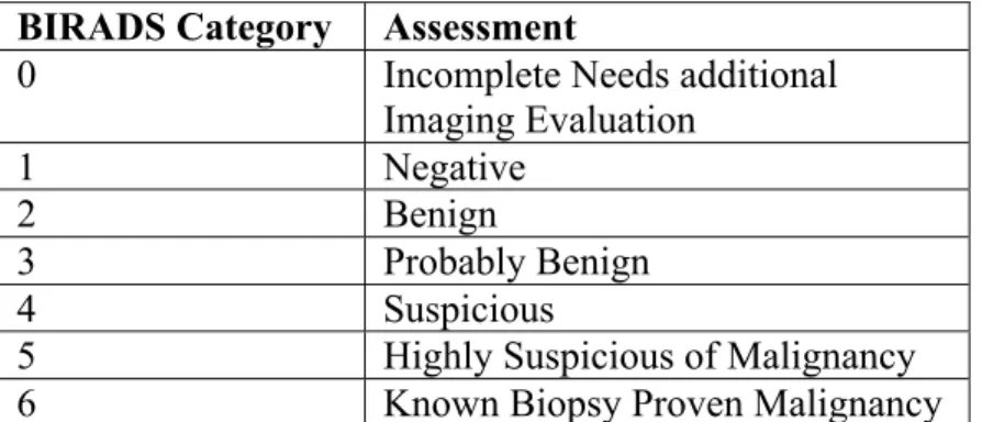
A radiologist analyzes the images produced by the mammogram for lesions. The radiologist interprets the image of the breast as either benign or suspicious of malignancy and in need of follow-up. The results are reported using the Breast Imaging Reporting and Data System (BI-RADS). BIRADS is a standardized method accepted by The American College of Radiology (ACR) for reporting mammography results 11. The Mammography Quality Standards Act (MQSA) of 1997 requires that results of all mammograms to be reported using the BI-RADS reporting system. Mammograms are determined to be positive or negative based on the radiologists’ interpretation using BI-RADS. A mammogram is scored on a scale from 0-6. The scoring assessment by BI-RADS category is displayed in Table 1.
Table 1. Breast Imaging Reporting and Data System (BI-RADS) BIRADS Category Assessment
0 Incomplete Needs additional
Imaging Evaluation
1 Negative
2 Benign
3 Probably Benign
4 Suspicious
According to the BIRADS classification schema, a mammogram is considered to be positive if the BI-RADS score is 0, 4, or 5. A BI-RADS score of 3 with recommendation for biopsy or further evaluation is also considered as a positive result. A score of 1, 2 or 3 with no recommendation for biopsy or further evaluation is considered negative 12.
Diagnostic Mammography
A diagnostic mammogram is performed on women who have a positive screening mammogram result or those who present with breast symptoms (pain, palpable lump/mass upon examination, change in breast size and/or shape, or nipple discharge or thickening). A diagnostic mammogram may include additional images and views of the breast tissue when compared to a screening mammogram.
Diagnostic Follow-up and Resolution of Screening Mammography The importance of diagnostic follow-up
The screening literature largely focuses on maximizing breast cancer screening rates through the use of community-based interventions 13. Two important, but less studied dimensions of screening effectiveness that impact disease mortality are diagnostic follow-up time and diagnostic resolution 14. Approximately 10% of screening mammograms require additional follow-up imaging and about 2% require a confirmatory biopsy 15.
Defining Follow-up and Diagnostic Resolution
progression from screening mammography to diagnostic resolution and finally diagnostic follow-up.
Though diagnostic follow-up time is important it is difficult to fully enumerate. There are some challenges inherent in accurately identifying a positive cancer diagnosis or a
benign/negative result in administrative databases. These include a lag in the reporting times of cancer registries, patients lost to follow-up either via failure to comply with follow-up recommendations or receiving care at a different facility or in a different site where capture may not be possible.
Current Recommendations for Follow-up after a Positive Screening Mammogram Breast imaging and reporting data system
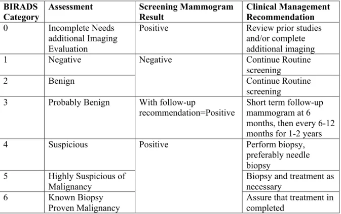
Though the BIRADS assessment provides guidelines for follow-up assessment, it does not specify a timeframe for all follow-up assessments outside of the ‘probably benign’ assessment category. The CDC guidelines do not differentiate the follow-up timeframe. All positive screening mammogram results should be resolved within 60 days. Table 2 briefly summarizes the recommendations based on BIRADS assessment categories.
Screening Mammogram
Diagnostic Follow-up (Diagnostic Mammogram, Breast
Ultrasound, MRI, Biopsy)
Diagnostic Resolution (Return to screening
or receive breast cancer diagnosis)
Time Time
Table 2. Categorization of Mammogram using BIRADS and Clinical Management Recommendation
BIRADS
Category Assessment Screening Mammogram Result Clinical Management Recommendation
0 Incomplete Needs
additional Imaging Evaluation
Positive Review prior studies
and/or complete additional imaging
1 Negative Negative Continue Routine
screening
2 Benign Continue Routine
screening 3 Probably Benign With follow-up
recommendation=Positive Short term follow-up mammogram at 6 months, then every 6-12 months for 1-2 years
4 Suspicious Positive Perform biopsy,
preferably needle biopsy
5 Highly Suspicious of Malignancy
Biopsy and treatment as necessary
6 Known Biopsy
Proven Malignancy
Assure that treatment in completed
Adapted from Eberl et al 200616
The National Comprehensive Cancer Network
The National Comprehensive Cancer Network (NCCN) is an organization consisting of various member institutions such as cancer hospitals with the mission of improving the quality and effectiveness of cancer care. NCCN consensus guidelines for clinical
management of patients undergoing breast cancer screening via mammography are arranged by BIRADS category (Figures 3 & 4)17. According to these guidelines, women with a
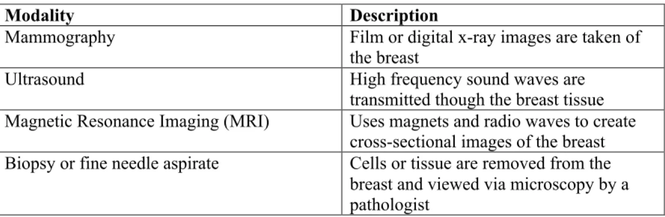
Both NCCCN and BIRADS provide recommendations for each level of clinical assessment, including benign and suspicious malignancy. The only assessment category that is given a definitive time table is probably benign or BIRADS category 3. This category is recommended for follow-up at 6 months post-screen. In Figures 1 and 2, the category ‘mammographic evaluation’ often includes both screening and diagnostic imaging. Imaging may consist of screening mammography, diagnostic mammography, breast ultrasound, breast MRI, and other specialized imaging techniques. Definitions for these procedures are
presented in Table 3.
Table 3. Study Definitions of Diagnostic Follow-up Procedures
Modality Description
Mammography Film or digital x-ray images are taken of
the breast
Ultrasound High frequency sound waves are
transmitted though the breast tissue Magnetic Resonance Imaging (MRI) Uses magnets and radio waves to create
cross-sectional images of the breast Biopsy or fine needle aspirate Cells or tissue are removed from the
Figure 5. Conceptual Model of the Impact of Patient and Healthcare System Factors on Diagnostic Follow-up after a Positive Screening Mammogram
Screening mammogram
performed
Individual Age, race, ethnicity, education, insurance status,
breast density, rural/urban residence Results reported Diagnostic Workup •Additional imaging •Biopsy Healthcare System Facility, imaging modality,
screening performance measures (true positive/false negatives), referrals/logistics negative positive
Return to screening per recommendations
Delays in Follow-up after a Positive Mammogram
In the extant literature, the reported time from positive screening mammogram until diagnostic follow-up studied ranges from a few days to 6 months 21-28. Currently however, the only U.S. guideline regarding the time in which follow-up should occur comes from the Centers for Disease Control and Prevention (CDC). The CDC Performance Indicators recommends that women with a positive screening mammogram complete diagnostic work-up within 60 days 19, 29. Much of the existing literature uses this designation to assess delay in follow-up after a positive screening mammogram. For example, a national study of a breast and cervical cancer screening program found that 20% of women did not receive diagnostic follow-up within 60 days of a positive screening mammogram 29. Other studies of follow-up after a positive screening mammogram result report that 20-40% of women fall outside of the CDC guidelines or receive no follow-up after a positive screening mammogram result 19, 29-32. Diagnostic Follow-up Pathways
should be assessed on the various pathways to diagnostic follow-up while taking their individual personal factors into account.
To our knowledge, one study has addressed the issue of multiple pathways or series of procedures for diagnostic follow-up and its impact on timing 33. The most common diagnostic follow-up procedures from this study and their definitions are presented in Table 3. Using data from five mammography registries in the Breast Cancer Surveillance
Consortium (BCSC), the authors looked specifically at variations in diagnostic pathways by imaging modality (digital or film screening mammography). The authors found that
differences in diagnostic pathways after positive screening mammogram between the imaging modalities were minimal. Variability in diagnostic pathways was observed across facilities performing mammograms indicating recommendation of follow-up by specific radiologists may play a role in patient’s follow-up pathways after a positive screening mammogram result.
Disparities in Diagnostic Follow-up after Positive Screening Mammogram
Untimely follow-up after a positive breast cancer screening mammogram may contribute to disparities in breast cancer outcomes and subsequently mortality. Specifically, follow-up times of 3-6 months have been associated with larger tumor size and reduced survival 14, 34, 54, 55.
Factors Contributing to Time to Diagnostic Follow-up after a Positive Screening Mammogram
A multitude of factors may contribute to diagnostic follow-up time after a positive screening mammogram result. Prior studies have investigated demographic patient factors such as race, education, and rural/urban residence that influence diagnostic follow-up time, often with limited ability to examine racial subgroups due to either small sample sizes or homogenous populations 56. Studies also identify a variety of healthcare system barriers (i.e. adequate communication and referral by physician, facility type and size, etc.) that minority women and women of low socioeconomic status experience in the cancer care continuum 46, 57.
Conceptual Model to Inform Follow-up after a Positive Screening Mammogram
Disparities in Diagnostic Follow-up after Positive Screening Mammogram
Though breast cancer mortality rates have declined over the past 10 years, mortality disparities exist for women of minority groups and those of low socioeconomic status (SES). Women of low-SES and minority, particularly African-American women, are less likely to receive screening mammography 34, present with more advanced disease, and are more likely to receive delayed care and experience increased wait time, than others after a positive mammogram 21-24, 35-50. Studies suggest extended time until follow-up varies by racial/ethnic group with African American women experiencing the longest delays when compared to White women 44, 49, 51. Additionally, persons living in rural areas have greater barriers (i.e. distance to facility, limited service availability) to receiving cancer care 52, 53.
Untimely follow-up after a positive breast cancer screening mammogram may contribute to disparities in breast cancer outcomes and subsequently mortality. Specifically, follow-up times of 3-6 months have been associated with larger tumor size and reduced survival 14, 34, 54, 55. Below, we describe factors previously studied in the literature that may be important in diagnostic follow-up.
Insurance
Though the literature does not identify precise reasons for these disparities, the literature does point to two main issues that may contribute to these disparities. One is that facilities may represent what the literature terms vulnerable populations, or in other words, the facility serves a majority of uninsured patients or low-income patients such as patients with Medicaid 59. These facilities may have limited resources such as staff or adequate imaging machines to accommodate patients. The other idea is that though most insurance providers provide an annual screening mammogram as a covered service, what the insurance provides for follow up services may vary. Some insurance providers may require approval or may require that a patient use a certain physician. These sorts of requirements could possibly add to the time needed to follow-up a positive screening mammogram.
In the following sections we briefly describe some common insurance categories. These insurance categories will be of interest in the study.
Medicare
in full once every 12 months. Digital and film mammography are both covered 60. Women 40 and over who have Medicare are covered for screening mammograms provided the facility and health care provider accept Medicare. Women between the ages of 35 and 39 can receive one baseline mammogram for comparison to future images once they become eligible for routine screening. Medicare does not cover MRI for screening purposes 60. Diagnostic mammography is covered where deemed medically necessary and women with part B must pay a deductible for diagnostic mammograms as well as 20% of the Medicare approved amount that a doctor can be paid for performing mammogram 60. Medicare offers a Part C plan or Medicare Advantage Plan that beneficiaries can buy into. This type of plan is offered and administered by a private insurance company.
Medicaid
National Breast and Cervical Cancer Early Detection Program (NBCCEDP) In order to reduce the breast cancer burden experienced by low-income women the CDC-funded National Breast and Cervical Cancer Early Detection Program (NBCCEDP) has provided cancer screening services, patient navigation assistance, and follow-up diagnostics and treatment to underserved women 11, 20. Women eligible for participation in North
Carolina are between the ages of 40 and 64, have a household income less than 250% of the federal poverty level, and are uninsured or underinsured (not enrolled in Medicaid or Medicare Part B) 62. The program includes a patient navigation component to ensure adequate and timely access to services and follow-up if needed, which Medicaid does not offer to patients. North Carolina offers the program to eligible women through health departments, community health centers, hospitals, and some physician offices. Currently more than 12,000 women participate in the NC program (10% of eligible North Carolinians), and many use NBCCEDP as their sole source for medical screening 63. This group of women represents a critical demographic that is often missing from other breast cancer studies as they do not qualify for Medicaid but are generally not able to afford private insurance coverage. It is also possible that this group of women may be misclassified as uninsured or ‘other’ insurance. Most studies (unless they specifically use NBCCEDP data) will not have information on this group of women.
Private insurance
Traditional fee for service insurance is generally offered through place of
A Health Maintenance Organization (HMO) is a type of private insurance plan that generally provides coverage within a specified network of providers and medical facilities. The providers and facilities contract with the insurance provider for medical services. Services received outside of the network are generally not covered by the plan. Most members of HMOs pay a fixed fee and select a primary care physician.
A Preferred Provider Organization (PPO) plan provides features found in both traditional private insurance plans and HMO plans. They often have premiums that are less than a traditional plan but may include co-insurance and/or co-pays that are due at the time of service.
Other Factors Thought to Influence Follow-up after a Positive Screening Mammogram Breast density
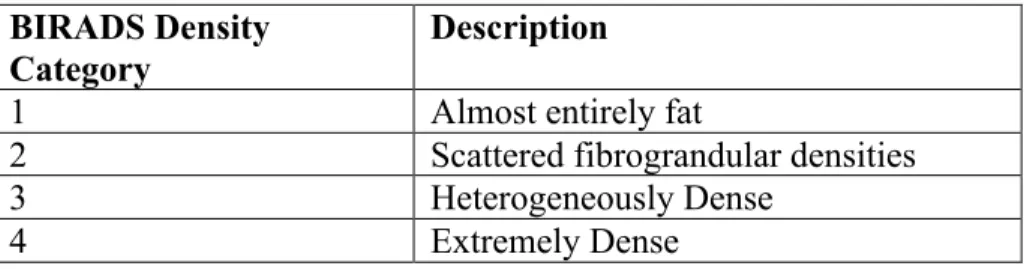
Breast density measures and classifies the composition of tissue within the breast. Specifically, it refers to the amounts fat and fibrous tissue that can be viewed in the breast via mammogram. A dense breast exhibits more fibrous tissue than fatty tissue. BIRADS
tissues that provide the lining of the ducts. The majority of breast cancers will originate in the epithelial cells. There is some information to suggest that high breast density and the
activation of the epithelial cells in the breast 67, 68. Additionally, studies suggest indeterminate results of screening mammography (not clearly positive or not clearly negative) and high breast density may lead to increased follow-up times 32. These factors, whether at the demographic or healthcare system level, may impact time to diagnostic follow-up after a positive screening mammogram. Figure 6 displays a comparison of the four levels of breast density as classified using the BIRADS system.
Table 4. Summary of Breast Density Classification BIRADS Density
Category
Description
1 Almost entirely fat
2 Scattered fibrograndular densities
3 Heterogeneously Dense
4 Extremely Dense
Figure 6. Comparison of BI-RADS Assessment Categories for Breast Density 69
Race
Socioeconomic status
Socioeconomic factors such as education, income, and access to healthcare facilities, not only impact breast cancer diagnosis and treatment, but are also shown to impact follow-up after a positive screening mammogram. Low-income and underinsured women present with more advanced stages of breast cancer 36, 39, 40 and have higher breast cancer mortality rates 35-38 and delayed diagnosis 71. Income may influence health-seeking behavior because of a patient’s ability or inability to pay for care. Higher socioeconomic status is associated with increased access to health care and increased access to resources that would facilitate health care access 39, 57. Studies show that women who understand their risk and the procedures required are more likely to seek care and comply with recommendations 47.
Family history and previous breast biopsy
Facility-level factors
Facilities performing mammography can exhibit a multitude of variations. Women can receive breast imaging services at facilities as varied as hospitals, outpatient clinics, breast centers, mobile mammography units, physician practices, and health departments. Additionally, services offered by each facility and protocols followed may vary. For instance some facilities have radiologists on sight to read and interpret images and provide immediate referral while some facilities send images to radiologists at other locations 23. At some facilities the images are read while the woman waits and at others the images are read in batches. This may require subsequent visits which could be time consuming, financially taxing, or impede follow-up. The type of equipment and technology used at facility may also vary. Some facilities only use film while some only use digital imaging. Studies have shown variability in facility in screening outcomes particularly among facilities that serve
vulnerable populations 72. Imaging modality
Digital mammography is more expensive than film mammography. Many practices and mammogram facilities are switching to digital mammography. In some areas,
particularly rural areas, digital mammography may not be available to patients. Digital mammography may also perform better for women less than 50 years of age and
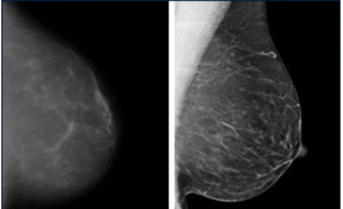
pre-menopausal women 74. Figure 7 shows a mammogram image of the breast. The mammogram on the left side is film mammography. The image on the right is digital mammography. This image appears clearer when compared to the image taken using film mammography.
Figure 7. Visual Comparison of Digital and Film Mammography 75
Patient residence
Summary of the Literature
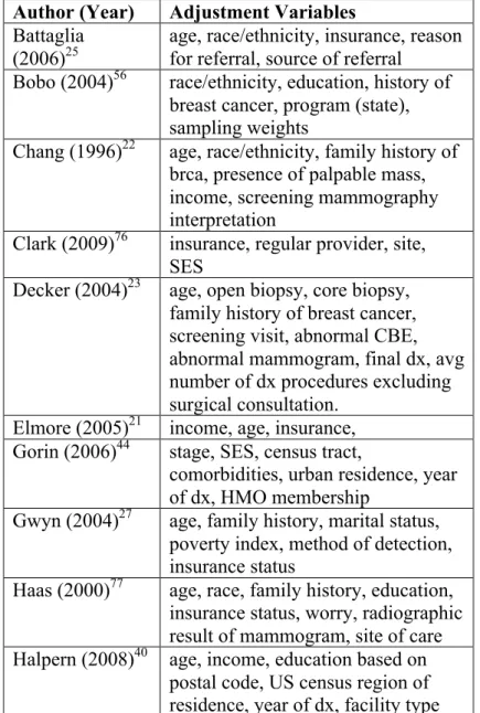
A summary of factors contributing to diagnostic follow-up time used previously in the literature is presented in Table 5. This summary represents the most common covariates included in adjustment sets of the models. The most common variables adjusted for are: age, race/ethnicity, history of breast cancer, socioeconomic status, and insurance. Table 6
Table 5. Adjustment Variables used in the Literature on Diagnostic Delay after a Positive Mammogram
Author (Year) Adjustment Variables Battaglia
(2006)25
age, race/ethnicity, insurance, reason for referral, source of referral
Bobo (2004)56 race/ethnicity, education, history of breast cancer, program (state), sampling weights
Chang (1996)22 age, race/ethnicity, family history of brca, presence of palpable mass, income, screening mammography interpretation
Clark (2009)76 insurance, regular provider, site, SES
Decker (2004)23 age, open biopsy, core biopsy, family history of breast cancer, screening visit, abnormal CBE, abnormal mammogram, final dx, avg number of dx procedures excluding surgical consultation.
Elmore (2005)21 income, age, insurance, Gorin (2006)44 stage, SES, census tract,
comorbidities, urban residence, year of dx, HMO membership
Gwyn (2004)27 age, family history, marital status, poverty index, method of detection, insurance status
Haas (2000)77 age, race, family history, education, insurance status, worry, radiographic result of mammogram, site of care Halpern (2008)40 age, income, education based on
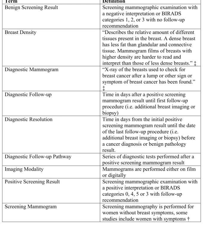
Table 6. Summary of Key Terms in this Study of Time Until Diagnostic Follow-up after a Positive Screening Mammogram
Term Definition
Benign Screening Result Screening mammographic examination with a negative interpretation or BIRADS
categories 1, 2, or 3 with no follow-up recommendation
Breast Density “Describes the relative amount of different tissues present in the breast. A dense breast has less fat than glandular and connective tissue. Mammogram films of breasts with higher density are harder to read and
interpret than those of less dense breasts.” ‡ Diagnostic Mammogram “X-ray of the breasts used to check for
breast cancer after a lump or other sign or symptom of breast cancer has been found.” ‡
Diagnostic Follow-up Time in days after a positive screening mammogram result until first follow-up procedure (i.e. additional breast imaging or biopsy)
Diagnostic Resolution Time in days from the initial positive screening mammogram result until the date of the last follow-up procedure (i.e.
additional breast imaging or biopsy) before a cancer diagnosis or benign pathology result.
Diagnostic Follow-up Pathway Series of diagnostic tests performed after a positive screening mammogram result
Imaging Modality Mammograms are performed either on film
or digitally
Positive Screening Result Screening mammographic examination with a positive interpretation or BIRADS
categories 0, 4, 5 or 3 with follow-up recommendation
Screening Mammogram Screening mammography is performed for women without breast symptoms, some studies include women with symptoms † †From Rosenberg 2006 78
REFERENCES
1. Services CfMM. Is my test, item, or service covered? Mammograms. Available at: http://medicare.gov/coverage/mammograms.html. Accessed 2013.
2. Cancer Facts & Figures 2011. American Cancer Society. Available at:
http://www.cancer.org/acs/groups/content/@epidemiologysurveilance/documents/doc ument/acspc-029771.pdf.
3. Institute NC. Breast Cancer Screening PDQ. Available at:
http://www.cancer.gov/cancertopics/pdq/screening/breast/healthprofessional/page1/A llPages/Print.
4. Nelson HD, Tyne K, Naik A, et al. Screening for breast cancer: an update for the U.S. Preventive Services Task Force. Ann Intern Med. Nov 17 2009;151(10):727-737, W237-742.
5. Nickson C, Mason KE, English DR, Kavanagh AM. Mammographic screening and breast cancer mortality: a case-control study and meta-analysis. Cancer Epidemiol Biomarkers Prev. Sep 2012;21(9):1479-1488.
6. Olsen AH, Njor SH, Vejborg I, et al. Breast cancer mortality in Copenhagen after introduction of mammography screening: cohort study. BMJ. Jan 29
2005;330(7485):220.
7. Nasca PC, Pastides H. Fundamentals of Cancer Epidemiology. 2nd ed. Sudbury, MA: Jones and Bartlett Publishers; 2008.
8. Kalager M, Adami HO, Bretthauer M, Tamimi RM. Overdiagnosis of invasive breast cancer due to mammography screening: results from the Norwegian screening
program. Ann Intern Med. Apr 3 2012;156(7):491-499.
9. Breen N, Yabroff KR, Meissner HI. What proportion of breast cancers are detected by mammography in the United States? Cancer Detect Prev. 2007;31(3):220-224. 10. Weaver DL, Rosenberg RD, Barlow WE, et al. Pathologic findings from the Breast
Cancer Surveillance Consortium: population-based outcomes in women undergoing biopsy after screening mammography. Cancer. Feb 15 2006;106(4):732-742. 11. Force USPST. Screening for breast cancer: U.S. Preventive Services Task Force
recommendation statement. Ann Intern Med. Nov 17 2009;151(10):716-726, W-236. 12. Society AC. American Cancer Society Guidelines for the Early Detection of Cancer,
Breast Cancer. Available at:
13. Institute NC. Breast Cancer. Available at:
http://www.cancer.gov/cancertopics/types/breast, 2013.
14. Lantz PM, Richardson LC, Macklem DJ, Shugarman LR, Knutson DB, Sever LE. Strategies for follow-up and treatment services in state breast and cervical cancer screening programs. Womens Health Issues. Jan-Feb 1999;9(1):42-49.
15. Richards MA, Westcombe AM, Love SB, Littlejohns P, Ramirez AJ. Influence of delay on survival in patients with breast cancer: a systematic review. Lancet. Apr 3 1999;353(9159):1119-1126.
16. Network NCC. NCCN Clinical Practice Guideilnes in Oncology: Breast Cancer Screening and Diagnosis. Available at:
http://www.nccn.org/professionals/physician_gls/pdf/breast-screening.pdf, 2013. 17. Caplan LS, May DS, Richardson LC. Time to diagnosis and treatment of breast
cancer: results from the National Breast and Cervical Cancer Early Detection Program, 1991-1995. Am J Public Health. Jan 2000;90(1):130-134.
18. Ryerson AB, Benard VB, Major AC. The National Breast and Cervical Cancer Early Detection Report: 1991-2002 National Report; 2005.
19. Elmore JG, Nakano CY, Linden HM, Reisch LM, Ayanian JZ, Larson EB. Racial inequities in the timing of breast cancer detection, diagnosis, and initiation of treatment. Med Care. Feb 2005;43(2):141-148.
20. Chang SW, Kerlikowske K, Napoles-Springer A, Posner SF, Sickles EA, Perez-Stable EJ. Racial differences in timeliness of follow-up after abnormal screening mammography. Cancer. Oct 1 1996;78(7):1395-1402.
21. Decker KM, Harrison M, Chateau D. Influence of direct referrals on time to diagnosis after an abnormal breast screening result. Cancer Detect Prev. 2004;28(5):361-367. 22. Mojica CM, Bastani R, Boscardin WJ, Ponce NA. Low-income women with breast
abnormalities: system predictors of timely diagnostic resolution. Cancer Control. Apr 2007;14(2):176-182.
23. Battaglia TA, Roloff K, Posner MA, Freund KM. Improving follow-up to abnormal breast cancer screening in an urban population. Cancer. 2007;109(S2):359-367. 24. Dennis CR, Gardner B, Lim B. Analysis of survival and recurrence vs. patient and
doctor delay in treatment of breast cancer. Cancer. Mar 1975;35(3):714-720.
26. Rojas M, Mandelblatt J, Cagney K, Kerner J, Freeman H. Barriers to follow-up of abnormal screening mammograms among low-income minority women. Cancer Control Center of Harlem. Ethn Health. Sep 1996;1(3):221-228.
27. Richardson LC, Royalty J, Howe W, Helsel W, Kammerer W, Benard VB. Timeliness of breast cancer diagnosis and initiation of treatment in the National Breast and Cervical Cancer Early Detection Program, 1996-2005. Am J Public Health. Sep 2010;100(9):1769-1776.
28. Lobb R, Allen JD, Emmons KM, Ayanian JZ. Timely Care After an Abnormal Mammogram Among Low-Income Women in a Public Breast Cancer Screening Program. Arch Intern Med. Mar 2010;170(6):521-528.
29. Eheman CR, Benard VB, Blackman D, et al. Breast cancer screening among low-income or uninsured women: results from the National Breast and Cervical Cancer Early Detection Program, July 1995 to March 2002 (United States). Cancer Causes Control. Feb 2006;17(1):29-38.
30. Jones B, Dailey A, Calvocoressi L, et al. Inadequate Follow-up of Abnormal Screening Mammograms: Findings From the Race Differences in Screening
Mammography Process Study (United States). Cancer Causes & Control. 2005/09/01 2005;16(7):809-821.
31. Hubbard RA, Zhu W, Horblyuk R, et al. Diagnostic imaging and biopsy pathways following abnormal screen-film and digital screening mammography. Breast Cancer Res Treat. Apr 2013;138(3):879-887.
32. Ryerson AB, Miller JW, Eheman CR, Leadbetter S, White MC. Recent trends in U.S. mammography use from 2000-2006: a population-based analysis. Prev Med. Nov 2008;47(5):477-482.
33. Bradley CJ, Given CW, Roberts C. Race, socioeconomic status, and breast cancer treatment and survival. J Natl Cancer Inst. Apr 3 2002;94(7):490-496.
34. Byers TE, Wolf HJ, Bauer KR, et al. The impact of socioeconomic status on survival after cancer in the United States : findings from the National Program of Cancer Registries Patterns of Care Study. Cancer. Aug 1 2008;113(3):582-591.
35. Du XL, Fang S, Meyer TE. Impact of treatment and socioeconomic status on racial disparities in survival among older women with breast cancer. Am J Clin Oncol. Apr 2008;31(2):125-132.
36. Newman LA, Griffith KA, Jatoi I, Simon MS, Crowe JP, Colditz GA. Meta-analysis of survival in African American and white American patients with breast cancer: ethnicity compared with socioeconomic status. J Clin Oncol. Mar 20
37. Ward E, Halpern M, Schrag N, et al. Association of insurance with cancer care utilization and outcomes. CA Cancer J Clin. Jan-Feb 2008;58(1):9-31.
38. Halpern MT, Ward EM, Pavluck AL, Schrag NM, Bian J, Chen AY. Association of insurance status and ethnicity with cancer stage at diagnosis for 12 cancer sites: a retrospective analysis. Lancet Oncol. Mar 2008;9(3):222-231.
39. Ashing-Giwa KT, Gonzalez P, Lim JW, et al. Diagnostic and therapeutic delays among a multiethnic sample of breast and cervical cancer survivors. Cancer. Jul 1 2010;116(13):3195-3204.
40. Kerlikowske K. Timeliness of follow-up after abnormal screening mammography.
Breast Cancer Res Treat. 1996;40(1):53-64.
41. Press R, Carrasquillo O, Sciacca RR, Giardina EG. Racial/ethnic disparities in time to follow-up after an abnormal mammogram. J Womens Health (Larchmt). Jul-Aug 2008;17(6):923-930.
42. Gorin SS, Heck JE, Cheng B, Smith SJ. Delays in breast cancer diagnosis and treatment by racial/ethnic group. Arch Intern Med. Nov 13 2006;166(20):2244-2252. 43. Kerner JF, Yedidia M, Padgett D, et al. Realizing the promise of breast cancer
screening: clinical follow-up after abnormal screening among Black women. Prev Med. Aug 2003;37(2):92-101.
44. Yabroff KR, Washington KS, Leader A, Neilson E, Mandelblatt J. Is the Promise of Cancer-Screening Programs Being Compromised? Quality of Follow-Up Care after Abnormal Screening Results. Medical Care Research and Review. September 1, 2003 2003;60(3):294-331.
45. Karliner LS, Patricia Kaplan C, Juarbe T, Pasick R, Perez-Stable EJ. Poor patient comprehension of abnormal mammography results. J Gen Intern Med. May 2005;20(5):432-437.
46. Hoffman HJ, LaVerda NL, Levine PH, et al. Having health insurance does not eliminate race/ethnicity-associated delays in breast cancer diagnosis in the District of Columbia. Cancer. Aug 15 2011;117(16):3824-3832.
47. Shavers VL, Brown ML. Racial and ethnic disparities in the receipt of cancer treatment. J Natl Cancer Inst. Mar 6 2002;94(5):334-357.
48. Maly RC, Leake B, Mojica CM, Liu YH, Diamant AL, Thind A. What Influences Diagnostic Delay in Low-Income Women with Breast Cancer? Journal of Womens Health. Jul 2011;20(7):1017-1023.
50. Athas WF, Adams-Cameron M, Hunt WC, Amir-Fazli A, Key CR. Travel distance to radiation therapy and receipt of radiotherapy following breast-conserving surgery. J Natl Cancer Inst. Feb 2 2000;92(3):269-271.
51. Nattinger AB, Kneusel RT, Hoffmann RG, Gilligan MA. Relationship of distance from a radiotherapy facility and initial breast cancer treatment. J Natl Cancer Inst.
Sep 5 2001;93(17):1344-1346.
52. Kothari A, Fentiman IS. Diagnostic delays in breast cancer and impact on survival.
International Journal of Clinical Practice. Apr 2003;57(3):200-203.
53. Sainsbury R, Johnston C, Haward B. Effect on survival of delays in referral of patients with breast-cancer symptoms: a retrospective analysis. Lancet. Apr 1999;353(9159):1132-1135.
54. Bobo JK, Shapiro JA, Schulman J, Wolters CL. On-schedule mammography rescreening in the National Breast and Cervical Cancer Early Detection Program.
Cancer Epidemiol Biomarkers Prev. Apr 2004;13(4):620-630.
55. Mandelblatt JS, Yabroff KR, Kerner JF. Equitable access to cancer services. Cancer.
1999;86(11):2378-2390.
56. Hsia J, Kemper E, Kiefe C, et al. The importance of health insurance as a determinant of cancer screening: evidence from the Women's Health Initiative. Prev Med. Sep 2000;31(3):261-270.
57. Schuur JD, Shah A, Wu Z, Forman HP, Gross CP. The impact of Medicaid coverage and reimbursement on access to diagnostic mammography. Cancer. Dec 1
2009;115(23):5566-5578.
58. Services CfMM. Medicare Program - General Information Available at:
http://www.cms.gov/Medicare/Medicare-General-Information/MedicareGenInfo/index.html, 2013.
59. Society AC. Medicare Coverage for Cancer Prevention and Early Detection. Available at:
http://www.cancer.org/healthy/findcancerearly/cancerscreeningguidelines/medicare-coverage-for-cancer-prevention-and-early-detection, 2013.
60. Assistance NDoM. What is Medicaid. Available at: http://www.ncdhhs.gov/dma/medicaid/, 2013.
61. Assistance NDoM. Breast Imaging Procedures Medicaid and Health Choice Clinical Coverage Policy. Available at: http://www.ncdhhs.gov/dma/mp/1k1.pdf, 2013. 62. National Breast and Cervical Cancer Early Detection Program (NBCCEDP): About
63. Henson RM, Wyatt SW, Lee NC. The National Breast and Cervical Cancer Early Detection Program: a comprehensive public health response to two major health issues for women. J Public Health Manag Pract. Spring 1996;2(2):36-47.
64. N.C. Division of Public Health CDaIS. North Carolina Breast and Cervical Cancer Control Program. Available at: http://bcccp.ncdhhs.gov/.
65. Tangka FK, Dalaker J, Chattopadhyay SK, et al. Meeting the mammography
screening needs of underserved women: the performance of the National Breast and Cervical Cancer Early Detection Program in 2002-2003 (United States). Cancer Causes Control. Nov 2006;17(9):1145-1154.
66. Carney PA, Miglioretti DL, Yankaskas BC, et al. Individual and combined effects of age, breast density, and hormone replacement therapy use on the accuracy of
screening mammography. Ann Intern Med. Feb 4 2003;138(3):168-175.
67. Kerlikowske K, Ichikawa L, Miglioretti DL, et al. Longitudinal measurement of clinical mammographic breast density to improve estimation of breast cancer risk. J Natl Cancer Inst. Mar 7 2007;99(5):386-395.
68. Tice JA, Cummings SR, Smith-Bindman R, Ichikawa L, Barlow WE, Kerlikowske K. Using clinical factors and mammographic breast density to estimate breast cancer risk: development and validation of a new predictive model. Ann Intern Med. Mar 4 2008;148(5):337-347.
69. Boyd NF, Rommens JM, Vogt K, et al. Mammographic breast density as an intermediate phenotype for breast cancer. Lancet Oncol. Oct 2005;6(10):798-808. 70. Li T, Sun L, Miller N, et al. The association of measured breast tissue characteristics
with mammographic density and other risk factors for breast cancer. Cancer Epidemiol Biomarkers Prev. Feb 2005;14(2):343-349.
71. Fedewa SA, Edge SB, Stewart AK, Halpern MT, Marlow NM, Ward EM. Race and Ethnicity are Associated with Delays in Breast Cancer Treatment (2003-2006).
Journal of Health Care for the Poor and Underserved. Feb 2011;22(1):128-141. 72. Bradley CJ, Neumark D, Shickle LM, Farrell N. Differences in Breast Cancer
Diagnosis and Treatment: Experiences of Insured and Uninsured Women in a Safety-Net Setting. Inquiry-the Journal of Health Care Organization Provision and
Financing. Fal 2008;45(3):323-339.
73. Goldman LE, Walker R, Hubbard R, Kerlikowske K, Breast Cancer Surveillance C. Timeliness of abnormal screening and diagnostic mammography follow-up at facilities serving vulnerable women. Med Care. Apr 2013;51(4):307-314.
75. Clark CR, Baril N, Kunicki M, et al. Addressing social determinants of health to improve access to early breast cancer detection: results of the Boston REACH 2010 Breast and Cervical Cancer Coalition Women's Health Demonstration Project. J Womens Health (Larchmt). May 2009;18(5):677-690.
CHAPTER 2: SPECIFIC AIMS
The advantages of early detection gained through screening may be diminished if positive mammogram test results do not lead to timely diagnosis and treatment. Current CDC guidelines recommend women receive diagnostic follow-up and begin treatment if necessary within 60 days of a positive screening mammogram result. Despite this, disparities in both diagnostic follow-up time (defined as the time from a positive screening result to the first diagnostic exam/procedure) and diagnostic resolution time (defined as the time from a positive screening result to the last work-up procedure leading to breast cancer diagnosis or benign result) continue to exist.
Minorities, those of low socioeconomic status, those living in rural areas, Medicaid recipients, and the uninsured are more likely to experience increased wait time for diagnostic resolution of a positive screening mammogram result. Understanding disparities in diagnostic follow-up and resolution among underserved women is crucial since this group of women present with advanced stages of breast cancer and have higher breast cancer mortality rates compared to insured non-low income women.
the radiologists’ assessment of the imaging exam and recommendation for follow-up, and pathologic cancer outcomes from the North Carolina Central Cancer Registry and from abstracted pathology reports. We accomplished the overall objective of the study with the following specific aims:
1. Assess the relationship between insurance status and initial diagnostic follow-up after a positive screening mammogram in a large, racially diverse population.
Hypothesis: Among women with a positive screening mammogram result, women reporting Medicaid and the uninsured will experience an increased time to diagnostic work up and diagnostic resolution when compared with women with private insurance.
2. Compare the diagnostic follow-up pathways observed after a positive screening mammogram result by insurance status.
Hypothesis: Among women with a positive screening mammogram, the number and type of subsequent imaging/procedures does not differ by insurance status.
2a. Examine differences in the diagnostic follow-up pathways by insurance status and assessed the association between insurance status and receipt of biopsy
CHAPTER 3: METHODS Overview
A retrospective cohort design was used to examine the association between insurance status and time until diagnostic follow-up after a positive screening mammogram result. Results from this study assessed whether the amount of time from a positive screening mammogram until diagnostic follow-up varied by insurance status as well as enumerated the different clinical pathways women may experience to resolve a positive screening result. We used data from the Carolina Mammography Registry from the years 1995-2010 for women who were at least 40 years of age, and had a positive screening mammogram result.
CMR has collected prospective data from participating mammography facilities in North Carolina since 1994. The CMR data include information on sociodemographic characteristics of women, reason for the breast imaging visit, breast cancer risk factors, and result of the imaging exam performed, the screening findings, and management
recommendations of the radiologist. CMR data also include patient insurance status at the time of the mammogram as well as breast cancer outcomes and pathology results from the North Carolina Central Cancer Registry.
Data Source: Carolina Mammography Registry
CMR provides quality improvement reports to participating facilities to help with reporting requirements of the Mammography Quality and Standards Act (MQSA) 81. CMR is also an active member of the Breast Cancer Surveillance Consortium (BCSC), a national research program that pools data from six mammography registries in the US 82. CMR data has been used widely to assess the utility and performance of mammographic screening for breast cancer research 64, 78, 83-94.
http://www.cmr.unc.edu
Figure 8. Breast Cancer Surveillance Sites 95
pathology information from hospital pathology reports that are collected through the Lineberger Comprehensive Cancer Center Rapid Case Ascertainment (RCA) program. The RCA program provides accelerated identification of cancer patients throughout the state.
Figure 9. Imaging Facility Coverage for the Carolina Mammography Registry by North Carolina County
Created by the CMR Project Manager http://www.cmr.unc.edu
opposed to recruiting and enrolling a new sample of women to follow prospectively. Unlike other data sources, (i.e. Cancer Registry, Medicare, or Medicaid claims data alone) CMR contains information on the radiologists’ interpretation of the screening mammogram as well as woman-level risk factor information such as breast density. Other data sources may contain only a medical billing code to indicate that a mammogram was completed making it necessary to collect and abstract medical records to obtain the screening mammogram test result.
Data Acquisition and Dataset Creation
CMR data from 1995-2009 were collected under a waiver of consent as approved by the Institutional Review Board (IRB) at The University of North Carolina at Chapel Hill (UNC). CMR data from 2010 to the present were collected with an opt-out option. This ancillary studied has received approval from the UNC IRB (IRB #11-1606).
Source Population
Data describing the source population, before application of study inclusion and exclusion criteria, are presented in Table 7. The left-hand side of Table 7 displays some general characteristics of screening mammograms in the CMR data for the years 1995-2010. Currently, the CMR contains patient and radiologist data from 1995-2010, with cancer registry data and pathology-associated outcomes through 2011. The right hand side of Table 7 displays demographic information for women with positive screening mammograms in the CMR data. The numbers in the table represent exams, not individual women.
population is White followed by nearly 18% of the population reporting their race as Black or African-American. Only 10% have less than a high school education. The majority report having private insurance followed by Medicare (16%).
Table 7. Demographic Characteristics of CMR Study Population (1995-2010) Characteristic All Screening
Mammograms n=1,853,944 n (%) Positive Screening Mammogram Result n=172,787 n (%) Age
18-39 113,948 (6.1) 11,313 (6.5) 40-49 375,991 (20.3) 42,110 (24.4) 50-59 426,864 (23.0) 38,454 (22.2) 60-69 314,196 (16.9) 29,534 (17.1) 70+ 270,714 (14.6) 51,376 (29.7) Race
Black 326,440 (17.6) 42,437 (24.6) White 1,357,379 (73.0) 111,813 (64.7)
Other 21,810 (1.2) 1,941 (1.1)
Missing 148,315 (8.0) 16,596 (9.6) Educational Status
<High School 185,103 (10.0) 17,201 (10.0) High
School/GED
381,912 (20.6) 35,886 (20.8) Some college 296,631 (16.0) 27,298 (15.8) College
Graduate
341,417 (18.4) 31,065 (18.0) Missing 648,881 (35.0) 61,337 (35.5) Insurance
Medicare 303,064 (16.3) 27,039 (15.6) Medicaid 72,303 (3.9) 7,532 (4.4)
BCCCP 26,118 (1.4) 2,716 (1.6)
Study Population
This study included data on breast imaging examination and pathology result through 2010 and cancer diagnosis through 2011. The study population consisted of female North Carolina residents included in CMR from 1995-2010 who were at least 40 years of age, had a positive screening mammogram result and who did not have a personal history of breast cancer. Women less than 40 years of age were excluded since they have not historically been recommended for routine mammography screening 4. There were no exclusions based on race, ethnicity, or geographic region. Only women were included in the study as male breast cancer is clinically considered a separate pathology. Also, men are not currently
recommended to receive routine screening mammography for breast cancer prevention 1. Facility Exclusions
Exclusions at the facility level were implemented for this study. We excluded the first two years of data for each facility, a total of about 11,000 exams. Facilities included in the CMR standard facility exclusion list were excluded, a total of 6 facilities. Facilities that do not use the CMDS system to collect data were excluded from these analyses, a total of 13 facilities. CMDS is CMR’s custom radiology information software for data capture,
during the study period we required that a woman’s first positive screening mammogram occur at least 365 days before the end date of facility participation.
Definition of Screening Mammography in the CMR
Study eligibility is limited to women undergoing a screening mammogram and only women with a positive screening mammogram were included in the study population. At the time of the mammogram the radiologist recorded whether the examination was a screening or diagnostic mammogram. Using BCSC definitions, a screening mammogram is defined as routine bi-lateral views of the breast among women without breast symptoms such as pain or lump 12. The radiologist reports information on the type of mammogram performed by answering the following questions: “What is the reason for this visit?” and “What exams were performed at this visit?” The response options are: Asymptomatic (screening
mammogram); Symptomatic, problem solving, diagnostic follow-up; Continued follow-up following abnormal mammography; Short-term follow-up (mostly 6 month follow-up; Biopsy; Other. We considered a response of “asymptomatic (screening mammogram)” and “Screening 2-view mammogram” respectively, as indication of screening mammography. Identification of a Positive Screening Mammogram in the CMR
A positive screening mammogram was defined using BCSC definitions 10. To be considered positive the radiologist had to assign the screening mammogram a Breast Imaging Reporting and Data System (BI-RADS) assessment of 0 (needs additional imaging), 4
women with more than one positive screening mammogram, one positive screen was randomly selected.
Table 8. Categorization of Screening Mammogram Result Using the Breast Imaging and Reporting Data System (BIRADS)
BIRADS Category
Assessment Screening
Mammogram Result
0 Incomplete Needs additional Imaging Evaluation
Positive
1 Negative Negative
2 Benign
3 Probably Benign without recommendation for immediate follow-up
Negative
3 Probably Benign with recommendation for immediate follow-up
Positive
4 Suspicious Positive
5 Highly Suspicious of Malignancy
6 Known Biopsy Proven
Malignancy Adapted from Eberl et al 2006 16
Exposure Assessment: Insurance Status
using the acronym BCCCP in North Carolina) is a program that provides free breast and cervical cancer screening to women who are uninsured 62.
In CMR insurance is coded as separate dichotomous variables. For the purposes of this study we created a new categorical variable using the existing insurance variables to accommodate women who reported with multiple insurance providers by checking more than one box on the form. These categories were:
Medicare Only: Women in this category selected Medicare as the sole source of insurance paying for the screening mammogram.
Medicare & Private: Women in this category selected Medicare and either Managed Care or Private as the sources paying for the screening mammogram. These women selected no other insurance categories.
Medicaid Only: Women in this category selected Medicaid as the sole source of insurance paying for the screening mammogram. These women selected no other insurance categories.
Medicare & Medicaid: Women in this category selected Medicare and Medicaid as the sources of insurance paying for the screening mammogram. These women selected no other insurance categories.
Private: Women in this category selected either private insurance or Managed Care as the sources of insurance paying for the screening mammogram. These women selected no other insurance categories.
NBCCEDP: Women in this category selected BCCCP and Other. No women in the study sample selected only BCCCP.
Women who did not select a box for this question were categorized as having an unknown insurance status. Table 9 displays the CMR variables used in exposure assessment.
Table 9. Variables Used in Exposure Assessment CMR
Variable Name
Description Value
BCCCP Insurance=BCCCP? Y=Yes N=No INSHMO Is the patient’s
insurance an HMO? Y=Yes N=No MEDICAD Covered by
Medicaid? Y=Yes N=No MEDICAR Covered by
Medicare?
Y=Yes N=No NOINS Not covered by
insurance?
Y=Yes N=No OTHINS Covered by other? Y=Yes
N=No
PRIINS Private? Y=Yes
N=No
Outcome Assessment: Time until Initial Diagnostic Follow-up
The primary outcome of interest was time until initial diagnostic follow-up defined as the time in days from the initial positive screening mammogram result until the date of the first/initial follow-up procedure (i.e. additional breast imaging or biopsy) before a cancer diagnosis or benign pathology result. After the examination the radiologist reports findings from the screening mammogram, lists other imaging studies needed to resolve the screening assessment if the result is positive, and provides recommendations for follow-up.
the CMR data. (Table 10) CMR variables and their associated dates were used to calculate follow-up time in days after a positive screening mammogram.
Statistical Analyses
We performed exploratory analysis of the data for distributions among each covariate. For continuous variables we studied graphical representations and for categorical variables we created tabular representations to view the distributions of each variable. Error checks for missing or implausible values were performed. In the event that the date for a follow-up examination (such as a confirmatory biopsy), occurred before the screening mammogram that value was re-coded as missing.
We provided descriptive statistics of the study population and described the time in days from a positive screening mammogram until initial follow-up for the total population and by insurance status. We summarized screening mammography use, and described the series of examinations and procedures (diagnostic follow-up pathways) the patient
experienced to follow-up a positive screening mammography result. All p values were two-sided and a value of p<0.05 was considered statistically significant. Statistical analyses were performed using SAS Software version. 9.3 96.
Covariate Adjustment
Assessment of confounding
explanatory variables determined by the literature to impact time until diagnostic follow-up after a positive screening mammogram 25, 98, 99. In the DAG (Figure 10) the variables Breast Density, Imaging Modality and Facility are colliders. Adjusting on these variables could potentially be a source of confounding and selection bias when assessing the effect of insurance status on time until diagnostic follow-up. This would occur if the association of insurance status and time until diagnostic follow-up changes when adjusting for the colliders 98. To assess the extent of this phenomenon we ran multiple models including and excluding these variables. Selected variables were examined using likelihood ratio tests 98.
Based on our interpretation of the directed acyclic graph (DAG) the following variables were considered as potential confounders in the association between insurance status and time until diagnostic follow-up following a positive screening mammogram result: age, race, facility, imaging modality, and breast density. For comparison with prior studies, we also considered variables commonly adjusted for in the literature. These variables included: Age, Race, Education, Family history of breast cancer, and Patient residence (Previously presented in Table 5). Urban/Rural patient residence, facility type, and distance to nearest facility were not found to be statistically significant predictors of the outcome and were not included in the final adjusted models. The following covariates were included in the adjusted models: age, race, education, breast density, and family history of breast cancer.
Covariates