Introduction
There are an estimated 13,430 new cases of laryngeal squamous cell carcinomas (LXSCC) diagnosed each year in the U.S [1]. Advanced stage LXSCC is treated with radiation or chemoradiation, with current strategies
favoring organ- sparing approaches to reduce comorbidity [2]. Despite well- established multimodality protocols, locoregional failure remains at 20–30% [3]. Whereas the detection of human papillomavirus (HPV) is an important predictor of clinical outcome in oropharyngeal squamous cell carcinomas (OPSCC) [4], HPV is much less prevalent
ORIGINAL RESEARCH
Nonpromoter methylation of the CDKN2A gene with active
transcription is associated with improved locoregional
control in laryngeal squamous cell carcinoma
Miriam M. Ben-Dayan1, Thomas J. Ow1,2, Thomas J. Belbin1,3, Joshua Wetzler1, Richard V. Smith2,
Geoffrey Childs1, Brenda Diergaarde4, D. Neil Hayes5, Jennifer R. Grandis6,7, Michael B. Prystowsky1 & Nicolas F. Schlecht8
1Department of Pathology, Albert Einstein College of Medicine, Montefiore Medical Center, Bronx, New York
2Department of Otorhinolaryngology-Head and Neck Surgery, Albert Einstein College of Medicine, Montefiore Medical Center, Bronx, New York 3Dicipline of Oncology, Faculty of Medicine, Memorial University of Newfoundland, St. John’s, Newfoundland
4Department of Epidemiology, University of Pittsburgh Cancer Institute, Pittsburgh, Pennsylvania
5Department of Otolaryngology/Head and Neck Cancer Surgery, University of North Carolina, Chapel Hill, North Carolina 6Departments of Otolaryngology Head and Neck Surgery, University of California, San Francisco, California
7Department of Clinical and Translational Science Institute, University of California, San Francisco, California
8Departments of Epidemiology & Population Health and Medicine, Albert Einstein College of Medicine, Bronx, New York
Keywords
CDKN2A, gene transcription, larynx cancer, methylation, p14(ARF), p16(INK4a), tumor biomarkers
Correspondence
Nicolas F. Schlecht, Albert Einstein College of Medicine, 1300 Morris Park Avenue, Bronx, NY 10461. Tel: 718 430 2307; Fax: 718 839 7988; E-mail: [email protected]
Funding Information
This study was partially supported by National Institute of Dental & Craniofacial Research (DE023941) and National Cancer Institute grants (CA131648 and CA115243), and the departments of Otorhinolaryngology-Head and Neck Surgery and Pathology at Montefiore Medical Center. The University of Pittsburgh head and neck cancer study was supported by National Cancer Institute grants (CA097190 and CA047904).
Received: 9 September 2016; Revised: 14 October 2016; Accepted: 21 October 2016
Cancer Medicine 2017; 6(2):397–407
doi: 10.1002/cam4.961
Abstract
We previously reported a novel association between CDKN2A nonpromoter methylation and transcription (ARF/INK4a) in human papillomavirus associated oropharyngeal tumors. In this study we assessed whether nonpromoter CDKN2A methylation in laryngeal squamous cell carcinomas (LXSCC) conferred a similar association with transcription that predicted patient outcome. We compared DNA methylation and ARF/INK4a RNA expression levels for the CDKN2A locus using the Illumina HumanMethylation27 beadchip and RT- PCR in 43 LXSCC tumor samples collected from a prospective study of head and neck cancer patients treated at Montefiore Medical Center (MMC). Validation was performed using RNAseq data on 111 LXSCC tumor samples from the Cancer Genome Atlas (TCGA). The clinical relevance of combined nonpromoter CD-KN2A methylation and transcription was assessed by multivariate Cox regression for locoregional recurrence on a subset of 69 LXSCC patients with complete clinicopathologic data from the MMC and TCGA cohorts. We found evidence of CDKN2A nonpromoter hypermethylation in a third of LXSCC from our MMC cohort, which was significantly associated with increased ARF and INK4a RNA expression (Wilcoxon rank- sum, P = 0.007 and 0.003, respectively). A similar association was confirmed in TCGA samples (Wilcoxon rank- sum test P < 0.0001 for ARF and INK4a). Patients with CDKN2A hypermethylation or high ARF/INK4a expression were significantly less likely to develop a locoregional recurrence compared to those with neither of the features, independent of other clinicopatholgic risk factors (adjusted hazard ratio=0.21, 95% confidence inter-val:0.05–0.81). These results support the conclusion that CDKN2A nonpromoter methylation is associated with increased ARF and INK4a RNA expression, and improved locoregional control in LXSCC.
Cancer Medicine
in LXSCC [5], and there are no molecular or genomic biomarkers to guide treatment selection for LXSCC patients.
Recently, our group identified a novel association between methylation in the nonpromoter region of CDKN2A and increased p16(INK4a) expression in HPV- positive OPSCC [6]. The CDKN2A gene, located at chromosome 9p21, encodes two distinct tumor suppressor proteins, INK4a and ARF. While the transcripts overlap, they each incor-porate a unique first exon and different reading frames [7]. Furthermore, whereas both CDKN2A proteins are known to induce cell cycle arrest and are commonly dis-rupted during tumorigenesis [7], p16(INK4a) expression is not common in LXSCC and is not predictive of clinical outcome [8]. However, the expression of ARF is being assessed as a therapeutic alternative in HPV- negative head and neck cancer [9]. We hypothesized that nonpromoter hypermethylation of CDKN2A and associated transcription may occur in a subset of less aggressive LXSCC.
To test this hypothesis, we examined patients with LXSCC enrolled in an ongoing research program at a large urban health center to measure CDKN2A nonpro-moter methylation and transcription in tumors. We assessed whether increased methylation and expression of the CDKN2A region was associated with locoregional control in these patients. We validated our findings in an inde-pendent sample of LXSCC tumors with genetic, epigenetic, and transcriptomic data from the Cancer Genome Atlas (TCGA; http://cancergenome.nih.gov/) [10].
Methods
Patient tissue samples
Initial analyses were based on 43 histologically confirmed LXSCC tumors obtained by biopsy or surgical resection from patients undergoing treatment at MMC in Bronx, NY between 2002 and 2010. All patients provided written consent for participation in this study under a protocol approved by the Institutional Review Board at MMC. All tumors were assessed histologically to ensure a majority of tumor cells in samples. Tumor samples were snap- frozen in liquid nitrogen within 30 min and kept at −80°C until DNA and RNA analysis. This cohort served as our discovery sample set.
All tumor samples were tested for HPV- 16 RNA using a Real- Time Reverse Transcription- Polymerase Chain Reaction (qRT- PCR) assay described previously [11]. Tumors with high E6 and/or E7 RNA expression (∆CT ≥100- fold compared to GAPDH) were considered HPV- positive. Presence of p16(INK4a) protein was also assayed using a clinically available immunohistochemistry (IHC) antibody (BD Biosciences, San Jose, CA; Cat. no. 551154) [8]. Briefly, p16 staining was done for formalin- fixed and
paraffin- embedded tumor section. Tumors were considered p16- positive if >50% of the tumor cells exhibited strong nuclear staining.
Methylation analyses
The protocol for measuring DNA methylation using micro-arrays was as described previously [11]. For methylation analysis, HumanMethylation27 beadchip (Illumina, San Diego, CA) beta values were transformed to normalized M- values using an established protocol [12]. The previ-ously described nonpromoter region included one CpG within exon 3 (cg11653709) and three CpGs within the preceding intron (cg07752420, cg09099744, and cg10895543) [6]. In addition to the nonpromoter region, we assessed methylation levels for 3 CpG loci within the ARF promoter of CDKN2A (cg00718440, cg03079681, and cg26673943). We also tested adjacent normal tissues from 48 LXSCC patients but found consistently low levels of methylation overall in all samples (data not shown).
RNA expression analyses
Expression levels for ARF and INK4a were quantified from total RNA (100 ng) of 29 LXSCC tumors using real- time reverse transcriptase- PCR (qRT- PCR) Taqman® probes and the TaqMan RNA- to- Ct™ 1- Step Kit (Applied Biosystems, Waltham, MA). Resulting cycle threshold (Ct) values were normalized to GAPDH and subsequently converted to relative expression (2−ΔΔCT) values.
As an additional platform, whole- genome expression microarray data from a prior analysis of head and neck tumors were also assessed [11] and 21 of the LXSCC sam-ples used for the methylation analysis were used in this analysis. TRIzol (Ambion, Austin, TX) extracted RNA (500 ng) for each tumor sample was amplified and biotin labeled using the Illumina TotalPrep RNA Amplification Kit (Ambion, Austin, TX). Messenger RNA expression levels were subsequently analyzed by RNA hybridization to the Illumina HumanHT- 12- v3 Expression BeadChip (Illumina, San Diego, CA). Microarray probes for the CDKN2A gene were identified using the RefSeq database release 17 and UniGene build 188. RNA expression levels were assessed by a probe targeting ARF and a combined CDKN2A probe. In addition, a probe corresponding to the INK4b gene in the adjacent CDKN2B region was also identified and used to measure corresponding expression of that transcript.
TCGA data and analyses
methylation, RNA expression, and patient clinical data were mined for 111 LXSCC tumor samples from the TCGA database. Per TCGA protocol, only samples with >60% tumor cells were included for analysis. One CpG (cg12840719) located within the same downstream non-promoter region of the CDKN2A gene was captured by the HumanMethylation 450K beadchip (Illumina, San Diego, CA) array used by TCGA. We previously showed this region to exhibit consistent methylation patterns across neighboring CpGs [6].
Other data extracted from TCGA included RNA expres-sion and genetic mutation events corresponding to the CDKN2A gene. Reads Per Kilobase per Million mapped reads (RPKM) for ARF, INK4a, Cylcin A, Cyclin E, and INK4b from TCGA were used to measure gene expression levels generated by RNAseq [10]. HPV expression status was determined using E6 and E7 RNAseq data [10]. In addition, level 3 copy number variation (CNV) data for 9p21 and the CDKN2A locus, and level 3 somatic muta-tion data for CDKN2A and TP53 were mined for all TCGA LXSCC samples [10].
Statistical analyses
For efficiency and purposes of statistical validation, the MMC discovery cohort samples were divided into two groups based on CDKN2A methylation levels (hypo- and hyper- methylated) using Euclidean distance hierarchical clustering with complete linkage using all 4 CpG loci. The corresponding methylation heatmap was visualized using the supraHex v1.8.0 R package [13]. To allow for statistical comparison, grouping of the TCGA samples was based on the distribution of M- values for the single down-stream CDKN2A CpG (cg12840719) assayed by the HumanMethylation 450K beadchip assuming a cutoff for hypermethylation based on the same percentile threshold of 32% observed in our discovery sample of LXSCC tumors from MMC (M = −0.048). CpG loci located at the pro-moters of ARF and INK4a transcripts were also assessed from TCGA data.
With respect to the expression data, MMC samples were grouped based on median RNA values for each transcript excluding LXSCC tumors with no detectable RNA, with relative expression thresholds of 49 (ΔΔCT) for ARF and 166 (ΔΔCT) for INK4a. The cut- off selection for ARF and INK4a expression in TCGA samples was similarly based on median RNA levels excluding tumors with no detect-able RNA (with a corresponding RPKM of 171 for ARF and 35 for INK4a). This was done to differentiate between tumors with high and low RNA expression.
Wilcoxon rank sum tests were used to determine the statistical significance of observed differences in CDKN2A transcript levels between tumors with hypo- or
hypermethylated CDKN2A. Box- and- Whisker plots were generated using the ggplot2 v2.0.0 R package [14]. We used contingency tables to assess the associations between CDKN2A locus methylation/expression status (i.e., com-paring tumors with high vs. low CDKN2A methylation/ expression) and clinicopathologic factors at diagnosis, and tested using Chi- square or Fisher Exact test statistics, where appropriate. All tests were two- sided.
In addition to the 29 patients followed at MMC, infor-mation on clinical outcome was ascertained for 69 LXSCC patients originating from three collaborating TCGA tissue collection sites: the University of Pittsburgh (n = 23), Vanderbilt University (n = 9), and the University of North Carolina (UNC) at Chapel Hill (n = 8). Response to treat-ment was based on locoregional recurrence, defined as a recurrence of cancer at the primary site or regional lymph nodes. The association between CDKN2A locus methylation/ expression and time to locoregional recurrence was assessed using Kaplan–Meier survival analyses, and strata were com-pared using Log- rank test statistics. Multivariable Cox regres-sion models were built, adjusting a priori for gender, age (<60 vs. ≥60 years) and regional lymph node metastases [15] [node negative (N0) vs. regional lymph node metastasis (N+)]. Other clinicopathologic variables assessed included treatment (coded as unimodality treatment [radiation or surgery], surgery plus postoperative adjuvant treatment [radiation or chemoradiation], and primary chemoradiation), overall stage [16] and tumor size [15] (I/II vs. III/IV), smoking status (current vs. former smoker), and HPV status. All models were stratified by sample cohort (MMC vs. TCGA) in order to adjust for possible bias associated with tissue acquisition protocols for the different studies and methylation platforms. A parsimonious model was derived using backward elimination for variables with P < 0.05. Effect modification (e.g., with clinical factors and study site) was tested for by including cross- product terms into the model. The potential for confounding was also assessed using a change in point criterion [17]. Proportional hazards assumptions were tested using log- log plots, observed versus expected plots, and goodness- of- fit tests. Kaplan–Meier plots were generated using the Survival v2.38.3 R package[18], and multivariable Cox regression analyses were performed using Stata v.14 (StataCorp, College Station, TX).
Results
A subset of LXSCC tumors exhibit CDKN2A nonpromoter methylation
data for four CpGs within the downstream nonpromoter region of CDKN2A (Fig. 1). Two clusters of tumors were identified with approximately a 2- to- 1 ratio with hypo- and hypermethylation.
By clustering the methylation values for the four non-promoter CpGs, we were able to compare LXSCC tumors with and without CDKN2A methylation. The clinical characteristics for the MMC patients included in our analyses are summarized in Table S1. Hypermethylated LXSCC tumors were more likely to have p16(INK4a) protein expression as measured by IHC compared to hypomethylated tumors (Fisher’s Exact test P = 0.029). In contrast, the prevalence of HPV was much lower and showed no significant association with CDKN2A methyla-tion (P = 1.0). No significant differences were observed with respect to other clinicopathologic factors between the hyper- and hypomethylated groups.
CDKN2A nonpromoter methylation is associated with ARF and INK4a RNA expression
As we did previously for OPSCC, we also examined the relationship between CDKN2A nonpromoter methylation and RNA expression in LXSCC. We measured RNA levels for INK4a and ARF by qRT- PCR in 29 LXSCC tumors. The clinical characteristics for the patients with qRT- PCR data are summarized in Table S2. Increased levels of both ARF and INK4a were found in tumors with CDKN2A hypermethylation compared to those without (Wilcoxon rank- sum test P = 0.007 and 0.003, respectively; Fig. 2).
RNA levels for these transcripts were also measured using the Illumina expression array as an alternative platform for validation. Of the 33 LXSCC tumors analyzed using the Illumina expression array, 26 were also assayed by qRT- PCR. Consistent with the qRT- PCR results, ARF and Figure 1. Cluster analysis of laryngeal tumor samples based on CDKN2A nonpromoter methylation. The M- values of 4 CpGs from the downstream nonpromoter region of CDKN2A were heirarchically clustered for 43 primary LXSCC tumors. Each leaf in the dendrogram represents a sample, and each row represents an individual CpG loci. Two main clusters were identified based on the dendrogram above the heatmap. Samples in the left, mostly green cluster, were categorized as the hypomethylated group (n = 29), and the samples in the right, mostly red cluster, were categorized as the hypermethylated group (n = 14). HPV RNA, p16(INK4a) protein expression (by IHC), and smoking status for each sample are shown below the heatmap. White squares indicate HPV RNA negative, p16(INK4a) protein negative and noncurrent smokers, respectively. Black squares indicate HPV RNA positive, p16(INK4a) protein positive and current smokers, respectively. Missing data are denoted by an “X”. A color key matching color with corresponding M- value is shown in the upper left corner. HPV, human papillomavirus; IHC, immunohistochemistry.
CDKN2A RNA levels were increased in tumors with non-promoter CDKN2A hypermethylation (P < 0.0001 for both probes; Fig. S1).
ARF and INK4a RNA levels were also plotted against the methylation M- values for each of the four nonpro-moter CpG loci tested on the Illumina methylation array revealing significant positive correlations with ARF and INK4a transcription (Figs S2 and S3). Methylation of CpG loci within the promoter region of ARF were also meas-ured, but these showed no evidence of methylation and no correlation with corresponding RNA expression (data not shown). Together, these data indicate that increased ARF and INK4a RNA levels are associated with CDKN2A nonpromoter hypermethylation.
TCGA LXSCC tumors validate our methylation and expression results
To validate our results from the MMC cohort, we analyzed CDKN2A nonpromoter methylation and RNA levels for 111 LXSCC tumors available in the TCGA database. Table S3 summarizes the clinical characteristics for the patients in TCGA. Unlike the Illumina HumanMethylation27 bead-chip array used in our MMC study, which assayed four downstream nonpromoter CDKN2A CpG loci, collection of DNA methylation data for TCGA samples utilized the Illumina HumanMethylation 450K beadchip platform, which assayed only one novel CpG within the region of interest. For consistency, we selected an M- value cutoff based on the distribution in methylation observed for the MMC cohort (see Methods for cutoff); ARF and INK4a RNA levels were plotted against CpG M- values to dem-onstrate the correlation between hypermethylation and increased RNA levels for CDKN2A expression (Fig. S4).
We compared the CDKN2A RNA levels obtained by RNAseq for 75 samples that were classified as hypo-methylated and 36 samples that were hyperhypo-methylated (Fig. 3). The hypermethylated LXSCC tumors exhibited an increase in both ARF and INK4a RNA compared to hypomethylated tumors (Wilcoxon rank- sum test P < 0.0001 for both transcripts), confirming the results seen in our MMC cohort. Alternative cutoffs were evalu-ated, but moving the methylation threshold by one standard deviation (equivalent to −1.7 M- values) did not affect our results. In addition, we were able to assess the methylation levels for the promoter regions of both CDKN2A transcripts, which were uniformly unmethyl-ated, ruling out the possibility that the lack of CDKN2A RNA in hypomethylated tumors was due to promoter methylation. As such, data from TCGA LXSCC tumors validated our MMC findings that CDKN2A nonpromoter methylation was associated with ARF and INK4a RNA expression.
Tumors with high CDKN2A expression exhibit CDKN2A and/or TP53 mutations
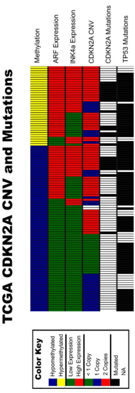
Given that the CDKN2A locus encodes two tumor sup-pressors, and that both transcripts are elevated in LXSCC tumors, we wanted to examine the presence of genetic variations across this locus. Data available from TCGA allowed for the assessment of genetic alterations, including 9p21 CNV, CDKN2A mutation status, and p53 mutation status (Fig. 4). As expected, tumors with homozygous deletions for CDKN2A were only observed in the group with low CDKN2A RNA, and in the majority of the hypomethylated LXSCC. However, 58% of hypomethylated tumors with low CDKN2A RNA expression had at least one copy of CDKN2A. Moreover, at least one copy was retained for all tumors with high levels of ARF and INK4a RNA, and 75% of the hypermethylated tumors retained both copies. Many tumors with high levels of CDKN2A RNA also exhibited mutations within the CDKN2A (22%) and/or TP53 (89%) loci. All the CDKN2A mutations were seen within the first exon of the INK4a transcript (exon 1α) and the shared exon 2, and there were no mutations within the first exon of the ARF transcript (exon 1β). These data suggest that despite high levels of ARF and INK4a RNA, mutations within the CDKN2A and TP53 loci may be a hindrance toward fully functional tumor suppressors.
RNA levels of E2F targets remain unaffected by CDKN2A nonpromoter methylation
Since INK4a acts as an inhibitor of E2F transcription factors, we also examined the RNA expression levels of two E2F targets, Cyclin A and Cyclin E. RNAseq data were obtained from TCGA data for 111 laryngeal tumors, and we compared RNA levels of Cyclin A and Cyclin E for 75 tumors with CDKN2A hypomethylation and 36
tumors with CDKN2A hypermethylation (Fig. S5). We found no significant differences in Cyclin A or Cyclin E RNA expression between the two groups. While these analyses did not directly measure the functionality of INK4a, the results suggest normal E2F activity for both groups of laryngeal tumors.
RNA expression of INK4b is also associated with CDKN2A nonpromoter methylation
There is a possibility that the association between CDKN2A methylation and RNA expression may be a consequence of chromatin conformation with more of a regional effect on gene expression. As a preliminary test for this hypoth-esis, we examined the RNA expression for an adjacent locus (CDKN2B) found at chromosome 9p21. We assessed RNA levels for the INK4b gene, which is transcribed from the CDKN2B locus, in both MMC and TCGA cohorts. For the MMC samples, RNA levels for INK4b were obtained from the Illumina expression array, and found to be sig-nificantly increased in tumors with CDKN2A hypermeth-ylation (Wilcoxon rank- sum, P = 0.015; Fig. S6A). For TCGA samples, RNAseq data were collected for INK4b, which also showed a significant increase in INK4b RNA expression for tumors with CDKN2A hypermethylation (Wilcoxon rank- sum, P < 0.0001; Fig. S6B). Our finding of increased INK4b RNA levels in tumors with nonpro-moter CDKN2A hypermethylation suggests an open chro-matin conformation for at least a portion of the 9p21 locus.
CDKN2A methylation/expression is associated with locoregional control
In order to examine the relationship between CDKN2A methylation/expression and clinical outcome, we worked with collaborators at the University of Pittsburgh [BD and JRG], Vanderbilt University [JL], and the University of North Carolina [NH], to retrieve clinical outcome data for a subset of 40 LXSCC patients included in the TCGA (Table S4). Using the same expression cutoffs established for the initial TCGA analyses, we identified 24 hypometh-ylated and 16 hypermethhypometh-ylated tumors (Fig. S7). While the TCGA subcohort was more likely to be male, Caucasian and non- Hispanic, and less likely to be HPV positive compared to our MMC cohort, no significant associations were seen between nonpromoter CDKN2A methylation and clinicopathologic factors. RNA levels of ARF and INK4a were significantly higher in the hypermethylated vs. hypomethylated tumors from this subgroup (Wilcoxon rank- sum test P < 0.0001 and P = 0.005, respectively).
To evaluate the relationship between nonpromoter CDKN2A methylation, RNA expression and tumor Figure 4. Copy number variation and mutation data for TCGA LXSCC
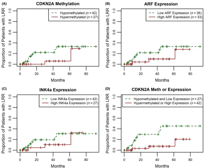
progression, we assessed the risk of locoregional recur-rence (LRR) for the combined cohort of (n = 69) LXSCC patients from MMC, Pittsburgh, Vanderbilt, and UNC (median follow- up of 26 months). Figure 5A shows the Kaplan–Meier plots for LRR comparing LXSCC patients with respect to nonpromoter CDKN2A methylation status.
Patients with CDKN2A hypermethylation in the tumor had a lower risk of LRR when compared to those with hypomethylated tumors, although the results were not significant (Log- rank test P = 0.11). We observed similar differences when we assessed risk of LRR by CDKN2A transcript levels using median cutoff levels for ARF and Figure 5. Increased risk of local regional recurrence is associated with CDKN2A hypomethylation and low RNA expression. Recurrence was assessed for 69 LXSCC patients in the combined MMC, Pittsburgh, Vanderbilt and UNC cohort. (A) A Kaplan–Meier was generated for 42 hypomethylated (green, dashed line) and 27 hypermethylated tumors (red, solid line). The plotted results indicate a decreased risk of recurrence in the hypermethylated group (Log- rank, P = 0.11). (B) Patients were divided by ARF expression. The Kaplan–Meier was generated comparing 36 tumors with low ARF expression (green, dashed line) and 33 tumors with high ARF expression (red, solid line). High ARF expression was defined as a relative expression level
INK4a RNA, although these were also not significant (Log- rank test P = 0.18 for low vs. high ARF expression; Fig. 5B, and P = 0.31 for INK4A expression; Fig. 5C). When we further classified patients based on their com-bined tumor CDKN2A locus methylation/expression status, comparing patients with either hypermethylated CDKN2A or high RNA levels of ARF/INK4a to those with CDKN2A hypomethylation and low ARF+INK4a expression, we found a stronger association with risk of LRR (Log- rank test P = 0.01; Fig. 5D).
To assess if CDKN2A hypermethylation and high ARF/ INK4a expression was associated with lower risk of LRR after adjustment for confounding factors, multivariable cox proportional hazard regression models were con-structed. After backward selection to identify significant confounders, a model adjusting for treatment and other relevant/significant clinicopathologic factors (e.g., age, gender, and nodal status at diagnosis) showed that LXSCC patients with nonpromoter CDKN2A hypermethylation, high ARF expression or high INK4a expression were sig-nificantly less likely to develop a LRR compared to patients lacking both nonpromoter CDKN2A methylation and RNA expression (adjusted hazard ratio = 0.21, 95% CI: 0.05–0.81; Table 1). Given the potential impact of treatment modali-ties on locoregional control, we also assessed the subset (n = 48) of patients who received surgery as a component of their treatment, which was the most commonly used modality (Fig. S8). As with the expanded cohort, detec-tion of nonpromoter CDKN2A hypermethyladetec-tion or high ARF/INK4A expression was associated with a significantly lower risk of LRR (Log- rank test, P = 0.01). Our statisti-cal analyses on clinistatisti-cal outcome indicate that a combined marker of CDKN2A methylation or expression is associ-ated with a decreased risk of LRR for LXSCC.
Discussion
In this study, we demonstrate that methylation of the terminal intron/exon of the CDKN2A tumor suppressor gene in LXSCC is associated with increased expression of CDKN2A transcripts, ARF and INK4a, which are known to stabilize cell cycle proteins p53 and RB, respectively [19]. In addition, we found that hypermethylation and transcription of this locus was significantly associated with improved locoregional control in LXSCC.
We previously identified a significant relationship between downstream CDKN2A hypermethylation (at the same locus) and increased RNA expression that was also correlated with HPV infection in OPSCC [6]. Whereas HPV is prevalent in OPSCC, LXSCC is not typically asso-ciated with HPV infection [20]. Despite this, we observed a significant proportion of LXSCC exhibited hypermeth-ylation of the same nonpromoter region of CDKN2A,
and increased levels of RNA expression, suggesting that the association was not limited to HPV- associated disease.
The CDKN2A locus is frequently altered in head and neck cancer, either through complete deletion of both transcripts or microdeletion of exons specific to ARF and/ or INK4A, or through mutation producing nonfunctional proteins [10]. When we compared the genetic and epige-netic data from TCGA, we found that while CDKN2A was frequently mutated in LXSCC tumors, the mutations occurred exclusively within exon 1α (which is unique to INK4a) and exon 2, which is shared by both transcripts and essential for ARF- mediated MDM2 nucleolar transloca-tion [21]. No mutatransloca-tions were found within exon 1β, which is unique to ARF. In addition, a number of tumors with high CDKN2A expression also exhibited a loss of one copy of CDKN2A. This is of relevance since ARF cannot stabilize p53 without both alleles [22], and combined with exon 2 mutations, suggests the presence of a nonfunctional ARF- MDM2- p53 pathway. Furthermore, when we looked at the RNA expression of two downstream targets of the INK4a- CDK- Rb- E2F pathway, Cyclin A and Cyclin E [23, 24], we found similar levels in tumors independent of CDKN2A methylation status. This suggests the presence of active E2F protein despite high levels of INK4a RNA.
Whereas loss of the Rb and p53 pathways may drive tumorigenesis in LXSCC, high levels of ARF RNA may Table 1. Multivariable cox regression model evaluating the association between CDKN2A nonpromoter methylation or expression and time to locoregional recurrence.
Variable N
Hazard
Ratio 95% CI P- value
Methylated or high expression
Negative 25 –
Positive 41 0.21 (0.05, 0.81) 0.024
Age
<60 years old 28 –
≥ 60 years old 38 0.55 (0.12, 2.44) 0.430
Gender
Male 48 –
Female 18 0.30 (0.03, 2.76) 0.290
Nodal metastases
N0 32 –
N+ 34 1.17 (0.28, 4.89) 0.830
Treatment
Unimodality1 28 –
Surgery + adjuvant2 25 1.48 (0.27, 8,13) 0.650
Chemo- radiation 13 5.00 (0.91, 27.43) 0.064
Model stratified on institution (MMC or TCGA). Hazard ratios were mu-tually adjusted for all other variables shown.
1Unimodality, radiation alone or surgery alone. 2Adjuvant, postoperative radiation or chemoradiation.
enhance patient response to treatment by inducing apop-tosis in tumor cells independent of p53 [25, 26]. In par-ticular, the overexpression of ARF exon 1β alone may be sufficient to induce p53- indepent apoptosis [26]. Since exon 1β was found to be intact in all of TCGA LXSCC samples, we infer that tumors with high levels of ARF RNA may be generating some level of tumor suppression despite the absence of an intact p53 pathway, which improved locoregional control.
Unlike promoter methylation, which is associated with transcriptional silencing of genes [27], CpG island hyper-methylation in nonpromoter regions has been associated with transcriptional activation [28]. CpG methylation pat-terns can affect protein- DNA binding interactions and alter chromatin structures that control corresponding gene expression [29]. By altering protein- DNA interactions, such as methylation- sensitive CTCF protein binding, CpG methylation can play a role in chromatin looping [30], resulting in open and active regional chromatin transcrip-tional regulation. CTCF protein binding within the CDKN2A/B locus (including at a site adjacent to our area of interest) has previously been associated with decreased ARF, INK4a, and INK4b transcription [31]. RNA expression of INK4b was associated with CDKN2A nonpromoter methylation in both the MMC and TCGA cohorts, suggesting a region of open chromatin and active transcription.
Although we did not investigate the potential mecha-nisms for the associations between CDKN2A nonpromoter methylation and RNA expression in LXSCC tumors, we did validate our findings using the publicly available TCGA data and were able to establish a clinical relevance for the observations. Using TCGA data, we found that neither CDKN2A metylation status nor RNA expression was reflec-tive of CNV except in the case of complete deletion (<1 copy), which made up less than half the cases without CDKN2A methylation and high RNA expression. Therefore, it is unlikely that the observed associations were driven by CNV. It should be noted that the combination of our institutional samples with TCGA necessitated comparing data from different methylation and expression platforms, which required statistical adjustment to compare high versus low methylation and RNA expression. However, given that the same relative associations were found across multiple assays and specimens from the different studies, this suggests our findings are robust. Furthermore, the association between CDKN2A nonpromoter methylation or expression and LRR remained significant after adjust-ment for clinicopathologic factors.
It should also be noted that this initial study did not assess correlation with protein levels (other than clinical p16 staining by IHC). While we did not see a significant association between p16 protein expression and prognosis
in non- OPSCC [8], others have shown p16(INK4a) expres-sion to be associated with improved progresexpres-sion- free sur-vival in LXSCC [32]. Further independent validation is mandated to establish the clinical utility of our findings, and future studies will also need to include an assessment of p14(ARF) protein expression.
In conclusion, we show that nonpromoter hypermeth-ylation of the CDKN2A downstream locus correlates with increased ARF and INK4a mRNA expression, and that activation of these CDKN2A transcripts is associated with improved locoregional control in LXSCC. Additional work is needed to elucidate the exact mechanism driving increased nonpromoter CDKN2A methylation and expres-sion, and how this affects response to treatment in LXSCC.
Acknowledgments
The authors thank Dr. James Lewis Jr. for his help with obtaining follow- up data for patients treated at Vanderbilt University.
Conflict of Interest
The authors report no conflicts of interest.
References:
1. Siegel, R. L., K. D. Miller, and A. Jemal. 2016. Cancer statistics, 2016. CA Cancer J. Clin. 66:7–30.
2. Weber, R. S., B. A. Berkey, A. Forastiere, et al. 2003. Outcome of salvage total laryngectomy following organ preservation therapy: the Radiation Therapy Oncology Group trial 91- 11. Arch. Otolaryngol. Head Neck Surg. 129:44–49.
3. Cooper, J. S., T. F. Pajak, A. A. Forastiere, et al. 2004. Postoperative concurrent radiotherapy and chemotherapy for high- risk squamous- cell carcinoma of the head and neck. N. Engl. J. Med. 350:1937–1944.
4. Ang, K. K., J. Harris, R. Wheeler, et al. 2010. Human papillomavirus and survival of patients with
oropharyngeal cancer. N. Engl. J. Med. 363:24–35. 5. Castellsague, X., L. Alemany, and M. Quer, et al. 2016.
HPV Involvement in Head and Neck Cancers: Comprehensive Assessment of Biomarkers in 3680 Patients. J. Natl Cancer Inst. 108:djv403.
6. Schlecht, N. F., M. Ben-Dayan, N. Anayannis, et al. 2015. Epigenetic changes in the CDKN2A locus are associated with differential expression of P16INK4A and P14ARF in HPV- positive oropharyngeal squamous cell carcinoma. Cancer Med 4:342–353.
7. Sherr, C. J. 2001. The INK4a/ARF network in tumour suppression. Nat. Rev. Mol. Cell Biol. 2:731–737. 8. Salazar, C. R., N. Anayannis, R. V. Smith, et al. 2014.
predicts head and neck cancer survival. Int. J. Cancer 135:2404–2412.
9. Yarbrough, W. G. 2002. The ARF- p16 gene locus in carcinogenesis and therapy of head and neck squamous cell carcinoma. Laryngoscope 112:2114–2128.
10. Cancer Genome Atlas. 2015. N. Comprehensive genomic characterization of head and neck squamous cell carcinomas. Nature 517:576–582.
11. Lleras, R. A., R. V. Smith, L. R. Adrien, et al. 2013. Unique DNA methylation loci distinguish anatomic site and HPV status in head and neck squamous cell carcinoma. Clin. Cancer Res. 19:5444–5455.
12. Mancuso, F. M., M. Montfort, A. Carreras, A. Alibes, and G. Roma. 2011. HumMeth27QCReport: an R package for quality control and primary analysis of Illumina Infinium methylation data. BMC Res.Notes 4:546.
13. Fhag, J. 2014. supraHex: an R/Bioconductor package for tabular omics data analysis using a supra- hexagonal map. Biochem. Biophys. Res. Commun. 443:285–289. 14. Wickham, H. 2009. ggplot2: Elegant Graphics for Data
Analysis. Springer-Verlag, New York.
15. L. Sobin, M. G. , C. Wittekind, eds. 2009. TNM classification of malignant tumours. 7th ed. John Wiley & Sons, Inc, Hoboken, NJ.
16. S. B. Edge, C. C. Compton, A. G. Fritz, F. L. Greene, and A. Trotti, eds. 2009. American Joint Committee on Cancer Staging Manual. 7th ed. Springer, New York. 17. Greenland, S., and J. M. Robins. 1986. Identifiability,
exchangeability, and epidemiological confounding. Int. J. Epidemiol. 15:413–419.
18. Grambsch TMTaPM. 2000. Modeling Survival Data: Extending the Cox Model. Springer, New York. 19. Chin, L., J. Pomerantz, and R. A. DePinho. 1998. The
INK4a/ARF tumor suppressor: one gene–two products– two pathways. Trends Biochem. Sci. 23:291–296. 20. Torrente, M. C., J. P. Rodrigo, M. Jr Haigentz, et al.
2011. Human papillomavirus infections in laryngeal cancer. Head Neck 33:581–586.
21. Zhang, Y., and Y. Xiong. 1999. Mutations in human ARF exon 2 disrupt its nucleolar localization and impair its ability to block nuclear export of MDM2 and p53. Mol. Cell 3:579–591.
22. Eischen, C. M., J. R. Alt, and P. Wang. 2004. Loss of one allele of ARF rescues Mdm2 haploinsufficiency effects on apoptosis and lymphoma development. Oncogene 23:8931–8940.
23. Girard, F., U. Strausfeld, A. Fernandez, and N. J. Lamb. 1991. Cyclin A is required for the onset of DNA replication in mammalian fibroblasts. Cell 67:1169–1179. 24. Jackson, P. K., S. Chevalier, M. Philippe, and M. W.
Kirschner. 1995. Early events in DNA replication require cyclin E and are blocked by p21CIP1. J. Cell Biol. 130:755–769.
25. Hemmati, P. G., B. Gillissen, C. von Haefen, et al. 2002. Adenovirus- mediated overexpression of p14(ARF) induces p53 and Bax- independent apoptosis. Oncogene 21:3149–3161.
26. Saadatmandi, N., T. Tyler, Y. Huang, et al. 2002. Growth suppression by a p14(ARF) exon 1beta adenovirus in human tumor cell lines of varying p53 and Rb status. Cancer Gene Ther. 9:830–839.
27. Esteller, M.. 2007. Epigenetic gene silencing in cancer: the DNA hypermethylome. Hum. Mol. Genet. 16 Spec No 1: R50–R59.
28. Falzone, L., R. Salemi, S. Travali, et al. 2016. MMP- 9 overexpression is associated with intragenic
hypermethylation of MMP9 gene in melanoma. Aging (Albany NY). 8:933–944.
29. Ballestar, E., and A. P. Wolffe. 2001. Methyl- CpG- binding proteins. Targeting specific gene repression. Eur. J. Biochem. 268:1–6.
30. Kang, J. Y., S. H. Song, J. Yun, et al. 2015. Disruption of CTCF/cohesin- mediated high- order chromatin structures by DNA methylation downregulates PTGS2 expression. Oncogene 34:5677–5684.
31. Hirosue, A., K. Ishihara, K. Tokunaga, et al. 2012. Quantitative assessment of higher- order chromatin structure of the INK4/ARF locus in human senescent cells. Aging Cell 11:553–556.
32. Chung, C. H., Q. Zhang, C. S. Kong, et al. 2014. p16 protein expression and human papillomavirus status as prognostic biomarkers of nonoropharyngeal head and neck squamous cell carcinoma. J. Clin. Oncol. 32:3930–3938.
Supporting Information
Additional supporting information may be found in the online version of this article:
Figure S1. ARF and all CDKN2A variants are overex-pressed in laryngeal tumors with downstream CDKN2A methylation.
Figure S2. ARF expression is associated with CDKN2A downstream methylation. Relative ARF expression levels were plotted against M- values for each CpG assayed in the downstream region of the CDKN2A locus.
Figure S3. INK4a expression is associated with non-promtoer CDKN2A methylation.
Figure S4. ARF and INK4a expression are associated with CDKN2A nonpromoter methylation in TCGA laryn-geal tumors.
Figure S6. INK4b expression is increased in hypermeth-ylated laryngeal tumors from MMC and TCGA Cohorts.
Figure S7. ARF and INK4a expression are increased in hypermethylated laryngeal tumors from TCGA.
Figure S8. Nonpromoter CDKN2A hypomethylation and low ARF/INK4a expression in tumors is associated with an increased risk of local regional recurrence in laryngeal cancer patients treated with surgery.
Table S1. Clinical data for the montefiore medical center cohort.
Table S2. Clinical data for the patients from the mon-tefiore medical center cohort with qRT- PCR data.