INTRODUCTION: The intestinal epithelium is one of the most rapidly dividing tissues in the mammalian organism, with near complete replacement of the epithelial monolayer every 7-10 days (Wright et al., 1984). The start site of this impressive cellular renewal and division begins with a pool of intestinal epithelial stem cells (IESCs) that reside at the base of the crypts. The crypts are small depressions in the intestine that extend to the lamina propria and house both the IESCs and supporting niche cells at its base (Cheng et al., 1974). As the IESCs divide, their progeny migrate upward to the top of the crypt towards the lumen, into what is termed the transit-amplifying zone. Once in this zone, the transit
amplifying cells mature into one of four well defined post-mitotic cell lineages: absorptive enterocytes, goblet cells, enteroendocrine cells, and Paneth cells (Figure 1). With the exception of the Paneth cells, all of these cell types continue to migrate upward into the large finger-like villi, where they live for several days before dying and being sloughed off at the tip of the villus (Cheng et al., 1974). Each cell type in the villus has a distinct role; absorptive enterocytes are responsible for absorbing nutrients and transferring them to the bloodstream, goblet cells secrete a protective mucus layer over the epithelium, and enteroendocrine cells produce
important signaling hormones (Cheng et al., 1974). Paneth cells are unique in that they migrate downward from the transit-amplifying zone back to the crypt base where they secrete antimicrobial peptides and serve as niche cells to the stem cells (Sato et al., 2011). This rapid
cycle of tissue renewal, coupled with well-defined post-mitotic lineages, make the intestinal epithelium an ideal model for the study of factors that regulate the potency and self renewing capability of the IESCs, which to date remain poorly understood. Mutations or deficiencies in these properties of stem cells, collectively considered “stemness,” lie at the heart of many gastrointestinal disorders. Establishing a better understanding of the processes and transcription factors that regulate stemness is critical to developing novel cellular and gene-based therapies for those afflicted with GI disorders and cancers.
The Sry-related HMG box (Sox) is a group of highly conserved transcription factors that is broadly expressed across tissue systems, particularly during embryogenesis. Interestingly, knockout experiments of many Sox factors demonstrate a failure to undergo organogenesis and implicate this group of transcription factors as potent regulators of stem cell maintenance and differentiation (Gracz et al., 2011). Sox4 causes lethality at embryonic day 10.5 in knockout mice due to malformations in the endocardial ridge (Schilham et al., 1996). In addition, Sox4 has been shown to play an important role in B-cell, T-cell, and neuronal proliferation and differentiation, providing further evidence that Sox4 has crucial regulatory functions in the maintenance and differentiation of stem cell populations (Schilham et al., 1996, 1997, Bergsland et al, 2006). Recently, in situ hybridization has been used to show that Sox4 is expressed at the base of intestinal crypts, in the known stem cell compartment (Figure 2). Given the role of Sox4 in other tissues and its localization to the stem cell zone, these preliminary data collectively suggest Figure 3. Cartoon gene diagram
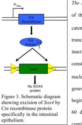
depicting action of Cre recombinase carrying out site-specific deletion of Sox4.
Figure 3. Cartoon gene diagram depicting action of Cre recombinase carrying out site-specific deletion of Sox4.
that Sox4 may be playing a critical role in maintaining and regulating the differentiation of the IESCs.
METHODS:
The Sox4fl/fl:VilCre Mouse Model: A genome-wide knockout of Sox4 results in embryonic lethality. To study the role of Sox4 in the adult intestine, we generated conditional transgenic knockout mice using the Cre-lox recombination system (Marioue et al., 2004). In our model, Cre-recombinase expression is driven by a villin promoter, a gene that is expressed exclusively in the intestinal epithelium. This system allows for an intestinal specific knockout of Sox4 (Figure 3). To attempt to discern the role of Sox4 in the epithelium, this mouse model will be examined using a variety of techniques including RNA and protein analysis.
The APCmin mouse model:. The APC gene is an component of the destruction complex which targets cytoplasmic β -catenin for degradation. This mouse model contains a truncation in one copy of the APC gene Following random inactivation of the remaining copy of APC, Wnt signaling is constitutively turned on and β-catenin is localized to the nucleus where it can act as a transcription factor, promoting genes for proliferation and cell growth. In this model, mice begin to display multiple intestinal neoplasias as early as 50-60 days, with more severe and damaging tumor growth coming at about 14 weeks (Moser et al. 1990, Su et al. 1992). Tissue Processing: Knockout mice with littermate controls for immunohistochemistry and quantification were sacrificed at 8-9 weeks after birth. Intestines were dissected out and divided into three equal length segments, with segment 1 representing the most proximal section and segment 3 the most distal. These sections were then flushed with ice cold Phosphate Buffered Saline (PBS) and fixed in 4% Paraformaldehyde (PFA). After fixing for 24 hours at 4°C, tissue was transferred to a 30% sucrose solution before it was filleted open to expose the lumen and give a flat intestinal segment. The flattened segments were then rolled up in a manner similar to a swiss-roll, and frozen in optimal cutting temperature embedding media (OCT). The swiss-rolls were then sectioned at a thickness of 6-8 µm,
Immunohistochemistry and quantification: Staining was performed according to previously described protocols (Gracz et al. 2013). Primary antibodies used were raised against murine Ki67, lysozyme, Ep-CAM, Mucin-2, Chromogranin-A, and Sox4 and visualized with Cy3 conjugated
secondary antibodies. Nuclear staining was done using bisbenzimide, following primary and secondary antibody incubation. For quantification, crypts counted had to meet certain criteria; crypts had to be open to the luminal space and extend their base to the lamina propria or muscle layer. For goblet cells and enteroendocrine cells, which exist in both the crypt and the villus, images for quantification were taken and cell number was quantified separately in the crypt and in the villus. Imaging analysis software (ImageJ version 1.48a) was used for cell quantification and morphometric analysis.
Quantitative Real-Time PCR (qRT-PCR): After sacrificing mice at 8-12 weeks of age, 0.5 cm segments of jejunal segment number 2 and
proximal and distal colon were dissected out and placed into RNAlater (Qiagen) for 24 hours at 4° C. RNA was then extracted and purified, and cDNA was made using the iScript cDNA Synthesis Kit (Bio-Rad). qRT-PCR was performed with SsoFast Probes Supermix with ROX. qRT-PCR data was analyzed using StepOne Plus Software. Tumor dissociation and flow cytometry
APCmin mice between 14-16 weeks were sacrificed, and the intestines were removed and flushed as normal. The intestines were then opened longitudinally, and tumors were dissected out under a dissecting microscope.
These tumors were minced with a razor blade to a paste-like consistency and placed in an EDTA chelation buffer for 45 minutes at 4° C. These chunks of tissue were then spun down at 1500 rpm for 5 minutes and resuspended in pre-warmed Hank’s Balanced Salt Solution (HBSS, Gibco) containing dispase and DNase. This suspension was placed at 37° C and shaken lightly every 1.5 minutes, with samples taken to observe dissociation progress after each step. After 6-10 minutes of this enzymatic digestion, the cells were put through a 40 µm filter directly into ice cold PBS containing 10% Fetal Bovine Serum (FBS). The single cells were then washed twice with cold PBS, and resuspended in IESC media (DMEM, 10% FBS, B27, N2, Pen/Strep, Y27632, NAC). Cells in IESC media were stained for 1 hours with EpCAM antibody (EpCAM FITC, Biolegend), and washed twice. Live/dead cell exclusion stains were added 10 minutes before flow cytometry. All flow cytometry was performed using the Beckman-Coulter Cyan machine in the Lineburger Cancer Center.
RESULTS
Characterizing Sox4 expression patterns
SOX4 was expressed at both a high and low level in different cells of the crypt (Figure 4). The cell position of each subset was quantified and showed that Sox4 low expressing cells were primarily at the crypt base, around position +1 to +3 and becoming less frequent towards the top of the crypt. The Sox4 high expressing cells, however, were found most frequently at cell position +5, a position that is consistent with that of the ‘reserve’ ISC population (Van Landeghem et al., 2012).
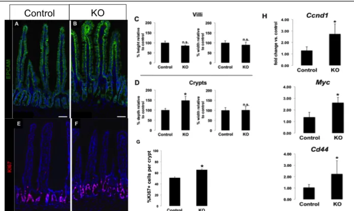
Loss of Sox4 results in crypt hyperplasia and hyperproliferation
To test whether or not an increase in cell proliferation was responsible for the oversized crypts, slides were stained with the proliferative marker KI67. The percentage of KI67 positive crypt cells were quantified and this showed that there was increased proliferation in the crypts of Sox4 knockout mice compared to controls (Figure 5). While this increase was obvious in many crypts, it appeared more subtly in others, but overall was statistically significant (n=3 animals, 25 crypts per animal, p<0.05). Additionally, it was qualitatively observed that there appeared to be more Ki67 positive cells lower in the crypt near the stem cell zone and higher towards the top of the crypt, although cell position has yet to be quantified.
Sox4 regulates Wnt signaling
Due to the significant in KI67 positive proliferating cells, it was hypothesized that Sox4 may be involved in regulating Wnt signaling, a well studied pathway that controls cellular growth and proliferation14. Quantitative Real-Time PCR on several direct Wnt target genes such as Myc, Cyclin D-1, and CD44 confirmed that there was increased Wnt signaling in the knockout animals. This result was also observed in tissue from both the proximal and distal colon sections. These data suggest that Sox4 is acting as an inhibitor of proliferation, at least partially by down-regulating Wnt signaling.
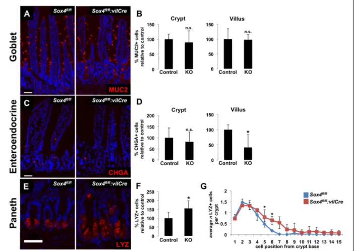
Sox4 regulates secretory lineage allocation in the intestinal epithelium
Investigating the secretory lineages revealed an interesting pattern, marked by a significant decrease in enteroendocrine cells and a concomitant increase in the number of Paneth cells (Figure 6). No significant change in the number of goblet cells was observed and goblet cell numbers per crypt and per villus was highly variable across biological replicates.
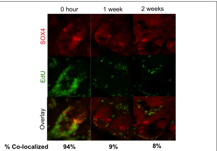
Sox4 high expressing cells are not label-retaining
To directly interrogate whether or not Sox4 expressing cells were acting in the label-retaining cell described in Buczacki et al. 2013, cells were labeled with the thymidine analog EdU over a 28 day period using subcutaneously implanted osmotic mini-pumps. Over this 28 day period, every cell that enters S phase of the cell cycle is labeled with the thymidine analog which can later be visualized with the (Click-it EdU Kit, Invitrogen). The label is then allowed to wash out, such that all cells that divide lose their fluorescent label, and those that are long-lived and dividing at a slow rate remain labeled.
Sox4 is highly expressed in small intestinal adenomas and is correlated with decreased EpCAM expression
To begin to investigate the role of Sox4 in tumorigenesis, tissue from APCmin mice was stained for Sox4, showing that it is highly expressed in cells of small intestinal adenomas. Interestingly, when stained simultaneously with Epithelial Cell Adhesion Molecule (EpCAM), it can be seen that cells highly expressing Sox4 appear to have a severely decreased expression of EpCAM (Figure 8). These same cells also begin to collapse from a polarized epithelium into an unorganized tissue mass and blend with the mesenchyme previously in the deeper tissue layers. This process is suggestive of a potential EMT and the invasion that is necessary for tumor metastasis.
Flow cytometry can also be used to validate the transition of cells from a high to low EpCAM expression profile. Following dissociation of tumors to single cells, a histogram of cells expressing EpCAM at different levels confirms that many cells are showing decreased levels of EpCAM. This flow cytometric analysis will allow us to derive an appropriate gating strategy for using fluorescent activated cell-sorting (FACS) to sort out these cell populations for subsequent RNA analysis.
CONCLUSIONS: The expression pattern of Sox4 protein suggests that it may be acting in a context-dependent manner, in two different cell types. First, it showed that Sox4 was being expressed at a low level in the stem cells, which occupy the base of the crypt at positions +1 +3. The identity of cells in which Sox4 is expressed at a high level is a more complicated question. Early label-retaining studies propose a quiescent, slowly-dividing stem cell at the +4 position,
and a recent study using a novel Cyp1a1-H2B-YFP mouse model to more precisely locate the label-retaining cell found it predominantly at position +3, but in a variety of positions from +1 to +6. It is also well known that the transit amplifying cells occupy positions +6 and up in the crypt. Given these facts, it remains possible that Sox4 is acting at a high level in either a long-lived, label-retaining, reserve stem cell, or in the rapidly-dividing transit amplifying progenitor cells.
The increased levels of KI67 by immunofluorescent quantification and qRT-PCR, combined with Sox4 expression in the stem cells, strongly suggest that Sox4 is involved in regulating the proliferation of intestinal epithelial stem cells. More specifically, the data suggests that in normal tissue, Sox4 is limiting proliferation by down-regulating genes that regulate cell cycle entry and growth through the Wnt pathway.
Quantification of secretory cells in knockout animals reveals a lineage allocation defect where cells are preferentially fated towards Paneth cells rather than enteroendocrine cells in the absence of SOX4. This result is striking in light of the existence of the label-retaining secretory precursor cell that gives rise to both enteroendocrine and Paneth cell lineages described in Buczacki et al. 2013. This result lends support to the hypothesis that Sox4 high cells may be acting in a label-retaining secretory precursor, pushing this cell towards an enteroendocrine fate rather than a Paneth cell fate.
transiently in the label-retaining secretory precursor and we have simply not caught these cells at the right time in their lifespan. However, given the data it seems more likely that Sox4 is regulating a short-lived transit amplifying progenitor cell. This joint function of regulating proliferation and differentiation is consistent with roles shown in other tissues (Schilham et al., 1996, 1997, Bergsland et al, 2006).
In the cancer context, it appears as though Sox4 may have a role unique from its function in normal intestinal epithelium homeostasis. The correlation between increased Sox4 levels and decreased EpCAM levels suggests that a possible EMT may be occurring in our mouse model of colorectal cancer. While it is well established that EMT is an essential program in colorectal cancer metastasis, the driver of this program remains unknown. The highly aberrant expression
of Sox4 in the adenomas combined with previous work in breast tissue implicating Sox4 as a master regulator of EMT, provides tanatalizing clues that Sox4 may be responsible for promoting the EMT program in colorectal cancer. While further study in this area is certainly needed, confirmation of this finding would have profound impact on colorectal cancer research and would identify Sox4 as a therapeutic target to manage tumor progression and metastasis.
References:
1. Wright N, Allison, M. The Biology of Epithelial Cell Populations. Oxford: Clarindon, 1984. 2. Cheng H. Origin, differentiation and renewal of the four main epithelial cell types in the mouse small intestine. II. Mucous cells. Am J Anat 141: 481-501, 1974.
3. Cheng H. Origin, differentiation and renewal of the four main epithelial cell types in the mouse small intestine. IV. Paneth cells. Am J Anat 141: 521-535, 1974.
4. Cheng H, and Leblond CP. Origin, differentiation and renewal of the four main epithelial cell types in mouse small intestine. I. Columnar cell. Am J Anat 141: 461-479, 1974.
5. Cheng H, and Leblond CP. Origin, differentiation and renewal of the four main epithelial cell types in mouse small intestine. III. Entero-endocrine cells. Am J Anat 141: 503-519, 1974.
6. Cheng H, and Leblond CP. Origin, differentiation and renewal of the four main epithelial cell types in the mouse small intestine. V. Unitarian Theory of the origin of the four epithelial cell types. Am J Anat 141: 537-561, 1974.
8. Gracz AD, and Magness ST. Sry-box (Sox) transcription factors in gastrointestinal physiology and disease. Am J Physiol Gastrointest Liver Physiol 300: G503-515, 2011.
9. Schilham MW, Oosterwegel MA, Moerer P, Ya J, de Boer PA, van de Wetering M, Verbeek S, Lamers WH, Kruisbeek AM, Cumano A, and Clevers H. Defects in cardiac outflow tract formation and pro- B-lymphocyte expansion in mice lacking Sox-4. Nature 380: 711-714, 1996. 10. Bergsland M, Werme M, Malewicz M, Perlmann T, and Muhr J. The establishment of neuronal properties is controlled by Sox4 and Sox11. Genes Dev 20: 3475-3486, 2006.
11. Schilham MW, Moerer P, Cumano A, and Clevers HC. Sox-4 facilitates thymocyte differentiation. Eur J Immunol 27: 1292-1295, 1997.
12. Marjou FE, Robine, S. Tissue-specific and inducible Cre-mediated recombination in the gut epithelium. Genesis 39: 186-193, 2004.
13. Gracz AD, Puthoff BJ, Magness ST. Identification, isolation, and culture of intestinal epithelial stem cells from murine intestine. Methods mol bio 879: 89-107, 2012.
14. Gregorieff A, Pinto D, Begthel H, Destree O, Kielman M, and Clevers H. Expression pattern of Wnt signaling components in the adult intestine. Gastroenterology 129: 626-638, 2005.
15. Buczacki A, Zecchini H, Winton DJ. Intestinal label-retaining cells are secretory precursors expressing Lgr5. Nature 000; 0-5, 2013.
18. Su LK, Kinzler LK, Vogelstein B, Preisinger AC, Moser AR, Luongo C, Gould KA, Dove WF. Multiple intestinal neoplasia cause by a mutation in the murine homolog of the APC gene. Science 256: 668-670, 1992.
19. Vervoort SJ, Boxtel RV, Coffer PJ. The role of SRY-related HMG box transcription factor 4 (SOX4) in tumorigenesis and metastasis: friend or foe? Oncogene 32: 3397-3409, 2013.
20. Loboda, A, Nebozhyn, MV, Watters JW, Buser CA, Shaw PM, Huang PS, Veer LV, Tollenaar RA, Jackson DB, Agrawal D, Dai H, Yeatman TJ. EMT is the dominant program in human colon cancer. BMC Medical Genomics 4: 9-19, 2011.