of BenzEalanthracene using a Synthetic Iron Porphyrin
Catalyst to Model Cytochrome P-450. (Under the direction of
DR. AVRAM GOLD)
Oxidations of the PAH benzEalanthracene were carried
out using ro-chloroperoxybenzoic acid as monooxygen donor and
the 2,6-chlorotetraphenyl-substituted iron porphyrin as
catalyst. These reactions were performed in two different
modes. The first used the oxoferryl porphyrin cation
radical as direct oxidant, essentially allowing only one
turnover of catalyst, whereas the second used the
high-valent complex in a multi-turnover, catalytic mode.
Phenoxathiin hexachloroantimonate was used as a model
one-electron oxidant to compare its product profile to the other
modes. Identical products (or substitution at the same
carbon) in the other modes would provide support for a
one-electron pathway. ^®0-labelled mCPBA was used in oxidations
to provide direct evidence for involvement of the peroxyacid
as the monooxygen donor, and ^®0 incorporation in the
oxidized PAH was observed. Analysis of the product profiles
in the three different oxidations supports one-electron
oxidation as the major pathway operating. The products of
the porphinatoiron-mediated oxidation are also postulated to
depend on the relative amounts of substrate and available
ACKNOWLEDGEMENTS
I would like to thank Dr. Avram Gold for giving me the
opportunity to work in his laboratory on this project, and
for all his help and guidance throughout. I also greatly
appreciate the time spent by both Dr. Jayaraj and Dr.
Sangaiah in assisting me with procedures and explaining the
chemistry involved. I am also grateful to Dr. Ball and Dr.
Aitken for taking the time to read this document and
TABLE OF CONTENTS
Page
ACKNOWLEDGMENTS...iii
LIST OF FIGURES...v
Chapter I. INTRODUCTION...1
II. LITERATURE SEARCH...8
Metabolic Activation of PAH...8
Metabolism of BenzCalanthracene...12
One-electron oxidation of PAH...13
Catalytic Cycle of Cytochrome P-450...17
Model Oxidations Using Synthetic Catalysts...20
III. EXPERIMENTAL METHODS...26
Instrumentation and Laboratory Materials...26
Porphyrin Synthesis...26
Synthesis of TPP ( 2, 6-Cl) H2...27
Metallation of TPP(2, 6-Cl)H2...28
Synthesis of FeTPP ( 2, 6-Cl )0S02CF3...28
Oxidations Using Porphyrin Catalysts...29
Oxidation with the compound I analogue...29
Catalytic or Multi-turnover Oxidations...30
Acetylation of Oxidation Reaction Mixtures.30
One-electron Oxidations...31Preparation of Phenoxathiin SbClg...31
Oxidation using Phenoxathiin SbCl5...31
IV. RESULTS...32
^H NMR and UV-visible spectra of BCaDA...32
Oxidation of BCaDA using Fe(IV) cation
radical as direct oxidant...32Oxidation of BCalA using ^®0-mCPBA...42
Catalytic Oxidations of BtaDA...45
Phenoxathiin SbClg oxidation...50
V. DISCUSSION AND CONCLUSION...53
Summary...62
1. TPP(2, 6-Cl)Fe(III)...4
2. BenzC a] anthracene...4
3. Expected Oxidation Products...7
4. Mammalian Pathways in PAH metabolism...9
5. The NIH Shift...11
6. Possible Metabolic Fates of an Arene Oxide...11
7. Catalytic Cycle of Cytochrome P-450...IS
8. ^®0-lncorporation into Norbornene Oxide...18
9. Proposed Pathway for the Epoxidation of Olefins...22
10. Proposed Orientation of Porphyrin and Substrate...22
11. Ligand Exchange...25
12. ^H NMR Spectrum of BE a] A...33
13. UV-Visible Spectrum of BCalA...34
14. a) FAB Mass Spectrum of 7, 12-0CD3-B[a3A...35
b) ^H NMR Spectrum of 7, 12-0CD3-BCa]A...35
15. a) FAB Mass Spectrum of 7-0CD3-12-0H-BCa]A...36
b) ^H NMR Spectrum of 7-0CD3-12-0H-BCa]A...36
16 a) UV-Visible Spectrum of 7, 12-0CD3-BCa]A...37
b) UV-Visible Spectrum of 7-0CD3-12-0H-BCa]A...37
17. ^H NMR Spectrum of 8-, and 11-acetoxy-BCa]A...39
18. EI Mass Spectrum of 8-, and 11-acetoxy-BCa]A...40
19. UV-Visible Spectrum of 8-, and ll-acetoxy-BCalA...41
20. a) EI Mass Spectrum of 8-,and 11-acetoxy BEalA
labelled with ^^O-mCPBA and acetylated overnight...44
21. ^H NMR Spectrum of y-OCDg-BCaDA...46
22. EI Mass Spectrum of 7-OCD3-B[a3A...47
23. Accurate Mass for 7-0CD3-BCa]A...48
24. a) UV-Visible Spectrum of 7-0CD3-Bta]A...49
b) UV-Visible Spectrum of 7-Cl-B[a]A...49
25. ^H NMR Spectrum of 7-Cl-B[a]A...51
26. EI Mass Spectrum of 7-Cl-B[a3A...52
27. BCalA/Porphyrin Interaction...58
28. Oxygen Rebound Mechanism...59
The metabolism of polycyclic aromatic hydrocarbons
(PAH) is mediated by a class of heme proteins known as
cytochromes P-450, and activation by these enzymes is
thought to be requisite for PAH to exert carcinogenic or
mutagenic effects. The P-450 complex functions as a
monooxygenase and carries out oxidative reactions on a wide
variety of xenobiotics. Hydroxylation by cytochrome P-450
involves the uptake of two electrons from NADPH with the
concomitant reduction of one atom of O2 to water; the other
oxygen atom is incorporated into the substrate (White andCoon 1980). While this conversion usually produces a more
excretable metabolite, it can also result in the formation
of reactive intermediates that are either toxic or
carcinogenic.
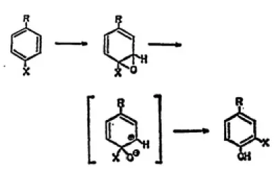
In mammalian liver homogenates, initial insertion of an
oxygen across the double bond of an aromatic system to form
an arene oxide has been shown to be a key step in the
activation of PAH (Jerina and Daly, 1974). The arene oxide
can undergo one of two enzyme-catalyzed reactions.
trans-subject to non-enzymatic isomerization to a phenol (Diinges,
1973). This rearrangement, which can result in retention of
substituents at the oxidized position via a 1,2-migration,
is termed the NIH shift (Jerina and Daly, 1974).
Cytochrome P-450 can also carry out oxidations of
hydrocarbons in the presence of iodosyl benzene and
peroxyacid monooxygen donors (Hrycay and O'Brien, 1974).
Since O2 or reducing cofactors are not involved, cytochrome
P-450 is functioning in these reactions as a peroxidase, an
enzyme that utilizes H2O2 as the ultimate electron acceptor.
Horseradish peroxidase (HRP) and its intermediates have been
extensively characterized. During catalysis, HRP is
initially oxidized by two electrons to a green species known
as compound I. It has been postulated that one of the two
oxidation equivalents in compound I is stored as low spin
ferryl iron (Fe<III)-->Fe<IV)), while the other oxidation
equivalent is stored as a porphyrin centered n-cation
radical. The techniques of Mossbauer spectroscopy and EPR
lend support to this theory. From MSssbauer studies it was
suggested that the iron is in the Fe(IV) state (Moss,
Ehrenberg and Bearden, 1969), while EPR data have been
Due to this presumed similarity, there have been
numerous studies performed using synthetic catalysts with
monooxygen donors to model the active center of cytochrome
P-450. Among the many studies, only a few have rigorously
demonstrated the presence of a compound I analogue (Gold et
al., 1988; Groves et al., 1981, 1986). While substituted
porphyrins in biomimetic oxidations do not approach the
complexity of an enzymatic protein, insight into the
mechanism of oxidation can be gained. Therefore, model
oxidations on selected PAH were carried out employing
porphyrin complexes shown to form observable compound I
analogues when a peroxyacid is supplied as the monooxygen
source.
The synthetic catalyst employed was
2,6-dichlorophenyl-substituted iron porphyrin. Formation of the compound I
analogue CTPP(2,6-Cl)FeG3+ (Fig. 1) has been confirmed by
spectroscopic and resonance studies. This porphyrin
possesses strongly electron withdrawing ligands on its
periphery and its ferryl-porphyrin radical spin are strongly
parallel coupled. Presence of chlorine on the phenyl rings
should render the cation radical intermediate more strongly
oxidizing. The specific ramifications of the spin coupling
characteristics pertaining to reactivity are still not
clear, but it is important to note that all known compound I
o
Fig. 1. Structure of
2,6-dichlorophenyl-substituted iron porphyrin.
TPP(2,6-Cl)Fe(III)
o
o
o
o
Fig. 2. Benz[a]anthracene, an alternant
having antiferromagnetic coupling, to see what effect this
has on reaction pathway and product profiles.
BenzCalanthracene, an alternant PAH containing four fused
benzene rings (Fig. 2), was selected as substrate for the
chemical oxidations.
One objective of this study was to determine whether the reaction pathway proceeds via direct electrophilic attack by the oxo oxygen on the PAH periphery, or initial
one-electron charge transfer, in which the PAH is oxidized
to a cation radical. To help clarify this point the oxidations were performed in two different ways, and the major products that resulted were compared. The first
employed the oxoferryl porphyrin cation radical as a direct
oxidant, generating the green compound I analogue, to which
the substrate was subsequently added. The second set of oxidations was carried out in a truly catalytic mode using a small concentration of porphyrin catalyst and excess
substrate, to which was added an approximate 100-fold excess of ro-chloroperoxybenzolc acid (mCPBA). This allows the
porphryln to "turn over", and simulates the enzymatic
reaction which cytochrome P-450 catalyzes in a biological
compound is an effective electron acceptor, and is known to
carry out one-electron transfer exclusively. Data generated
from these studies will lend insight into the likelihood of
one-electron transfer in the other modes if identical
products are obtained.
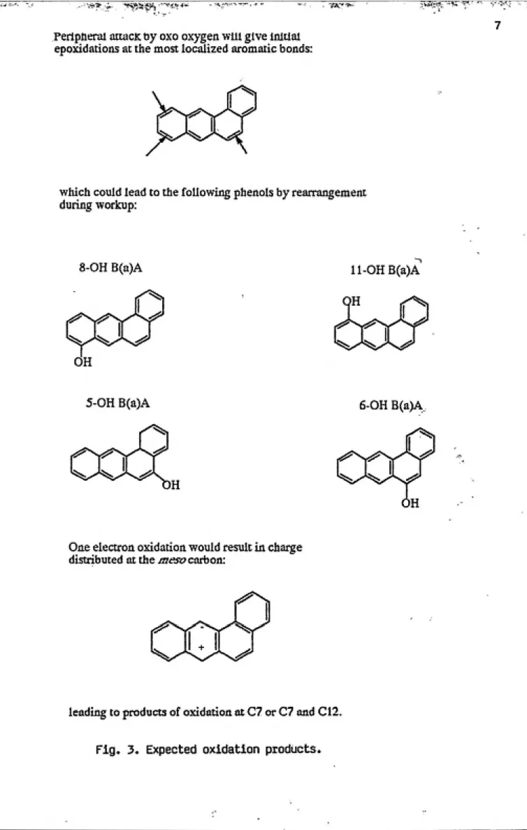
The regioselectivity of oxidation is determined by
steric constraints that the iron(V)-oxo complex encounters
when it reacts with the PAH, as well as the charge
distribution properties of the aromatic system (see Fig. 3).
For this reason, distinguishing between these two pathways and proving which one is operating is not always
straightforward. We wished to compare the product
distributions obtained under both sets of conditions using
the porphyrin catalyst with the product distribution obtained by the known one-electron oxidant as a means of
which could lead to the following phenols by rearrangement
during workup:
8-OH B(a)A ll-OHB(a)A
5-OH B(a)A 6-OH B(a)A
One electron oxidation would result in charge
distributed at the mesocarboa:
leading to products of oxidation at C7 or C7 and C12.
Metabolic Activation of PAH
Polycyclic aromatic hydrocarbons are compounds
containing carbon and hydrogen with fused benzene rings in linear, angular and cluster arrangements. They are
universal products of the combustion of organic matter, and
their occurence in the environment is due to both natural
and anthropogenic sources (Blumer, 1976).
The enzymes responsible for bioactivation of PAH and
other xenobiotics are localized primarily in the endoplasmic reticulum of hepatocytes. Of these proteins, the most
important are the oxidative enzymes in the cytochrome P-450 system, also termed the mixed-function oxygenase (MFO)
system. Other enzymes involved in mammalian PAH metabolism
include epoxide reductase, epoxide hydrolase, glutathione transferase, UDP-glucuronyl transferase, and
sulfotransferase, and these pathways are shown in Fig. 4. In general, the activity of these enzymes renders the
substrate more polar and facilitates excretion from the cell
and body. In some cases, however, the intermediates become
electrophilic (electron deficient) and bind to nucleophilic
c^
H H
I SG
rcductat*
Glutathione
S-«poxid«
trant{«ras«
P-450
02
H
•poxido hydroioso
H2O
ZmOH
non-•nzymatic covoiont binding to ONA, RNA
and protoin
^-Sulfates
O-GlucuronidesFig. 4. Major pathways involved in the mammalian
metabolism of polycyclic aromatic hydro¬
certain nucleic acid adducts are considered to have the
potential to initiate malignant transformation. Thus, the
same enzymes that serve as detoxifiers can also act to increase the toxicity of certain xenobiotics.
Most PAH are not reactive or carcinogenic as the parent
compound and require activation to be converted to a species
that is biologically active with no further metabolic
transformation, called an ultimate carcinogen. One
oxidative pathway of PAH and olefins involves addition of
oxygen across the C-C double bond. This reaction is carried
out by a monooxygenase, and results in the formation of an
epoxide. Important indirect evidence for an epoxide as an
intermediate is the demonstration of an NIH shift (Fig. 5),
that often accompanies rearrangement of an arene oxide to a
phenol. The NIH shift involves the intramolecular migration
of ring substituents, from the position where the oxygen is
introduced to a neighboring position on the ring. The
strained three-membered ring of the epoxide intermediate
results in a highly reactive species, and upon opening to an
electrophilic carbonium ion it seeks nucleophilic sites
within the cell.
The extent of binding of this electrophile will depend
on the rate of formation of arene oxides and the rates of
hydration (both enzymatic and non-enzymatic) to
dihydrodiols, conjugation with glutathione, and
isomerization to phenols (Fig. 6). Where oxidation products
4
Fig. 5. The NIH shift. This migration and
retention of substituents occurs during
the monooxygenase-catalyzed formation
ofphenols.<st
4,.
iii>^
\«
.0^'
EnzymaifeHydration
9f
S35$5
a"
N3Ha:
Fig. 6. Three possible metabolic fates of an
arene oxide: 1) Isomerization to a phenol
2) Hydration to a dihydrodiol 3) Conjugation
formed during isomerization usually depend on the stability
of the corresponding carbonium ion intermediate (Jerina et
al., 1971).
Metabolism of BenzEalanthracene
The bacterium Beijerinckla sp. strain B-836 metabolizes
benzCalanthracene to four cis-dihydrodiols, formed by the
incorporation of both atoms of molecular oxygen into the
aromatic nucleus, and carried out by a dioxygenase. Fungi,
in contrast, oxidize PAHs via cytochrome P-450 monooxygenase
and epoxide hydrolase-catalyzed reactions to
trans-dihydrodiols. The fungus C. elegans was shown to oxidize
BCaDA predominantly to tra/Js-S, dihydroxy-8, 9-dihydrobenzCalanthracene (Cerniglia et al., 1980).
BenzCalanthracene is metabolized by mammalian
microsomal preparations and by a purified monooxygenase system to yield trans-5,6-dihydrodiol and trans-&,
9-dihydrodiol as major metabolites. rj-a/3s-3, 4-,-10, 11-,-1, 2-dihydrodiols are also formed in small amounts (Sims, 1970; Thakker et al., 1979). In general, BCalA and its
metabolites are considered to be weak carcinogens. However
one metabolite, the trans-3, 4-dihydrodiol, is known to possess mutagenic and tumorigenic capabilities. This
congener has been observed to be oxidized by the cytochrome
P-450 monooxygenase system to products that are 10 times
more mutagenic in Salmonella typhimurium TA 100 than are
derivatives. Based on mutagenic, tumorigenic, and carcinogenic evidence, the tJ-ans-3, 4-dihydrodiol-l,
2-epoxides (bay region diol 2-epoxides) are indicated to be the ultimate carcinogenic metabolites of benzCa3anthracene (Wood
et al., 1977). This metabolite is formed on a three-sided
peripheral indentation called a "bay region." The diol epoxide arises by further enzyme-catalyzed oxidation of the double bond adjacent to the existing tj^ans—dihydrodiol. These results are in accord with studies on benzo[a3pyrene that identified a bay region dihydrodiol epoxide as this compound's ultimate carcinogen (Sims and Grover, 1981).
Metabolic activation of PAH by monooxygenation, which is formally a two-electron oxidation, involves formation of vicinal diol-epoxides as the ultimate carcinogen. Another major pathway which has recently become the focus of
considerable interest involves one-electron oxidation,
resulting in the formation of radical cations, which, like the diol-epoxides, can react with cellular nucleophiles
(Miller and Miller, 1981, Cavalieri and Rogan, 1984).
One-electron Oxidation of PAH
A PAH radical cation is generated by removal of a n electron from the aromatic system. The ease of formation of this species is a function of the compound's first
ionization potential (IP), which is the energy required for
complete removal of an electron from the highest occupied
Cytochrome P-450 acting in the presence of NADPH and
dioxygen is not considered to act primarily as a
one-electron oxidant, yet cases have been observed <Augusto et al., 1982; Rauckman et al., 1982). The one-electron pathway
appears to be more efficiently catalyzed in the presence of hydroperoxide cofactors. It has been demonstrated that the
oxidation of benzoCaDpyrene carried out by liver microsomes
yields different product distributions depending on whether
the hydroxylation is supported by NADPH or cumene
hydroperoxide (Capdevila, Estabrook, and Prough, 1980).
While the major products obtained with NADPH were phenols,
presumably derived from epoxidation and rearrangement, the
metabolite profile for cumene hydroperoxide showed a
preponderance of three quinone isomers. The quinones were likely formed from a one-electron oxidation of the
hydrocarbon, since these products are known to be derived also via non-enzymatic one-electron oxidations. These
results suggest that the reaction mechanism operating in the
presence of the organic hydroperoxide may differ from that
functioning in an NADPH-supported oxidation. For this reason caution must be used in equating NADPH-supported reactions of cytochrome P-450 and hydroperoxide-dependent
reactions of hemeproteins.
Peroxidases, which are enzymes that catalyze the
oxidation of inorganic or organic substrates at the expense
of a hydroperoxide, primarily catalyze electron transfer
aliphatic hydroxylation or olefin epoxidation, reactions
that proceed via direct insertion of oxygen across C-C double bonds. Initial electron transfer occurs between
substrate and enzyme, but subsequent reactions of the
resulting cation radical may occur in solution, and the
oxidation products will depend on the solution chemistry and
the nucleophilic species present (Ortiz de Montellano,
1986).
One specific peroxidase, called lignin peroxidase, is
produced by the fungus P, chrysosporlum, and acts to degrade
lignin, a plant polymer of randomly linked phenylpropane
units. In the presence of Yi2P2.' lignin peroxidase catalyzes
one-electron oxidations of lignin-related compounds, and due
to its low substrate specificity, it was expected to oxidize
aromatics and heteroaromatics as well. It was found that
purified lignin peroxidase did indeed carry out one-electron oxidation of PAH that had ionization potentials < a value of
7.55 eV (Hammel et al., 1986).
The lignin peroxidase-catalyzed oxidation of the PAH
pyrene was carried out in aqueous medium. Under these
conditions, the expected one-electron oxidation products are
the 1,6- and 1,8-quinones, resulting from nucleophilic
attack of water at carbons possessing the highest positive charge density (Cavalieri and Rogan, 1985). Through gas chromatography and mass spectrometry analysis the identity
experiments proved that water was the source of the quinone
oxygens (Hammel et al., 1986).
Previous studies supported the involvement of lignin peroxidase in the initial degradation steps of
benzo[a3pyrene to its expected quinone derivatives
(Haemmerli et al., 1986). Once formed, the benzoCaDpyrene
cation radical reacts with water to yield a mixture of 1,5-,
3,6-, and 6,12-diones. These results and those discussed
above are consistent with lignin peroxidase plus H2O2 acting
as a fairly non-specific oxidation reagent for aromaticpollutants, proceeding via a cation radical mechanism.
Horseradish peroxidase-mediated binding of PAH to DMA
is another reaction known to occur by one-electron
oxidation. Once formed, PAH radical cations bind covalently
to nucleophiiic sites of biological macromolecules including DMA. Again, studies of PAH with known ionization potential
have shown that compounds must possess an IP below a
specific threshold in order to be oxidized and activated by
this mechanism (Cavalieri et al., 1983).
Irrespective of the fate of the cation radical, evidence has accumulated indicating that only PAH with relatively low IP can be activated biologically by one-electron oxidation. The carcinogenicity of PAH with relatively high IP would be due to the formation of
bay-region diol epoxides (Conney, 19S2). The existence of a few
inactive PAH with low IP indicate that this characteristic
type of PAH include perylene and anthanthrene. These weak
or inactive PAH are known to have their positive charge
delocalized over several carbons, indicating highly
localized charge is also a requisite for activation by this mechanism. Positive charge localized on one or two carbons renders those positions more reactive toward nucleophiles.
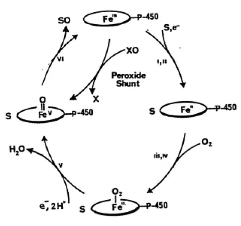
Catalytic Cycle of Cytochrome P-450
The active site of cytochrome P-450 contains a single iron protoporphyrin IX prosthetic group, and it is here that dioxygen is bound, reduced and activated. An overview of its catalytic cycle is shown in Fig. 7, and includes the following steps: 1) Binding of substrate, resulting in a high spin ferric complex ; 2) One electron reduction of iron(III) to iron(II); 3) Binding of dioxygen; 4) A second one electron reduction; 5) Heterolysis of the 0-Q bond and concomitant production of the active oxidizing species and
H2O; 6) Two electron oxidation of substrate producing
oxidized product and returning the enzyme to its resting
ferric state (Ortiz de Montellano, 1986).
Within this cycle, it has been found that the binding
and reduction of molecular O2 can be circumvented by the
1-450
S,er
XO
J Peroxide
yf Shunt
lll,IV
e.2H s
ͣ
450
Fig. 7. An overview of the catalytic cycle
of cytochrome P-450.
ArCOOH
T
18
Fig. 8. Scheme showing 0 incorporation into norbornene oxide, proving the oxygen
1-8
as monooxygen donors for the study of oxidative reactions
catalyzed by cytochrome P-450.
Although the active oxidizing species of cytochrome
P-450 has never been observed, the stoichiometry leading to
dioxygen activation and the shunt pathway using monooxygen
donors are identical to the chemistry characterized for
peroxidase enzymes. Therefore, insight into the nature of
the active oxidizing species has been sought through
knowledge of an extensively characterized peroxidase, namely
horseradish peroxidase (HRP). This protein reacts with H2O2
to generate an enzymic intermediate (HRP I or compound I)
that is a monooxygen complex two electrons oxidized above
the ferric resting state. The accepted view is that the
two-electron oxidation of the enzyme to compound I involves
heterolysis of the peroxy 0-0 bond, with one electron
removed from the iron atom, the second electron abstracted
from the porphyrin ring to form a n-cation radical. The
resulting chemical species can thus be described as an
oxoferryl porphyrin n cation radical. Compound I abstracts
one electron from the substrate to form a compound II
intermediate, which is subsequently reduced back to the
resting enzyme by an additional one-electron oxidation of a
second molecule of substrate (Van Wart and Zimmer, 1985).
Like horseradish peroxidase, the catalytic cycle of
cytochrome P-450 is thought to involve a ferryl ion species
as the active oxygen transfer agent. Support for the
peroxy acids and other single oxygen donors effect oxygen
transfer in a manner similar to the fully reconstituted
enzyme system.
Metabolism of PAH in mammalian, bacterial and fungal systems involves a wide variety of enzymes and cofactors and isozymes of cytochrome P-450 (Cerniglia, 1984). Comparing enzymatic product profiles to those obtained in biomimetic oxidations using synthetic catalysts has limitations.
Nevertheless, the fundamental aspects of the mechanism of
action of cytochrome P-450 can be elucidated through the use
of porphyrin catalysts that exhibit similar reactivity.
Model Oxidations using Synthetic Catalysts
Synthetic tetraarylporphyrins have been used
extensively to model the natural heme prosthetic group of cytochrome P-450. The bulky aryl substituents protect the
meso carbons of the porphyrin ring from oxidation, and reduce the likelihood of heme agglomeration in solution. That these synthetic catalysts could carry out oxidations in the same manner as cytochrome P-450 was an important detail
that had to be established. The first evidence came from
Groves and Nemo (1979), where they described oxidative reactions of alkenes catalyzed by porphyrin complexes. It was demonstrated that iodosylbenzene reacted readily with
iron porphyrin species in the presence of olefins to produce
In a later study by the same investigators, the
preparation and characterization of the high-valent iron porphyrin complex was reported (Groves and Nemo, 1981), using the sterlcally encumbered porphyrin complex iron tetramesityl-porphyrin chloride [Fe(TMP)Cl]. Employing m-chloroperoxybenzoic acid as oxygen donor, Fe(TMP)Cl was oxidized and converted to an apparent green intermediate, indicative of compound I analog formation. NMR, visible, Mossbauer and EPR data of the intermediate lend support to an iron(IV) porphyrin cation radical formulation. In
addition, in the presence of H2^®0, the unlabeled oxygen of
the catalytically active complex was exchangeable with the
heavier isotope, as indicated by 99% incorporation of ^®0
into the product epoxide (Fig. 8). These results are
consistent with an oxoiron intermediate, and tend to
disprove a free or metal coordinated peroxyacid as the active intermediate.
A more recent study explored preferential oxidation of the two isomers of stilbene using various synthetic
porphyrin catalysts, indicating a marked cis-trans
selectivity (Groves and Nemo, 1983). While cis-stilbene
reacted to produce cis-stilbene oxide, tr^ans-stilbene was found to be unreactive. This selectivity was postulated to result from nonbonding interactions between the phenyl rings of tj~aJ7S-stilbene and the iron porphyrin phenyl groups. The
degree of c±s/trans selectivity was dependent on the extent
I Ci
<!::>
L
6
\-j
ͣ
A
a
Fig. 9, Proposed scheme for the epoxidation of
olefins carried out by Fe (TMP)Cl and
iodosylbenzene.Fig. 10. Postulated approach for the iron-oxo
porphyrin, and is a good indication that oxygen transfer
must have occured at or very close to the iron center as
would be expected for an iron-oxo intermediate (Fig. 9). As
established in previous work (Groves and Nemo, 1981), the
iron IV porphyrin cation radical is the high valent species
involved in the epoxidation of the olefins in question.
Based on a mechanism derived from molecular orbital theory,
stereoelectronic effects on the oxygen transfer from iron to
the C-C double bond have been postulated to arise from a
side-on approach of substrate to allow favorable overlap
between the p orbitals of the Fe-bound oxygen and the olefin
(Fig. 10).
Investigations such as these emphasize the importance
of considering steric interactions between the synthetic
catalyst and organic substrate when evaluating product
distributions.
A study by Gold et al., 1988, investigated the
oxidation of benzCalanthracene in an mCPBA/porphinatoiron
system where the peroxyacid was proven to be unreactive in
the absence of catalyst. It was observed using
2,4,6-trimethoxyphenyl-, and 2,6-dichlorophenyl-substituted
catalysts that BEalA was oxidized predominantly to the
7,12-quinone. These results lend support to oxidation of the PAH
meso carbons via charge transfer. Taking steric
considerations into account, oxygen transfer via
feasible, and a charge transfer oxidation pathway would have
to be followed.
As indicated in the description of the enzymatic
profiles, there is no evidence of fungal, bacterial, or
mammalian enzymatic oxidation at the two meso carbons. The
difference in the product profiles could be the result of a
lower oxidation potential for the cytochrome P-450 oxoferryl
porphyrin cation radical than for the model complex. If the
oxidation potential is below that required to react with the
substrate via charge transfer, electrophilic substitution
would be the preferred pathway for the enzyme.
Many biomimetic studies on model compounds are carried
out in CH2C12* There is some uncertainty about the validity
of these studies due to the present knowledge that compound
I formation is not entirely clean when pure CH2CI2 is used
as a reaction medium. A polar co-solvent, such as
deuterated methanol (CD3OD), is also required, and was used
in the oxidations outlined in this report. This species
adds polarity to an otherwise non-polar solvent system,
facilitating ionization of the iron atom and exchange of the
axial ligand, eventually resulting in compound I formation.
In the case of our catalyst, (see Fig. 1), the axial ligand
is the triflate anion (0S02CF3~), which is weakly
coordinated to the iron, and electron-withdrawing chlorines
are present on the phenyl rings. Both of these
characteristics lead to facile compound I formation. The
formation via coordination of the peroxy acid, taking the
place of chlorine (or triflate) as the axial ligand, and
emphasizing the importance of ligand exchange in this
process.
O
II
CI „X-„-,- P 9
I
RCOO- 11 oFe'" ::;;=r Fe"' —^ Cl^ " ^"^^
a
CHAPTER III
EXPERIMENTAL METHODS
Instrumentation and Lab Materials
^H nuclear magnetic resonance (NMR) spectra were
obtained at the UNC Department of Chemistry on a Varian
XL-400 at XL-400 MHz. Chemical shift values are reported relative
to tetramethylsilane (TMS). UV-visible spectra were
recorded on a Spectronic 1201 spectrophotometer (Milton Roy
Company). Mass spectral data were taken using a direct
insertion probe on a VG 70S 250SEQ mass spectrometer in the
EI mode at 70eV at the UNC Department of Environmental
Sciences and Engineering.
Preparative thin layer chromatography (TLC) was
performed using Silica Gel PK5F (20cm x 20cm) 500 jam plates
(Whatman), while analytical TLC was conducted using silica
gel on aluminum (Aldrich). Column chromatography was carried
out using Merck Silica gel, grade 60, 230-400 mesh, 60A.
BenzCalanthracene was commercially available (Aldrich)
and was purified by column chromatography on silica using
benzene/hexane as eluant. m-Chloroperoxybenzoic acid was
obtained from Fisher Scientific Co. and used as received.
^®0-mCPBA was prepared by photolysis of ro-chlorobenzaldehyde
FeTPP(2,6-Cl)0S02CF3 was synthesized as described in the
methods section. Reagent grade solvents were employed, and
were dried and distilled prior to use where indicated. Any
routine chemicals were purchased from one of the following
sources: American Scientific Products; Aldrich Chemical
Company, Inc. ; Sigma Chemical Company; and Fisher
Scientific.
Porphyrin Synthesis
1) Synthesis of TPP(2.6-Cl)H7
This reaction was carried out under argon atmosphere by
evacuating a manifold system and filling with argon. 1.6 L
of CH2CI2 <0.2X ethanol, 3.2 mL added as preservative) was
placed in a 3-necked flask with reflux condenser and two
septa attached. 2,S-Dichlorobenzaldehyde (2.S g, 16 mM) was
added before 1.1 mL distilled pyrrole (16 mM) was placed
into the reaction mixture through a septum via syringe.
Finally, 1 mL BF3-etherate was added via syringe and stirred
2 hours to form the porphyrinogen (Hoffman et al., 1990).
The UV spectrum was checked after 2 hours ( max=511
nm, shoulder at 490 nm corresponding to porphyrinogen).
After addition of p-chloranil (2.85 g), the reaction was
refluxed for 2.5 hours to obtain the porphyrin free base
TPP(2,6-Cl)H2 (514 nm, 416 nm). The free base was allowed
through a medium frit to obtain a solid, which was
subsequently purified using an aluminum oxide column with
CH2CI2 as eluant. Fractions that were pure by UV spectra
and analytical TLC were pooled to give a final yield of 430
mg (11%).
2) Metallation of TPP(2.6-Cl)Ho
The solution of 210 mg free base in 85 mL DMF under
argon was heated to reflux and excess FeCl2«4H20 was added;
this proceeded approximately 17 hours overnight. The next
day an additional 100 mg FeCl2 was added and the reaction
heated 30 minutes, then refluxed under air for 2 hours.
DMF was distilled off under vacuum using a liquid
nitrogen collecting trap, and the remaining solid
redissolved in a minimal amount of CHCI3 and chromatographed
on a silica column using CHCI3 as eluant. A band was
collected containing a mixture of TPP(2,6-Cl)FeCl and
TPP(2,6-Cl)FeOH. To convert any OH complex to chloro
complex, H2SO4 was dropped onto NaCl, generating HCl gas
which was bubbled through a solution of the iron porphyrin
in 200 mL CH2C12- The solvent was evaporated on a rotary
evaporator, and the solid was dried and weighed. Yield was
180 mg FeTPP(2,6-Cl)Cl (86*/.).
3> Synthesis of FeTPP(2.6-Cl)0S02CF3
90 mg FeTPP(2,6-Cl)CI dissolved in 30 mL dry
6 mL THF in a Schlenk tube in a glove box under argon.
FeTPP(2,6-Cl)Cl has a characteristic UV spectrum with
max=4ia nm and 508 nm. After 3 hours, the UV spectrum was
taken ( max=395 nm, 511 nm) which corresponds to
FeTPP(2,6-Cl)0S02CF3. The THF was removed by bulb-to-bulb
distillation using a liquid nitrogen trap. The residue was
returned to the glove box, dissolved in a minimal amount of
dry CH2Cl2» and filtered to remove insoluble silver salts.
The solvent was removed, yielding 102 mg of pure triflate
complex (98.9%). This species was subsequently used in all
oxidations where a porphyrin catalyst was employed.
Oxidations Using Porphyrin Catalysts
Oxidation with the green Compound I analog
This oxidation was carried out at -80°C in an
ethanol/dry ice bath under argon atmosphere. 16 mg
ro-chloro-peroxybenzoic acid <.09 mmoles) in 0.5 mL CD3OD was added to 16 mg FeTPP(2,6-Cl)Tf (.02 mmoles) in 2 mL CH2CI2 to
generate the green compound I analog (oxoferryl porphyrin
cation radical). B[a]A (50 mg, .22 mmoles) in 2 mL CH2CI2
was added, and a color change from green to brown was
observed. After 1 h, 5 mL saturated Na2C03 solution was
added to neutralize the mCPBA, CHCI3 was added, and the
organic layer was extracted and dried over anhydrous sodium
sulfate. The solvent was evaporated on a rotary evaporator
minimal amount of CHCI3) was applied to preparative TLC
plates (500pm) to separate the oxidation products.
Catalytic or Multi-turnover Oxidations
These oxidations used an approximate 100-fold excess of
mCPBA over the porphyrin catalyst to generate multi-turnover
conditions. Again 40-50 mg BCalA and 7 mg FeTPP(2,6-Cl)Tf
(.006 mmoles) were stirred together at -80'*C under argon
before 80 mg mCPBA (.46 mmoles) was added to serve as oxygen
donor. The reaction was allowed to proceed for 1 hour and
the subsequent workup was identical to the other oxidative
mode in which the porphyrin was used as catalyst.
Acetylation of Oxidation Reaction Mixtures
Due to the observed instability of some phenols and
dihydrodiols which prevented accurate characterization of
the reaction products, the reaction mixtures were acetylated
following oxidation. Through the addition of pyridine and
acetic anhydride any phenols or dihydrodiols formed are
acetylated and stabilized.
The oxidation workup was amended as follows:
After reaction completion, before the addition of
bicarbonate, the solvent was removed by blowing reaction
mixture with argon. The residue was redissolved in a
minimal amount of pyridine and an equal volume of acetic
anhydride (1:1 volume ratio) was added, and the mixture was
bicarbonate, and the same subsequent steps were followed to
obtain the reaction product mixture.
One-electron Oxidations
Preparation of Phenoxathiin hexachloroantimonate
This reaction was performed at room temperature in the
glove box under argon atmosphere, following the procedure of
Gans et al, 1981. 600 mg of antimony pentachloride in 10 mL
dried CH2CI2 was added to a solution of 500 mg phenoxathiin in 15 mL CH2C12. The reaction mixture was allowed to stir
for 15 minutes, during which time the cation radical
hexachloroantimonate precipitates as a violet
microcrystalline powder. The solution was filtered and
dried and the solid was weighed. Average yield was 830 mg
oxidant (75*/.).
Oxidation using Phenoxathiin hexachloroantimonate
This reaction was performed at room temp, under argon
atmosphere. An equimolar amount of phenoxathiin
hexachloroantimonate (62 mg, .12 mmoles) in 5 mL dry CH2CI2 was added to 27 mg B[a]A (.12 mmoles) in 5 mL CH2CI2. The
reaction was quenched after 30 min. with 10 mL methanol, and
the solvent was removed at 55''C on a rotary evaporator. The
resulting solid mixture was dried under oil pump vacuum, and
separation of the oxidation products was carried out using
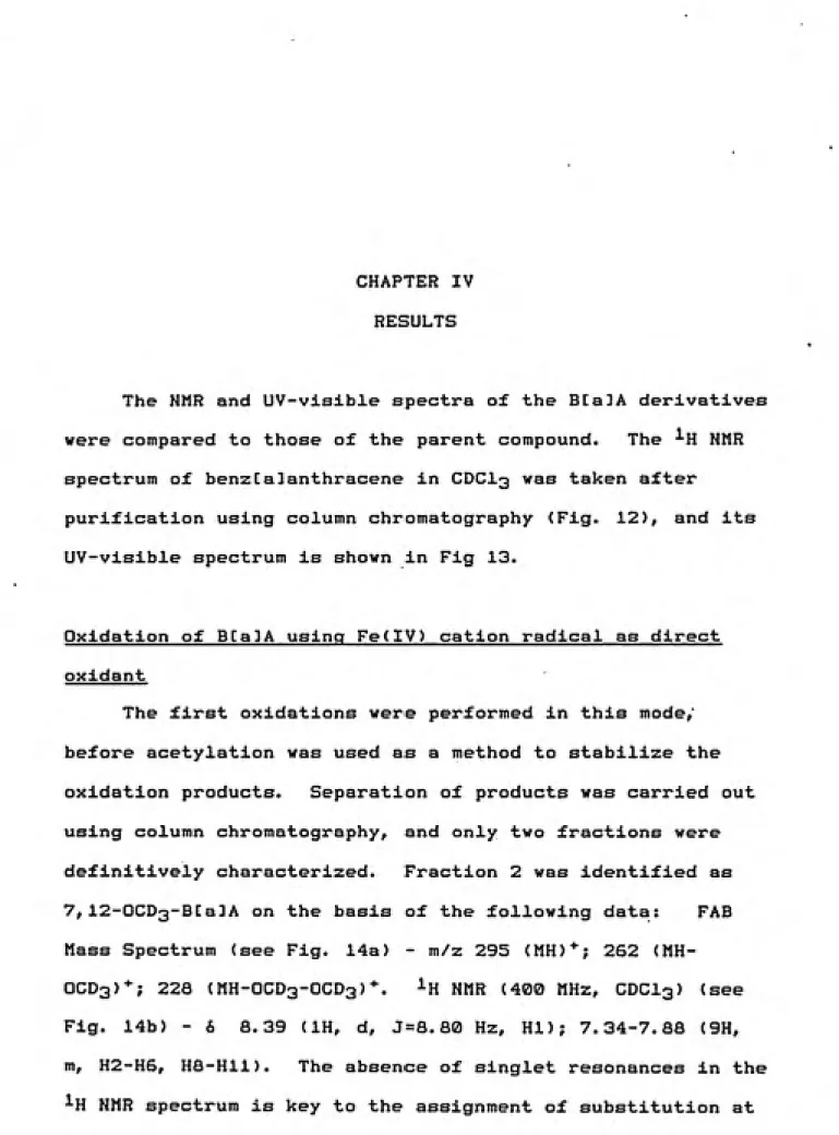
CHAPTER IV RESULTS
The NMR and UV-visible spectra of the BCaDA derivatives
were compared to those of the parent compound. The ^H NMR
spectrum of benzCalanthracene in CDCI3 was taken after
purification using column chromatography (Fig. 12), and its
UV-visible spectrum is shown in Fig 13.
Oxidation of BEaJA using Fe(IV) cation radical as direct
oxidant '
The first oxidations were performed in this mode,'
before acetylation was used as a method to stabilize the
oxidation products. Separation of products was carried out
using column chromatography, and only two fractions were
definitively characterized. Fraction 2 was identified as
7,12-OCD3-BCa3A on the basis of the following data: FAB
Mass Spectrum (see Fig. 14a) - m/z 295 (MH)*; 262
(MH-OCD3)*; 22a (MH-OCD3-OCD3)*. ^H NMR (400 MHz, CDCI3) (see
Fig. 14b) - & 8.39 (IH, d, J=8. 80 Hz, HI); 7.34-7.88 (9H,
m, H2-H6, HS-Hll). The absence of singlet resonances in the
1
285
200nm 500nm
109!
98.
38.
78.
60. 58.
48,
20,
18,
93
75
262
228
185
U9
i|4n#iU
,l1
1
12 1.8
ͣ
4ipiUlh4
186 156 288 258 386 358
486 458 568
mFig.14a. FAB Mass Spectrum of Fraction
2-7,12-OCD -B[a]A.
1
14b. H NMR Spectrum (400 MHz, CDCl^) of
Fraction 2.
»j«l»SiV»''»»»'Vw«>f'*«
ͣͣ
«
ͣͣ
«"< v^iK Y'f •'•»"< <-NM«rv.>'**'-'*'"'^^**»
y
.aX„J
-T---,---1-^ , ""I—T—T---1---r—]—J—-r—1---r- - 1'—i—T r i p-t r -1 —i"- -|...f i
1081 33
78 S8 58. 48. 38, 28
18J
6
244
1$
uLu
-4
215
ilUi
278
188 156 268 258
llL, iT
«1 1.8
356 468 456 566 RRSS
Fig. 15a. FAB Mass Spectrum of Fraction
3-7-0CD3-12-0H-B[a]A.
15b. ^H NMR Spectrum of Fraction
3-(400 MHz, CDCl^).
^^•**Uk>VAWv,/VuA
V^*w^A^vJ>/V>'.-*v'*V
Fig. 16a. UV-visible Spectrum (CHCl^)
of 7,12-OCD3-B[a]A.
i 269
200nn 500™
Fig. 16b. UV-visible Spectrum (CHC1„)
of 7-OCD^, 12-0H-B[a]A.
the C7 and C12 positions. Fraction 3 was confirmed to be
7-0CD3-12-0H-B[a]A on the basis of the following: FAB Mass
Spectrum (see Fig. 15a) - m/z 278 (MH)"^; 244 (MH-OCD3)*; 226
(MH-OCD3-OH)*. ^H NMR (400 MHz, CDCI3) (see fig. 15b) - &
8.73 (IH, d, J = 9.20 Hz, HI); 8.4-7.58 (9H, m, H2-H6, H8-Hll). As in the case of fraction 2, the C7,C12 substitution
is based on the absence of singlet resonances in the ^H NMR.
The UV-visible spectra of Fractions 2 and 3 are shown in
Fig. 16a and b, respectively.
Oxidations followed by acetylation in this mode were
performed twice as described in Chapter III. After work-up
the products were separated on two preparative 500 pm TLC
plates developed in CHCI3. While mostly unreacted BCalA was
recovered as the first band (635i and 86% for the first and
second experiments), the predominant amount of product was
collected as band 3 (20X and 7%, respectively, based on mCPBA added). The remaining bands accounted for trace percentages. The lower yield of the second oxidation
resulted from the use of a two-fold excess of mCPBA over the
porphyrin compared to an initial 8-fold excess. The
assignment of band 3 as a mixture of 8-acetoxy-BEa]A and
11-acetoxy-BCa]A was based on the following spectral data. ^H
a: ON
1
H <
,
» — d> M o
X H Ol rt + u) <n
— Ul -J o
-J
-» •-»!
I- a\
» C M
ͣ
=--- VD
G a o
.
W O P> 00
ͣ
H rt t3
3 o~——
H. ..
cK' =_
HOW UV
1 3 a\
"
i»
to ••
Jr-? -ias.
wSO
to M ~
1 pi o
•
O-|-»
2'§*S
ͣ
- fBh
H <D NJ H r+ o
-E- CD
1
W
h"""
n tx) 3
N>-.
H-O g
CO O.
~
H &9
CO =
n>.. 1
-5?
-'
$0 00 vo
O W H M ͣ
•
» en
n >t>- W H NJ
It
O. w o
y M
m ••
£ ^---lU
OS w
s»
F=- vo H
o i-h OV-O. • H M O W «> + oo
=1 U)
B :_ H* a\ o
g
—— „. 1 1 ,^1 to ••
O Ol
ͣ
J vo
04
^ o.- ͣ ͣ M *? 00 00 --- _ »s.
1
CO
- Ki f?>
E
to"---^ r-1 (^
> =---O Jvj
g
ro'
' *^»^ to
S-en
1—'
NJ-ͣ
^^-i o. K3
^^
1
fo — ai
5 s
1
1
M"
,___1
O. to Q
ifc 9
K)' ro
J
> • O. lO' to •s-^ 00- -J o
s^
s%
CJ'o- , . .
o
M 1 111 11 11 II1 11111 11 111 11111 1111 r 11 M 1 11 M n 111 n |tti i | i i i i] n 111 in-rpi ii-i-itttti i i rpTTT| 11 irfT-rrͣq
OiUQ0l->K-'N)l>0h.>0)U)ifeitkUlU1UI0vm<J-J>J 00
ON)u>o)-^i->cnvou)a>N}(noibcsu)^H(Jivo w
^EdMEdHnnnnnnGdnnnMEQnMnn n
Souioio^o^a^aimoioio^atmcnmma^atata^ o\
200nm SOOnm
Mass Spectrum (see Fig. 18) - m/z 286 (M)*; base peak 244
(M-[0=C=CH23); 215 (M-acetate). Its UV-visible spectrum is
shown in Fig. 19. The identification of the band as a
mixture of 8- and 11-acetoxy isomers rests on the presence
of two singlets in the region of H7 and two acetate methyl
singlets.
Oxidation of BCalA using l§0-mCPBA
To prove that the acetoxy derivatives stem from
monooxygen transfer from mCPBA to the iron porphyrin
species, ^®0-mCPBA was employed in the following oxidations,
and its incorporation was measured by EI mass spectrometry.
B[a]A (30 mg, .13 mmoles) in 1.5 mL CH2CI2 was added to
the oxoferryl porphyrin cation radical generated by mixture
of ^®0-mCPBA (6.5 mg, .04 mmoles) in 0.5 mL CD3OD with
FeTPP(2,6-Cl)Tf (16 mg, .02 mmoles) in 3 mL CH2CI2. Thegreen color that was apparent in the previous oxidations was
not as markedly apparent, so 3.5 mg more of the peroxy acid
was added. This reaction was not expected to be as
efficient due to the lower known activity of labelled mCPBA,
as indicated by titration experiments with iodine, and
smaller yields were predicted. The reaction was allowed to
proceed 1 h at -80''C under argon, then acetylated overnight*
The reaction was worked up following standard protocol, and
the two predominant oxidation products were weighed (Bands 2
and 3; 4/i and 8% yield, respectively). Band 2 was
spectrum (data not shown). The presumed acetoxy mixture
(Band 3) was submitted for mass spectral analysis to
determine ^®0 vs. ^^0 Incorporation.
EI Mass Spectrum of Band 3 (see Fig. 20a) - m/z 286,
288 (M)* (B[a3A-^^0, l^O-acetate); 244, 246
(M-16o,18o=c=CH2>. (M)* peaks present In a 1:1 ratio
Indicating 50 '/. ^®0-acetate and 50 '/. ^^0-acetate. It Is
possible that during acetylatlon, nucleophlllc exchange of
ISo-acetate with ^^0-acetate from the acetic anhydride
occured. To determine whether or not this was the case, the
oxidation was repeated, but the acetylatlon was carried out
for a much shorter period.
B[a]A (25 mg, .11 mmoles) was oxidized using the same
amounts of mCPBA and catalyst as before. Acetylatlon was
performed using acetyl chloride and pyridine, and the
pyridine salt precipitated out so rapidly that the reaction
mixture could not be stirred. Comparable yields were
obtained for this experiment. Again, band 2 was identified
as 7-0CD3-BCa3A on the basis of its mass spectra (data not
shown). The EI mass Spectrum of Band 3 (see Fig. 20b)
yielded ^®0 and ^^0 acetoxy peaks in a 2:1 ratio, indicating
a higher proportion of acetoxy derivatives that contain ^®0
from the labelled mCFBA, supporting the hypothesis that
286 3.4B6 3.2E6 L3.0E6 2.8Be L2.eE6 2. 4Be L2.2Ee L2.0Ee L1.8E6 i.ese .1.4E6 i.2Ee 1.0E6 8. OSS 6.0E5 4.0E5 2.0E5
i'V^'\^fll-'h"fi'...f .i„|„.,|.,„|.„.,,„v-i , , ͣ, I .
) 280 300 320 340 360 380 O.OEO
M/Z
20b.
Fig. 20a. EI Mass Spectrum of 8-, and 11-acetoxy-B[a]A labelled with 1 Q
0-mCPBA and acetylated overnight. 20b. The same fraction
from a repeat experiment, but acetylated only 5 min.
File:Vllll Scan:46 Acq: 3-JUH-91 17:49:46 +1:40 70SEQ EI+ Function:Magnet Bpl:4444294 110:75166808
File Text:BRUST I-49-BIII EI DFROBB 100_ 4.2E6 4.0E6 i3.8E6 l3.6E6 L3.3E6 3.1E6 .2. 9Ee 2.7E6 U2.4E6 L.2.2Ee 2.0E6 1.8E6 1.6E6 1.3E6 1.1E6 •8.9E5 L6.7E5 L4.4E5
l2 . 2E5
Catalytic oxidation of BenzEaJanthracene
These experiments were done according to the procedure
described, and the oxidation products were resolved using
preparative TLC. Yields were determined based on moles of
hydrocarbon used as substrate, which was the limiting
reactant. As shown by the higher yields and lower amount of
unreacted B[a]A recovered, these oxidations were more
efficient than those using the oxoferryl porphyrin cation
radical in a single turnover. The major fraction collected
was Band 2, which migrated very close to the parent
compound. Yields of 25% and &3'/. for this derivative in the
first and repeat experiment were obtained. The higher
efficiency of the repeat experiment is unexplained, since
identical amounts of reactants were used. Band 2 was
confirmed to be 7-OCD3-B[a3A on the basis of the following
spectral data: ^H NMR (400 MHz, CDCI3) (see Fig. 21) - &
8.97 (IH, s, H12); 8.81 (IH, d, J=a. 41 Hz, HI); 8.28 (IH,
dd, J=6. 5, 2.91 Hz, HID; 8.14 (IH, d, J = 9. 7 Hz, H6); 8.11
(IH, dd, J=6. 21, 2.91 Hz, H8); 7.84 (IH, d, J = 8. 65 Hz, H4);
7.66 (IH, d, J = 9.31 Hz, H5); 7.50-7.65 (4H, m, H2, H3, H9,
H10). EI Mass Spectrum (see Fig. 22) - m/z 261 (M)*; 243
(M-CD3); 228 (M-CD3-O); 215 (M-CD3-O-C); Accurate Mass of
261.1237 observed within 1.4 ppm of mass calculated for the
elemental composition Cj^gHj^j^D30 (see Fig. 23). Its UV
|i 1II ] II r
9. I
II I I I I Ill| I I I I I II I I I I II I I I I I I I I I 11 I II I I I I I I I [II I I I I I II I I I I I I I I I I I I I I I I I I I I I I I ll| I I I I I II11 I II 11 I IM I I I I I l[
9.0 8.8 8.6 8.4 8.2 8, 0 7. 8 7.6 7.4 7.2 PPRi. 0
953 90 85 80j 75J 70 65 60 55^ 50i 45 40 35 30 25 20. 15 10. 5. 0. 57 1^ 108 84 71 llj'M.ll 60 luiiL 95 131 80
ili|la^llll|iiji|
1001^
(M-CDo-0-C) 215 189Ul|iJ|lui|ll>.]|ill)ll..iJI.I.|.l.l|l..,|.l.l|ll..,,. ,|llll|ll.l|.„l|ll.Ml.l,
.l.l|l...|.l.l|ll..,,.,,nll|ll.l,.„l|ll.l|..,|140 160 180
202
if_^
(M-CD3-O)
228
200
2^0
1^
(M)""
261 T~-rk
60 6.0E6 L5.6E6 L5.3E6 L5.0E6 4.7E6 -4.4E6 L4.1E6 L3.8E6 3.4E6 L3.1E6 L2.8E6 L2.5E6 L2.2E6 L1.9E6 LI.6E6 L1.3E6 L9.4E5 L6.3E5 L3.1E5 T—h 280 LO.OEO M/ZL^t
261.12365S
261.123656
-0.5 15 5 0 0 0
10.0 20,0 25 20 5 3 3
ͣ
Oa PPM CaXs. Mmmm DBS C H 2H H O
-0.2 -0.6 261.123501 14,0 17 11 2 3 -0.4 -1.4 261.123295 14.5 19 11 3 1
1.2 4.5 261.124844 13,5 19 13 2 1 1.4 5.3 261.125049 13.0 17 13 1 3
-1.7 -6.5 261.121953 15.0 17 9 3 3
-1.9 -7.3 261.121747 15.5 19 9 4 t
2.7 10.5 261.126392 12.5 19 15 1 t 2.9 11.3 261.126598 12.0 17 15 3 -3.3 -12.5 261.120404 16.0 17 7 4 3 -3.S -13.2 261.120199 16.5 19 7 5 1
4.3 16.4 261.127940 11.5 19 17 1
-4.8 -18.4 261.118856 17.0 17 5 5 3
5.1 19.5 261.128752 12,0 15 9 5 1 3
6.6 25.4 261.130301 11,0 15 11 4 1 3
7.8 29.8 261.131432 16,5 18 ͣ 7 5 2
8.2 31.4 261.131849 10,0 15 13 3 1 3 -8.3 -31.8 261.115364 12.0 18 15 1 1
9.3 35.7 261,132981 15.5 18 9 4 2 9.7 37.3 261.133397 9.0 15 15 2 1 3 -9.8 -37.T 261.113816 13.0 18 13 1 1 1
Fil«:vfl527 scaa.i 3Moa,7J proH,3,7,8.56*,fl.3,8.fl«,f,r) Acq: 2-MAX-H 81:55:11 +2:39
70SEQ EI+ Function:Voltage Bpl:2897082 TIC:53H8596
File Taxt:I-35-BII ACC MASS 261
100 261.^1238 7.0E6 90J L6.3E6 BOJ 1 L5.6E6 70J L4.9E6 60J
'.:ͣͣͣ L4.2E6
50J L3.5E6 40J L2.8E6 30J L2.IE6 20J Ll.4E6 lOJ
/ 1
17.0E5oJ / V
LO.OEO
260.8214 2^1
M/Z
rile:V0927 Soan:4 3MO(l,7) PKD(7,3,7,0.50%,0.0,0.00%,F,F) SPEC(Heights,Centroid) Acq: 2-MRY-91 »
70SEQ EI+ Function:Voltage Bpl:2897082 TIC:5311859S
File Text:I-35-BII ACC MASS 261
100-, 261.|1237 ^7.0E6 90J ͣ L6.3E6 80J L5.6E6 70^ L4.9E6 60. L4.2B6 50j L3.SE6 iOl L2.8E6 30^ L2.IE6 20i — Ll.4K6 10. L7.0E5 oi LO.OBO
260.8214 2il
M/Z
Fig. 2Aa.
UV-visible Spectrum
of 7-OCD3-B[a]A.
(methanol)
200 rm
Fig 24b,
UV-visible Spectrum
of 7-Cl-B[a]A.
(methanol)
284
resonance corresponding to H7 in the ^H NMR requires that
the substitution occur at C7.
Phenoxathiin hexachloroantimonate Oxidation
This reaction was carried out as described in Chapter
III; after quenching the reaction with methanol, the
oxidation products were separated on two preparative 500 pm
TLC plates developed in CHCI3. Predominant band scraped
from plate was Band I (23.5 mg according to NMR integration
relative to phenoxathiin, 87% yield). The assignment of
Band I as 7-Cl-benz[a]anthracene was confirmed by the
following: ^H NMR (400 MHz, CDCI3) (see Fig. 25) - 6 9.04
(IH, s, H12); 8,74 (IH, d, J = 8. 01 Hz, HI); 8.49 (IH, d,
J = 8.66 Hz, HID; 8.31 (IH, d, J= 9.38 Hz, H6); 8.06 (IH, d,
J=8. 09 Hz, H8); 7.83 (IH, d, J = 7. 77 Hz, H4); 7.71 (IH, d,
J = 9.45 Hz, H5); 7.50-7.70 (4H, m, H2, H3, H9, H10). Before
the sample was submitted for EI analysis, the substituted
hydrocarbon was crystallized out of methanol to separate it
from the phenoxathiin, which remained in solution. EI Mass
Spectrum (see Fig. 26) - m/z 262, 264 (M)*; 226 (M-HCl); 200
(M-HCI-C2H2 group). The ratio of m/z 262: m/z 264 is 3:1,
correct for the expected '^Sci:'^^Cl isotopic distribution.
H12
fK. .\__.._..._-_,
pfienoxatMin
,H3
H9,H10
j ' I
ͣ
I—I—I—I—i—I—I—I--1—
0:0 ';' :9.:b. .'
ͣ
". . 9.0
i—I—i—r—!—I—I—i—i—I—I—I—
8.5 8.0 7.5
1—I—I—I—I—I—
7. 0 PPM b . !•)
O.HT
.W
ͣ
Oi 00
o.L *
o-ro o O o. o NH o o. M I
to ",
ov I—I
(D n o
« H 00 n- + w
a s CO o o
X
u> »ss O tX)(Q ••H D M (D N} W ft H
W
-to
O U) (-»
to U) H vc M
ilk .• O) in en H O O 00 w + Ol O U) u> en to G
IO +
""l'""l""l' I I I I I I I I I I M I
CHAPTER V
DISCUSSION AND CONCLUSION
Identification of predominant fractions obtained from
the oxidations was used as an indication of the major
pathways involved. Electron abstraction and
addition-rearrangement are both expected to yield specific oxidation
products based on the charge distribution properties of
benz[a]anthracene. Highest positive charge of PAH cation
radicals is localized at the PAH meso positions (C7 and
C12), and substitution at these carbons is support for the
one-electron oxidation pathway. Electrophilic attack is
expected at relatively localized arene bonds (such as
C5-C6). A table summarizing the products obtained from the
three different types of oxidations is shown in Table 1.
Initially, the oxidation products of benzCalanthracene
under single turnover conditions were not stabilized by
acetylation and were separated by silica column
chromatography. As the column was eluted, B[a]A derivatives
were screened using UV-visible spectroscopy. However, as we
learned from later results, the UV spectra of
Qgjd^tlve Mode
1. Single turnover
2. Multi-turnover
M^ior FrociuctCs^
CH3C0
ll-acetoxy-B[a]A
CH3CO
8-acetoxy-B[a]A
OCD3
7-OCD3-BWA
3. One-electron Oxidation
7-Ci-B[a]A
Table 1. Major products obtained from the three
from the parent hydrocarbon. Even though no phenol
derivatives were isolated or identified, they may have been
present and pooled with the unreacted BCalA on the basis of
their UV spectra. Two fractions were characterized.
7,12-OCD3-BCa3A resulted from one-electron oxidation of the PAH
and nucleophilic attack of OCD3, probably first at C7,
followed by a second oxidation with attack at C12.
QCD3-12-0H-BCa]A was probably formed from initial oxidation to
7-OCD3 product followed by a second oxidation resulting from
exposure to air during workup. Acetylation, which was
carried out in subsequent oxidations, not only stabilizes
any phenol or dihydrodiol derivatives, but ensures that the
derivatives characterized were formed under the specified
conditions (an argon atmosphere with mCPBA the only source
of oxygen).
Oxidation by the known one-electron oxidant
phenoxathiin hexachloroantimonate was carried out to confirm
that substitution would occur at C7 (as predicted by HOMO theory). As predicted, the NMR and mass spectral data confirm that in a CH2CI2 medium, the cation radical
abstracts an electron from benzCalanthracene, and the
chloride ion (Cl~), acting as a nucleophile, attacks the carbon possessing the greatest positive charge. Formation of 7-Cl-B[a3A resulted in a stable species and further oxidation did not occur.
Oxidation in the single turnover mode produced
carried out. Whether these derivatives were the result of
one-electron oxidation or electrophilic attack is hard to
determine. The latter pathway involves formation of an epoxide which was not demonstrated (but could be through
observation of an NIH shift). However, one-electron
oxidation followed by oxygen rebound could also proceed via
an epoxide intermediate, a possibility that is considered
below in greater detail. There were no products identified
resulting from substitutions at C5 or C6, representing
oxidation of the most localized bond, yet it could be the
case that steric interactions arising from the spatial relationship between the substrate and porphyrin would not favor oxidation at these sites.
We favor the postulate that the same oxidative pathway, namely one-electron oxidation, is operating in the single
and multi-turnover modes, and that reaction condition
variables account for the difference in product profiles.
Since the charge distribution of the B[a]A radical cation and any steric constraints should apply equally in both single turnover and catalytic cases, the difference in product profiles between the two porphyrin modes can be
thought of in terms of the relative concentrations of
substrate and catalyst.
In the case of the single turnover oxidations, the
oxygen rebound to occur. The regiochemistry of cage
recombination of the oxoferryl porphyrin cation radical with
the PAH cation radical is influenced by the requirement of
side-on approach for oxygen transfer. In the case of
benzCalanthracene, the carbons at the terminal ring of the
molecule <C8-C11), may be the only region accessible to
oxidation (see Fig. 27). The initial product of oxygen
rebound can be formally considered to be an 8,9- or
10,11-oxide which will rearrange to an 8- or 11-phenol,
respectively (see. Fig. 28). Acetylation yields the
corresponding acetoxy derivative.
The catalytic oxidations used a lower concentration of
porphyrin relative to substrate with an excess of mCPBA,
leading to more competitive complexation and consequently to
a transient porphyrin/PAH complex. The PAH cation radical
may be formed as in the single turnover mode, but may escape
from the cage before oxygen rebound occurs, leading to
products of attack by nucleophiles in the reaction medium.
In the CH2CI2/CD3OD solution, this results in attack of
methanol-d4 at C7, which is the most electrophilic site on
the periphery of the benzCa3anthracene cation radical,yielding 7-methoxy-d3-benz[a]anthracene. In agreement with
this theory, the major product formed was 7-OCD3-BCa3A.
The ^°0-labeling experiments proved that the oxygen
incorporated into the substrate in the single turnover
oxidations was derived from ^®0-mCPBA. The peroxyacid forms
^^0-r\
\ /
CI
V /
Fig. 27. Proposed orientation of B[a]A when
PtFe'^^b
ifV
IV-*- PFe'\=0
^=.q
/TT".
H
N^A.
O2 rebound
H
Q
V^f^
H-H
- "°a
Fig. 28. Oxygen rebound mechanism via an
J-oQ bond occurs, and an [ ^°0]-oxo-iron Intermediate is
formed which is the active oxidizing species (see Fig. 29). Hydrophobic and hydrophllic characteristics of residues at the active site of cytochrome P-450 govern how a specific substrate would interact with the enzyme. The substituted porphyrin that we employ as catalyst is a scant model
relative to a complex protein. Although definitive
conclusions concerning monooxygenase activity of the iron porphyrin cannot be drawn from these experiments and
applicability of these specific results as a model for the reactivity of cytochrome P-450 in a biological system may be uncertain, this work Is the first systematic examination of PAH oxidation by the compound I analogue thought to be
O
(.18q18qjj
a
+ PFe(III) PFe(III)
II
OPtFe(IV) + RC^»0
18/^-II
Ft Fe(IV)
^f
CH3C
acetic anhydride
pyridine
1ft
SUMMARY
We have carried out oxidations of benzCalanthracene
using a well-characterized high-valent iron porphyrin
complex, and have repeated the oxidations under
multi-turnover conditions in which the high-valent species is not
an observable intermediate. We have identified one-electron
oxidation as the major pathway operating under
multi-turnover, truly catalytic conditions. Differences in
product profiles under the single turnover and catalytic
modes are interpreted in terms of kinetic effects resulting
from the changes in reaction conditions. Further
experiments should be pursued using other high-valent iron
porphyrin species with varying degrees of substitution,
different substituents on the phenyl rings, and different
oxidation potentials. In addition, the oxidations should be
performed using a variety of polycyclic aromatic
hydrocarbons to gain further knowledge about
structure/reactivity relationships between the synthetic
REFERENCES
Aasa, R., T. Vanngdrd, and H.B. Dunford. 1975. EPR studies on compound I of horseradish peroxidase. Biochimica and Biophvsica Acta 391: 259-264.
Augusto, O., H.S. Beilan, P.R. Ortiz de Montellano. 1982. The catalytic mechanism of cytochrome P-450.
Spin-trapping evidence for one-electron substrate oxidation. J. Biol. Chem. 257: 11288-11295.
Capdevila, J., R.W. Estabrook, and R.A. Prough. 1980.
Differences in the mechanism of NADPH- and cumene
hydroperoxide-supported reactions of cytochrome P-450
Archives of Biochemistry and Biophysics 200, no. 1:
186-195.
Cerniglia, C.E. 1980. Review Biochemical Toxicology 3, 321.
Cerniglia, C.E. 1984. Microbial metabolism of polycyclic
aromatic hydrocarbons. In Advances in Applied Microbiology, Volume 30, 31-57: Academic Press.
Cavalieri, E.L., E.G. Rogan, R.W. Roth, R.K. Saugier, and A.
Hakam. 1983. The relationship between ionization
potential and horseradish peroxidase/hydrogen
peroxide-catalyzed binding of aromatic hydrocarbons to DNA. Chem-Biol. Interactions 47: 87-109.
Cavalieri, E.L., and E.G. Rogan. 1985. Role of radical
cations in aromatic hydrocarbon carcinogenesis.
Environmental Health Perspectives 64: 69-84.
Conney, A.H. 1982. Induction of microsomal enzymes by foreign chemicals and carcinogenesis by polycylcic
aromatic hydrocarbons. Cancer Res. 42: 4875-4917.
Dunges, W. 1973. Liver microsomal hydroxylation induces
migration of an aromatic methyl group. Nature New
Biology 243: 60-61.
Gans, P., J. Marchon, and C.A. Reed. 1981. One-electron
oxidation of chloroiron (III) tetraphenylporphyrin. Evidence for porphyrin cation radical in the oxidized
product. Noveau J. de Chemie 5, no.4-1981: 203.
Gold, A., K. JayaraJ, R. Sangaiah, L.M. Ball. 1988
Porphinatoiron -mediated oxidation of polycyclic
aromatic hydrocarbons. Chem. Biol. Interactions 68:
![Fig. 2. Benz[a]anthracene, an alternant](https://thumb-us.123doks.com/thumbv2/123dok_us/8336273.2212913/9.933.248.714.192.1043/fig-benz-a-anthracene-an-alternant.webp)


![Fig. 13. UV-visible Spectrum (methanol) of Benz[a]anthracene,](https://thumb-us.123doks.com/thumbv2/123dok_us/8336273.2212913/39.1188.0.1188.14.888/fig-uv-visible-spectrum-methanol-of-benz-anthracene.webp)


