Abstract:
Intravenous chemotherapy, the most common method of chemotherapy delivery, has several limitations including non-specific biodistribution, limited circulation time, and high susceptibility for multi-drug resistance. In the treatment of non-small cell lung cancer (NSCLC), administration of docetaxel (DX) and paclitaxel (PX) using a nanoparticle system offers numerous improvements to the standard of care therapies. This nanoparticle (NP) formulation, composed of Brij 78, Vitamin E TPGS, and a Miglyol 812 oil core (“BTM”) in addition to free drug, accomplishes passive targeting to the tumor site through the enhanced permeation and retention (EPR) effect. It also exhibits decreased drug resistance by circumvention of Pgp efflux pumps, and stealth properties through PEGylation. However, the BTM NPs are limited by the low solubility of PX and DX in the Miglyol core (<10% wt/wt), low percent drug encapsulation within the NPs, and rapid drug release in-vivo with >90% of drug released in 1-2 hours.1 To overcome this limitation, lipid-ester prodrugs of DX and PX were synthesized to increase the solubility of DX and PX in the Miglyol core. The solubility of the prodrugs in Miglyol was an order of magnitude greater that the DX and PX. More importantly, our results showed a marked increase in pro-drug accumulation at the tumor site compared to standard of care therapies of Taxotere® and Taxol®.2 A limitation of the lipidized prodrugs pertained to their slow rate of
Introduction:
Lung cancer claims more lives than any other cancer nationwide. Estimates for 2015 predict lung and bronchus cancers will account for 28% and 26% of cancer-related deaths for men and women, respectively. Of these cancers, non-small cell lung cancer (NSCLC) composes 83% of all cases. The standard of care (SOC) for advanced stage NSCLC patients, for which the 5-year survival rate is only 4%, is chemotherapy, targeted therapies, or a combination of both.3 Paclitaxel (PX) is an FDA-approved drug that interrupts cell division by preventing microtubule breakdown during mitosis. Currently, PX is administered intravenously in combination with carboplatin for treatment of NSCLC.4
The shift from intravenous administration of free drug to targeted therapies aims to reduce non-specific toxicity and increase therapeutic efficacy by limiting the amount of drug distributed to healthy tissue. Nanoparticles act as a vehicle for drug delivery. The particle shields the drug from healthy tissue during transport and increases drug accumulation at the tumor via the Enhanced Permeation and Retention (EPR) effect. The first FDA-approved PX nanoparticle formulation, Abraxane ®, delivers PX as an albumin-bound particle, and was shown to deliver 1.5 times the maximum tolerated dose (MTD) of free PX without increased non-specific toxicity.5
However, the improvement in rates of progression free survival and overall survival in phase III trials were not shown to be statistically significant.6
Incorporation of PX into nanoparticles, such as Abraxane, has been limited by low solubility of PX in most hydrophobic solvents. This leads to low dosing and rapid release of PX. Therefore, there is a need for a formulation that can more efficiently dose PX and provide a higher therapeutic index (ratio of toxicity to the tumor to toxicity to the host) than the current SOC therapies.2
For some cancers originating in the peritoneal cavity, such as ovarian and pancreatic cancers, new targeted therapies include intraperitoneal (IP) injections. Such cancers are often characterized by solid, dense tumors with limited vasculature and high interstitial pressure. These characteristics hinder intravenously administered drugs from entering the tumor, and subsequently from spreading throughout the tumor. IP injection therapy offers delivery of drug directly at the tumor site, both increasing drug penetration of the tumor and reducing non-specific toxicity. Similar to nanoparticles, these therapies are limited by low dosing ability, rapid release profiles, and excipient toxicity. Currently, IP therapy is used to treat debulked stage III ovarian cancer patients who have a small residual tumor. However, many complications arise due to the use of an IP catheter, such as infection, fever, and peritoneal tissue damage.7 Therefore, a slow-releasing, one-time injectable depot can be an ideal alternative.
Materials and Methods:
Materials:
Polyoxyl 20-stearyl ether (Brij 78) was purchased from Uniqema (Wilmington, DE, USA). D-alpha-tocopheryl polyethylene glycol-1000 succinate (Vitamin E TPGS) was purchased from Eastman Chemicals (Kingsport, TN, USA). Miglyol 808 and Miglyol 812 were purchased from Sasol (Witten, Germany). N-octyl 2-pyrrolidone (NOP) was purchased from Sigma Aldrich (St. Louis, MO, USA). Paclitaxel (PX) was purchased from LC Laboratories (Woburn, MA, USA). Isopropanol (IPA) was purchased from Fischer Scientific (Fair Lawn, NJ, USA).
Determination of drug solubility in NOP and Miglyol
The solubility of multiple drugs and prodrugs was tested in three solvents: NOP, Miglyol 808, and Miglyol 812. Each solubility test began with addition of 10 mg of drug and 100 mg of solvent to a 7-mL glass vial. Solutions were obtained by vortexing and/or gently heating the sample. Increased amounts of drug or solvent were added in 10 mg and 100 mg increments, respectively, as needed.
Formation of Placebo NOP NPs
All excipients (Brij 78, Vitamin E TPGS, Brij 78-PEG1000) were weighed into a 7-mL clear glass vial, followed by addition of 70:30 NOP:Miglyol 808. The vial was then placed in a 65°C water bath until to allow all excipients to melt, followed by vortexing to create a homogenous mixture. Finally, 1-mL of warm DI H2O was added to the vial and the microemulsion was formed by stirring at 1000 RPM at room temperature for 20 minutes.
Characterization of NOP Placebo NPs
The particle diameter and zeta potential of NOP placebo NPs were characterized using a Malvern Zetasizer Nano ZS. Samples were diluted by a hundred-fold and a thousand-fold in phosphate-buffered saline (PBS) to test stability. The nanoparticles were also lyophilized using an AdVantage 2.0 BenchTop Lyophilizer and tested for particle size after reconstitution in DI water.
Cytotoxicity of NOP and Placebo NOP NPs
The cytotoxicity of NOP and NOP-based placebo NPs in A549 human lung adenocarcinoma cells, MLg healthy mouse lung cells, MRC5 healthy human lung cells, and HuVEC healthy human endothelial cells was determined using a CellTiter-Glo Luminescent Cell Viability Assay ® and compared to the placebo BTM NPs. Cells were incubated with NOP, NOP placebo NPs, and BTM placebo NPs at a series of different concentrations ranging from 30.5 ng/mL to 500 μL/mL for 72hrs without replenishment of media.
Release Study and Quantification of PX-loaded NOP Depot The PX/NOP solution (x mg) was injected into a 1cm3
time point, the support was removed and placed into a new vial containing fresh medium. The release medium was removed using a rotary evaporator and drug was reconstituted in 1 mL of acetonitrile (ACN). The amount of drug was quantified using high performance liquid chromatography (HPLC) using a Finnigan Surveyor HPLC system (Thermo Finnigan San Jose, CA) with a Photodiode Array (PDA) plus detector, autosampler, and LC pump plus with an Inertsil® ODS-3 column (4μm, 4.6×150mm, [GL Sciences, Torrance, CA]) at 40°C. Chromatographic separation was achieved by gradient elution using mobile phase consisting of 0.1% triflouroacetic acid (TFA) in water and 0.1% (TFA) in acetonitrile (ACN) (95:5 v/v). The flow rate was 1.0 mL/minute and the total run time was 28 minutes for each 25 μL injection. The wavelength was set to 249 nm for peak analysis.
Results:
Determination of drug solubility in NOP and Miglyol
NOP was a superior solvent for all drugs and prodrugs tested, most notably DX and PX (Table 1). These results indicate that a nanoparticle composed of an NOP core would have the potential to carry a much higher dose of PX and DX than existing NP formulations, such as the BTM NP formulation in which Miglyol 812 is the oil core.
Table 1: Solubility of Various Drugs and Prodrugs in NOP, Miglyol808, Miglyol812 (mg/mL)
Solubility testing was done visually, and solubility of drugs in solvents is reported in mg/mL.
Formation of NOP Placebo NPs
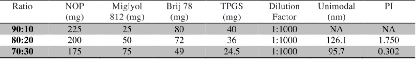
The NOP placebo NPs were fabricated as described above using NOP/Miglyol 812 (70:30 wt/wt) as the oil phase, and Brij78, Vitamin E TPGS, and Brij78-PEG1000 as surfactants. This formulation was optimized through testing of various ratios of NOP:Miglyol 812, surfactant, and levels of PEGylation. For each trial, the particle size distribution and stability upon dilution was examined. A ratio of 70:30 NOP:Miglyol 812 as the oil core produced the most stable microemulsion (Table 2). A ratio of 2:1 Brij78:Vitamin E TPGS, as used in the BTM formulation, was initially used and proved to be successful with the NOP placebo NPs. No significant difference was observed at various levels of PEGylation. Equal parts of Brij78 and Brij78-PEG1000 was chosen for use in further experiments. The final formulation is shown in Table 3 below.
Docetaxel Paclitaxel Camptothecin SN38 C12:DX C18:DX Br-C16:DX Br-C16:PX
NOP 1000 1000 < 21.4 59.8 1026 684 256 342
Miglyol 808
42.7 27.7 < 5.63 < 0.74 84.0 84.0 568 568
Miglyol 812
Table 2: Optimization of NOP Placebo NP Formulation
Quantities of Brij 78 and TPGS are amounts needed to form microemulsion
Table 3: NOP Placebo NP Formulation
Characterization of NOP Placebo NPs
The particle diameter and zeta potential of the NOP placebo NPs was measured at various dilutions (Table 4). The particles exhibited a particle diameter of approximately 90 nm and a zeta potential of -7.9 mV.
Table 4: Particle Diameter of NOP Placebo NPs
Particle sizes obtained using Malvern Zetasizer Nano ZS. PI is the abbreviation for polydispersity index, a measure of unimodality of the sample.
Cytotoxicity of NOP and NOP-based Placebo NPs
An in vitro cytotoxicity study for NOP, placebo NOP NPs, and placebo BTM NPs was conducted with A549, MLg, and MRC5 cell lines using a CellTiter-Glo Luminescent Cell Viability Assay. As shown in Figure 1, both pure NOP and NOP placebo NPs were significantly less toxic than the placebo BTM NPs. Since NOP has only been used for veterinary purposes as of yet, this study was the first to examine its potential biocompatibility. From these results, it can be concluded that the NOP NPs do not introduce any additional excipient toxicity compared to the BTM NPs, a formulation whose biocompatibility has already been demonstrated in previous studies.1,2
Future studies will directly compare the toxicity of NOP to Miglyol 812.
Ratio NOP
(mg)
Miglyol 812 (mg)
Brij 78 (mg)
TPGS (mg)
Dilution Factor
Unimodal (nm)
PI
90:10 225 25 80 40 1:1000 NA NA
80:20 200 50 72 36 1:1000 126.1 1.750
70:30 175 75 49 24.5 1:1000 95.7 0.302
Ingredient Function Concentration (mg/mL)
70:30 NOP:Miglyol 812 Oil 192.3
Brij 78 Surfactant 19.2
Vitamin E TPGS Surfactant 19.2 Brij 78 – PEG1000 Stealth 19.2
1:100 Dilution (PBS) 1:1000 Dilution (PBS) Sample # Unimodal (nm) PI Unimodal (nm) PI
1 94.86 0.231 88.90 0.257
2 93.25 0.246 89.24 0.259
Figure 1: Cytotoxicity Assay of NOP and NOP NPs
Concentrations reflect the concentration of solvent (NOP of Miglyol812) in the NP formulations 0 20 40 60 80 100 120 30.5ng/ m l 122ng/ m l 488ng/ m l 1.95ug/ m l 7.8ug/ m l 31.25ug/ m l 125ug/ m l 500ug/ m l Cell Viability (%)
A549 Cell Line
NOP BTM NPs NOP NPs 0 20 40 60 80 100 120 30.5ng/ m l 122ng/ m l 488ng/ m l 1.95ug/ m l 7.8ug/ m l 31.25ug/ m l 125ug/ m l 500ug/ m l Cell Viability (%)
MLg Cell Line
NOP BTM NPs NOP NPs 0 20 40 60 80 100 30.5ng/ m l 122ng/ m l 488ng/ m l 1.95ug/ m l 7.8ug/ m l 31.25ug/ m l 125ug/ m l 500ug/ m l Cell Viability (%)
MRC5 Cell Line
NOP BTM NPs NOP NPs 0 20 40 60 80 100 120 30.5ng/ m l 122ng/ m l 488ng/ m l 1.95ug/ m l 7.8ug/ m l 31.25ug/ m l 125ug/ m l 500ug/ m l
Cell Viability (%)
HuVEC Cell Line
Release Study and Quantification of PX-loaded NOP Depot
Release studies testing both 50% loaded NOP and 75% PX-loaded NOP as potential depots were performed using a polyurethane foam support to contain the depot and imitate in vivo injection site. For these release studies 50:50 IPA:H2O was used as the release medium. This medium was chosen due to the low water solubility of PX (<1
μg/mL).8
The medium was replenished at each time point to ensure that sink conditions were maintained at all times. The lower limit of quantification for PX generated from the HPLC standard curve was 244 ng/mL, which
represents less than 0.01% release from the depot. The results showed a large burst release of 43.4 + 3.3% of the total encapsulated drug within the first six hours of the study, followed by an additional 0.2% release over the remainder of the 4-week period (Figure 2).
0.00 5.00 10.00 15.00 20.00 25.00
0 100 200 300 400 500
% Release
Time (Hours)
Figure 3: Cumulative % Release of UNC3230/NOP Depot in 25:75 IPA:H20
30.00 35.00 40.00 45.00 50.00 55.00
0 25 50 75 100 125 150 175 % Release
Time (Hours)
Figure 2: Cumulative % Release of PX/NOP Depots in 50:50 IPA:DI H20
Release Study and Quantification of UNC3230A-loaded NOP Depot
A potential new
chemotherapeutic, UNC 3230-A, was also tested in an NOP depot. The small-molecule drug targets the lipid kinase PIP5K1C, a target in the treatment of many diseases, including prostate cancer.9
The solubility of UNC 3230-A in NOP was 35% wt/wt. The release study was also performed using a foam support and 50:50 IPA:DI H2O as the release medium. The medium was replenished at each time point to ensure that sink conditions were maintained at all times. The lower limit of quantification for UNC 3230-A generated from the standard curve was 488 ng/mL,
which represents less than 0.01% release from the depot. Results from this study demonstrated a large burst release of 44.5 + 13.1% of the total encapsulated drug within the first six hours of the study, followed by continued release over the remainder of the 4-week period (Figure 3) for a final cumulative release of 83.9 + 3.4%. To determine if the release of UNC 3230-A from NOP could exhibit a slower burst release and a zero-order release kinetics following the burst release, the release medium was changed to 25:75 IPA:DI H2O and a second release study was performed. The results from this study showed that the depot exhibited a smaller burst release of 8.28 + 0.22% of the total encapsulated drug in the first eight hours of the study, followed by an additional 10.1 + 0.8% release steadily over the remaining 17 days (Figure 4).
Discussion:
The solubility of docetaxel (DX) and paclitaxel (PX) in N-octyl 2-pyrrolidone (NOP) (1000 mg/mL) is significantly greater than in Miglyol 808 and 812 (42.7 mg/mL). Existing nanoparticle (NP) formulations involving Taxols, such as the “BTM” formulation involving Miglyol 808, are limited by low drug encapsulation and the use of lipidized prodrugs to overcome this limitation. The superior solvent ability of NOP offers potential for a nanoparticle formulation with high dosing of free drug. This is preferable over a prodrug that requires hydrolysis at the tumor site to release the active drug.
Optimization of a NOP placebo NPs yielded a formulation that maintained the use of biocompatible surfactants Brij 78 and Vitamin E TPGS. In addition, there was also flexibility to replace the Brij 78 with various ratios of Brij 78-PEG1000 to provide stealth properties. These NPs
0 10 20 30 40 50 60 70 80 90 100
0 25 50 75 100 125 150 175
% Release
Time (Hours)
(approximately 90 nm) were stable upon a hundred-fold and a thousand-fold dilution, and showed low polydispersity (< 0.260) when analyzed for particle diameter.
Cytotoxicity of pure NOP and the placebo NOP NPs was examined relative to the placebo BTM NPs using four different cell lines: A549, MRC5, MLg, and HuVEC. The results showed that, generally, pure NOP was the least toxic to cells at a given concentration, followed by the placebo NOP NPs, and placebo BTM NPs. Increased toxicity of NPs over pure NOP is likely due to excipients (i.e. surfactants). The similar profiles of placebo NOP NPs and placebo BTM NPs are promising for the use of NOP in vivo. BTM NPs have been previously studied and shown to be biocompatible, whereas this was the first toxicity assay for NOP and NOP NPs.
Initial experiments in which the placebo NOP NPs were loaded with paclitaxel were not successful. We hypothesize that the high affinity of PX for NOP may have resulted in a breakdown of the NPs as the surfactants were forced to compete for solubility in NOP. One way to overcome this is by using a stabilizing agent, such as a polymer which could help form a stable drug-loaded formulation. These studies are currently underway.
Pure PX-loaded NOP was tested for potential use as an injectable, intraperitoneal depot. The viscous nature of 50+% wt/wt PX/NOP allows for injection while solidification upon contact with water allows for a solid, stationary depot. Both 50% and 75% wt/wt PX/NOP were tested as depots and demonstrated a high burst release profile, releasing 43.4 + 3.3% within six hours and only an additional 0.2% release over the remainder of the 4-week period. The addition of a biodegradable polymer to control drug release from a depot is a proven technique, and is the next step in this optimization process.10 A second drug, UNC 3230-A, was introduced as a depot candidate with a high solubility of 350 mg/mL in NOP. This 35% wt/wt UNC3230A/NOP depot was tested in two media (list here), each indicating a considerable burst release and subsequent steady release over time. While this formulation would also benefit from the addition of a polymer, it would likely not require as much modification as the PX formulation. It is clear that both PX and UNC 3230-A depots warrant further studies and future in vivo work, as they succeed in releasing over 40 and 80% of encapsulated drug in vitro, respectively.
Conclusions:
Acknowledgements:
Dr. Rahima Benhabbour
Dr. Chris Luft (DeSimone Lab) Group Members:
Amy Webster Dylan Glatt Shelby Hudson Charlotte Wells Aditya Shah
References:
1. Feng L, Wu H, Ma P, Mumper RJ, Benhabbour SR. Development and optimization of oil-filled lipid nanoparticles containing docetaxel conjugates designed to control the drug release rate in vitro and in vivo. International Journal of Nanomedicine. 2011; 6:2545-2556.
2. Peng L, Schorzman AN, Ma P, et al. 2′-(2-bromohexadecanoyl)-paclitaxel conjugate nanoparticles for the treatment of non-small cell lung cancer in an orthotopic xenograft mouse model. International Journal of Nanomedicine. 2014; 9:3601-3610.
3. American Cancer Society. Cancer Facts & Figures 2015. Atlanta: American Cancer Society; 2015.
4. Schiller, Joan H. Current Standards of Care in Small-Cell and Non-Small-Cell Lung Cancer.
Oncology 2001; 3-13.
5. Miele E, Spinelli GP, Miele E, Tomao F, Tomao S. Albumin-bound formulation of paclitaxel (Abraxane® ABI-007) in the treatment of breast cancer. International Journal of
Nanomedicine. 2009;4:99-105.
6. Socinski M, Bondarenko I, Karaseva, N, et al. Weekly Nab-Paclitaxel in Combination With Carboplatin Versus Solvent-Based Paclitaxel Plus Carboplatin as First-Line Therapy in Patients With Advanced Non-Small-Cell Lung Cancer: Final Results of a Phase III Trial.
Journal of Clinical Oncology. 2012; 30:2055-062.
7. Lu Z, Wang J, Wientjes MG, Au JL-S. Intraperitoneal therapy for peritoneal cancer. Future
oncology (London, England). 2010;6(10):1625-1641.
8. Konno, T., Watanabe, J. and Ishihara, K. (2003), Enhanced solubility of paclitaxel using water-soluble and biocompatible 2-methacryloyloxyethyl phosphorylcholine polymers. J. Biomed. Mater. Res., 65A: 209–214.
9. Flemming, Alexandra. "Cancer: Lipid Kinase PIP5K1α as a New Target in Prostate Cancer." Nature Reviews Drug Discovery 13.10 (2014): 723.