© 2020 by the Serbian Biological Society How to cite this article: Todorović JS, Terzić Šupić ZJ, Mazić SD, Suzić Lazić 265 JM, Žikić DD, Nešić DM. The effects of intracerebroventricularly applied ghrelin
on thymocytes and thymic architecture in rats of different ages. Arch Biol Sci. 2020;72(2):265-70.
The effects of intracerebroventricularly applied ghrelin on thymocytes and thymic
architecture in rats of different ages
Jovana S. Todorović1, Zorica J. Terzić Šupić1, Sanja D. Mazić2, Jelena M. Suzić Lazić3, Dejan D. Žikić4 and Dejan M. Nešić2,*
1Institute of Social Medicine, Faculty of Medicine, University of Belgrade, Serbia 2Institute of Medical Physiology, Faculty of Medicine, University of Belgrade, Serbia
3Department of Cardiology, University Clinical Center “Dr Dragiša Mišović – Dedinje”, Faculty of Medicine, University of
Belgrade, Serbia
4Institute of Biophysics, Faculty of Medicine, University of Belgrade, Serbia
*Corresponding author: [email protected]
Received: April 16, 2020; Revised: May 1, 2020; Accepted: May 3, 2020; Published online: May 11, 2020
Abstract: Ghrelin positively influences the total number of thymocytes and size of the thymus in 14-, 20- and 24-month-old rats. We examined the effect of centrally-applied ghrelin on thymus weight and structure in Wistar rats of different ages. The study included 30 male Wistar rats of three age groups: peripubertal (5-week-old rats), young (2 months) and adult (6 months). The animals of each age group were divided into control and experimental groups that were administered intrac-erebroventricularly (ICV) 1 μg ghrelin/5 μL saline daily for five consecutive days. Following treatment, the thymuses were isolated, weighed and processed for stereological analysis by the point-counting method. The average weights of the thymuses were significantly higher in the ghrelin groups with respect to control rats (5 weeks: 444.90±18.03 vs 365.00±18.63; 2 months: 354.30±13.77 vs 257.00±9.60; 6 months: 365.00±15.90 vs 225.00±7.03, p<0.01). The absolute volume of lymphoid tissue was significantly higher in the ghrelin groups (5 weeks: 392.85±16.94 vs 294.48±33.37; 2 months: 309.30±12.10 vs 216.62±10.72; 6 months: 222.70±11.41 vs. 114.33±16.48, p<0.01). Ghrelin treatment restored thymic structure by increasing medullary cel-lularity, improving thymic medullary architecture and providing a clearer delineation between the cortex and medulla. This study shows the positive effects of centrally applied ghrelin on suppression of thymus atrophy, its weight and architecture.
Keywords: ghrelin; thymus; intracerebroventricularly (ICV); lymphoid tissue; thymocytes
INTRODUCTION
The thymus is an organ that consists of two separate areas: the true thymic epithelial space (TES) and the perivascular space (PVS) [1,2]. The TES is comprised of the cortex and the medulla where thymopoiesis, the process of maturation and activation of T-cells, occurs [3-8]. The thymus shows the highest level of function during fetal and neonatal periods, whereas in adult-hood its function decreases, which is followed by its involution [9-11]. Although the thymus is the primary site for the formation of new T-cell lymphocytes, it does not have hematopoietic precursor cells, which is why progenitor cells are constantly recruited from the blood [9]. The aging-associated involution of the
thymus leads to a reduction in the total number of thymocytes and increasing production of adipocytes in thymic tissue [12]. The reduction in the number of thymocytes negatively influences the process of gen-eration of T-cells and the consequent age-associated decline in health [12,13]. Previous animal studies have shown a decrease of 45% in total thymic weight and in the number of thymocytes in 8-month-old animals [3].
system [4,14,15]. The best-described association be-tween the two systems is during sepsis in which the activation of the immune system leads to fever, muscle catabolism, lower appetite and consequent weight loss [14]. Immune cells respond to corticosteroids, insulin, prolactin, growth hormone, insulin-like growth factors, opioids, substance P and somatostatin [14].
The growth hormone secretagogue receptor R1a (GHS-R1a), the receptor for ghrelin, is expressed in immune cells [12] as well as in multiple tissues: neuronal, pancreatic, lymphoid, reproductive and gastrointestinal [14]. Additionally, it is expressed in many cancer cells and could be associated with cancer development and metastasis [16]. In plasma, ghrelin is present in acylated and deacylated forms. The acylated form of ghrelin has been shown to have inhibitory effects on mRNA and protein expression of proinflammatory cytokines, interleukin 1 beta (IL-1β), interleukin 6 (IL-6) and tumor necrosis factor alpha (TNFα), activated T-lymphocytes, monocytes, dendritic cells and Natural killer (NK) cells [14,17].
Previous studies have shown that infusion of ghrelin positively influences the total number of thymocytes and size of the thymus in 14-, 20- and 24-month-old rats, and that thymic adiposity is reduced [12,18]. Ghrelin infusion was also shown to be able to reverse, at least partially, the involution of the thymus associ-ated with age [18]. Along with the increases in the number of thymocytes and of thymus weight, ghrelin was shown to influence the architecture of the thymus into more defined cortical and medullary region with, more pronounced boundaries between these regions and significant loss in the total number of adipocytes [14]. The exact mechanism by which ghrelin influences thymogenesis is not well examined, but it was proposed that ghrelin increases the number of progenitors cells from the lymph [18]. Caloric restriction, which leads to increased ghrelin levels, also positively influences the total number of T-cells in the thymus [14]. Along with the positive influence on thymogenesis, ghrelin has adipolytic effects, as do growth hormone, insulin-like growth factor-I (IGF-I) and keratinocytes growth-factor (KGF) [14].
In light of these data and the lack of results concern-ing the response of the thymus to ghrelin treatment, the aim of our work was to examine whether repeti-tive intracerebroventricular (ICV) administration of
ghrelin to male rats of different ages affects thymus weight and structure. The aim of this study was to examine the influence of centrally applied ghrelin on total thymus mass and thymic architecture in Wistar rats of different ages.
MATERIALS AND METHODS
Ethics statement
All experimental protocols were approved by the lo-cal Animal Care Committee (Faculty of Medicine, University of Belgrade from September 10, 2009), and conformed to the ethical terms of reference stated in the United States NIH guidelines (Guide for the Care and Use of Laboratory Animals (1985), DHEW Pub-lication no. (NIH) 85-23: Office of Science and Health Reports, DRR/NIH, Bethesda, MD).
Experimental animals
The study included 30 male Wistar rats of different ages: peripubertal (5-week-old animals; body weight: 142.7±7.0 g, n=10), young (2 months old: 231.3±8.0 g, n=10), adult (6 months old: 456.7 ± 8.0 g, n=10). During the experiment, the animals were held in individual metabolic cages, with constant laboratory conditions, including room temperature (22±2ºC), and a 12-h light:dark cycle, and were accustomed to daily handling. The rats were given a balanced diet, which contained 20% proteins, 50% corn starch, 10% sucrose, 10% corn oil, 5% cellulose 5 % vitamins and minerals (prepared by the Veterinarski Zavod Subotica, Subotica, Serbia). Food and water were available to the animals ad libitum [19,20].
Experimental protocol
Animal preparation
into a lateral cerebral ventricle, 1 mm posterior and 1.5 mm lateral to the bregma, and 3 mm below the cortical surface. A small stainless-steel anchor screw was placed at a remote site on the skull. The cannula and screw were cemented to the skull by dental acrylic (Simgal, Galenika, Belgrade, Serbia). Following surgery, the animals received a single dose of buprenorphine s.c. 0.28 mg/kg (Buprenex; Reckitt Benckiser Healthcare, Slough, UK). The correct placement of the cannula was confirmed at the end of the experiment by a 5-μL injection of Trypan blue dye [20,21].
Animal treatment
After the recovery period of three days, animals of the same age were randomly divided into two groups, experimental and control. Each group consisted of five animals. The first group was treated with an ICV administration of 0.15 nmol of rat ghrelin (Global Peptide, USA), dissolved in 5 μL of phosphate buffered saline (PBS) every 24 h for 5 consecutive days. The dose of ghrelin used in this experiment was based on our previous studies related to ICV ghrelin actions [19-21]. The second group was a control group, with rats treated in the same manner but injected with 5 μL of solvent only. The ICV injections were given at 10:00 a.m. All animals were killed by decapitation 1 h after the last ICV injection, under deep pentobarbital anesthesia (Thiopental Sodium, 45 mg/ kg, i.p.). The thymuses were carefully removed, separated from the surrounding lymphoid tissue and weighed, and then fixed in Bouin’s solution for 48 h. The tissue specimens were dehydrated in a graded series of ethyl alcohol, cleared in xylene and embedded in paraffin. They were serially cut into 5-μm-thick sections, identified and stained with hematoxylin-eosin for microscope examination. An Olympus BX50 light microscope was used; this microscope has a digital camera and is equipped with image processing software (Micro Im-age, Ver. 4.0, Olympus Optical Co. GmbH, Germany).
Stereological analysis
Stereological analysis was performed by the data numbering method [22,23], which includes a grid (net) that is randomly placed on a thymus section, and the counting is performed in compartments. The first part of the stereological analysis was performed
at 40X enlargement with a grid with 130 points, and the total number tested was 100. The examination included numbering of the data. The second part of the stereological analysis was performed at 100X magnification with an emersion and test grid of 42 dots. Three compartments were examined: the external cortex, internal cortex and medulla. A total of 60 fields were analyzed in each section. The net was positioned parallel to the external cortex. For the analysis of the internal cortex, the net was positioned on the cortical side of the cortical-medullar border. The total number of thymocytes (No) in the cortex and in the medulla, and the total number of thymocytes in the inner and outer cortex were calculated.
Statistical analysis
Statistical analysis was performed using GraphPad Prism 5.0. The differences between the experimental and control groups were examined using the Kruskal-Wallis test. The data are presented as the mean±standard deviation (SD) for 10 animals per group, with a P value<0.05 considered as statistically significant.
RESULTS
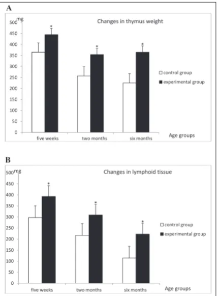
The effects of ICV ghrelin injections on thymus weight in rats of different ages are shown in Fig. 1A. ICV application of ghrelin (1 µg/5µL PBS) significantly increased thymus weight regardless of the age of the animals. Comparison of age-matched animals showed that the increases were 18% (p<0.01) in 5-week-old rats, 28% (p<0.01) in 2-month-old rats and 39% (p<0.01) in 6-month-old rats.
The absolute volume of lymphoid thymus tissue in the ghrelin-treated animal group was significantly higher, by 32.1% (P<0.01) in 5-week-old rats, 30.0% (P<0.01) in 2-month-old rats, and 49.0% (P<0.01) in 6-month-old rats (Fig. 1B). The total number of thymocytes was significantly higher in ghrelin-treated 2-month-old (227%; p<0.01) and 6-month-2-month-old (232%; p<0.01) animals than in age-matched control rats (Table 1).
cortex was increased by 53.2% (P<0.05) in 5-week-old rats, and by 138.3% (P<0.01) in 2-month-old rats, but not in 6-month-old rats, as compared to matching controls. The total number of thymocytes in the outer cortex was also significantly higher in animals in the 2-month-old ghrelin group by 177.2% (P<0.01), but not in 6-month-old nor in 5-week-old rats.
The total number of thymocytes in the medulla was significantly higher in the ghrelin group in 5-week-old rats by 68.9% (P<0.01), and in 6-month-old rats by 355.8% (P<0.01); their number increased in the 2-month-old ghrelin group but it was not significant (Table 1).
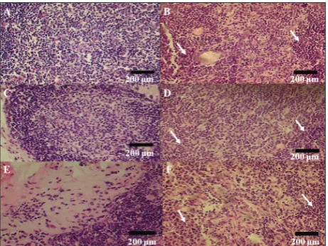
Fig. 2 shows the cross-sectional images of thymuses in animals from the ghrelin (B, D, F) and control (A, C, E) groups in each examined age group, 5 weeks, 2 and 6 months , respectively. Centrally applied ghrelin increased the number of cells in the cortex and the medulla. Additionally, ghrelin treatment yielded more and better-defined cortical and medullary regions
and boundaries, and was responsible for a signifi-cant loss of adipocytes (Fig. 2 B, D, E, arrows). Fig. 3. shows the thymocytes in the medulla of thymuses in animals from the ghrelin (B, D, F) and control (A, C, E) groups in each examined age group, 5 weeks, 2
Fig. 1. The effects of ICV ghrelin injections on thymus weight (A) and absolute lymphoid tissue (B) in rats of different ages. All data are expressed as the mean±SD; n=10 animals per group,*P<0.01 vsage-matched control rats.
Table 1. The effects of ghrelin treatment on thymuses in animals of different age.
5-week-old
animals 2-month-old animals 6-month-old animals Non-lymphoid tissue (mg)
Control group 67.48±35.75 40.56±6.44 110.71±17.71
Experimental group 52.01±6.63 45.00±3.58 142.72±12.12
Tnt inner cortex
Control group 226.00±47.83 143.60±15.36 79.02±12.80
Experimental group 346.30±34.96* 342.20±14.01* 75.78±16.03 Tnt in outer cortex
Control group 84.12±18.09 51.66±5.45 14.52±2.86
Experimental group 110.00±10.30 143.20±13.30* 14.98±2.677 Tnt in total cortex
Control group 310.10±65.37 195.00±19.85 94.00±14.28
Experimental group 456.30±34.19 485.50±25.06* 90.31±18.65 Tnt in medulla
Control group 61.50±4.18 72.60±7.83 56.97±17.26
Experimental group 103.90±8.68* 124.20±17.40 259.70±19.34* tnt total thymus
Control group 414.00±68.98 267.80±18.91 151.00±30.20
Experimental group 517.80±32.43 609.60±30.24* 350.00±25.15*
The values are the mean±SD; n=10 animals per group; Tnt – total number of thymocytes; *P<0.01 vs age-matched control rats.
and 6 months , respectively. As can be seen, ghrelin treatment improved thymic medullary architecture by providing for a clearer delineation between the cortex and medulla and increasing medullary cellularity (Fig. 2 B, D, E, arrows).
DISCUSSION
Our study shows a significant positive influence of ghrelin administration on the weight of the thymus, the total number of thymocytes and on thymic ar-chitecture. Although there was a significant decrease in the average weight of thymuses of animals in the two older age groups as compared to animals in the youngest age group, the weights of the thymuses in each experimental group of different age was signifi-cantly higher when compared to matching controls. Although thymic involution is an age-dependent process and cannot be completely reversed, the ghrelin infusions can slow down this process, which could benefit the immune system. This was also shown in previous studies [16,22]; however, although previous studies suggested a positive influence of ghrelin on the thymus, this influence was shown in animals that were older than 10 months of age. Our study suggests that a positive influence of ghrelin can be observed
in younger animals as well [17]. Partial restoration of complete thymic architecture might be all that is possible as age produces DNA damage, as well as lipid and protein damage, which leads to a decrease in the function of cellular repair pathways and an increase in oxidative damage, with aging also associated with changes in metabolism [20,23].
Studies have shown the effect of ghrelin administra-tion on thymic architecture and its positive influence on both the cortex and the medulla [23,24,25]. How-ever, our study failed to show a significant influence of ghrelin administration on thymic cortex, but shows its influence on the medulla. The absence of a clear effect on the cortex could mean the lack of influence on the T-progenitor cells whose early development takes place in the cortex while later stages of T-cell development occur in the medulla. Further studies confirming the positive influence of ghrelin on thymic cortex are therefore necessary for the confirmation of the potential role of ghrelin in rejuvenation of the thymus. Ghrelin appears to influence early thymocyte progenitors (ETP) to restock the lymphoid tissue of the thymus through linlowsca1+ckit+(LSK) and common lymphoid progenitor (CLP) cells from the bone marrow and to increase its population [22,24], which leads to accumulation of ETP cells in the thymus, providing for a greater T-cell output [23,24].
The potential use of ghrelin for thymic rejuvenation lies in its possible significance from a global public health perspective. The human thymus and human immune system in general are shown to markedly decline in functionality after the ages of 40-50, ren-dering the majority of individuals after this age more prone to infectious, as well as autoimmune diseases and cancers. There is a necessity to improve the immune system of people over 40 years of age [3,9], and to that end, in addition to ghrelin, multiple approaches have been considered, including steroid ablation, cytokine treatment (KGF, Il-7, IL-22) and growth hormone infusion [3,25].
In conclusion, this study shows the positive ef-fects of centrally applied ghrelin on the suppression of aging-related thymus atrophy, total thymus weight and thymic architecture. Also, centrally applied ghre-lin increased thymus cortex and medullar cellularity, and contributed to a well-defined corticomedullary
junction. Ghrelin may have potential therapeutic ef-fect in elderly and immune-compromised patients as a result of an increase in lymphocyte counts and could be considered as a potential agent for stimulation of the immune system.
Funding: This work was supported by The Ministry of Educa-tion, Science and TechnologicalDevelopment of the Republic of Serbia [Grant No. III 41025, Contract No. 175042 (2011-2014), and Grant No. 175046].
Author contributions: JT: was involved in the study design, data collection, data analysis, drafting the article, writing the article and approving the final version of the article. SM was involved in study supervision, data analysis, drafting the article, writing the article and approving the final version of the article. ZTS was supervised the study and was involved for the study design, data gathering, data analysis and writing of the final version of the article. JSL was involved in the study design, study supervision, data analysis, writing of the final version of the article and approval of the final version of the article. DZ was involved in data gathering, data interpretation, writing the draft of the article and approval of the final version of the article. DN supervised the study, wrote the article and approved the final version of the article.
Conflict of interest disclosure: The authors declare that they have no conflict of interest.
REFERENCES
1. Hale LP. Histologic and Molecular Assessment of Human Thymus. Ann Diagn Pathol. 2004;8(1):50-60.
2. Boehm T. Thymus development and function. Curr Opin Immunol. 2008;20:178-84.
3. Ventevogel MS, Sempowski GD. Thymic rejuvenation and aging. Curr Opin Immunol. 2013;25(4):516-22.
4. Rezzani R, Nardo L, Favero G, Peroni M, Rodella LF. Thymus and aging : morphological, radiological , and functional over-view. Age (Omaha). 2014;36:313-51.
5. Muñoz JJ, García-Ceca J, Alfaro D, Stimamiglio MA, Cejalvo T, Jiménez E, Zapata AG. Organizing the thymus gland. Ann N Y Acad Sci. 2009;1153:14-9.
6. Zhang L, Sun L, Zhao Y. Thymic epithelial progenitor cells and thymus regeneration : an update. Cell Res. 2007;17:50-5. 7. Manley NR, Rothman Richie E, Blackburn CC, Condie BG, Sage J. Structure and function of the thymic microenviron-ment Nancy. Front Biosci. 2011;16:2461-77.
8. Jin R, Zhang J, Chen W. Thymic Output : Influence Fac-tors and Molecular Mechanism. Cell Mol Immunol. 2006;3(5):341-50.
9. Holländer GA, Krenger W, Blazar BR. Emerging strate-gies to boost thymic function. Curr Opin Pharmacol. 2010;10(4):443-53.
10. Zdrojewicz Z, Pachura E, Pachura P. The Thymus: A For-gotten, But Very Important Organ. Adv Clin Exp Med. 2016;25(2):369-75.
11. Dominguez-Gerpe L, ReyMendez M. Evolution of the Thy-mus Size in Response to Physiological and Random Events Throughout Life. Microsc Res Tech. 2003;476:464-76. 12. Albarran-Zeckler RG, Sun Y, Smith RG. Physiological roles
revealed by ghrelin and ghrelin receptor deficient mice. Pep-tides. 2011;32(11):2229-35.
13. Shanker A. Is thymus redundant after adulthood ? Immunol Lett. 2004;91:79-86.
14. Baatar D, Patel K, Taub DD. Molecular and Cellular Endo-crinology The effects of ghrelin on inflammation and the immune system. Mol Cell Endocrinol. 2011;340(1):44-58. 15. Savino W, Postel-vinay MC, Smaniotto S, Dardenne M. The
Thymus Gland : a Target Organ for Growth Hormone. Scand J Immunol. 2002;55:442-52.
16. Okada Y, Sugita Y, Ohshima K, Morioka M, Komaki S, Miyo-shi J, Abe H. Signaling of ghrelin and its functional receptor, the growth hormone secretagogue receptor, promote tumor growth in glioblastomas. Neuropathology. 2016;36(6):535-43. 17. Oztas B, Sahin D, Kir H, Eraldemir FC, Musul M, Kuskay
S, Ates N. The effect of leptin, ghrelin, and neuropeptide-Y on serum Tnf-Α, Il-1β, Il-6, Fgf-2, galanin levels and oxida-tive stress in an experimental generalized convulsive seizure model. Neuropeptides. 2017;61:31-7.
18. Dixit VD, Yang H, Sun Y, Weeraratna AT, Youm Y, Smith RG, Taub DD. Ghrelin promotes thymopoiesis during aging. J Clin Invest. 2007;117(10):2778-90.
19. Nesic DM, Stevanovic DM, Stankovic SD, Milosevic VL, Tra-jkovic V, Starcevic VP, Severs WB. Age-dependent modula-tion of central ghrelin effects on food intake and lipid metab-olism in rats. Eur J Pharmacol. 2013;710(1-3):85-91. 20. Nešić D, Stevanović D, Đelić M, Mazić S, Trbojević-Stanković
J, Soldatović I, Stajić-Trošić J, Starčević V. The effects of cen-trally applied ghrelin on appetite and metabolic parameters during aging. Dig J Nanomater Biostruct. 2014; 9(4):1439-49. 21. Radoman K, Živković V, Nikolić T, Stojić I, Raičević D,
Jeremić J, Srejovic I, Jakovljevic V. Differences between α-linolenic and linoleic acid supplementation on the redox status and cardiodynamic parameters of male and female Wistar albino rats. Arch Biol Sci. 2018;70(2):223-31. 22. Aspinall R, Mitchell W. Reversal of age-associated thymic
atrophy: Treatments, delivery, and side effects. Exp Gerontol. 2008;43:700-5.
23. Taub DD, Murphy WJ, Longo DL. Rejuvenation of the aging thymus: growth hormone-mediated and ghrelin-mediated signaling pathways. Curr Opin Pharmacol. 2010;10(4):408-24. 24. Youm YH, Yang H, Sun Y, Smith RG, Manley NR,
Vandan-magsar B, Dixit VD. Deficient ghrelin receptor-mediated signaling compromises thymic stromal cell microenvi-ronment by accelerating thymic adiposity. J Biol Chem. 2009;284(11):7068-77.