ARTIGO ORIGINAL
Metabolic Bone Disease of Prematurity in Very Low
Birthweight Infants: Retrospective Observational Study
Doença Metabólica Óssea da Prematuridade em
Recém-Nascidos de Muito Baixo Peso: Estudo
Observacional Retrospetivo
Raquel COSTA1, Catarina FRANCO2, Nádia SANTOS1, Patrícia MAIO1, Filipa VIEIRA3, Sónia ANTUNES4,
Laura MARTINS4, Madalena Lopo TUNA5
Acta Med Port 2019 Jul–Aug;32(7–8):536–541 ▪ https://doi.org/10.20344/amp.10994
1. Serviço de Pediatria. Hospital Espírito Santo de Évora. Évora. Portugal. 2. Serviço de Pediatria. Hospital Divino Espírito Santo. Ponta Delgada. Portugal.
3. Serviço de Pediatria. Hospital São Francisco Xavier. Centro Hospitalar Lisboa Ocidental. Lisboa. Portugal. 4. Serviço de Neonatologia. Hospital Espírito Santo de Évora. Évora. Portugal.
5. Serviço de Neonatologia. Hospital São Francisco Xavier. Centro Hospitalar Lisboa Ocidental. Lisboa. Portugal. Autor correspondente: Raquel Costa. [email protected]
Recebido: 27 de junho de 2018 – Aceite: 05 de abril de 2019 | Copyright © Ordem dos Médicos 2019 ABSTRACT
Introduction: Metabolic bone disease of prematurity consists in a decrease of bone matrix mineral content, in comparison with the level expected for gestational age. Screening of this condition is based on serum alkaline phosphatase and phosphate levels. The aim of this study is to evaluate the prevalence of metabolic bone disease of prematurity, to assess the aspects associated with a higher risk of this disease and to describe the growth of newborns with birth weight below 1500 g and metabolic bone disease of prematurity.
Material and Methods: Observational, retrospective, multicenter and descriptive study in three neonatal intensive care units in Portugal, from May 1st 2016 to April 30th 2017. A convenience sample of very low birthweight newborns was obtained. Demographic,
clinical, and laboratory variables were described in newborns with and without metabolic bone disease of prematurity.
Results: A total of 53 newborns were included in this study: 30 males, 16 with gestational age ≤ 28 weeks. Five cases of metabolic
bone disease of prematurity were diagnosed. In this group, the majority of patients was male and presented a lower gestational age and birth weight, in comparison with the group without metabolic bone disease of prematurity. The average duration of parenteral nutrition was higher in newborns with metabolic bone disease of prematurity and the calcium/phosphate ratio was lower than the recommended values. Growth was similar in both groups. No patient with metabolic bone disease of prematurity underwent physical rehabilitation.
Discussion: The prevalence of metabolic bone disease of prematurity was 9.43%, which is lower than what is described in the
liter-ature. However, only 50% of newborns completed the screening according to the recommendations. The main risk factors identified
concur with the literature.
Conclusion: Metabolic bone disease of prematurity is a frequent but underdiagnosed comorbidity in very low birthweight newborns. It is essential to screen newborns at risk for this condition, using biochemical markers, as well as structure nutritional interventions and physical stimulation in order to avoid short and long-term consequences of this disease.
Keywords: Bone Diseases, Metabolic; Infant, Extremely Low Birth Weight; Infant, Premature; Infant, Premature, Diseases; Nutritional Status
RESUMO
Introdução: A doença metabólica óssea da prematuridade consiste numa diminuição da matriz óssea, relativamente ao nível espe-rado para a idade gestacional. O rastreio baseia-se no doseamento sérico da fosfatase alcalina e fósforo. O objetivo deste estudo é avaliar a prevalência da doença metabólica óssea da prematuridade, analisar os aspetos associados a maior risco para esta doença e descrever o crescimento estaturo-ponderal dos recém-nascidos com peso ao nascer inferior a 1500 g, com doença metabólica óssea da prematuridade.
Material e Métodos: Estudo multicêntrico, retrospetivo, observacional e descritivo em três unidades de apoio perinatal diferenciado, entre 1 de maio de 2016 e 30 de abril de 2017; foi obtida uma amostra de conveniência de recém-nascidos com muito baixo peso ao
nascer. Descrevem-se as variáveis demográficas, clínicas e laboratoriais dos recém-nascidos com e sem doença metabólica óssea
da prematuridade.
Resultados: Neste estudo foram incluídos 53 recém-nascidos: 30 do sexo masculino, 16 com idade gestacional ≤ 28 semanas. Foram
diagnosticados cinco casos de doença metabólica óssea da prematuridade. Neste grupo, a maioria dos doentes era do sexo masculino e apresentavam idade gestacional e peso ao nascer inferior aos do grupo sem doença metabólica óssea da prematuridade. A duração média de nutrição parentérica foi superior nos recém-nascidos com doença metabólica óssea da prematuridade e a relação cálcio/ fósforo utilizada foi inferior às recomendações nacionais. A evolução estaturo-ponderal foi semelhante nos recém-nascidos com e sem doença. Nenhum doente com doença metabólica óssea da prematuridade teve intervenção por medicina física e reabilitação.
Discussão: A prevalência de doença metabólica óssea da prematuridade foi de 9,43%, valor inferior ao descrito na literatura. Contudo,
apenas 50% dos recém-nascidos cumpriram o rastreio de acordo com as recomendações. Os principais fatores de risco identificados
estão de acordo com a literatura.
ARTIGO ORIGINAL Costa R, et al. Metabolic bone disease in VLBW, Acta Med Port 2019 Jul–Aug;32(7–8):536–541
INTRODUCTION
Metabolic bone disease of prematurity (MBDP) devel-ops due to a decrease in bone matrix compared to the expected level in infants with similar length/age.1–4
Despite the quality improvement in healthcare, namely regarding nutritional support, MBDP remains as a relevant comorbidity in very low birth weight (VLBW) infants as well as in those presenting with mainly a chronic respiratory or gastrointestinal condition, leading to long hospital stays in the neonatal intensive care unit (NICU).2,5
The real incidence of this pathology is still unknown,
mainly due to the lack of consensus regarding its definition
and the reduced number of studies, even though a 55%
incidence in VLBW infants has been estimated.1,3 Even
today, there are no prevalence studies on this disease in Portuguese NICU and therefore the national reality remains undetermined.
Bone formation begins around the sixth week of gestation, while the third trimester remains critical for foetal bone mineralisation, which depends on calcium (Ca) and phosphate (P) placental transport,6 while calcium-phosphate
metabolism is regulated by the action of vitamin D and parathyroid hormone (PTH).5 Apart from Ca/P homeostasis,
mechanical stimuli are also crucial in bone formation.7
Antenatal factors affecting foetal bone formation and postnatal factors associated with a disorder of bone homeostasis are the major known risk factors for the
development of MBDP.2
Antenatal risk factors include (i) genetic factors, (ii) maternal smoking during pregnancy, (iii) placental pathology, (iv) maternal hypovitaminosis D and (v) the use of medications (namely magnesium sulphate),3,6 while
postnatal factors include (i) gestational age (GA) < 28 weeks, (ii) birth weight (BW) < 1,500 g, (iii) male gender, (iv) medications affecting bone metabolism (diuretics, corticosteroids, methyl-xanthine and sodium bicarbonate), (v) immobilisation and (vi) nutritional factors.3,6
Mechanical stimulation is expected to be higher in utero than the regular foetal movements against the maternal abdominal wall. Therefore, the immobilisation of preterm infants is a major postnatal risk factor for the development of MBDP, while higher bone mineralisation has been obtained with interventions based on regular physical stimulation of patients.7,8
Postnatal nutritional factors include delayed introduction of progressive enteral feeds, long-term parenteral nutrition, namely more than four weeks, suboptimal molar Ca/P ratio
in parenteral nutrition (< 1.3:1), use of unfortified human milk (HM) and vitamin D deficiency.3,6
As regards Ca/P ratio in parenteral nutrition, even though
still non-consensual according to the current scientific
evidence, a molar 1.3:1 ratio would be the most adequate, due to the fact that it seems associated with the highest mineral retention in preterm infants.3,6,9 Nevertheless, molar
Ca/P ratio should be maintained between 0.8 – 1 : 1 within
the first days of life in VLBW and mainly in LBW infants, in
order to prevent hypophosphataemia that could arise from the optimisation of caloric and protein intake.10
HM fortification is indicated in preterm infants with BW
<1,500 g, as HM contains a suboptimal amount of Ca and
P in order to ensure postnatal growth. The fortification
should be started at least when 100 mL/kg/day enteral intake is reached and should be maintained up to hospital discharge.11 It could be based on a standard formula or
could be ‘tailored’, based on each patient’s requirements.3,6 Ca/P metabolism is regulated by vitamin D and deficiency
is related to lower bone mineral retention.5 According to
the national recommendations, preterm infants should be supplemented with 800 – 1000 IU of vitamin D per day.11
MBDP range from a silent asymptomatic disease to rickets and pathological fractures, while growth retardation, failure to thrive and short stature are some of the most frequent late manifestations of the disease.8
As a clinically silent disease, frequently only presenting with late manifestations, screening is recommended and should be aimed at infants with risk factors, from the fourth week of life onwards and should be repeated (every two weeks) as long as risk factors remain,3–6 in order to allow
for an early diagnosis together with an adequate and timely intervention, in order to prevent clinical manifestations in the long term. With the presence of an established disease, monitoring is based on the same biochemical markers.3–6,12
Early diagnosis and monitoring are obtained by the use of biochemical markers (serum P and alkaline phosphatase [ALP]). Combined interpretation of both results has shown a
100% sensitivity and a 70% specificity in detecting MBDP.3,12,13
Therefore, the early presence of MBDP is suggested by serum ALP > 900 IU/L associated with hypophosphataemia < 5.5 mg/dL [< 1.8 mmol/L] or ALP > 600 IU/L with an increasing trend and persistent hypophosphataemia < 5.5
mg/dL in serial measurements.12,13 Serum Ca levels are
usually maintained within a normal range due to the action of PTH and therefore these are not an early marker of the
disease.3 Imaging markers of the disease correspond to
late changes and are not indicated as routine screening.3
When diagnosis is obtained, the approach to patients is usually aimed at correcting postnatal risk factors associated with the admission to the NICU. These should include a nutritional approach aimed at improving the supply of Ca and P either in parenteral nutrition, enteral nutrition or with supplementation, according to the national
recommendations.8,9 An effort should be made to reduce
the duration of parenteral and to provide enteral nutrition as soon as possible, reducing hang time of enteral feeding. In addition, medications associated with an increase in bone turnover should be revised and removed, whenever possible.3,6 Physical medicine strategies should also be included in patient’s daily care, with the benefits that have
already been described.7
ARTIGO ORIGINAL
Costa R, et al. Metabolic bone disease in VLBW, Acta Med Port 2019 Jul–Aug;32(7–8):536–541
This study was aimed at assessing the prevalence of MBDP and describing the issues that are associated with an increased risk of the disease. In addition, it was aimed at describing the growth and development of infants with birth weight (BW) < 1,500 g presenting with MBDP.
MATERIAL AND METHODS
This was a multicentric retrospective, observational and descriptive study carried out between 1 May 2016 and 30 Apr 2017 in three perinatal units; a non-randomised convenience sample of premature infants with BW < 1,500 g has been included in the study, while deceased patients or patients that were transferred to other NICUs have been excluded from the study, as well as patients who were not followed at the participating intensive care units upon discharge. Patients presenting with congenital malformations, neuromuscular disorders, inborn errors of metabolism and other bone disorders were also excluded.
Patient’s socio-demographic characteristics, in addition to maternal smoking during pregnancy, placental pathology, gestational age and birth weight, the use of medications affecting bone metabolism was also evaluated (corticosteroids, diuretics, methylxantines and sodium bicarbonate) as well as nutritional factors (duration of parenteral nutrition; Ca/P ratio in parenteral nutrition9; enteral feeding hang time; HM fortification and dose of
vitamin D) were evaluated,11 in addition to any Physical
Medicine and Rehabilitation (PMR) approach. This retrospective study was based on clinical data and the study of maternal hypovitaminosis D were not available as this is not systematically evaluated in pregnancy.
Disease screening is recommended and is based
on serum ALP and P serial measurements. The first
measurement of these biochemical parameters should be carried out by the fourth week of chronological age and repeated every two weeks. The presence of MBDP was based on the biochemical markers with the highest
sensitivity and specificity: ALP > 900 IU/L and P < 5.5 mg/
dL [< 1.8 mmol/L], or ALP > 600 IU/L trending up and serum phosphate levels persistently < 5.5 mg/dL in serial measurements.3,12,13
Each patient’s anthropometric data (body weight and length) throughout the study period were evaluated. Z-score calculation was based on corrected age (Fenton and WHO [World Health Organization] growth charts up to the 50th week of postmenstrual age and beyond that age, respectively).14,15 Data regarding weight and length were
continuously evaluated at the NICU up to hospital discharge and those beyond that were available from the Neonatology outpatient clinic.
The statistical analysis was carried out by use of SPSS, version 23® software, including a descriptive analysis aimed
at the characterisation of patients, the variables known as risk factors and patient’s growth. The study was previously approved by the different Ethics committees and was included in a research project with a prospective study, apart from a retrospective analysis with data collected
anonymously. An informed consent was obtained from the parents of each patient included in the prospective study.
RESULTS
A total of 83 infants with BW <1,500 g were born during the study period and 30 out of these, with similar characteristics regarding birth weight and gestational age, were excluded from the study (nine deceased, 11 who were not followed at the participating hospitals and 10 transferred to other NICU).
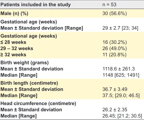
A total of 53 patients were included in the study, mostly male and within mean GA of 29 ± 2.7 weeks (range 23 – 34 weeks), mostly within the 29 – 32 week range (49%) and an average BW of 1,118.6 g ± 261.3 g (range 625 – 1,491 g). Patient’s characteristics are shown in Table 1.
Five patients were diagnosed with MBDP based on the biochemical markers that were described (serial measurements of serum P and ALP levels) (9.43%). However, a routine measurement of serum ALP and P by the fourth week of chronological age was only carried out in 15 patients and a serial analytical evaluation was only obtained in 10 patients up to hospital discharge. Therefore, a 50% incidence of MBDP was found in VLBW infants complying with the screening.
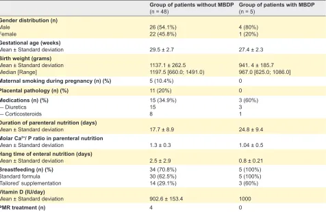
Male patients more frequently presented with the factors associated with higher risk of MBDP (Table 2) and lower BW and GA were on average found in patients with MBDP when compared to those without the disease. The presence of placental pathology and maternal smoking during pregnancy were most frequently found in patients without MBDP (antenatal risk factors).
A higher mean duration of parenteral feeding was found in patients with MBDP (24.8 days ± 9.4 days vs. 17.7 days ± 8.9 days), while a higher hang time of enteral feeding was found in the group of patients without MBDP (2.5 days ± 2.9 days vs. 0.8 days ± 0.21 days). A lower than recommended molar Ca/P ratio was used in parenteral nutrition (< 1.3:1)
Table 1 – Characteristics of the patients included in the study (n = 53) regarding gestational age, birth weight, body length and head circumference
Patients included in the study n = 53
Male (n) (%) 30 (56.6%)
Gestational age (weeks)
Mean ± Standard deviation [Range] 29 ± 2.7 [23; 34]
Gestational age (weeks)
≤ 28 weeks
29 – 32 weeks
≥ 32 weeks
16 (30.2%) 26 (49.0%) 11 (20.8%)
Birth weight (grams) Mean ± Standard deviation
Median [Range] 1118.6 ± 261.31148 [625; 1491]
Birth length (centimetre) Mean ± Standard deviation
Median [Range] 36.7 ± 3.4937.5; [29.0; 46.5]
Head circumference (centimetre) Mean ± Standard deviation
ARTIGO ORIGINAL Costa R, et al. Doença metabólica óssea em RNMBP, Acta Med Port 2019 Jul–Aug;32(7–8):536–541
in the group of patients with MBDP. Most patients in our
group were breastfed with fortified breast milk and/or
supplemented according to the national recommendations,11
based on a standard formula and/or ‘tailored’ to each patient’s requirements by the hospital nutrition department. The supplementation with vitamin D also complied with the national recommendations in both groups.11
Sodium bicarbonate was not prescribed to any patient, while caffeine citrate was prescribed to all the patients, according to the national recommendations for the prevention of apnoea of prematurity. Diuretics were prescribed to 60% of the patients with MBDP (Table 2).
It should be mentioned that no patient with MBDP in our group underwent any physical medicine treatment (Table 2).
A similar growth trend was found in both groups, with negative z-score (Table 3) and, on average, a lower z-score was found in the group of patients with MBDP, both regarding body weight and length variables (weight: –1.29 ± 1.0 vs. –1.37 ± 0.92; length: –1.30 ± 2.32 vs.
–1.69 ± 2.48).
DISCUSSION
MBDP remains as an important comorbidity in VLBW infants and in infants with chronic disorders, mainly in those with longer stays at the NICU.
A 9.43% prevalence of MBDP was found in our group
of patients, well below what has been found as affecting more than half of the VLBW infants.1–3,5 This low prevalence
is partly explained by the absence of a systematic biochemical screening in the presence of risk factors and suggests that MBDP has been underdiagnosed. In fact, the presence of MBDP correspond to 50% of the patients, when infants submitted to a screening and complying with the recommendations are considered. A high number of exclusions could also have had a contribution on the small number of diagnoses, particularly in patients with gastrointestinal pathology and patients in need to be
transferred to other units for specific surgical management.
Relevant nutritional risk factors are involved in this subgroup of patients, namely long parenteral nutrition courses and long hang time of enteral feeding.3–6
Male patients with lower GA and BW are associated with MBDP, according to literature3–6 and these characteristics
have been more frequently found in our group of patients. A higher rate of maternal smoking and placental pathology was found in the group of patients without MBDP, in contrast to what has been described in literature, in addition to the hang time of enteral feeding.3–6 According to the authors,
these risk factors were not found more frequently in the subgroup of patients with MBDP due to the small number of patients in the study.
Nutritional issues are the major physiopathological
Table 2 – Risk factors for MBDP
Group of patients without MBDP
(n = 48) Group of patients with MBDP (n = 5)
Gender distribution (n)
Male
Female 26 (54.1%)22 (45.8%) 4 (80%)1 (20%)
Gestational age (weeks)
Mean ± Standard deviation 29.5 ± 2.7 27.4 ± 2.3
Birth weight (grams)
Mean ± Standard deviation
Median [Range] 1137.1 ± 262.51197.5 [660.0; 1491.0) 941. 4 ± 185.7967.0 [625.0; 1086.0]
Maternal smoking during pregnancy (n) (%) 5 (10.4%) 0
Placental pathology (n) (%) 11 (20%) 0
Medications (n) (%)
— Diuretics — Corticosteroids
15 (34.9%) 15 8
3 (60%) 3 1
Duration of parenteral nutrition (days)
Mean ± Standard deviation 17.7 ± 8.9 24.8 ± 9.4
Molar Ca2+/ P ratio in parenteral nutrition
Mean ± Standard deviation 1.3 ± 0.3 1.04 ± 0.5
Hang time of enteral nutrition (days)
Mean ± Standard deviation 2.5 ± 2.9 0.8 ± 0.21
Breastfeeding (n) (%)
Standard formula ‘Tailored’ supplementation
34 (70.8%) 30 (62.5%) 14 (29.1%)
5 (100%) 5 (100%) 3 (60%)
Vitamin D (IU/day)
Mean ± Standard deviation 902.6 ± 153.4 1000
PMR treatment (n) 4 0
Characteristics of patients as regards known risk factors of MBDP: gender; gestational age; birth weight; maternal smoking during pregnancy; placental pathology; use of medications
affecting bone metabolism*; duration of parenteral nutrition; molar Ca/P ratio in parenteral nutrition; breast milk fortification; dose of vitamin D supplementation and Physical Medicine
and Rehabilitation (PMR) treatment.
ARTIGO ORIGINAL
Costa R, et al. Doença metabólica óssea em RNMBP, Acta Med Port 2019 Jul–Aug;32(7–8):536–541
principles of the disease, with an important role in its development and management. Nutrition therapy is critical to the approach to patients, aimed at the prevention and treatment of MBDP. In fact, a longer duration of parenteral nutrition was found in patients with MBDP and a lower molar Ca/P ratio than the national recommendation was used in this group (< 1.3:1).9 The national recommendations were complied with as regards breast milk fortification and
supplementation with vitamin D.11
In addition to adequate nutritional strategies, rehabilitation interventions are crucial to the prevention and management of the disease;3,7,16,17 however, no patient with
MBDP in our group underwent motor physiotherapy. Withdrawal of medications associated with higher bone turnover was not feasible in the three patients with MBDP due to the relevance of the treatment of comorbidities
(bronchopulmonary). However, the identification of the disease has led to the replacement of fortified breast milk
with a standard formula by a ‘tailored’ supplementation, based on the higher mineral requirements of these patients.
The evaluation of somatometric data is crucial to the evaluation of the growth of patients admitted to NICU. The major clinical manifestations of the disease are associated
with the impact of MBDP on growth and development.18
In our group of patients, the progression of body weight and length in both groups has shown, on average, lower z-score values in patients with MBDP. However, it is worth mentioning that a small number of patients diagnosed with MBDP have been included in the study and the follow-up period (less than 12 months) was too short to allow for an accurate evaluation of the impact of the disease on the patient’s growth.
This study was limited by the small convenience sample, non-representative of the population. The participation of other NICUs, namely those within surgical centres dealing with abdominal pathologies and receiving patients with multiple risk factors, would correspond to a higher number of patients. The application of a systematic screening would estimate a more accurate prevalence, closer to the values described in literature1–3,5 and leading to higher knowledge
on the national reality of the MBDP. The small number of patients diagnosed with MBDP, in addition to the short study period are limitations to reaching any conclusions on
the characterisation of the risk factors. Therefore, only a continuous evaluation of anthropometric data could clarify the effects of MBDP on the growth of these patients.
CONCLUSION
Newborn infants admitted to the NICU frequently present with MBDP, even though it is still scarcely discussed
nationwide. The findings of this study suggested that MBDP
is underdiagnosed and do not correspond to the real prevalence of the disease. A systematic screening should be carried out in VLBW infants by using low-cost biochemical markers as a starting point for diagnosis.19 There is a need
for further studies with representative samples in a wider range of NICUs.
The early identification of the disease is crucial
to ensure a timely approach. Nutritional and physical stimulation interventions are the major strategies aimed at controlling the disease and should be included as a routine in healthcare of newborn infants.20 Laboratorial monitoring
and intervention attitudes should be maintained even upon discharge from the hospital, in patients with an established disease or presenting with risk factors. The evaluation of late clinical manifestations should be closely carried out in the Neonatology outpatient setting, due to the negative impact on these patients.
ACKNOWLEDGMENTS
The authors wish to acknowledge the Neonatology departments, Nutrition departments and Rehabilitation and Physical Medicine & Rehabilitation departments at the
Hospital Espírito Santo de Évora, Hospital São Francisco Xavier and Hospital Divino Espírito Santo de Ponta Delgada.
HUMAN AND ANIMAL PROTECTION
The authors declare that the followed procedures were according to regulations established by the Ethics and Clinical Research Committee and according to the Helsinki Declaration of the World Medical Association.
DATA CONFIDENTIALITY
The authors declare that they have followed the protocols of their work centre on the publication of patient data.
Table 3 – Trend of body weight and length in patients with and without MBDP
Patients without MBDP
(n = 48) Patients with MBDP (n = 5)
Weight
Grams
Mean ± Standard deviation 2,526 ± 1,765 2,189 ± 1,397
z-score*
Mean ± Standard deviation –1.29 ± 1.0 –1.37 ± 0.92
Length
Centimetre
Mean ± Standard deviation 49.93 ± 10.13 47.48 ± 8.23
z-score*
Mean ± Standard deviation –1.30 ± 2.32 –1.69 ± 2.48
ARTIGO ORIGINAL Costa R, et al. Doença metabólica óssea em RNMBP, Acta Med Port 2019 Jul–Aug;32(7–8):536–541
CONFLICTS OF INTEREST
The authors declare that there were no conflicts of
interest in writing this manuscript.
FINANCIAL SUPPORT
The authors declare that the study was carried out
with the financial support of the Sociedade Portuguesa de Neonatologia (Bolsa Milupa DN-ELN).
REFERENCES
1. Choban P, Dickerson R, Malone A, Worthington P, Compher C. A.S.P.E.N. Clinical Guidelines. J Parenter Enter Nutr. 2013;37:714–44. 2. Bozzetti V, Tagliabue P. Metabolic bone disease in preterm newborn: an
update on nutritional issues. Ital J Pediatr. 2009;35:20.
3. Nallagonda S, Nallagonda M, Deorukhkar A. Metabolic bone disease of prematurity — an overview. Paediatr Child Heal. 2017;27:14–7. 4. Manfredini VA. Metabolic bone disease of prematurity: a review of
minerals supplementation and disease monitoring. J Neonatal Biol. 2015;4:1–4.
5. Rustico SE, Calabria AC, Garber SJ. Metabolic bone disease of prematurity. J Clin Transl Endocrinol. 2014;1:85–91.
6. Rehman MU. Metabolic bone disease in the preterm infant: current state and future directions. World J Methodol. 2015;5:115.
7. Land C, Schoenau E. Fetal and postnatal bone development: reviewing the role of mechanical stimuli and nutrition. Best Pract Res Clin Endocrinol Metab. 2008;22:107–18.
8. Machado A, Rocha G, Silva Isabel A, Alegrete N, Guimarães H. Bone fractures in a neonatal intensive care unit. Acta Med Port. 2015;28:204. 9. Secção de Neonatologia da SPP. Nutrição parentérica no recém-nascido: 1a revisão do Consenso Nacional, 2008. Acta Pediatr Port.
2008;39:125–34.
10. Mihatsch W, Fewtrell M, Goulet O, Molgaard C, Picaud JC, Senter T. ESPGHAN/ESPEN/ESPR/CSPEN guidelines on pediatric parenteral nutrition: calcium, phosphorus and magnesium. Clin Nutr. 2018;37:2360–5.
11. Macedo I, Alexandrino AM, Pissarra S, Cardoso M. Nutrição entérica na
criança nascida pré-termo : revisão do consenso nacional. Acta Pediatr Port. 2014;45:326–39.
12. Harrison CM, Johnson K, McKechnie E. Osteopenia of prematurity: a national survey and review of practice. Acta Paediatr. 2008;97:407–13. 13. Abdallah EA, Said RN, Mosallam DS, Moawad EM, Kamal NM,
Fathallah MG. Serial serum alkaline phosphatase as an early biomarker for osteopenia of prematurity. Medicine. 2016;95:1–5.
14. Fenton TR, Kim JH. A systematic review and meta-analysis to revise the Fenton growth chart for preterm infants. BMC Pediatrics.2013;13:59. 15. World Health Organization. Child growth standards. [consultado 2018
jan 20]. Disponível em: http://www.who.int/childgrowth/en/.
16. Rigo J, Pieltain C, Salle B, Senterre J. Enteral calcium, phosphate and vitamin D requirements and bone mineralization in preterm infants. Acta Paediatr. 2007;96:969–74.
17. Rauch F, Schoenau E. Skeletal development in premature infants: a review of bone physiology beyond nutritional aspects. Arch Dis Child Fetal Neonatal Ed. 2002;86:F82–5.
18. Embleton N, Wood CL. Growth, bone health, and later outcomes in infants born preterm. J Pediatr. 2014;90:529–32.
19. Ramón AM, Espuelas CF, Calmarza PC, Gracia SR, Del Cacho MJ. Factores de riesgo y marcadores bioquímicos de la enfermedad metabólica ósea del recién nacido prematuro. Rev Chil Pediatr. 2017;88:487–94.