Seven new species of Selaginella subg. Stachygynandrum
(Selaginellaceae) from Brazil and new synonyms
for the genus
Iván A. Valdespino1, Gustavo Heringer2, Alexandre Salino3, Luiz A. de Araújo Góes-Neto3, Jorge Ceballos4
1 Departamento de Botánica, Facultad de Ciencias Naturales, Exactas y Tecnología, Universidad de Panamá, Apartado Postal 0824-00073, Panama 2 Pós-Graduação em Botânica, Departamento de Biologia Vegetal, Universidade Federal de Viçosa, CEP 36.570-000 Viçosa, Minas Gerais, Brazil 3 Departamento de Botânica, Instituto de Ciências Biológicas, Universidade Federal de Minas Gerais, CP 486, 31270-901, Belo Horizonte, MG, Brazil 4 Smithsonian Tropical Research Institute, Apartado Postal 0843-03092, Panama
Corresponding author: Iván A. Valdespino ([email protected])
Academic editor: Pavel Stoev | Received 11 March 2015 | Accepted 18 May 2015 | Published 16 June 2015
Citation: Valdespino IA, Heringer G, Salino A, Góes-Neto LAA, Ceballos J (2015) Seven new species of Selaginella subg.
Stachygynandrum (Selaginellaceae) from Brazil and new synonyms for the genus. PhytoKeys 50: 61–99. doi: 10.3897/ phytokeys.50.4873
Abstract
We describe seven new species of Selaginella subg. Stachygynandrum (S. alstonii, S. blepharodella, S. crinita, S. mucronata, S. mucugensis, S. saltuicola, and S. sematophylla) from Brazil and discuss their possible af-finities and conservation status. Scanning electron micrographs of stem sections, leaves, and spores are provided to illustrate the new taxa. In Selaginella alstonii and S. saltuicola vegetative growth from strobilus tips is reported and discussed. Four of the new species are from the Espinhaço Mountain Range associ-ated with Campos Rupestres (montane savannah/rocky fields) vegetation. Three of these (i.e., Selaginella blepharodella, S. crinita, and S. mucugensis) were collected in the northern part of the range in Chapada Diamantina, state of Bahia, while S. alstonii is from the southern part of the range in the state of Minas Gerais. Selaginella mucronata is found in Atlantic Rainforest vegetation in the state of Espírito Santo, whereas S. saltuicola inhabits Cerrado (tropical savannah) vegetation in the state of Mato Grosso. Se-laginella sematophylla is the most widely distributed of the new species and was collected in Espírito Santo, Minas Gerais, and Rio de Janeiro states in Campos Rupestres and Atlantic Rainforest vegetation. Selaginella alstonii occurs in rocky caves, S. blepharodella, S. crinita, S. mucugensis, and S. sematophylla seem adapted to seasonally dry places, living on sandy or humid soils, S. mucronata occupies humid, for-est understory, and S. saltuicola is adapted to wet places associated with rocks or logs in waterfalls. Of the seven new species, six are considered local endemics (except for S. sematophylla) because of their restricted http://phytokeys.pensoft.net
Copyright Iván A. Valdespino et al. This is an open access article distributed under the terms of the Creative Commons Attribution License (CC BY 4.0), which permits unrestricted use, distribution, and reproduction in any medium, provided the original author and source are credited.
Iván A. Valdespino et al. / PhytoKeys 50: 61–99 (2015)
62
currently known distributions to one or two localities within a single state in Brazil. Additionally, we propose new synonymy for S. palmiformis (syn. = S. bahiensis subsp. manausensis, ≡ S. manausensis) and S. vestiens (syn. = S. fragillima); the last species is endemic to Brazil, recorded in the states of Goiás and Minas Gerais. Finally, based on literature discussed and this study, we conclude that the number of well-documented Brazilian Selaginella species is 61, of which 58 are native and three introduced and natural-ized. These statistics are likely to change with further work on Selaginella from Brazil.
Resumen
Describimos siete nuevas especies de Selaginella subg. Stachygynandrum (Selaginella alstonii, S. blepharode-lla, S. crinita, S. mucronata, S. mucugensis, S. saltuicola y S. sematophylla) de Brasil y discutimos sus posibles afinidades y estado de conservación. Micrografias electrónicas de barrido de secciones de los tallos, hojas y esporas se proveen para ilustrar los nuevos taxa. Igualmente, se describe y discute el crecimiento vegetativo a partir del ápice de los estróbilos en Selaginella alstonii y S. saltuicola. Cuatro de las especies nuevas pro-ceden de la Cadena del Espinhaço asociadas a vegetación de Campos Rupestres (sabana montana). Tres de éstas (i.e., Selaginella blepharodella, S. crinita y S. mucugensis) fueron recolectadas en la parte norteña de la Cadena del Espinhaço en la Chapada Diamantina, estado de Bahia, mientras que S. alstonii se registra para la parte sureña en el estado de Minas Gerais. Selaginella mucronata se encuentra en vegetación de Bosques Lluviosos del Atlántico en el estado de Espírito Santo, mientras que S. saltuicola habita vegetación de Cerrado (sabana tropical) en el estado de Mato Grosso. De las nuevas especies, Selaginella sematophylla es la más ampliamente distribuida y se ha recolectada en los estados de Espírito Santo, Minas Gerais y Río de Janeiro en vegetación de Campos Rupestres y Bosques Lluviosos del Atlántico. Selaginella alstonii crece sobre rocas en cuevas, mientras que S. blepharodella, S. crinita, S. mucugensis y S. sematophylla parecen estar adaptadas a lugares estacionalmente secos, creciendo sobre suelos arenosos o húmedos; a su vez, S. mucro-nata crece en el sotobosque de bosques húmedos y S. saltuicola está adaptada a vivir en lugares húmedos asociada a rocas o troncos en cascadas. De las siete nuevas especies, seis son consideradas tentativamente endémicas locales (con la excepción de S. sematophylla) debido a su distribución restringida a una o dos localidades dentro de un sólo estado de Brasil. Adicionalmente, proponemos nuevos sinónimos para S. palmiformis (syn. = S. bahiensis subsp. manausensis, ≡ S. manausensis) y S. vestiens (syn. = S. fragillima), la cual se confirma como endémica de Brasil donde se registra para los estados de Goiás y Minas Gerais. Finalmente, de acuerdo con este estudio y la literatura discutida, estimamos que el número de especies bra-sileñas de Selaginella debidamente documentadas es de 61, de las cuales 58 son nativas y tres introducidas y naturalizadas. Esta estadística muy probablemente cambiará conforme se realicen estudios adicionales sobre Selaginella en Brasil.
Keywords
Atlantic Rainforest, Chapada Diamantina, Chapada dos Guimarães, Espinhaço Mountain Range, Mucugê, Serra do Sincorá
Palabras clave
Bosques Lluviosos del Atlántico, Chapada Diamantina, Chapada dos Guimarães, Cadena del Espinhaço, Mucugê, Serra do Sincorá
Introduction
although some are adapted to live in dry, desert-like areas and some are circumboreal (Jermy 1990, Valdespino 1993a, Mickel et al. 2004).
Alston et al. (1981) recorded 45 species and two subspecies of Selaginella from Brazil, while Hirai (2015) listed 56 taxa, including two subspecies and three introduced species. As part of ongoing work on Selaginella by the senior author and a study of this genus in the state of Minas Gerais conducted by Heringer (2011) under the supervision of Salino, we now describe seven new taxa from Brazil: S. alstonii G. Heringer, Salino & Valdespino, S. blepharodella Valdespino, S. crinita Valdespino, S. mucronata G. Heringer, Salino & Valdespino, S. mucugensis Valdespino, S. saltuicola Valdespino, and S. sematophylla Valdespino, G. Heringer & Salino, and place them in subg. Stachygynandrum (P. Beauv.) Baker following Jermy’s (1986, 1990) infrageneric classification.
Three of the new species Selaginella blepharodella, S. crinita, and S. mucugensis are reported from three localities (i.e., Pico das Almas in Serra do Rio de Contas and Ibicoara and Mucugê in Serra do Sincorá) of Chapada Diamantina in the state of Bahia, whereas S. alstonii was collected in Santo Antônio do Itambé in the state of Minas Gerais. These localities are within the Espinhaço Mountain Range, which is dominated by “Campos Rupestres” (montane savannah/rocky fields) vegetation (Melo 2000, São-Pedro and Feio 2011) and recognized as an important biodiversity and endemism center (Harley and Simmons 1986, Melo 2000, Rapini et al. 2008, Bünger et al. 2014). Selaginella mucronata was collected in Castelo, Parque Estadual do Forno Grande, a locality that has highland remnants of the rich, biodiverse Atlantic Rainfor-est vegetation in the state of Espírito Santo, southeastern Brazil (Meirelles and Gold-enberg 2012, Silva-Soares and Scherrer 2013). Selaginella saltuicola is recorded from Chapada dos Guimarães, a high plateau in the state of Mato Grosso (Oliveira-Filho and Martins 1991) in the Central-West region of Brazil, where the species-rich (Ratter et al. 1997) “Cerrado” (tropical savannah) vegetation is dominant (Oliveira-Filho and Martins 1991) and waterfalls, caves, and ponds are common. Finally, S. sematophylla seems to be the most widely distributed species of all the spike mosses newly described herein, as it is recorded from Campos Rupestres vegetation in the localities of São Sebastião do Paraíso and Parque Estadual de Serra Nova, part of the Espinhaço Moun-tain Range, in the state of Minas Gerais and in mountane areas with some remnants of Atlantic Rainforest vegetation such as Pedra do Garrafão in Santa Maria do Jetibá, state of Espírito Santo and Santo Antônio do Imbé in the state of Rio de Janeiro. Because of their restricted currently documented distributions to one or two localities within a single Brazilian state, six of these new species, except for S. sematophylla, are tentatively considered local endemics.
Iván A. Valdespino et al. / PhytoKeys 50: 61–99 (2015)
64
& Grev.) Spring (Alston et al. 1981). Furthermore, S. gynostachya Valdespino and S. sandwithii Alston, reported from Brazil by Góes-Neto et al. (2015) should be added to Hirai’s account as well. Accordingly, there are 58 well-documented native Brazilian Selaginella species and if we were to take into account the introduced taxa listed by Hirai (2015), i.e., S. kraussiana (Kunze) A. Braun [native of Africa and Macaronesia (Alston et al. 1981)], S. plana (Desv. ex Poir.) Hieron. [native of Southeast Asia and Indonesia (Valdespino 1993b), and S. vogelii Spring [native of Africa (Stefanović et al. 1997)], then a total of 61 species of Selaginella would be recorded for Brazil. These statistics are likely to change as work on Brazilian Selaginella continues.
Material and methods
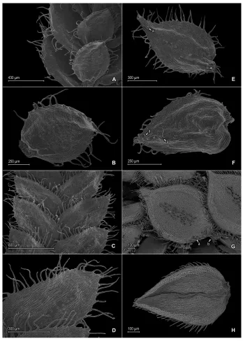
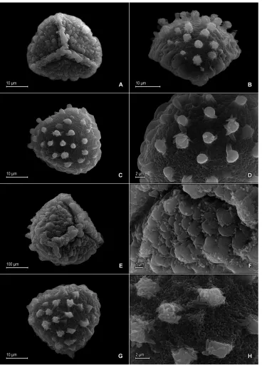
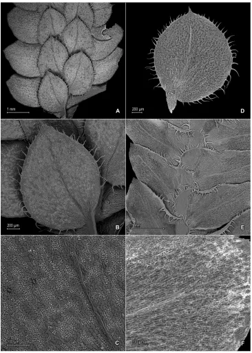
Herbarium specimens were examined from B, BHCB, BM, CAS, CESJ, COL, G, GH, INPA, K, MG, MO, NY, P, PMA, QCA, R, RB, UC, US, and W (Thiers 2015) and samples for Scanning Electron Microscopy (SEM) were taken from selected collections to document upper and lower surfaces of stems and leaves, as well as spore morphology. Although for each of the new species an effort was made to secure megaspore and mi-crospore samples to determine sculpturing pattern, color, and diameter, these were not always available or, in some cases, were too immature to be utilized for those purposes. The SEM samples were prepared, viewed, and photographed at different magnifications using a Zeiss Model Evo 40 at 20–30 KV following standard techniques as described by Valdespino (1995) and Valdespino et al. (2014). Digitized SEM images were post-pro-cessed with Adobe Photoshop and assembled according to species in multipart figures.
Taxonomy
Selaginella alstonii G. Heringer, Salino & Valdespino, sp. nov.
urn:lsid:ipni.org:names:77147598-1
Figures 1, 2
Diagnosis. Selaginella alstonii resembles S. acanthostachys Baker, from which it differs by having the upper surfaces of the lateral leaves glabrous (vs. hairy near basiscopic margins), median leaves acuminate to short-aristate (vs. long-aristate) with each acu-men (arista) ¼ or less the lamina length (vs. arista ⅓–½), with the outer and inner hyaline margins about the same width (vs. outer margin almost twice as wide as the inner one), and non stoloniferous stems (vs. stoloniferous).
Type. BRAZIL. Minas Gerais: Santo Antônio do Itambé, Parque Estadual do Pico do Itambé, 18°23'50,4"S, 43°19'55,5"W, 1676 m, 5 Oct 2006, T.E. Almeida et al. 533 (holotype: BHCB!; isotype: PMA!).
Iván A. Valdespino et al. / PhytoKeys 50: 61–99 (2015)
66
Figure 1. Selaginella alstonii G. Heringer, Salino & Valdespino. A Section of upper surface of stem B Upper surface of median leaf C Section of lower surface of stem D Lower surface of lateral leaf. A–D taken from isotype, Almeida et al. 533 (PMA).
Iván A. Valdespino et al. / PhytoKeys 50: 61–99 (2015)
68
Habitat and distribution. Selaginella alstonii is epipetric on rocky caves in Cam-pos Rupestres vegetation; the type and paratype were collected at an elevation range of 1676–1810 m. The species is known only from Parque Estadual do Pico do Itambé in Serra do Espinhaço, Minas Gerais, Brazil, where it may be a local endemic.
Etymology. Selaginella alstonii is named for Arthur Hugh Garfit Alston (1902– 1958), a British pteridologist and one of the world’s authorities on the genus Selaginella. Conservation status. There is limited information on the conservation status and range distribution of Selaginella alstonii. Nevertheless, given that the localities where this species is presently known are located within the Espinhaço Mountain Range, a habitat threatened by human activities (Rapini et al 2008), we tentatively consider it vulnerable (VU) according to IUCN (2012) categories and criteria.
Additional specimen examined (paratype). BRAZIL. Minas Gerais: Santo Antônio do Itambé, Parque Estadual do Pico do Itambé, 18°23'50,4"S, 43°19'55,5"W, 1810 m, 5 Oct 2006, Almeida et al. 535 (BHCB).
Discussion. Selaginella alstonii belongs to subg. Stachygynandrum and is character-ized by its oblong to oblong-lanceolate lateral leaves with acroscopic margins short-ciliate along proximal⅓–½ and elliptic to elliptic-lanceolate or ovate-elliptic median leaves with oblique bases (Fig. 1). Dried specimens of S. alstonii tend to develop a groove along midribs of lateral leaves (Fig. 1A), but it remains to be confirmed if this is a character observed in living plants or an artifact when plants are dried. The sur-faces of the median and lateral leaves of S. alstonii do not show conspicuous idioblasts when observed with a stereoscope, but on SEM micrographs, idioblast-like, papillate elongate cells are observed on the lower surfaces of lateral leaves, with papillae in 1 row over each cell lumen, parallel to the midribs (Fig. 1D). Additionally, in some median leaves, the outer bases have 2–4 short cilia. In some plants of S. alstonii, as well as in S. saltuicola (which see for discussion), we observed vegetative growth from the tips of some strobili.
Selaginella alstonii resembles S. acanthostachys from Colombia, Ecuador, and Peru; however the characters given in the diagnosis separate them. Among other species of Selaginella from Minas Gerais, S. alstonii may be confused with S. decomposita Spring because of their similar texture and shape of the lateral leaves. Selaginella decomposita, however, has an ascending to erect habit and is a more robust plant.
Selaginella blepharodella Valdespino, sp. nov.
urn:lsid:ipni.org:names:77147599-1
Figures 3, 4, 5
orbicular), 0.8–2.0 × 0.5–0.8 (vs. 2.0 × 1.5) mm, and upper surfaces of sporophylls with long or short cilia along distal ½ of the midribs (vs. upper surfaces glabrous).
Type. BRAZIL. Bahia: Ibicoara, [13°24'00"S, 41°18'00"W], 26 Aug 2009, P. Moraes & van der Werff 2933 (holotype: PMA!; isotypes: HUEFS-n.v., MO!, UC!).
Iván A. Valdespino et al. / PhytoKeys 50: 61–99 (2015)
70
Iván A. Valdespino et al. / PhytoKeys 50: 61–99 (2015)
72
surfaces hyaline to greenish, comprising elongate, sinuate-walled cells. Megasporangia in proximal portion in 2 ventral rows; megaspores light-yellow, rugulate-reticulate on proximal faces, reticulate on distal faces, with psilate-perforate microstructure on both faces, 200–230 μm diam. Microsporangia in 2 dorsal rows and, in distal portion, also in 2 ventral rows; microspores orange, verrucate-rugulate with granulate microstructure on proximal faces, broadly capitate to clavate (5B–D) or broadly baculate (if apices of projected elements broken off, Fig. 5G, H) with reticulate-perforate and echinulate microstructure on distal faces, ca. 30–38 μm diam.
Habitat and distribution. Selaginella blepharodella is presumed to be a local en-demic of the Serra do Sincorá, Espinhaço Range, state of Bahia, Brazil, where it is known from only two localities, growing on sandy soil or overhanging from rocks at 1400 m.
Etymology. The epithet of the new species derives from the Greek blepharis, meaning eyelash, ode meaning similar to and ella, Latin diminutive suffix; this refers to the long-ciliate leaf margins that resemble miniature eyelashes.
Conservation status. Selaginella blepharodella is known from only two collec-tions in Serra do Sincorá and may be expected to occur in places with similar vegeta-tion types in the Chapada Diamantina region of the Espinhaço Mountain Range. The Chapada Diamantina region and the Espinhaço Mountain Range, in general, are still subject to anthropomorphic pressure, including low-scale mining (Pedreira 2002), subsistence agriculture accompanied by the slash-and-burn methods, and plant extrac-tion for commerce (Rapini et al. 2008). Based on these threats and according to IUCN (2012) categories and criteria, this species is tentatively considered vulnerable (VU).
Additional specimen examined (paratype). BRAZIL. Bahia: Serra do Sincorá, 1400 m, Nov 1906, Ule 7298 (B, BM, PMA-fragment).
Iván A. Valdespino et al. / PhytoKeys 50: 61–99 (2015)
74
(Fig. 4G). These two species also share similar megaspore color; however Selaginella blepharodella can be separated from S. thysanophylla by the characters discussed in the diagnosis. Selaginella blepharodella differs further from S. thysanophylla by having megaspores 200–230 (vs. 150–200) μm, lateral leaves with acute (vs. rounded to suba-cute) apices, median leaves with the inner margins hyaline in a band 5–15 (vs. 20–25) cells wide at least along proximal ⅓ with long-acuminate (vs. apiculate) apices, each acumen 0.1–0.3 (vs. acumen 0.05–0.1) mm, and sporophyll apices each tipped by 2 cilia (Fig. 4F) [vs. 2 teeth; (Fig. 4H)].
In Brazil, Selaginella blepharodella does not seem to have close relatives, but it shares some characters, e.g., hyaline and ciliate leaf margins, with the newly described S. mucugensis and S. crinita (which see for comparison).
Selaginella crinita Valdespino, sp. nov.
urn:lsid:ipni.org:names:77147600-1
Figures 6, 7
Diagnosis. Selaginella crinita is morphologically similar to and may be confused with the Brazilian endemic, S. jungermannioides (Gaudich.) Spring, but differs in its lateral leaves long-ciliate throughout the basiscopic margins (vs. along proximal ¼ and then serrulate distally), median leaves with margins long-ciliate throughout (vs. along proxi-mal ¼, particularly on outer margins, otherwise short-ciliate to serrulate distally), and apices long-acuminate (vs. cuspidate to acuminate) with each acumen hyaline (vs. cusp or acumen green) tipped by 2–4 long cilia (vs. entire).
Type. BRAZIL. Bahia: Mun. Água Quente, Pico das Almas, Vertente Oeste, tril-ho do povoado da Sta. Rosa, 35 km W of the city, 13°31'S, 42°00'W, 1100–1300 m, 1 Dec 1988, R. Harley & N. Taylor 27048 (holotype: NY!; isotypes: BM-n.v., CEPEC-n.v., K-CEPEC-n.v., PMA!, SPF-n.v.).
2 rows over each cell lumen, with stomata in 1 or 2 rows along midribs where cells are shortly elongate and sinuate. Median leaves imbricate, ascending, ovate-lanceolate to ovate-elliptic, 1.0–1.5 × 0.4–0.7 mm; bases rounded to truncate; margins hyaline in a band 2–5 cells wide, the cells elongate and papillate parallel to margins, papillae in 1 or 2 rows over each cell lumen, long-ciliate throughout; apices gradually tapering into a long-acumen, each acumen 0.12–0.15 mm, tipped by 2–5 cilia; both surfaces without idioblasts, upper surfaces comprising quadrangular to rounded, sinuate-walled cells covered by 15–30 papillae, with stomata along midribs, lower surfaces comprising elongate, sinuate-walled cells, without stomata. Axillary leaves ovate-oblong to oblong, otherwise similar to lateral leaves. Strobili terminal on branch tips, compact, quadran-gular, 1.5–2.0 mm. Sporophylls monomorphic, without a laminar flap, ovate, 0.7–1.1 × 0.4–0.6 mm, each usually with a slightly developed and ciliate (cilia often caducous) keel along distal ½ of midribs; bases rounded; margins narrowly hyaline, long-ciliate; apices acute, tipped by 1 or 2 cilia; dorsal sporophylls with upper surfaces green and cells as in median leaves, except for the half that overlaps the ventral sporophylls, there hyaline with elongate, papillate, and slightly sinuate-walled cells, lower surfaces silvery green and comprising elongate, sinuate-walled cells; ventral sporophylls with both sur-faces hyaline to greenish, comprising elongate, sinuate-walled cells. Megasporangia in proximal portion in 2 ventral rows; megaspores white to creamy, rugulate-reticulate on proximal faces, reticulate-granular on distal faces, with granulate-echinulate and perfo-rate microstructure on both faces, 250–258 μm diam. Microsporangia in 2 dorsal rows and, in distal portion, also in 2 ventral rows; microspores orange, rugulate-verrucate on proximal faces, broadly clavate or broadly baculate (if apices of projected, echinulate elements broken off) [Fig. 7F] on distal faces, with echinulate microstructure on both faces, 25–33 μm.
Habitat and distribution. Selaginella crinita is known only from the type collec-tion from Pico das Almas, Serra do Rio de Contas, Bahia, Brazil, where it is probably a local endemic. It grows on shady rocky and sandy soil at 1100–1300 m.
Etymology. The specific epithet is derived from the Latin crinitus, meaning long haired; this refers to the many, long cilia along leaf margins.
Conservation status. There is insufficient data to definitively ascertain distribu-tional range, abundance, and possible threats to this species. Nevertheless, since its type locality is in the Chapada Diamantina region of the Espinhaço Mountain Range, which is threatened by anthropomorphic activities (Rapini et al. 2008), Selaginella crinita is tentatively considered vulnerable (VU), according to IUCN (2012) catego-ries and criteria.
Iván A. Valdespino et al. / PhytoKeys 50: 61–99 (2015)
76
Figure 6. Selaginella crinita Valdespino. A Section of upper surface of stem B Upper surface of median leaves C Section of lower surface of stem D Lower surface of lateral leaf A–D taken from holotype, Harley & Taylor 27048 (NY).
with S. blepharodella. Selaginella crinita is easily separated from S. blepharodella by its pros-trate (vs. decumbent to suberect) habit, median leaves margins hyaline in a band 2–5 (vs. 5–15) cells wide, lateral leaves with obtuse to rounded (vs. acute) apices, and the cells of upper surfaces of median and lateral leaves covered by 15–30 (vs. 5–15) papillae.
Figure 7. Selaginella crinita Valdespino. A Megaspore proximal face B Close-up of megaspore proximal face surface C Megaspore distal face D Close-up of megaspore distal face surface E Microspore proximal face F Microspore distal face A–F taken from holotype, Harley & Taylor 27048 (NY).
Iván A. Valdespino et al. / PhytoKeys 50: 61–99 (2015)
78
sparingly long-ciliate along proximal ⅓, otherwise denticulate). Selaginella crinita also differs from the newly described S. mucronata, which may be part of the “Selaginella jungermannioides group”, by its median leaves ovate-lanceolate to ovate-elliptic (vs. orbiculate to broadly elliptic), with stomata on upper surfaces along midribs (vs. dis-tributed throughout the leaf laminae), and apices long-acuminate (vs. mucronate or infrequently acute), as well as by having the cells on the upper surfaces of the lateral and median leaves covered by 15–30 (vs. 5–10) papillae.
Selaginella mucronata G. Heringer, Salino & Valdespino, sp. nov.
urn:lsid:ipni.org:names:77147601-1
Figures 8, 9
Diagnosis. Selaginella mucronata seems morphologically related to S. jungerman-nioides but differs from it in having the upper surfaces of the leaves slightly rugose (vs. smooth), lateral leaves with the basiscopic margins entire to serrulate distally (vs. basiscopic margins long-ciliate along proximal ⅛, otherwise entire), median leaf bases rounded (vs. oblique with a slightly developed outer lobe), and margins hyaline (vs. greenish) and long-ciliate throughout (vs. inner margins denticulate and outer margins sparingly long-ciliate along proximal ⅓, otherwise denticulate).
Type. BRAZIL. Espírito Santo: Castelo, Parque Estadual do Forno Grande, [20°32'29"S, 41°07'17"W], [1200 m], 28 Jun 2008, A. Salino, G. Heringer & V.A.O. Dittrich 13686 (holotype: BHCB!; isotype: PMA-fragment!).
infre-quently acute, each mucro 0.14–0.16 mm, ending in 1–3 teeth; both surfaces without idioblasts, upper surfaces comprising rounded to quadrangular, sinuate-walled cells, many of these covered by 5–10 papillae, with stomata throughout the laminae and some near submarginal region of the outer bases, lower surfaces comprising elongate, sinuate-walled cells, without stomata. Axillary leaves ovate or slightly cordiform, bases rounded or cordate, margins and apices similar to lateral leaves. Strobili terminal on branch tips, compact, quadrangular, 4.0–7.0 mm. Sporophylls monomorphic to slightly dimorphic, without a laminar flap, ovate to lanceolate, 1.3–1.9 × 0.7–0.9 mm, each with or without a slightly developed denticulate keel along distal ½ of the midribs; bases rounded; margins narrowly hyaline, serrulate to short-ciliate; apices acute, tipped by 1–3 teeth; dorsal sporophylls with upper surfaces green and cells as in median leaves, except for the half that overlaps the ventral sporophylls, there hyaline and with elongate, papillate, and slightly sinuate-walled cells, lower surfaces silvery green and comprising elongate, sinuate-walled cells; ventral sporophylls with both sur-faces silvery green and comprising elongate, sinuate-walled cells. Megasporangia in 2 ventral rows; megaspores creamy or light yellow, most observed immature, reticulate to reticulate-rugulate on proximal faces, reticulate on distal faces, with perforate mi-crostructure on both faces, 200–230 μm. Microsporangia in 2 dorsal rows; microspores orange, psilate-rugulate on proximal faces, capitate or baculate (if apices of projected elements broken off) [Fig. 9E, G, H] on distal faces, with granulate microstructure on both faces, 20–27 μm diam.
Habitat and distribution. Selaginella mucronata is known only from the type col-lection from Parque Estadual do Forno Grande, state of Espírito Santo, growing on rocks in understory of Atlantic Rainforest vegetation at 1200 m. It could be considered a local endemic given its limited distribution and the vegetational type.
Etymology. The epithet mucronata refers to the apices of the median leaves. Conservation status. The paucity of data available does not allow an assessment of abundance and possible threats to this species and, thus, we assign to it a Data Defi-cient (DD) conservation assessment according to IUCN (2012) categories and criteria. Discussion. Selaginella mucronata belongs to subg. Stachygynandrum and is char-acterized by its creeping habit, orbicular to broadly elliptic, long-ciliate, mucronate or infrequently acute median leaves with stomata distributed throughout the upper surfaces (Fig. 8A–C). Selaginella mucronata seems to be morphologically most similar to S. jungermannioides; however, the characters of leaf texture, margin type, and shape of median leaf bases discussed in the diagnosis distinguish these two species. Selaginella mucronata could be confused with S. crinita, another member of the “Selaginella jun-germannioides group,” which see for discussion.
The upper surfaces of Selaginella mucronata may be slightly corrugate (Fig. 8A–C), perhaps as a drying artifact, and because of this and its creeping habit it could be confused, among other Brazilian species, with S. flexuosa Spring and S. macrostachya (Spring) Spring. Selaginella mucronata differs chiefly from those two species in having the apices of median leaves mucronate or acute (vs. long-aristate) with each acumen 1/
dif-Iván A. Valdespino et al. / PhytoKeys 50: 61–99 (2015)
80
Iván A. Valdespino et al. / PhytoKeys 50: 61–99 (2015)
82
fers from S. flexuosa by acroscopic margins of lateral leaves long-ciliate along proximal ½–2/
3 and serrulate to entire distally (vs. denticulate along proximal ¼–½, otherwise entire distally) and the margins of the median and axillary leaves ciliate (vs. serrulate). It is further distinguished from S. macrostachya by its orbiculate to broadly elliptic (vs. cordate) median leaves with the outer bases glabrous (vs. tufted with short hairs) and lateral leaves with upper surfaces near basiscopic margins glabrous (vs. often with short, tooth-like hairs).
Selaginella mucugensis Valdespino, sp. nov.
urn:lsid:ipni.org:names:77147602-1
Figures 10, 11
Diagnosis. Selaginella mucugensis differs from S. blepharodella in having median leaves distant (vs. imbricate), ovate (vs. broadly-ovate to ovate-elliptic), with margins hyaline in a band 2–5 (vs. 5–15) cells wide with cilia 30–50 (vs. 130–180) μm long, stomata on upper surfaces on submarginal and marginal regions of the outer bases (vs. restrict-ed to midribs), apices acute to short-acuminate (vs. long-acuminate), the acumen, if present, 0.02–0.08 (vs. acumen 0.1–0.3) mm, and lateral leaves with basiscopic mar-gins entire along proximal ¼–½ and serrate to short-ciliate distally (vs. usually ciliate throughout).
Type. BRAZIL. Bahia: Mucugê, campo defronte ao cemitério, [ca. 13°00'S, 41°22'19"W], [ca. 984 m], 20 Jul 1981, A.M. Giulietti et al. [CFCR 1430] (holotype: NY!; isotypes: PMA- fragment!, SPF-n.v.).
band 2–5 cells wide, the cells elongate and papillate parallel to margins, papillae in 1 row over each cell lumen, shortly ciliate throughout; apices acute to short-acuminate, each acumen 0.02–0.08 mm, occasionally with 1 or 2 hairs on upper surfaces, tipped by 1–3 teeth; both surfaces without idioblasts, upper surfaces comprising rounded to quadrangular, sinuate-walled cells covered by 4–8 papillae, with stomata along midribs and some on submarginal and marginal regions of the outer bases, lower surfaces com-prising elongate, sinuate-walled cells, without stomata. Axillary leaves similar to lateral
Iván A. Valdespino et al. / PhytoKeys 50: 61–99 (2015)
84
Figure 11. Selaginella mucugensis Valdespino. A Microspore proximal face B Close-up of microspore proximal face surface C Microspore distal face D Close-up of microspore distal face surface A–D taken from holotype, Giulietti et al. [CFCR 1430] (NY).
leaves. Strobili terminal on branch tips, compact, quadrangular, 2.0–7.0 mm. Sporo-phylls monomorphic, without a laminar flap, ovate, 0.8–1 × 0.4–0.5 mm, each with a well-developed, frequently puberulous keel along the midribs; bases rounded; margins hyaline, short-ciliate to serrate; apices acute, tipped by 1 or 2 teeth; dorsal sporophylls with upper surfaces green and cells as in median leaves, except for the half that overlaps the ventral sporophylls, there hyaline with elongate, papillate, and slightly sinuate-walled cells, lower surfaces silvery green and comprising elongate, sinuate-sinuate-walled cells; ventral sporophylls with both surfaces hyaline, comprising elongate, sinuate-walled cells. Megasporangia frequently proximal in 2 ventral rows or the proximal megasporangia abortive and a few intermixed with microsporangia; megaspores lemon-yellow, mostly immature or absent, proximal faces not observed, reticulate on distal faces, 275–285 μm. Microsporangia in 2 dorsal rows and, in distal portion, also in 2 ventral rows; mi-crospores orange, gemmate-rugulate or broadly baculate-rugulate with psilate to echi-nulate microstructure on proximal faces, vermiculate with echiechi-nulate microstructure on distal faces, 30–40 μm.
Etymology. This species is named for the type locality.
Conservation status. At present, there is limited information available to allow a conclusive determination of the conservation status of Selaginella mucugensis. Never-theless, according to IUCN (2012) categories and criteria, we tentatively considered this species to be vulnerable (VU) on account that it is so far known from a single locality in the Espinhaço Mountain Range, which is threatened by human activities (Rapini et al. 2008).
Discussion. Selaginella mucugensis is a member of subg. Stachygynandrum and may be confused with S. blepharodella because they have similar leaf margins and indu-ment on the upper surfaces in the distal region of median leaves and sporophylls (Fig. 7B). In fact, these two species may prove to be sympatric in the Serra do Sincorá, where both were collected. According to Harley and Simmons (1986), this area is an important center of diversity of the Brazilian montane flora. Selaginella mucugensis is distinguished from S. blepharodella by the characters discussed under the diagnosis.
Selaginella saltuicola Valdespino, sp. nov.
urn:lsid:ipni.org:names:77147603-1
Figures 12, 13
Diagnosis. Selaginella saltuicola is morphologically close to S. prasina Baker but dif-fers from it by having median leaves on main stems ovate or ovate-elliptic (vs. oblong to oblong-elliptic), with acute (vs. obtuse) apices, distally entire (vs. toothed), inner margins entire (vs. dentate distally), narrowly hyaline (vs. green) with (vs. without) a band of 1–3 elongate and papillate cells, leaf bases rounded to oblique (vs. decurrent), strobili borne throughout the stems and weakly defined (vs. terminal and compact), with (vs. without) continuous, vegetative growth from the apices, sporophylls similar to (vs. well-differentiated from) vegetative leaves, and light-orange (vs. deep orange) megaspores.
Type. BRAZIL. Mato Grosso: Chapada dos Guimarães, Gorge of Véu de Noiva [ca. 15°24'21"S, 55°50'12"W], [ca. 720 m] 17 Oct 1973, G.T. Prance et al. 19126 (holotype: NY!; isotypes: INPA!, PMA-fragment!).
Iván A. Valdespino et al. / PhytoKeys 50: 61–99 (2015)
86
along distal ¼, basiscopic margins on upper surfaces greenish on lower surfaces, nar-rowly hyaline marginally in a band 2–4 cells wide, the cells as along acroscopic hyaline margins, entire or inconspicuously denticulate throughout; apices rounded to broadly acute, entire or tipped by 1–3 teeth; upper surfaces comprising rounded to quadrangu-lar, sinuate-walled cells, some of these on or near basiscopic and apical regions of the laminae covered by 2–4 papillae, without idioblasts and with stomata along margins, lower surfaces comprising elongate, sinuate-walled cells, some of these papillate and idioblast-like, papillae in 1 row over each cell lumen, with stomata irregularly distrib-uted along midribs, as well as on acroscopic half of the laminae and on both margins (visible in both surfaces of the laminae). Median leavesdistant or imbricate apically, ascending to spreading, ovate or ovate-elliptic, 0.6–0.9 × 0.4–0.5 mm; bases rounded or oblique, ventricose (i.e., swollen); margins narrowly hyaline in a band 1–3 cells wide, the cells elongate and papillate parallel to margins, papillae in 1 row over each cell lu-men, entire; apices acute, entire (not distinctly tipped by teeth or cilia); both surfaces without idioblasts, upper surfaces comprising rounded to quadrangular, sinuate-walled cells, many of these covered by 2–4 papillae, with stomata throughout the laminae and some near submarginal region of the outer bases, lower surfaces comprising elongate, sinuate-walled cells, without stomata. Axillary leaves similar to lateral leaves. Strobili borne throughout the stems, weakly defined, lax, flattened, 1.0–2.0 mm. Sporophylls similar to or slightly differentiated from vegetative leaves, monomorphic to subdimor-phic, without a laminar flap, ovate, 0.7–1.4 × 0.5–0.8 mm, each without a keel; bases rounded; margins narrowly hyaline, entire; apices acute, entire (not distinctly tipped by teeth or cilia); dorsal sporophylls with upper surfaces green and cells as in median leaves, except for the half that overlaps the ventral sporophylls, there hyaline to greenish hyaline with elongate, papillate, and slightly sinuate-walled cells, lower surfaces silvery green and comprising elongate, sinuate-walled cells; ventral sporophylls with both surfac-es hyaline to greenish hyaline, comprising elongate, sinuate-walled cells. Megasporangia few in 1 ventral row; megaspores light-orange, mostly absent, proximal and distal faces not observed, not measured. Microsporangia in 2 dorsal rows and in 1 ventral row or few and in axils of median leaves; microspores deep orange, areolate-fossulate with granulate microstructure on proximal and distal faces, 25–31 μm.
Habitat and distribution. Selaginella saltuicola is unique among other species here described by its apparent adaptation to very wet areas near waterfalls and perhaps even partially submerged in water along creek banks in Cerrado vegetation. At present, this species is known only from the high plateau of the Chapada dos Guimarães, Mato Grosso, Brazil, where it may be a local endemic, growing on wet rocks or wet logs at 600–720 m.
Etymology. The epithet of the new species is derived from the Latin saltus, mean-ing jump, drop or fall and cola, meanmean-ing dweller, inhabitant, and alludes to it habitat near “cachoeiras” (waterfalls).
and Mott 2009). Selaginella saltuicola may therefore be tentatively considered vulner-able (VU), according to IUCN (2012) categories and criteria, at least until additional distributional and conservation status studies can be carried out.
Additional specimens examined (paratypes). BRAZIL. Mato Grosso: Waterfall at first Igarapé after descending Chapada on road to Cuiabá, 600 m, 23 Oct 1973, Prance et al. 19336 (INPA, NY), 19337 (INPA, K, NY); Chapada dos Guimarães, Gorge of Véu de Noiva, 17 Oct 1973, Prance et al. 19123 (INPA, NY), 19127 (NY), 19128 (INPA, NY), 19136 (INPA, NY), 19138 (NY).
Discussion. Selaginella saltuicola belongs to subg. Stachygynandrum and is mor-phologically similar to S. prasina from Cuba, S. salazariae Valdespino from Panama, and S. undata Shelton & Caluff, from Cuba, because they share similar habit and over-all vegetative leaf morphology, stomata throughout upper surfaces of median leaves, and midribs of lateral leaves restricted to ca. ¼ below apices. However, S. undata (iso-type: Shelton & Caluff 4514, B!) falls within the morphological range of S. prasina and may be best considered conspecific with the latter. Selaginella saltuicola differs from S. prasina by the characters of median leaf shape, apex type, inner margin color and projections, leaf base shape, strobilus morphology, and megaspore color, as discussed in the diagnosis, as well as by having ovate, ovate-elliptic, or ovate-oblong (vs. obovate) axillary leaves and many cells on the upper surfaces of median leaves covered by 2–4 (Fig. 12B) [vs. without (Fig. 12E)] papillae. It differs from Selaginella salazariae in its median leaves ovate or ovate-elliptic (vs. obovate, obovate-elliptic, or broadly elliptic) with acute (vs. abruptly cuspidate to short-aristate) apices.
We note that Neotropical Selaginella species studied (i.e., S. prasina, S. salazariae, and S. saltuicola) that grow either partially underwater or constantly wetted by water-falls, rivers, or creeks have numerous stomata distributed over the upper surfaces of median leaves (Fig. 12A, B, E) and broadly acute to obtuse, rounded (Fig. 12A, C, D) or truncate lateral leaves (Fig. 12F). At present, it is not clear if the shared characters among those species might be the result of adaptation to a similar habitat (i.e., wet rocks or logs on waterfalls or stream banks) by convergent evolution or synapomor-phies that may phylogenetically relate them.
Iván A. Valdespino et al. / PhytoKeys 50: 61–99 (2015)
88
Figure 13. Selaginella saltuicola Valdespino. A Microspore proximal face B Microspore distal face A, B taken from holotype, Prance et al. 19126 (NY).
species such as S. finitima Mickel & Beitel, S. porphyrospora A. Braun, and S. tenella (P. Beauv.) Spring in mainland in the Neotropics (Valdespino 1995), S. orbiculifolia Shelton & Caluff from Cuba (Caluff and Shelton 2003), and S. wangpeishanii Li Bing Zhang, H. He & Q. W. Sun from China, which Zhang et al. (2014) termed TST (where T is for trophophyll = vegetative leaf, and S is for sporophyll) arrangement of microphylls. In a third condition, the second vegetative growth of the V/F/V pattern of the shoot becomes fertile and indeterminate in a “V/F/ V/indeterminate F growth pattern” or “V/F/V/F” pattern, found for example in Selaginella correae Valdespino from Panama (Valdespino 1993b), S. oregana D.C. Eaton from temperate zones in western North America (Valdespino 1993a), and S. tuberculata Spruce ex Baker (e.g., Steyermark 75483, NY!) from South America. This V/F/V/F pattern consists of a shoot with alternating vegetative leaves, sporophylls, and vegetative leaves along the stems and is reminiscent of the pattern found in some species of Huperzia (Lycopodiaceae). Valdespino (1995) suggested these alternating patterns of vegetative stems and fertile shoot formation could be an adaptive strategy of Selaginella, or it could be a response to damage to the growing apices. In any case, hormones may probably mediate this phenomenon, which seems to be more common and found across geographically and phylogenetically different Selaginella taxa than previously acknowledged. The ecologi-cal advantages of such variation, phylogenetic significance, and possible genetic and/or hormonal origin remain to be determined.
Selaginella sematophylla Valdespino, G. Heringer & Salino, sp. nov.
urn:lsid:ipni.org:names:77147604-1
Figures 14, 15
Iván A. Valdespino et al. / PhytoKeys 50: 61–99 (2015)
90
Type. BRAZIL. Minas Gerais: São Sebastião do Paraíso, Baú, [ca. 20°53'52"S, 46°57'33"W], 26 Apr 1945, A.C. Brade & A. Barbosa 17953 (holotype: MO!; iso-types: BM!, CESJ!, NY!, PMA-fragment!, RB-image!).
Figure 14. Selaginella sematophylla Valdespino, G. Heringer & Salino. A Section of upper surface of stem B Upper surface of median leaf C Section of lower surface of stem D Lower surface of lateral leaf A–D taken from paratype, Brade et al. [Beta 109] (R).
towards the center with granulate microstructure on proximal faces, rugulate-cristate or cristate with broad baculate-like projections and granulate microstructure on dis-tal faces, 28–40 μm.
Iván A. Valdespino et al. / PhytoKeys 50: 61–99 (2015)
92
Etymology. The epithet of the new species derives from the Greek, sema -tos, meaning sign, flag, mark and phyllon, meaning leaf; this refers to the presence of con-spicuous, hyaline idioblasts on upper leaf surfaces.
Conservation status. The distributional range of Selaginella sematophylla encom-passes three southeastern states of Brazil, but the vegetation types it inhabits are in peril; thus, we believe advisable to consider it vulnerable (VU), according to IUCN (2012) categories and criteria.
Additional specimens examined (paratypes). BRAZIL. Minas Gerais: Arre-dores de São Sebastião do Paraíso, Apr 1945, Brade et al. [Beta 109] (R); Baú, 26 Apr 1949, Brade 3461 (CESJ); Serra Nova, Rio Pardo de Minas, Parque Estadual de Serra Nova, 15°39'37,5"S, 42°45'53,7"W, 1000–1230 m, 13 Mar 2007, Salino et al. 11734 (BHCB). Espírito Santo: Santa Maria do Jetibá, Garrafão, Pedra do Gar-rafão, 20°10'24,5"S, 40°55'6,8"W, 1081 m, 28 Aug 2009, Salino et al. 14543 (BHCB, PMA). Rio de Janeiro: Santo Antônio do Imbé, Mandigueira, Apr 1932, Brade & Santos-Lima 11670 (R).
Discussion. Selaginella sematophylla is a member of subg. Stachygynandrum and is characterized by having stems 1-branched, lateral and median leaves with hyaline margins, and idioblasts on upper surfaces of median leaves (Fig. 14A, B), lower surface of lateral leaves (Fig. 14C, D), and on both surfaces of sporophylls.
In the past, specimens of S. sematophylla were identified as S. fragillima (= S. ves-tiens, which see for discussion). Selaginella sematophylla differs from S. vestiens by cell types on leaf surfaces, median leaf apex shape, and habit, as discussed in the diagnosis.
Selaginella palmiformis Alston ex Crabbe & Jermy, 1973
Selaginella palmiformis Alston ex Crabbe & Jermy, Amer. Fern J. 63: 141. 1973. - Type. Venezuela. Amazonas: Near Salto de Huá, in western foothills of Sierra Imeri, 800 m, E. Holt & E. Blake 490 (holotype: US!; isotypes: BM! [photo: NY!, QCA!], NY!). Selaginella manausensis Bautista, Bol. Mus. Paraense Emílio Goeldi, n.s., Bot. 45:
2. 1974. —Selaginella bahiensis Spring subsp. manausensis (Bautista) Jermy & Rankin, Bull. Brit. Mus. (Nat. Hist.) Bot. 9: 260. 1981. —Type: Brazil. Amazo-nas: Estrada Manaus-Itacoatiara, Km 64, picada I, 10 Oct 1968, Rodrigues, Coêlho & Monteiro 8588 (holotype: INPA-image!; isotype: MG!). Syn. nov.
Schul-Iván A. Valdespino et al. / PhytoKeys 50: 61–99 (2015)
94
tes & Cabrera 17058 (US). Guainía: Maimachi, Serranía del Naquén, Caño Culebra, 02°06'N, 68°11'W, 150 m, Madriñan & Barbosa 822 (MO, NY); Río Guainía, Caño Guarinuma, 150 m, 10 Oct 1977, Espina et al. 153 (COL). Vaupés: Río Guainía, near Sejal, June 1948, Schultes & López 10162 (GH, MO); Río Kananarí, Cerro Isibukurí, 250-700 m, 28 Oct 1951, Schultes & Cabrera 14465 (US); Río Piraparaná, 28 Aug 1952, Schultes & Cabrera 17076 (UC, US); Río Piraparaná (tributary of Río Apapo-ris), Caño Teemeeña, 00°15'S-25'N, 70°30'W, 5 Sep 1952, Schultes & Cabrera 17185 (GH p.p.), Schultes & Cabrera 17190 (US), 10 Sep 1952, Schultes & Cabrera 17369 (NY, US-2 sheets). VENEZUELA. Amazonas: Dpto. Atabapo, Alto Cunucunuma, 04°08'N, 65°35'W, 380 m, Feb 1992, Chaviel 385 (NY), between Culebra and slope of Duida, 03°44'N, 65°44'W, 210 m, 16 Feb 1985, Liesner 17568 (MO, NY, UC), camino entre Culebra y la falda del extremo N del Cerro Duida, SW of Comunidad de Culebra, 03°40'N, 65°45'W, 180–300 m, 28, 30 Jan and 1 Feb 1982, Steyermark et al. 125726 (NY, UC); Río Cunucunuma, alrededores de Akanaña, 03°27'N, 65°44'W, 170 m, Apr 1990, Fernández 7946 (MO-2 sheets, NY); Cerro Duida, base on N side opposite Culebra, 03°44'W, 65°44'N, 210–350 m, 10 Oct 1988, Liesner 24640 (MO, NY, UC), slopes of Mount Duida, 750 ft [229 m], 15 Nov [1928?], Tate 376 (NY), in saddle between Duida and Marahuaca near base of Duida, 03°34'N, 65°32'W, 1000 m, 25 Oct 1988, Liesner 25363 (MO, NY, UC), slope of Huachamacari, 03°39'N, 65°42'W, 750 m, 6 Mar 1985, Liesner 18382 (MO, UC), Cerro Huachamacarí, E slope, 03°49'N, 65°42'W, 600–700 m, 2 Nov 1988, Liesner 25604 (MO, UC), Caño Negro, Río arriba desde la confluencia con Río Cunucunuma, 03°40'N, 65°45'W, 8 Feb 1982, Steyermark 126269 (NY, UC); Río Cunucunuma, Río Orinoco, Playa Alta near river mouth, 100 m, 6 Nov 1950, Maguire et al. 29452 (NY, US); Dpto. Río Negro, slopes of Cerro Aracamuni, 01°24'N, 65°38'W, 600 m, 21 Oct 1987, Liesner & Delascio 22264A (MO, NY, UC); Dpto. Río Negro, Neblina Base Camp on Río Bario (= Río Mawarinuma), SE of camp, 00°49'50"N, 66°09'40"W, 140 m, 27 Jan 1985, Beitel & Buck 85065, Beitel & Buck 85066 (NY, UC), Beitel & Buck 85067 (NY, UC). PERU. Amazonas: Dist. Bagua, Imaza, Aguaruna de Putuim, W of Putuim Village, 04°55'S, 78°19'W, 680 m, 12 Jun 1996, Rodríguez et al. 968 (MO, NY), along road Imaza-Chiriaco, 05°03'24"S, 78°20'17"W, 400 m, 18 Mar 2001, van der Werff et al. 16181 (MO). Loreto: Prov. Maynas, Dist. Iquitos, carretera del Caserio del Varillal, km 10, trail from Varillal, ca. 160 m, 4 Oct 1983, Rimachi 7101 (NY). BRAZIL. Acre: Santa Lucia, km 40 on Transamazonica Highway E of Cruzeiro do Sul, 07°08'S, 72°33'W, 14 Oct 1987, Pruski et al. 3466 (NY). Amazonas: Manaus, Rio Turumã, 23 Aug 1949, Fróes 25063 (RB); Manaos [Manaus], Sep 1929, Huebner 67 (B-2 sheets); Rio Cuieiras, 50 km upstream, 3 Apr 1974, Campbell et al. P21811 (GH, K, MO, NY-2 sheets, R, S); Rio Urubú, between Serra da Lua and Iracema, 8 Aug 1979, Calderón et al. 2978 (NY-2 sheets), between Cahoeira Iracema and Manaus-Caracarai Road, 6 Jun 1968, Prance et al. 5017 (NY). WITHOUT COUNTRY [BRAZIL?]. Bartlett s.n. (W).
According to Alston et al. (1981) this species was restricted to the Sierras of the Ama-zonian part of Venezuela and Colombia; however, Smith et al. (2005) recorded it in the Department of Loreto, Peru at 100–200 m and here we registered it in Amazonas Department of that country, where it was collected at 680 m. Both Departments are located in the Peruvian Amazon region. We also confirm the distribution range of Selaginella palmiformis to include the states of Acre and Amazonas in Brazil. It can be surmised that this species is widespread in the Amazon River basin in South America and that it grows in lowland tropical rainforests and in premontane wet forests from 100 to 1000 m.
Alston et al. (1981) considered Selaginella manausensis a subspecies of S. bahiensis Spring (= S. bahiensis subsp. manausensis). As part of his ongoing monographic work on the “Selaginella flabellata (L.) Spring group” the senior author studied the types of S. bahiensis [BRAZIL. Bahia: In vicinia urbis Soteropoleos, Blanchet 2528 (holotype: G!; isotypes: photo BM!, G!, P-2 sheets!], S. manausensis, and S. palmiformis. Based on this we conclude that S. manausensis is not closely related to S. bahiensis but rather it is conspecific with S. palmiformis and, accordingly, it is synonymized here.
Selaginella vestiens Baker, 1883
Selaginella vestiens Baker, J. Bot. 21: 97. 1883. - Selaginella cladostachya Baker, J. Bot. 21: 97. 1883. - Type. Brazil. Goiás: Morro de Canto Gallo, Burchell [7006] (holotype: K!; isotype: B p.p.!).
Selaginella erythrospora A. Silveira, Bol. Commiss. Geogr. Geol. Est. Minas Geraes 5: 126. 1898. - Type: Brasil, Minas Geraes [Gerais], in rupibus, locis arenosis in Serra do Linheiro prope urbem S. João d’ El Rei, Apr 1897, A. Silveira s.n., No. 2383 in herb. Com. Geog. et Geolog. Civitatis Minas Geraes (holotype: R! [as He-barium Silveira No. 156]; isotypes: B!, BM! [as HeHe-barium Silveira No. 156]). Selaginella fragillima A. Silveira, Bol. Commiss. Geogr. Geol. Est. Minas Geraes 5:
127. 1898. - Type: Brasil, Minas Geraes [Gerais], in umbrosis sub rupibus in Serra de S. José d’ El Rei prope Aguas Santas, Mar 1898, A. Silveira s.n., No. 2622 in herb. Com. Geog. et Geolog. Civitatis Minas Geraes (holotype: R! [as Hebarium Silveira No. 149]; isotypes: B!, P-image!). Syn. nov.
Iván A. Valdespino et al. / PhytoKeys 50: 61–99 (2015)
96
CFSC12385 (NY); Serra do Cipó, Jun 1908, Damaizo s.n. (RB); Serra de Ouro Preto, Ule s.n. (B); Serra do Rio Grande, 1260 m, Diamantiha [Diamantina], Mexia 5799a (CAS, GH, MO, NY, UC); Serra do Espinhaço, ca. 18 km E. of Diamantina, Dia-mantina, 1050 m, 20 Mar 1970, Irwin et al. 27953 (NY), slopes of Serra da Piedade, ca. 35 km E of Belo Horizonte, near BR-31, 1800 m, 18 Jan 1971, Irwin et al. 28699 (NY); Without specific locality, Schwacke s.n. (B).
Discussion. Selaginella vestiens belongs to subg. Stachygynandrum and is charac-terized by its erect habit, stoloniferous stems, leaves seemingly monomorphic below first branch, and median leaves acuminate to aristate, ciliate, and broadly hyaline. Selaginella fragillima was a poorly known taxon that Alston et al. (1981) maintained as a distinct species. Our examination of type material of S. fragillima causes us to conclude that it is conspecific with S. vestiens, under which it is synonymized here. See comparison of Selaginella vestiens with S. sematophylla under the latter.
Alston et al. (1981) cited Ule 7298 (B!, BM!) from Bahia and Glaziou 11723 (BM!, P-image!, US!) from Rio de Janeiro as Selaginella vestiens. Ule 7298 is here as-signed to Selaginella blepharodella, while Glaziou 11723 morphologically does not fit S. vestiens; therefore, we exclude Bahia and Rio de Janeiro from the range of the latter species. Specimens of Selaginella vestiens here cited and those cited by Heringer (2011) are either from Goiás (i.e., type collection) or from Minas Gerais in Brazil.
Acknowledgements
Exact Sciences and Technology, as well as the Vice-president’s office of Research and Graduate Programs of the University of Panama provided logistical and institutional support. Salino, Heringer, and Góes-Neto thank the Coordenação de Aperfeiçoamento de Pessoal de Nível Superior (CAPES) and the Graduate Program in Plant Biology - Universidade Federal de Minas Gerais for scholarships to Heringer and Góes-Neto, and to the Conselho Nacional de Desenvolvimento Científico e Tecnológico (CNPq) for grants (Proc. 308520/2011–4 and 475096/2012–6), as well as to the Fundação Grupo Boticário for financial support for field trips in Brazil. Finally, we thank Alan R. Smith for his helpful review of the manuscript.
References
Alston AHG, Jermy AC, Rankin JM (1981) The genus Selaginella in tropical South America. Bulletin of the British Museum (Natural History) Botany 9(4): 233–330.
Bünger M, Stehmann JR, Oliveira-Filho AT (2014) Myrtaceae throughout the Espinhaço Mountain Range of central-eastern Brazil: Floristic relationships and geoclimatic controls. Acta Botanica Brasilica 28(1): 109–119. doi: 10.1590/S0102-33062014000100011 Caluff MG, Shelton G (2003) The musciform Selaginella species (Selaginellaceae) with broad
lateral leaves in the West Indies. Willdenowia 33: 425–437. doi: 10.3372/wi.33.33217 Góes-Neto LAA, Maciel S, Pietrobom MR, Valdespino IA (2015) Licófitas (Lycopodiophyta)
do Corredor de Biodiversidade do Norte do Pará, Brasil. Rodriguésia 66(1): 229–244. doi: 10.1590/2175-7860201566114
Harley RM, Simmons NA (1986) Florula of Mucugê, Chapada Diamantina, Bahia, Brazil: A descriptive check-list of a campo rupestre area. Royal Botanic Gardens, Kew, 1–228. Heringer G (2011) Selaginellaceae Willk. no estado de Minas Gerais, Brasil. Masters Thesis.
Instituto de Ciências Biológicas da Universidade Federal de Minas Gerais, Belo Horizonte, 1–98.
Hesse M, Halbritter H, Zetter R, Weber M, Buchner R, Frosch-Radivo A, Ulrich S (2009) Pollen terminology. An illustrated handbook. Springer, Wein and New York, 1–264. Hieronymus G (1901) Selaginellaceae. In: Engler A, Prantl K (Eds) Die Natürlichen
Pflanzen-familien 1(4). Leipzig, 621–715.
Hirai RY (2015) Selaginellaceae. Lista de Espécies da Flora do Brasil, Jardim Botânico do Rio de Janeiro, Brazil. http://floradobrasil.jbrj.gov.br/jabot/floradobrasil/FB92047 [accessed 12.01.2015]
IUCN (2012) IUCN Red List Categories and Criteria, Version 3.1. Second edition. Prepared by the IUCN Species Survival Commission. IUCN, Gland, Switzerland and Cambridge, United Kingdom.
Jermy AC (1986) Subgeneric names in Selaginella. Fern Gazette 13: 117–118.
Iván A. Valdespino et al. / PhytoKeys 50: 61–99 (2015)
98
Meirelles J, Goldenberg R (2012) Melastomataceae do Parque Estadual do Forno Grande, Espírito Santo, Brasil. Rodriguésia 63(4): 831–855. http://www.scielo.br/pdf/rod/v63n4/ a08v63n4.pdf
Melo E (2000) Polygonaceae da Cadeia do Espinhaço, Brasil. Acta Botanica Brasilica 14(3): 273–300. doi: 10.1590/S0102-33062000000300006
Mickel JT, Smith AR, Valdespino IA (2004) Selaginella. In: Mickel JT, Smith AR. The Pteri-dophytes of Mexico. Memoirs of the New York Botanical Garden 88: 550–602.
Oliveira-Filho AT, Martins FR (1991) A comparative study of five cerrado areas in south-ern Mato Grosso, Brazil. Edinburgh Journal of Botany 48(3): 307–332. doi: 10.1017/ S0960428600003036
Pedreira AJ (2002) Serra do Sincorá, Chapada Diamantina, BA: Beleza paisagística e paleopláceres de diamante, SIGEP 85. In: Schobbenhaus C, Campos DA, Queiroz ET, Winge M, Berbert-Born MLC (Eds) Sítios Geológicos e Paleontológicos do Brasil. DNPM/CPRM - Comissão Brasileira de Sítios Geológicos e Paleobiológicos (SIGEP), Brasília, 187–194.
Punt W, Hoen PP, Blackmore S, Nilsson S, Le Thomas A (2007) Glossary of pollen and spore terminology. Review of Palaeobotany and Palynology 143: 1–81. doi: 10.1016/j. revpalbo.2006.06.008
Quiles J (2015) Selaginella denticulata (L.) Spring (images). In: Quiles J (webmaster) Flora Sil-vestre del Mediterráneo: Fotografías de flora silSil-vestre del mediterráneo occidental. Valencia, España. http://www.florasilvestre.es/mediterranea/index.htm [accessed 03.04.2015] Rapini A, Ribeiro PL, Lambert S, Pirani JR (2008) A flora dos campos rupestres da Cadeia do
Espinhaço. Megadiversidade 4(1-2): 16–23. http://www.conservation.org.br/publicacoes/ files_mega4/02_a_flora_dos_campos_rupestres_da_cadeia_do_espinhaco.pdf
Ratter JA, Ribeiro JF, Bridgewater S (1997) The Brazilian cerrado vegetation and threats to its biodiversity. Annals of Botany 80: 223–230. doi: 10.1006/anbo.1997.0469
São-Pedro VA, Feio RN (2011) Anuran species composition from Serra do Ouro Branco, southernmost Espinhaço Mountain Range, state of Minas Gerais, Brazil. Check List 7(5): 671–680. http://www.checklist.org.br/getpdf?SL020-11
Schoute JC (1938) Morphology. In: Verdoorn F (Ed.) Manual of Pteridology. Nijhoff, The Hague, 1–64. doi: 10.1007/978-94-017-6111-6_1
Silva-Soares T, Scherrer PV (2013) Amphibians of Parque Estadual do Forno Grande, state of Espírito Santo, southeastern Brazil: Species composition and conservation. North-Western Journal of Zoology 9(1): 113–120. http://biozoojournals.ro/nwjz/content/v9n1/ nwjz.131502.Silva-Soares.pdf
Smith AR, León B, Tuomisto H, van der Werff H, Moran RC, Lehnert M, Kessler M (2005) New records of pteridophytes for the flora of Peru. Sida 21: 2321–2342.
Stefanović S, Rakotondrainibe F, Badré F (1997) Famille 14, Sélaginellacées. Flore de Madagascar et des Comores. Muséum National d’Histoire Naturelle, Paris, 1–68.
Strüssmann C, Mott T (2009) Sympatric amphisbaenids from Manso Dam region, Mato Grosso state, western Brazil, with the description of a new two-pored species of
Amphisbae-na (Squamata, Amphisbaenidae). Studies on Neotropical FauAmphisbae-na and Environment 44(1):
Thiers B (2015, continuously updated) Index Herbariorum: A global directory of public her-baria and associated staff. New York Botanical Garden’s Virtual Herbarium. http://sweet-gum.nybg.org/ih/ [accessed 06.02.2015]
Valdespino IA (1993a) Selaginellaceae. In: Flora of North America Editorial Committee (Eds) Flora of North America North of Mexico. Vol. 2. Pteridophytes and Gymnosperms. Oxford University Press, New York, 38–63.
Valdespino IA (1993b) Notes on neotropical Selaginella (Selaginellaceae), including new species from Panama. Brittonia 45(4): 315–327. doi: 10.2307/2807605
Valdespino IA (1995) A monographic revision of Selaginella P. Beauv. subgenus Heterostachys Baker in Central and South America. Doctoral Thesis. The City University of New York, New York, 1–405.
Valdespino IA (2015) Two new species and a new record of Selaginella (Selaginellaceae) from Bolivia. Novon 24: 96–105.
Valdespino IA, López C, Góes-Neto LAA (2014) Additions to Cuban Selaginella (Selaginel-laceae). Phytotaxa 184: 235–244. doi: 10.11646/phytotaxa.184.4.4
Williams S (1931) An analysis of the vegetative organs of Selaginella grandis Moore, together with some observations on abnormalities and experimental results. Transactions of the Royal Society of Edinburgh 57(1): 1–24. doi: 10.1017/S0080456800016616