Long-Term Non-Invasive ECG-Based Risk
Stratification of Sudden Cardiac Death:
Extended 5-Year Results
Elena Okisheva, Dmitry Tsaregorodtsev, and Vitaly Sulimov
1. Department of internal diseases No. 1, I.M. Sechenov 1st Moscow State Medical University, Moscow, Russian Federation 2. Bolshaya Pirogovskaya str., 6-1, 119991 Moscow, Russian Federation, Department of internal diseases No. 1.
Corresponding author:
Elena Okisheva.
Bolshaya Pirogovskaya str., 6-1, 119991 Moscow,
Russian Federation, Department of internal diseases No. 1. E-mail: [email protected]
Introduction
Sudden cardiac death (SCD) remains the leading cause of mortality (up to 60%) in patients with coronary heart disease (CHD) [1,2] and especially with history of myocardial infarction (MI). SCD is reported in approximately 12% of this population during the first year after MI. [1, 2] The majority of SCD cases are due to ventricular arrhythmias - ventricular tachycardia (VT) and ventricular fibrillation (VF).
Left ventricular ejection fraction (LVEF) is still the most important instrumental factor determining the nature of the primary SCD prevention. However, current SCD risk assessment criteria
based on the LVEF only are being criticized. The risk of SCD in patients with CHD and the single criterion “LVEF < 30%” is 2.5% annually. [3] In this situation, 80-90% of ICD implantations for primary prevention of SCD are futile during the device lifetime. However, 40% of SCD cases are reported in patients with cardiac diseases and LVEF > 40%. [4, 5] The use of ICDs for primary prevention in these patients is not covered by the current guidelines. Therefore, cost-effective identification of the risk of life-threatening arrhythmias and predictors of SCD is extremely important to administer appropriate preventive treatment. [6] Several non-invasive ECG-based methods have been proposed to determine predictors of life-threatening arrhythmias and SCD,
* Corresponding author. E-mail: [email protected]
Highlights
Background
To evaluate predictive value of heart rate turbulence (HRT), deceleration capacity (DC) and microvolt T-wave alternans (mTWA) for risk stratification for sudden cardiac death (SCD) in patients after myocardial infarction (MI) during 60 months of follow-up.
Methods
We studied 111 patients after MI occurred > 60 days (27 [9; 84] months) before enrollment (84 men; mean age 64.1±10.5 years). All subjects had 24-hour ambulatory ECG monitoring with HRT, DC and mTWA evaluation. Follow-up period was 60 months; primary endpoint was SCD, secondary endpoint included all non-sudden cardiovascular deaths.
Results
During follow-up, we registered 19 cases of SCD and 11 cases of non-sudden cardiovascular deaths (including 7 fatal MI and 3 fatal strokes). DC had high negative predictive value (97.4% for cause mortality and 93.7% for SCD). DC values below 4.15 for all-cause mortality and 2.0 for SCD significantly increased risk of all-all-cause mortality (OR 8.5, 95% CI 2.9 to 24.6, <0.001) and SCD (OR 9.6, 95% CI 3.2 to 28.5, р<0.001). Combined risk assessment at 12 months revealed that the most significant combination was HRT2 and mTWA100 > 53 mcV, which increased risk both of all-cause mortality (30.7-fold) and SCD (63.3-fold); however, at 60 months this predictive value for SCD decreased (OR = 20.8 (95% CI 2.8 to 114.0), p <0.001).
Conclusions
Evaluation of HRT, DC and mTWA during 24-hour ECG monitoring may define the high risk of cardiovascular mortality and SCD in post-MI patients especially during the first 12 months after the baseline examination.
Keywords: Sudden cardiac death; Risk stratification; T-wave alternans; Heart rate turbulence; Deceleration capacity
Citation: Okisheva E, Tsaregorodtsev D, and Sulimov V. Long-Term Non-Invasive ECG-Based Risk Stratification of Sudden Cardiac Death: Extended 5-Year Results International Cardiovascular Forum Journal. 2017; 11: 23-30. DOI: 10.17987/icfj.v11i0.393
ISSN: 2410-2636
including heart rate turbulence (HRT) and microvolt T-wave alternans (mTWA). [1, 7, 8-10] Microvolt TWA has been described as a useful predictor of cardiac events in patients with various ischemic and nonischemic pathologies with or without low ejection fraction including acute coronary syndrome. [8, 9] However, there is lack of evidence concerning the use of these methods for SCD risk stratification in the post-MI patients, and they have not been included in the ESC 2015 Guidelines for management of patients with ventricular arrhythmias and the prevention of SCD, [11] despite the inclusion into ACC/AHA/ESC 2006 Guidelines for patients with severe LV dysfunction (classes of recommendations: mTWA – IIA, HRT – IIB). In recent years, deceleration capacity (DC) – parameter reflecting the ability of sinus rhythm to slow down - has also being investigated. Bauer et al. evaluated two-year mortality after MI and showed that predictive value of DC was higher than that of the LVEF and HRV. [7]
We have previously shown significantly high predictive value of HRT and mTWA during short-term follow-up (12 months). [12] Due to the relatively short follow-up (1-3 years) in most studies, we decided to investigate the long-term value of aforementioned predictors as well as DC in post-MI patients, both independently and in combination with other SCD risk factors (depressed LVEF, HRV, QRS duration, the number of premature ventricular contractions (PVCs) and the presence of VT).
Methods
Design
Single-center, prospective observational cohort study with retrospective DC evaluation.
Patient groups
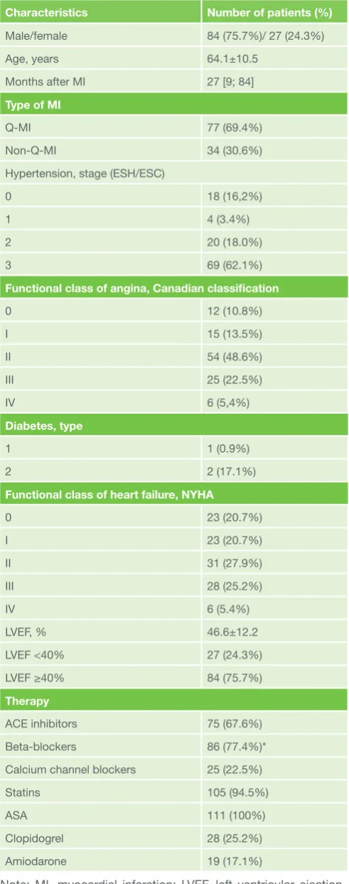
Between 2008 to 2010, 111 patients (age 64.1±10.5 years; 84 males and 27 females) with history of MI occurred 2 months to 36 years (median 27 [9;84] months) previously were enrolled into the study. Inclusion criteria were: MI more than 60 days prior to study entry, aged 18 years and over. Exclusion criteria were: permanent atrial flutter or atrial fibrillation, implanted pacemaker, II-III degree atrioventricular block, concomitant malignancies, hyperthyroidism, severe anemia (hemoglobin less than 90 g/l); co-administration of ivabradin or glycosides. Nineteen (17.1%) patients had percutaneous transluminal coronary angioplasty in the acute MI phase; and 45 (40.5%) had thrombolysis. Thirteen patients with Q-MI had not received revascularization treatment due to late admittance. Baseline characteristics are presented in Table 1. Detailed characteristics of the study population have been published previously. [12] Comparable control group included 60 individuals (44 males and 16 females, age 62.5±11.6 years) with no cardiovascular comorbidity.
The study complied with the Declaration of Helsinki. Study protocol was approved by the local ethics committee. All subjects signed informed consent prior to any study procedures.
Methods
All subjects had baseline examination including medical history, 12-channel ECG, echocardiography (EchoCG) with LVEF measurement, and 24-hour ambulatory ECG monitoring with HRV (SDNN and pNN50), HRT and mTWA evaluation, recording of PVCs number and episodes of sustained/unsustained VT.
Table 1.
Baseline clinical and demographic characteristics of post-MI patients. (n = 111)Characteristics Number of patients (%)
Male/female 84 (75.7%)/ 27 (24.3%)
Age, years 64.1±10.5
Months after MI 27 [9; 84]
Type of MI
Q-MI 77 (69.4%)
Non-Q-MI 34 (30.6%)
Hypertension, stage (ESH/ESC)
0 18 (16,2%)
1 4 (3.4%)
2 20 (18.0%)
3 69 (62.1%)
Functional class of angina, Canadian classification
0 12 (10.8%)
I 15 (13.5%)
II 54 (48.6%)
III 25 (22.5%)
IV 6 (5,4%)
Diabetes, type
1 1 (0.9%)
2 2 (17.1%)
Functional class of heart failure, NYHA
0 23 (20.7%)
I 23 (20.7%)
II 31 (27.9%)
III 28 (25.2%)
IV 6 (5.4%)
LVEF, % 46.6±12.2
LVEF <40% 27 (24.3%)
LVEF ≥40% 84 (75.7%)
Therapy
ACE inhibitors 75 (67.6%)
Beta-blockers 86 (77.4%)*
Calcium channel blockers 25 (22.5%)
Statins 105 (94.5%)
ASA 111 (100%)
Clopidogrel 28 (25.2%)
Amiodarone 19 (17.1%)
MTWA
MTWA was measured by modified moving average method [13] in two Holter leads with GE Healthcare (GE Healthcare, Fairfield, Connecticut, U.S.) GETEMED CardioDay Holter system, ie, recorder CardioMem CM3000 and software CardioDay® v. 2.2.0.3. The detailed technique has been described previously. [13] Absolute maximum value in 24 hours (mTWAmax), mTWA values at heart rate (HR) 100 bpm and at 05.00 AM were assessed in order to standardize measurement conditions.
HRT
Regarding HRT, turbulence onset (TO) (magnitude of sinus rhythm acceleration after PVC) and turbulence slope (TS) (intensity of subsequent sinus rhythm deceleration) were evaluated. Abnormal HRT values were considered as TO≥0%, and TS≤2,5 ms/RR.
DC
DC was calculated by appropriate software via the technique based on the determination of the difference between adjacent RR intervals described by Bauer et al. [7] According to the authors, DC reflects the influence of the parasympathetic autonomic nervous system on the heart. DC > 4.5 ms corresponds to the low SCD risk; DC 2.6 to 4.5 ms corresponds to the average risk; DC ≤ 2.5 ms reflects high risk. [7]
Duration of follow-up was 60 months. Primary endpoint was SCD; secondary endpoint included all non-sudden cardiovascular deaths; tertiary endpoints included non-fatal cardiovascular events and VTs. SCD was defined as death from an unexpected circulatory arrest usually due to cardiac arrhythmia occurring within an hour of the onset of symptoms. The cause of death outside the hospital was established based on the interview with relatives of the patients (personal or via telephone); all efforts were made to collect relevant medical documents.
Statistical analysis.
Data are presented as means ± standard deviations (SD) or as medians [25th; 75th percentiles]. Significance was assessed by two-sided t-test, Wilcoxon test or Mann-Whitney test with α=0.05. To assess sensitivity and specificity of absolute HRT, DC and mTWA values, and to define threshold values for the high risk group, ROC-analysis was performed. Significance of HRT, DC and mTWA values as independent risk factors was determined by univariate and multivariate Cox-regression analysis including clinical and ECG-based parameters with established risk predictive values. The relative risk was determined by Cox regression analysis. Due to the method of selection, risk of predefined outcomes was estimated using odds ratios adjusted for mTWA values. Survival was determined by Kaplan-Meier analysis. All calculations were performed with SPSS v21.0 (SPSS, Chicago, IL,USA).
Results
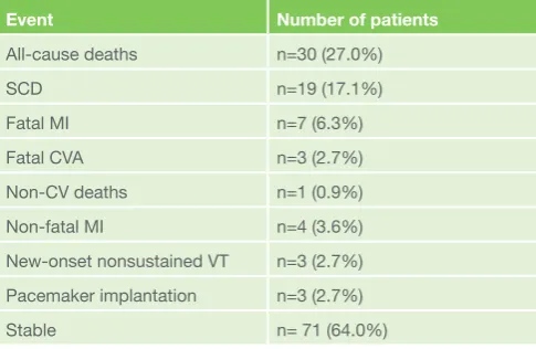
During the 60 months of follow-up, 19 cases of SCD (17.1%) and 11 cases of deaths from other causes (including 7 (6.3%) repeated fatal MIs and 3 (2.7%) fatal strokes) were recorded in post-MI patients. Among all-causes deaths, one case was not related to cardiovascular causes (death due to multiple organ failure after gastrectomy related to the ulcer bleeding; the cause of death was confirmed by autopsy). Four patients died in the hospital; autopsy revealed no structural changes that could be
fatal. In other SCD cases, no autopsy was performed; these were deaths occurring within 60 minutes of the onset of symptoms, as well as cases of death during sleep, i.e. events that met SCD criteria. Information about these deaths was obtained from the relatives of patients; all SCD cases were qualified by the pathologists and appropriate documents were collected. All fatal MIs and fatal strokes occurred in hospital and were confirmed by medical records. In addition, tertiary endpoints were recorded in several patients including nonfatal MIs and the new-onset VT (Table 2). Three patients received pacemakers (DDD mode) during follow-up due to onset of different cardiac blockades. All cases of implantation were performed within 3 months after the enrollment making impossible to assess the trends of mTWA, HRT and DC. However, clinical monitoring for these patients continued; two patients remained stable until the end of the follow-up. The third patient developed nonsustained VT 14 months after pacemaker implantation (confirmed by pacemaker’s records), which caused a loss of consciousness and fall. VT self-terminated within 30 seconds; further patient’s condition was stable. In the control group during the follow-up no deaths were recorded, and mTWA, DC and HRT showed no significant changes.
To evaluate the prognostic value of SCD predictors, all patients were divided into subgroups of survivors, all-causes deaths, and SCDs. HRT, mTWA, and DC values by subgroups shown in Table 3. The majority of deaths (11 of 19 in the SCD subgroup and 6 of 10 in the all-causes subgroup) occurred within the first 3 months after enrollment that did not allow us to assess the trends of SCD predictors. In 6 patients who completed at least one study visit before death, a progressive insignificant decrease in LVEF from 40.7 ± 17.0% to 36.8 ± 15.7% (p = 0.097), and no significant HRT, mTWA, or DC changes were observed. According to Cox regression analysis, factors that significantly influenced the risk of overall mortality included LVEF, DC, HRT, HRV (SDNN, pNN50), mTWA at 05.00 AM and the daily number of PVCs (Table 4).
Regarding SCD, the cut-off prognostic value of mTWA at 100 bpm was 52.5 µV (sensitivity 68.4%, specificity 60.0%) that was practically identical to the 12-months value (53.5 µV) (sensitivity 73.3%, specificity 64.6%), but the sensitivity and specificity
Table 2.
Endpoints in the study population during 60 months of follow-up.Event Number of patients
All-cause deaths n=30 (27.0%)
SCD n=19 (17.1%)
Fatal MI n=7 (6.3%)
Fatal CVA n=3 (2.7%)
Non-CV deaths n=1 (0.9%)
Non-fatal MI n=4 (3.6%)
New-onset nonsustained VT n=3 (2.7%) Pacemaker implantation n=3 (2.7%)
Stable n= 71 (64.0%)
decreased. [12] Analysis of the 60-months data revealed that mTWA at 100 bpm > 52.5 µV 3-fold increased the SCD risk (OR = 3.1 (95% CI 1.1-8.8), p = 0.03). Thus, mTWA at 100 bpm retained its significance during long-term follow-up.
The cut-off prognostic value of mTWA at 05.00 AM was 18.5 µV that was identical to the 12-months value with comparable sensitivity (56.7% and 60.0%, respectively) and specificity (60.8% and 63.8% respectively). The mTWA at 05.00 AM > 18.5 µV 2-fold increased the risk of death from cardiovascular causes (OR 2.3 (95% CI 1.1-5.5), p = 0.04) but not the risk of SCD, consistent with analysis of short-term data.
Analysis of the combinations of several risk factors revealed that at 60 months of follow-up the risk of cardiovascular mortality was the highest in patients with LVEF <40% + VT during 24-hour ECG monitoring (OR 22.9 (95% CI: 2.6-200.4), p = 0.0001), while SCD risk was the highest in patients with LVEF <40%, impaired HRT and mTWA at 100 bpm > 53 µV (OR 24.8 (95 CI 2.6-237.2%) p = 0.002) compared with any of these parameters alone (Fig. 1 and 2). Of note, the combination of LVEF <40% and VT during 24-hour ECG monitoring was only slightly inferior to the combination of LVEF <40%, impaired HRT and mTWA at 100 bpm > 53 µV. DC in combination with other non-invasive parameters moderately increased risk of both cardiovascular mortality and SCD.
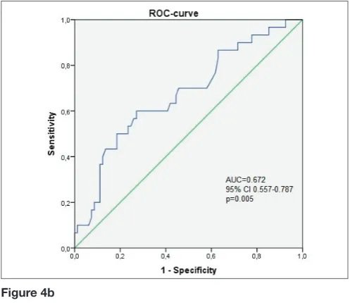
Significance of various combinations of risk factors regarding overall mortality and SCD risk after adjustment for clinical covariates is shown in Fig. 1 and 2. Survival curves are shown in Fig. 3 and ROC-curves for impaired HRT and mTWA combinations are shown in Fig. 4.
Discussion
To our knowledge, this is the first investigation of long-term risk stratification for mortality using mTWA, HRT and DC in post-MI patients.
In prospective clinical studies of >10,000 patients, mTWA identified patients at risk for fatal arrhythmia and cardiovascular
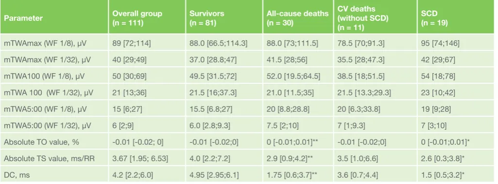
Table 3.
HRT, DC and mTWA values in the study population by outcomes.Parameter Overall group (n = 111) Survivors (n = 81) All-cause deaths (n = 30) CV deaths (without SCD)
(n = 11)
SCD (n = 19)
mTWAmax (WF 1/8), µV 89 [72;114] 88.0 [66.5;114.3] 88.0 [73;111.5] 78.5 [70;91.3] 95 [74;146] mTWAmax (WF 1/32), µV 40 [29;49] 37.0 [28.8;47] 41.5 [28;56] 35.5 [28;47.3] 42 [29;67] mTWA100 (WF 1/8), µV 50 [30;69] 49.5 [31.5;72] 52.0 [19.5;64.5] 38.5 [18;51.5] 54 [18;78] mTWA 100 (WF 1/32), µV 21 [13;36] 21.5 [16;37.3] 21.0 [11.5;35] 21.5 [13.3;29.3] 23 [10;42] mTWA5:00 (WF 1/8), µV 15 [6;27] 15.5 [6.8;27] 20 [8.8;28.8] 20 [6.3;33.8] 19 [9;28]
mTWA5:00 (WF 1/32), µV 6 [2;9] 6.0 [2.8;9.3] 7.5 [2;10] 7 [1;9.3] 7 [3;10]
Absolute ТО value, % -0.01 [-0.02; 0] -0.01 [-0.02;0] 0 [-0.01;0.01]** -0.01 [-0.02;0] 0 [-0.01;0.01]* Absolute ТS value, ms/RR 3.67 [1.95; 6.53] 4.0 [2.2;7.2] 2.9 [0.9;4.2]** 3.5 [1.0;6.6] 2.6 [0.3;3.8]* DC, ms 4.2 [2.2;6.0] 4.95 [2.95;6.1] 1.75 [0.6;3.7]** 3.6 [0.7;4.4] 1.5 [0.5;3.2]* Note: mTWA, microvolt T-wave alternans; mTWAmax, maximal microvolt T-wave alternans; HR, heart rate; WF, weighing factor; HRT, heart rate turbulence; ТО, turbulence onset; ТS, turbulence slope, SCD, sudden cardiac death.
*p<0.05 for SCD compared with other outcomes, ** p<0.05 for main group compared with control group, *** p <0.05 for the subgroup of survivors compared with overall mortality subgroup. All mTWA values are for the first lead.
Figure 1. Relative risk of overall mortality at 60 months for different risk factors combinations.
Note: DC, deceleration capacity; mTWA, microvolt T-wave alternans; HRT, heart rate turbulence; ТО, turbulence onset; ТS, turbulence slope; LVEF, left ventricular ejection fraction; PVC, premature ventricular contraction; VT, ventricular tachycardia; OR, odds ratio; CI, confidence interval.
Figure 2. Relative risk of SCD at 60 months for different risk factors combinations.
and total mortality [8] and met American Heart Association, American College of Cardiology, European Society of Cardiology, and/or Heart Rhythm Society requirements for class I (level of evidence A) and class IIa (level of evidence A) indications for arrhythmia risk assessment. [4] Goldberger et al. [1] stated that mTWA provides valuable information regarding the risk of cardiovascular mortality and SCD among patients with ischemic, dilated, and hypertrophic cardiomyopathies. Thus, a recent consensus guideline recommended mTWA monitoring in any suspected risk for fatal arrhythmia [8] but it has not been included in the ESC 2015 Guidelines for management of patients with ventricular arrhythmias and the prevention of SCD [11] due to the lack of evidence-based information.
Previously [12] we have demonstrated the significant predictive
value of HRT and mTWA both in the general population of post-MI patients and in patients with preserved left ventricular function at 12 months of follow-up. No baseline characteristic typically linked with poor outcomes was associated with high mTWA, suggesting that mTWA offers complementary prognostic information. However, the subgroup analysis of patients with different LVEF at 60 months of follow-up virtually all investigated predictors showed no significant difference between subjects with preserved and reduced LV function, except low DC that remained significance in the subgroup of patients with LVEF > 40% (cardiovascular mortality: OR 8.5, 95% CI 2.9-24.6; p <0.0001 for all patients, and OR 16.1, 95% CI: 3.3-78.2, p <0.0001 in patients with LVEF > 40%; SCD: OR 9.6, 95% CI 3.2-28.5; p <0.0001 for all patients, and OR 21.3, 95% CI 3.7-122.3, p <0.0001 in patients with LVEF> 40%). This finding may suggest the high importance of these predictors for SCD and cardiovascular mortality risk stratification in the short-term perspective, indicating the need for more close monitoring and more aggressive therapy in these patients, as well as optimization of antiarrhythmic therapy to improve myocardial electrical stability. This is consistent with the findings of MERLIN-TIMI study. [9]
Table 4.
Significance of independent risk factors Overall mortality riskRisk factor AUC Threshold value Sensitivity Specificity Р value
LVEF, % 0.720 46.5% 70.0% 67.6% <0.001 DC, ms 0.749 4.15 83.3% 60.8% <0.001 ТО, % 0.675 -0.005 63.3% 63.5% 0.005 pNN50, % 0.664 3.5% 63.3% 66.2% 0.009 TS, ms/RR 0.663 1.21 33.3% 94.6% 0.009 SDNN, ms 0.624 134 ms 86.7% 41.9% 0.048 Number of PVCs 0.644 399 53.3% 79.7% 0.022 QRS, ms 0.604 99 ms 53.3% 71.6% 0.097 mTWA at 05.00
AM, µV 0.568 18.5 µV 56.7% 60.8% 0.275 mTWA max, µV 0.523 67.5 93.3% 25.7% 0.709 mTWA at HR
100/min, µV 0.463 52.5 µV 50.0% 56.8% 0.554 Sudden cardiac death risk
LVEF, % 0,789 48,5% 89,5% 56,5% <0,001 DC, ms 0,747 2,0 63,2% 84,7% 0,001 ТО, % 0,737 -0,005 73,7% 62,4% 0,001 Number of PVCs 0,679 529 3,2% 80,0% 0,015 QRS, ms 0,676 99 ms 63,2% 70,6% 0,017 TS, ms/RR 0,674 3,96 84,2% 49,4% 0,018 mTWA at HR
100/min, µV 0,566 52,5 µV 68,4% 60,0% 0,037 pNN50, % 0,643 9,5% 84,2% 35,3% 0,052 mTWA max, µV 0,617 72,5 89,5% 29,4% 0,113 SDNN, ms 0,608 134 ms 84,2% 37,6% 0,142 mTWA at 05.00
AM, µV 0,546 8,5 µV 78,9% 34,1% 0,534 Note: AUC, area under the ROC-curve; DC, deceleration capacity; LVEF, left ventricular ejection fraction; SDNN, standard deviation of normal to normal RR intervals; pNN50, proportion of the number of pairs of successive NNs that differ by more than 50 ms divided by total number of NNs; mTWA, microvolt T-wave alternans; HR, heart rate; ТО, turbulence onset; ТS, turbulence slope; PVC, premature ventricular contraction.
Figure 3a. Cox regression for combination of HRT 2 and mTWA at HR 100 bpm > 53 mcV as a predictor of SCD at 12 months (А) [12] and 60 months (B). P values are for the log rank test.
Threshold values obtained in our study differed from findings in other studies [8, 14] that may be explained by differences in measurement and in characteristics of study populations, but cut-offs for mTWA at 05.00 AM (CV mortality) and mTWA at 100 bpm (SCD) at 12 and 60 months of follow-up were similar suggesting the good reproducibility of mTWA measurement during short-term and long-term monitoring; it is consistent with the results of Chan et al. [15] who showed that the predictive value of the baseline mTWA evaluation persists during 2-3 years, and therefore re-measurement mTWA is reasonable only every 2 years. In this study, duration of follow-up was 18 ± 11 months; our results for the follow-up period of 60 months confirm the conclusions of the authors [15] as repeated measurements at 3, 6 and 12 months were not significantly different.
Combined risk stratification at 60 months revealed that the risk of cardiovascular mortality was the highest in patients with LVEF <40% and the presence of VT, and the maximum risk of SCD was similar in patients with LVEF <40%, HRT 2 and mTWA at 100 bpm > 53 μV and patients with LVEF <40% and VT detection during ECG monitoring. This differs from the findings at 12 months, when the combination of HRT 2 and mTWA at 100 bpm > 53 μV defined the subgroup of the highest risk compared both with an isolated impairment of any indicator and other combinations, including those with LVEF < 40%. [12] This may be explained by the fact that most patients (15 of 19 in the SCD subgroup and 8 of 10 in the group of cardiovascular mortality) died within the first 12 months after enrollment. These patients were characterized by the maximum impairment of the studied parameters, and these dropouts resulted in the elimination of the highest-risk groups according to a combination of non-invasive predictors; in the remaining patients, “conventional” risk factors - the low LVEF and myocardial ectopic activity – became important. Again, this finding support the high short-term importance of these predictors.
In our study, the new predictors were more accurate than the conventional ones. While HRT has been used for over 10 years and is included in the International Guidelines for risk stratification in patients after myocardial infarction, data about the use of DC to predict adverse outcomes in high-risk groups are limited. [7, 16, 17, 18] However, DC has a number of advantages compared with conventional HRV: independent evaluation of parasympathetic (DC) and sympathetic system (acceleration capacity is used to assess the activity of the sympathetic system, but its prognostic value has not been demonstrated); simplicity of data presentation (only one component instead of multiple HRV parameters) and direct focus on the risk assessment, with a clear staging by the low, medium and high. [7] Several studies have shown prognostic significance of HRT 2 and DC (“severe autonomic failure”) for the SCD risk stratification in the post-MI patients and preserved LVEF [16, 17] as well as in patients with CAD and diabetes. [18] In our study, this combination also was quite good for assessment of the risk of both overall mortality (4.3-fold), and SCD (4.4-fold) at 60 months.
Combined risk assessment with HRT and mTWA was first used by the REFINE study authors, [14] and subsequently by the other two study groups [19, 20] published later than our 12-months results. [12] Despite the fact that these studies had relatively long follow-ups (1.5 to 4 years) and the number of patients (173
to 322), included patients early after MI, differences in mTWA measurement and cut-off values, [19, 20] their findings of high prognostic significance of HRT 2 + mTWA confirm our data. Our study was the first one that used DC in a combined assessment of the risk of adverse outcomes and had the long follow-up up to 5 years.
Thus, our findings support high predictive value of HRT, DC and mTWA values for SCD and CV mortality risk stratification. Of course, the main questions are the following: “What should physician do with the patient who has non-invasive electrophysiological SCD predictors” and “Whether these data affect the choice of treatment strategy?” Based on the high negative predictive value of mTWA, T.Ikeda (Ikeda T. Combination of Tests in Risk Stratification. Report for Cardiostim 2014 on behalf of ISHNE- International Society for Holter and Noninvasve Electrocardiology (oral presentation).-19.06.2014, Nice) proposed a new algorithm for selection of patients for ICD implantation. According to this algorithm, the standard LVEF evaluation should be followed by the mTWA measurement; if the test is negative drug therapy should be continued, otherwise, (positive or indeterminate test) Figure 4a. ROC-curves for for combination of HRT 2 and mTWA at HR 100 bpm > 53 mcV as a predictor of SCD (А) and all-cause mortality (B).
additional non-invasive examinations are performed (ventricular late potentials, HRV, unstable VT detection etc.), and in some cases - invasive electrophysiological study. There author considers ICD implantation justified only in case of positive result of the any additional tests. The more definitive answer may be received after the completion of a prospective REFINE-ICD study, which design includes randomization of patients with LVEF 36-50% and combined mTWA + HRT impairment for the standard treatment or ICD therapy. [5] In addition, long-term significance of noninvasive SCD predictors requires further confirmation in larger prospective studies.
Limitations
The single-center design, size of the study and number of events are limited; therefore, the findings require confirmation in a larger prospective study of high-risk subjects. This limitation was attenuated by adjustment for the well-known clinical and ECG risk factors. Despite the fact that post-MI population in our study was heterogenous and included patients with MI occurred from 2 months to about 30 years before enrollment, we analyzed possible effect of the MI onset on the noninvasive parameters; no significant influence was found; the heterogeneity of the previous MI treatment (invasive and non-invasive) was analyzed as well, and no significant impact was found. In addition, DC was evaluated retrospectively since this technique was unavailable at the study initiation.
Conclusions
Evaluation of HRT, DC and mTWA during 24-hour ECG monitoring may define the high risk of cardiovascular mortality and SCD in post-MI patients. The combination of abnormal HRT and mTWA100 > 53 μV is associated with the most significant increase in the risk of SCD during the first 12 months after the baseline examination. At 5 years of follow-up, value of noninvasive mortality predictors of the risk stratification is reduced compared with the importance of low LVEF and VT, but their contribution to a combined risk assessment remains significant. This suggest the high importance of these predictors for the short-term SCD and cardiovascular mortality risk stratification, indicating the need for more close monitoring and more aggressive therapy in these patients, as well as optimization of antiarrhythmic therapy to improve myocardial electrical stability or the selection of candidates for ICD implantation.
Declarations of Interest
V.S.: lectures and research work for Solvay Pharma, Sanofi, Nycomed, General Electric, Cordis a Johnson & Johnson; lectures for Boehringer Ingelheim, Bayer, Astra Zeneka. Elly Lilly; D.T.: lectures and research work for General Electric and Sanofi; E.O. received travel fees by General Electric, Boehringer Ingelheim and Sanofi.
Acknowledgements
The authors state that they abide by the “Requirements for Ethical Publishing in Biomedical Journals”. [21] This study received no grant from any funding agency in the public, commercial or not-for-profit sectors.
References
1. Goldberger JJ, Cain ME, Hohnloser SH , Kadish AH, Knight BP, Lauer MS, Maron BJ, Page RL, Passman RS, Siscovick D, Siscovick D, Stevenson WG, Zipes DP. American Heart Association/American College of Cardiology
Foundation/Heart Rhythm Society scientific statement on noninvasive risk stratification techniques for identifying patients at risk for sudden cardiac death: a scientific statement from the American Heart Association Council on Clinical Cardiology Committee on Electrocardiography and Arrhythmias and Council on Epidemiology and Prevention. Circulation. 2008;118:1497-1518. [PMID: 18833586 DOI: http://dx.doi.org/10.1161/ CIRCULATIONAHA.107.189375]
2. Sans S, Kesteloot H, Kromhout D. The burden of cardiovascular diseases mortality in Europe. Task Force of the European Society of Cardiology on Cardiovascular Mortality and Morbidity Statistics in Europe. Eur Heart J 1997; 18: 1231-18. [PMID: 9508543 DOI: http://dx.doi.org/10.1093/ eurheartj]
3. Buxton AE, Lee KL, Hafley GE, Pires LA, Fisher JD, Gold MR, Josephson ME, Lehmann MH, Prystowsky EN; MUSTT Investigators. Limitations of ejection fraction for prediction of sudden death risk in patients with coronary artery disease: lessons from the MUSTT study. J Am Coll Cardiol 2007, 18;50(12):1150-7. [PMID: 17868806 DOI: 10.1016/j.jacc.2007.04.095] 4. Zipes DP, Camm AJ, Borggrefe M, Buxton AE, Chaitman B, Fromer M,
Gregoratos G, Klein G, Moss AJ, Myerburg RJ, Priori SG, Quinones MA, Roden DM, Silka MJ, Tracy C, Smith SC Jr, Jacobs AK, Adams CD, Antman EM, Anderson JL, Hunt SA, Halperin JL, Nishimura R, Ornato JP, Page RL, Riegel B, Priori SG, Blanc JJ, Budaj A, Camm AJ, Dean V, Deckers JW, Despres C, Dickstein K, Lekakis J, McGregor K, Metra M, Morais J, Osterspey A, Tamargo JL, Zamorano JL; American College of Cardiology; American Heart Association Task Force; European Society of Cardiology Committee for Practice Guidelines. ACC/AHA/ESC 2006 guidelines for management of patients with ventricular arrhythmias and the prevention of sudden cardiac death. A report of the American College of Cardiology/American Heart Association Task Force and the European Society of Cardiology Committee for Practice Guidelines (Writing Committee to Develop Guidelines for Management of Patients With Ventricular Arrhythmias and the Prevention of Sudden Cardiac Death). J Am Coll Cardiol 2006;48:1064–1108. [PMID: 16949478 DOI: 10.1016/j.jacc.2006.07.010]
5. Wellens HJ, Schwartz PJ, Lindemans FW, Buxton AE, Goldberger JJ, Hohnloser SH, Huikuri HV, Kääb S, La Rovere MT, Malik M, Myerburg RJ, Simoons ML, Swedberg K, Tijssen J, Voors AA, Wilde AA1. Risk stratification for sudden cardiac death: current status and challenges for the future. Eur Heart J 2014. 35(25):1642-1651. [PMID: 24801071 DOI: 10.1093/eurheartj/ehu176]
6. Epstein AE, DiMarco JP, Ellenbogen KA, Estes NA 3rd, Freedman RA, Gettes LS, Gillinov AM, Gregoratos G, Hammill SC, Hayes DL, Hlatky MA, Newby LK, Page RL, Schoenfeld MH, Silka MJ, Stevenson LW, Sweeney MO, Smith SC Jr, Jacobs AK, Adams CD, Anderson JL, Buller CE, Creager MA, Ettinger SM, Faxon DP, Halperin JL, Hiratzka LF, Hunt SA, Krumholz HM, Kushner FG, Lytle BW, Nishimura RA, Ornato JP, Page RL, Riegel B, Tarkington LG, Yancy CW; American College of Cardiology/American Heart Association Task Force on Practice Guidelines (Writing Committee to Revise the ACC/AHA/NASPE 2002 Guideline Update for Implantation of Cardiac Pacemakers and Antiarrhythmia Devices); American Association for Thoracic Surgery; Society of Thoracic Surgeons. ACC/AHA/HRS 2008 Guidelines for Device-Based Therapy of Cardiac Rhythm Abnormalities: Executive Summary: A Report of the American College of Cardiology/ American Heart Association Task Force on Practice Guidelines (Writing Committee to Revise the ACC/AHA/NASPE 2002 Guideline Update for Implantation of Cardiac Pacemakers and Antiarrhythmia Devices) Developed in Collaboration With the American Association for Thoracic Surgery and Society of Thoracic Surgeons. J Am Coll Cardiol 2008;51(21);2085-2105. [PMID: 18498951 DOI: 10.1016/j.jacc.2008.02.032]
7. Bauer A, Kantelhardt J W, Barthel P, Schneider R, Mäkikallio T, Ulm K, Hnatkova K, Schömig A, Huikuri H, Bunde A, Malik M, Schmidt G.. Deceleration capacity of heart rate as a predictor of mortality after myocardial infarction: cohort study. Lancet 2006; 367:1674-1681. [PMID: 16714188 DOI: 10.1016/S0140-6736(06)68735-7]
8. Verrier RL, Klingenheben T, Malik M, El-Sherif N, Exner DV, Hohnloser SH, Ikeda T, Martínez JP, Narayan SM, Nieminen T, Rosenbaum DS. Microvolt T-wave alternans: physiologic basis, methods of measurement, and clinical utility — Consensus guideline by the International Society for Holter and Noninvasive Electrocardiology. J Am Coll Cardiol 2011;44:1309e1324. [PMID: 21920259 DOI: 10.1016/j.jacc.2011.06.029]
9. Bauer A, Malik M, Schmidt G, Barthel P, Bonnemeier H, Cygankiewicz I, Guzik P, Lombardi F, Müller A, Oto A, Schneider R, Watanabe M, Wichterle D, Zareba W. Heart rate turbulence: Standards of measurement, physiological interpretation, and clinical use. International Society for Holter and Noninvasive Electrophysiology consensus. J Am Coll Cardiol 2008; 52:1353-1365. [PMID: 18940523 DOI: 10.1016/j.jacc.2008.07.041] 10. Nieminen T, Scirica BM, Pegler JR, Tavares C, Pagotto VP, Kanas AF,
Sobrado MF, Nearing BD, Umez-Eronini AA, Morrow DA, Belardinelli L, Verrier RL. Relation of T-wave alternans to mortality and nonsustained ventricular tachycardia in patients with non-ST-segment elevation acute coronary syndrome from the MERLIN-TIMI 36 trial of ranolazine versus placebo. Am J Cardiol 2014 Jul 1;114(1):17-23. [PMID: 24852915 DOI: 10.1016/j.amjcard.2014.03.056]
ventricular arrhythmias and the prevention of sudden cardiac death: The Task Force for the Management of Patients with Ventricular Arrhythmias and the Prevention of Sudden Cardiac Death of the European Society of Cardiology (ESC). Eur Heart J 2015 Nov 1;36(41):2793-867. [PMID: 26320108 DOI: 10.1093/eurheartj/ehv316]
12. Sulimov V., Okisheva р., Tsaregorodtsev D. Non-invasive risk stratification for sudden cardiac death by heart rate turbulence and microvolt T-wave alternans in patients after myocardial infarction. Europace 2012. 14(12):1786-1792. [PMID: 22849973 DOI: 10.1093/europace/eus238] 13. Nearing BD, Verrier RL. Modified moving average analysis of T-wave alternans
to predict ventricular fibrillation with high accuracy. J Appl Physiol. 2002.92: 541–549. [PMID: 11796662 DOI: 10.1152/japplphysiol.00592.2001] 14. Exner DV, Kavanagh KM, Slawnych MP, Mitchell LB, Ramadan D, Aggarwal
SG, Noullett C, Van Schaik A, Mitchell RT, Shibata MA, Gulamhussein S, McMeekin J, Tymchak W, Schnell G, Gillis AM, Sheldon RS, Fick GH, Duff HJ; REFINE Investigators. Noninvasive risk assessment early after a myocardial infarction: The REFINE study. J Am Coll Cardiol. 2007;50:2275– 84. [PMID: 18068035 DOI: 10.1016/j.jacc.2007.08.042]
15. Chan PS, Kereiakes DJ, Bartone C, Chow T. Usefulness of microvolt T-wave alternans to predict outcomes in patients with ischemic cardiomyopathy beyond one year. Am J Cardiol 2008;102:280 –284. [PMID: 18638586 DOI: 10.1016/j.amjcard.2008.03.049]
16. Arsenos P, Gatzoulis K; Manis G; Dilaveris P; Gialernios T; Archontakis S; Tsiachris D; Aggelis A; Kartsagoulis E; Stefanadis C; APRET. Reduced deceleration capacity of heart rate risk stratifies patients presenting with preserved left ventricular ejection fraction (LVEF>35%) for sudden cardiac death (2011), ORAL PRESENTATION. Pacing and Clinical Electrophysiology, 34(11): 1307–1361. [DOI: 10.1111/j.1540-8159.2011.03251]
17. Bauer A, Barthel P, Müller A, Ulm K, Huikuri H, Malik M, Schmidt G. Risk prediction by heart rate turbulence and deceleration capacity in postinfarction patients with preserved left ventricular function retrospective analysis of 4 independent trials. J Electrocardiol. 2009 Nov-Dec;42(6):597-601. [PMID: 19853731 DOI: 10.1016/j.jelectrocard.2009.07.013]
18. Zuern CS, Rizas K, Eick C, Sterz K, Gawaz M, Bauer A. Prevalence and predictors of severe autonomic failure in patients with insulin-dependent type 2 diabetes mellitus and coronary artery disease: pilot study. J Electrocardiol. 2012 Nov-Dec; 45(6):774-9. [PMID: 22944520 DOI: 10.1016/j.jelectrocard.2012.07.010]
19. Hoshida K., Miwa Y., Miyakoshi M, Tsukada T, Yusu S, Yoshino H, Ikeda T. Simultaneous assessment of T-wave alternans and heart rate turbulence on holter electrocardiograms as predictors for serious cardiac events in patients after myocardial infarction. Circ J. 2013. 77(2):432-438. [PMID: 23059771]
20. Li-na R, Xin-hui F, Li-dong R, Jian G, Yong-quan W, Guo-xian Q. Ambulatory ECG-based T-wave alternans and heart rate turbulence can predict cardiac mortality in patients with myocardial infarction with or without diabetes mellitus. Cardiovasc Diabetol. 2012. 6;11:104. [PMID: 22950360 DOI: 10.1186/1475-2840-11-104]
![Figure 3a. Cox regression for combination of HRT 2 and mTWA at HR 100 bpm > 53 mcV as a predictor of SCD at 12 months (А) [12] and 60 months (B)](https://thumb-us.123doks.com/thumbv2/123dok_us/7827165.2088528/5.637.65.310.102.529/figure-cox-regression-combination-predictor-scd-months-months.webp)