Detection of Early Heart Failure with
Preserved Ejection Fraction (HFpEF) in
Metabolic Syndrome Patients Detected as
Part of a National Screening Programme in
Middle Aged Subjects
Jelena Čelutkienė
1,2, Aistė Monika Jakštaitė
1Jolita Badarienė
1,2, Svetlana Solovjova
1,2, Ieva
Slivovskaja
4, Rokas Navickas
2, Edita Kazėnaitė
2, Egidija Rinkūnienė
2, Alma Čypienė
2, Jonas
Misiūra
1,2, Ligita Ryliškytė
1,2, Aleksandras Laucevičius
1,2,3, Andrew Coats
51. Clinic of Cardiac and Vascular Diseases, Medical faculty, Vilnius University, Vilnius, Lithuania 2. Centre of Cardiology and Angiology, Vilnius University Hospital Santaros Klinikos, Vilnius, Lithuania 3. State Research Institute Centre For Innovative Medicine, Vilnius, Lithuania
4. Department of Rehabilitation, Physical and Sports Medicine, Vilnius University Hospital Santaros Klinikos, Vilnius, Lithuania 5. Department of Medical Sciences, IRCCS San Raffaele Pisana, Rome, Italy.
Corresponding author:
Prof. Jelena Čelutkienė,
Senior Researcher, Clinic of Cardiovascular Diseases, Faculty of Medicine, Vilnius University, Santariškių 2, LT-08661
E-mail: [email protected]
Abstract
Background
We wished to investigate if community detected Metabolic Syndrome (MetS) is associated with the burden of incipient HFpEF in the community.
Methods and Results
We prospectively studied 148 consecutive MetS patients identified from the Lithuanian High Cardiovascular Risk primary prevention programme and investigated them further for unknown HFpEF through cardiopulmonary stress testing as well as assessment of BNP levels and of arterial stiffness. Subjects with a peak VO2 value lower than 90% of predicted and/or BNP≥35 ng/l were categorized as having early phase HFpEF. For comparison of this early phase HFpEF with others already clinically diagnosed with HFpEF, patients with both established HFpEF and MetS were selected retrospectively from patients attending our cardiopulmonary stress testing laboratory (n=38). Two thirds of the screening programme-derived MetS population (n=96) demonstrated a reduced exercise capacity and/or an elevated BNP, indicating signs of early HFpEF. Both the clinically diagnosed HFpEF and the screening programme detected MetS group with early HFpEF demonstrated similarly decreased exercise tolerance evaluated by peak oxygen uptake (79.8 ± 22.1% vs 82.7 ± 14.0%, p>0.05). Analysis of arterial markers in the screening programme group revealed statistically significant differences of augmentation index values between groups with and without signs of early HFpEF (p=0.016).
Conclusions
A considerable proportion of patients having MetS may be diagnosed with previously undetected early stage HFpEF. The use of objective parameters of exercise capacity and neurohormonal activation might be effectively used for the early detection of HFpEF. Also early HFpEF in this setting is found to be associated with increased arterial stiffness.
Keywords: Heart failure with preserved ejection fraction, metabolic syndrome, arterial stiffness, early detection
Citation: Čelutkienė J, Jakštaitė AM, Badarienė J. et al. Detection of Early Heart Failure with Preserved Ejection Fraction (HFpEF) in Metabolic Syndrome Patients Detected as Part of a National Screening Programme in Middle Aged Subjects. International Cardiovascular Forum Journal 2018;13:9-15, DOI: 10.17987/icfj.v13i0.550
ISSN: 2410-2636 © Barcaray Publishing © 2018 Author(s). This is an Open Access article distributed under the terms of the Creative Commons Attribution CC-BY-4.0 license CC-BY-4.0 (http://creativecommons.org/ licenses/by/4.0/), which permits use, distribution and reproduction, provided the original work is properly cited. Published by Barcaray (International) Publishing.
Introduction
Metabolic syndrome (MetS) is a syndrome described as a clustering of metabolic risk factors including abdominal obesity, elevated blood glucose and triglycerides, reduced HDL cholesterol and hypertension. It has been described as being associated with an increased risk of type 2 diabetes, myocardial infarction, stroke, heart failure and cardiovascular mortality. In western populations and in the developing world the burden of MetS has reached epidemic proportions and is fast becoming one of the greatest global public health concerns with a prevalence of 34% in the general population [1].
Recent studies have highlighted the importance of the MetS and its components for the development and progression of heart failure [2,3,4,5]. MetS increases the risk of myocardial infarction and chronic ischaemia, both of which predispose to heart failure with reduced ejection fraction (HFrEF). It can also lead to heart failure with preserved ejection fraction (HFpEF) via different pathophysiological pathways. It has been shown that the prevalence of left ventricular (LV) diastolic dysfunction is significantly higher in patients with MetS compared to those without (35% vs. 9%) [6,7]. A large multicentre study which evaluated 6422 asymptomatic patients with LV dysfunction demonstrated the relevance of MetS as an independent predictor of diastolic dysfunction after adjustment for age, gender and myocardial hypertrophy. Furthermore, the degree of diastolic dysfunction was dependent on the number of individual MetS components [8].
A diagnosis of HFrEF is usually made after the patient presents with an acute decompensation, often with pulmonary oedema, and frequently after a period of silent LV remodelling, neurohormonal activation and sub-clinical fluid retention. Echocardiographic screening programmes in at-risk subjects can detect pre-symptomatic cases at high risk of HFrEF. The situation for HFpEF is however, more complicated, as the diagnosis can be harder to make in the early course of the syndrome, with reduced exercise tolerance and occasional dyspnoea being ascribed to the effects of advancing age and common co-morbidities, such as obesity and lung disease.
Currently, little is known about the early development and early clinical diagnosis of HFpEF in patients with metabolic syndrome, and thus opportunities to intervene early in MetS to reduce both the prevalence of HFpEF and its subsequent clinical course are being lost. Given the high prevalence of diastolic dysfunction in this population, we hypothesized that a significant number of patients with MetS may already have unrecognized HFpEF. The purpose of the study was thus to investigate the possibility of detecting an early phase of HFpEF by means of exercise capacity evaluation and biomarker testing. Additionally, we sought to examine the potential association between markers of arterial stiffness an early HFpEF within a community identified MetS population.
Methods
The study was approved by the local Ethics Committee and written informed consent was obtained from each patient. The investigation conforms with the principles outlined in the Declaration of Helsinki (Br Med J 1964; ii:177).
Study populations
a. Screening Programme Cohort with MetS:
We prospectively enrolled 148 patients with MetS (mean age 56.4 ± 6.6 years, 46 (31.1%) males) referred from the Lithuanian High Cardiovascular Risk (LitHiR) primary prevention programme, a nationwide screening programme for males between the ages of 40 and 55 years and females between the ages of 50 and 65 years and free of overt cardiovascular disease. The inclusion criteria were: metabolic syndrome (as defined below), and the availability of brain natriuretic peptide (BNP) test, cardiopulmonary stress test, echocardiography and arterial markers (pulse wave velocity [PWV], augmentation index [AI], common carotid artery intima-media thickness [IMT], Quality Carotid Stiffness [QCS] of common carotid artery [CCA], ankle brachial index [ABI], reactive hyperaemia index [RHI]).
b. Clinical HFpEF Cohort with MetS:
We recruited 38 patients (mean age 61 ± 10.5 years, 16 (42.1%) males) who had both a past clinical diagnosis of HFpEF (clinically diagnosed heart failure with left ventricular ejection fraction >50%) and who also satisfied our criteria for MetS (as described below). These subjects were recruited from patients who had been referred to the cardiopulmonary exercise laboratory for evaluation of heart failure, or for the differential diagnosis of dyspnoea. Seventy-one percent of this cohort had a history of hospitalisations due to cardiovascular diseases (CVD).
Definitions and Testing Protocols:
Definition of Metabolic Syndrome
We used the definition of MetS which was adapted by the National Cholesterol Education Program Adult Treatment Panel III recommendations. Participants were classified as having MetS if they satisfied at least 3 of the following 5 criteria:
1. Central (abdominal) obesity with a waist circumference >102 cm in men or >88 cm in women
2. Triglycerides of ≥1.7 mmol/l 3. Fasting serum glucose ≥5.6 mmol/l
4. HDL-cholesterol level ≤1.03 mmol/l in men and ≤1.29 mmol/l in women
5. Blood pressure of ≥130/85 mmHg.
Detection of early HFpEF
We defined early HFpEF as the presence of either an objective impairment of exercise tolerance (peak VO2 ≤90% of its predicted value on valid symptom-limited cardiopulmonary exercise testing) or an objective biomarker suggestive of increased myocardial wall stress (BNP level 35 ng/l or higher). Based on these two criteria the screening MetS cohort was divided into two subgroups: those with and those without signs of early HFpEF.
Cardiopulmonary stress test
calibrated before each test. The anaerobic threshold and VAT were determined by the “V-slope” method (VCO2/VO2 ratio) – the first curve inflection [9]1986.-Excess CO2 is generated when lactate is increased during exercise because its [H’] is buffered primarily by HCO: (22 ml for each meq of lactic acid. Peak oxygen consumption (VO2peak) was considered to be achieved if VO2 reached a plateau for at least 30 sec. in the presence of an increasing power output (W) [10]19&Z.-This study was undertaken to determine which of four com-monly used ventilatory or gas exchange indices provides the most accurate and reliable detection of the anaerobic threshold (AT. If the subject was exhausted before the attainment of VO2 peak, the VO2 value was accepted as a reasonable maximum if the heart rate was >85% of the predicted maximum rate and a respiratory quotient (RQ) >1.00 was recorded.
BNP testing
The Abbott ARCHITECT immunoassay analyzer was used for the diagnosis of BNP concentration in plasma (units of measure - ng/l).
Echocardiographic diagnosis of LV Hypertrophy
Two-dimensional echocardiography was performed using ultrasonic system equipped with a 1.0 – 5.0 MHz transducer (GE Vivid 4; GE Healthcare, New York, USA). The following measures were obtained: interventricular septal end-diastolic thickness (IVST), left ventricular posterior wall end-diastolic thickness (LPWT), and left ventricular end-diastolic dimension (LVDd). The following formulas were used: body surface area (BSA) = 0.0061 × body height (cm) + 0.128 × body weight (kg) - 0.1529 (m2), left ventricular mass (LVM) = 0.8 × 1.04 × [(IVST + LPWT + LVDd)3 - LVDd3] + 0.6 (g) and relative wall thickness (RWT) = (IVST + LPWT)/LVDd.
According to recommendations for chamber quantification, LV hypertrophy was diagnosed when LV mass index made ≥95 g/m² in women and ≥115 g/m² in men and/or relative wall thickness ≥0,42 [11]. In our study 68.4% of the clinically diagnosed and 60.8% of the Screening Cohort patients had LV hypertrophy.
Definition of LV Diastolic Dysfunction
Assessment of LV diastolic function included trans-mitral pulsed wave Doppler with evaluation of peak velocities of early diastolic flow (E) and peak flow of atrial contraction (A), E/A ratio, as well as tissue Doppler imaging parameters with early (E’) and late (A’) diastolic mitral medial and lateral annular velocities. The left atrial volume (LAV) was measured by the biplane area–length method and indexed to the BSA.
Diastolic dysfunction was defined according to the 2016 ESC Guidelines for the diagnosis and treatment of acute and chronic heart failure [12]. Impaired relaxation was described as E/A <1.0 and E/e‘ mean <13. Participants were considered as having pseudo-normal or restrictive diastolic dysfunction if the E/e‘mean ratio was ≥13. In case of E/A >1.0 and e‘ septal ≥8 cm/s and e‘ lateral ≥10 cm/s diastolic function was interpreted as normal.
Arterial Markers of Subclinical Atherosclerosis
Arterial stiffness measurements
Parameters of arterial stiffness and wave reflection were assessed by applanation tonometry (SphygmoCor, AtCor Medical, version
8.0, Sydney, Australia) and Fukuda Vascular Screening system VaSera VS-1000 (Fukuda Denshi Co. Ltd., Tokyo, Japan). Using SphygmoCor system, radial, carotid and femoral pressure waveforms were recorded for 20 seconds each with single transducer synchronized with ECG R wave, after obtaining high quality waveform. Carotid femoral pulse wave velocity (cfPWV), which represents more selectively central arterial wall stiffness, was calculated automatically as the distance divided by time (meters per second). Aortic pressure waveform with calculation of heart-rate 75 bpm adjusted aortic augmentation index (AIx@ HR75) was automatically derived from radial pressure waveform using previously validated transfer function. VaSera VS-1000 system was used for the measurement of ankle-brachial index (ABI), which rates the presence of peripheral artery disease. ABI was calculated automatically from the pressure curves.
Common carotid artery wall assessment
Carotid intima-media thickness (IMT) is an anatomical measure which is used for the detection of subclinical atherosclerosis, from early to late stages. High-resolution echo-tracking technology (Art.Lab; Esaote Europe, Maastricht, The Netherlands) was used for the evaluation of carotid IMT, calculated in μm, and the non-dimensional index QCS of CCA.
The measurements were performed at 1 cm proximal to the carotid bifurcation along 4 cm arterial segment.
Assessment of endothelial function
Reactive hyperaemia index, which evaluates endothelial function in the microcirculation, was measured by peripheral arterial tonometry (PAT) (EndoPAT 2000 system; Itamar Medical, Caesarea, Israel) in fasting patients.
Statistical analysis
Statistical analysis was performed using SPSS Statistics version 24.0 (IBM Corp., Armonk, NY, USA). Descriptive parameters are presented as mean ± standard deviation or in percentages. Two-tailed t-tests and Pearson’s chi-square tests were used to analyse the differences in means between groups which were considered to be statistically significant when p value < 0.05.
Results
in terms of the presence or absence of early HFpEF included a lower waist circumference (in women only) and a marginally higher HDL cholesterol. Perhaps surprisingly diastolic functional parameters did not significantly differ between the groups with and without early HFpEF, perhaps indicative of the range of pathophysiologies that can explain the symptoms and signs of HFpEF beyond echocardiographically determined diastolic dysfunction.
Importantly the patient group with clinically established HFpEF and the Screening Cohort subjects with signs of early HFpEF demonstrated similarly decreased exercise tolerance, with no significant difference in oxygen uptake (Table 1, Figure 1).
Markedly higher values of BNP, E/E’, LAVI and VE/VCO2 were observed in the clinical HFpEF group compared to the Screening Cohort with early HFpEF perhaps suggesting a later stage of HF with more abnormal diastolic dysfunction compared to the early HFpEF of the Screening Cohort. It could also be that a diagnosis of HFpEF may be more likely to be made if patients with the same symptoms and objective limitation demonstrate these changes on echocardiography. There was also a lower HDL-cholesterol and a higher VE/VCO2 in the clinical HFpEF
group that might suggest more advanced disease affecting the whole body as is described in the muscle syndrome of heart failure [13].
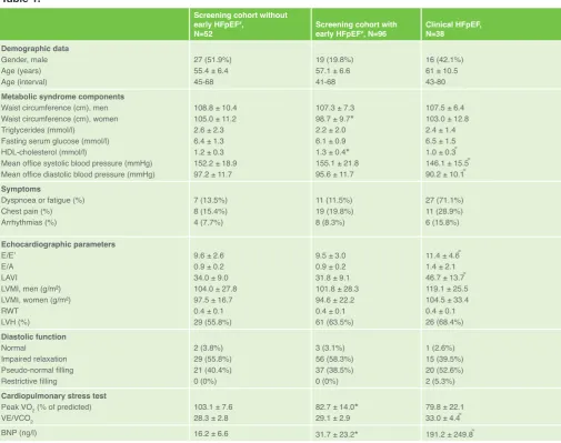
Table 1.
Screening cohort without early HFpEF#,
N=52 Screening cohort with early HFpEF#, N=96 Clinical HFpEF, N=38
Demographic data Gender, male Age (years) Age (interval)
27 (51.9%) 55.4 ± 6.4 45-68
19 (19.8%) 57.1 ± 6.6 41-68
16 (42.1%) 61 ± 10.5 43-80 Metabolic syndrome components
Waist circumference (cm), men Waist circumference (cm), women Triglycerides (mmol/l)
Fasting serum glucose (mmol/l) HDL-cholesterol (mmol/l)
Mean office systolic blood pressure (mmHg) Mean office diastolic blood pressure (mmHg)
108.8 ± 10.4 105.0 ± 11.2 2.6 ± 2.3 6.4 ± 1.3 1.2 ± 0.3 152.2 ± 18.9 97.2 ± 11.7
107.3 ± 7.3 98.7 ± 9.7* 2.2 ± 2.0 6.1 ± 0.9 1.3 ± 0.4* 155.1 ± 21.8 95.6 ± 11.7
107.5 ± 6.4 103.0 ± 12.8 2.4 ± 1.4 6.5 ± 1.5 1.0 ± 0.3 ͌ 146.1 ± 15.5 ͌ 90.2 ± 10.1 ͌ Symptoms
Dyspnoea or fatigue (%) Chest pain (%) Arrhythmias (%)
7 (13.5%) 8 (15.4%) 4 (7.7%)
11 (11.5%) 19 (19.8%) 8 (8.3%)
27 (71.1%) 11 (28.9%) 6 (15.8%)
Echocardiographic parameters E/E’
E/A LAVI
LVMI, men (g/m²) LVMI, women (g/m²) RWT
LVH (%)
9.6 ± 2.6 0.9 ± 0.2 34.0 ± 9.0 104.0 ± 27.8 97.5 ± 16.7 0.4 ± 0.1 29 (55.8%)
9.5 ± 3.0 0.9 ± 0.2 31.8 ± 9.1 101.8 ± 28.3 94.6 ± 22.2 0.4 ± 0.1 61 (63.5%)
11.4 ± 4.6 ͌ 1.4 ± 2.1 46.7 ± 13.7 ͌ 119.1 ± 25.5 104.5 ± 33.4 0.4 ± 0.1 26 (68.4%) Diastolic function
Normal
Impaired relaxation Pseudo-normal filling Restrictive filling
2 (3.8%) 29 (55.8%) 21 (40.4%) 0 (0%)
3 (3.1%) 56 (58.3%) 37 (38.5%) 0 (0%)
1 (2.6%) 15 (39.5%) 20 (52.6%) 2 (5.3%) Cardiopulmonary stress test
Peak VO2 (% of predicted)
VE/VCO2
103.1 ± 7.6
28.3 ± 2.8 82.7 ± 14.029.1 ± 2.9 * 79.8 ± 22.133.0 ± 4.4 ͌
BNP (ng/l) 16.2 ± 6.6 31.7 ± 23.2* 191.2 ± 249.8 ͌
HFpEF – heart failure with preserved ejection fraction; VE/VCO2 - ventilation/carbon dioxide production slope; LVMI - left ventricular mass index; RWT - relative wall thickness; LAVI – left atrial volume index; LVH - left ventricular hypertrophy; BNP - brain natriuretic peptide; # early HFpEF was considered if peak VO2 was
≤90% of predicted value or/and BNP was 35 ng/l or higher; * - statistically significant (p<0.05) difference between the Screening Cohort subjects with, versus those without, early HFpEF ;͌ - statistically significant (p<0.05) difference between the screening cohort with early HFpEF and the clinical HFpEF group.
Figure 1. Comparison of BNP and peak VO2 values in
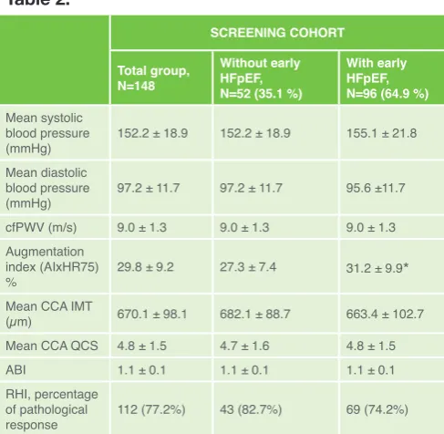
The analysis of arterial markers in the Screening Cohort revealed a statistically significant association of heart-rate 75 bpm adjusted aortic augmentation index (AIxHR75) with the detection of early HFpEF (p=0.016). There were no differences between mean systolic (p=0.416) and mean diastolic (p=0.440) blood pressure comparing those with and without early HFpEF. The data of all investigated markers of preclinical atherosclerosis are presented in Table 2. More than 75% of the entire Screening Cohort had impared endothelial function, showing how common this abnormality is within the MetS population.
Discussion
The metabolic syndrome has been identified as one of the major factors in the development of HF. A Japanese clinical study has revealed that the prevalence of MetS in HF patients is more than double compared with the general population. This is true for both HFrEF and for the less well studied HFpEF population, with a significantly higher prevalence of HFpEF in the MetS group in comparison with non-MetS group in this study [14]. The Framingham Heart Study showed that after adjustment for known risk factors each increase of 1 point in BMI increased the risk of heart failure (not differentiating HFrEF from HFpEF) by 5 percent for men and 7 percent for women [15].
To our knowledge, this is the first study that addresses the early detection of HFpEF in MetS patients using a combination of BNP and peak VO2. In our previous study of a large cohort of MetS patients [16], we showed that the vast majority of subjects had diastolic dysfunction, and even more than half of them showed a pseudo-normal pattern, consistent with elevated LV filling pressures. Thus, we postulated that in the group of patients, who have a high risk of HF development, a very early phase of HFpEF is already present and presumably can be detected by functional and biomarker testing. Indeed, in our prospectively enrolled MetS
population without established overt cardiovascular disease we found a sizeable subgroup (up to two thirds) of patients with impaired functional capacity, elevated neurohormonal activation and ultrasound evidence of elevated LV filling pressures. Assignment of an HF diagnosis to such patients is largely in accordance with more recently refined HFpEF definitions the recent ESC guidelines [12]; with the components of the diagnosis including an increased BNP level, and objectively documented impaired exercise capacity. An advantage of our report is the consistent use of cardiopulmonary exercise testing in order to elicit an objective decrease of exercise tolerance, and by the use of respiratory gas analysis establish it is truly cardiac related limitation. Clinically it is noted that in many subjects with HFpEF a loss of functional capacity can be ascribed to unfitness or age, and the true limitation due to cardiac disease frequently not being adequately appreciated by the patients themselves.
Looking to the subjective self-evaluation of symptoms, it has been shown that the use of self-rated health (SRH), assessed through a single question [17-18], is limited for patients with asymptomatic left ventricular diastolic dysfunction, for the reason that such subjects understate their complaints. A Swedish study has shown that the majority of participants with asymptomatic left ventricular diastolic dysfunction rated their health status as good or very good, despite the high prevalence of co-morbidities [19]. Therefore, there is a need for comprehensive diagnostic methods that would help to highlight the symptoms. Cardiopulmonary exercise testing is an established tool for the evaluation of the disease severity and prognosis in heart failure patients. Our study also suggests a clinically relevant value for its use in the early detection of reduced exercise tolerance in subjects enrolled in the screening and prevention program. Though only 11.5% of patients with the signs of early HFpEF had documented complaints of dyspnoea, the cardiopulmonary stress test revealed objectively impaired exercise tolerance in 83.3% of these patients.
The most recently published of the major guidelines for heart failure, the ESC HF guidelines suggest an elevated BNP level as a necessary component for the establishment of an HFpEF diagnosis. In parallel with the substantially increased BNP levels in clinically labelled advanced HFpEF patients, we were also able to show a modest but statistically significant elevation of BNP in the subgroup of MetS population with early HFpEF.
One can speculate that the group with undetected HFpEF could be differentiated even more precisely if there was no inverse impact of MetS on BNP – as has been previously demonstrated BNP values are significantly lowered in persons affected by MetS [20].
Several hypotheses attempt to explain the inverse relationship between BNP levels and obesity. The renal theory states that BNP levels are depressed because of higher glomerular filtration rates which lead to more efficient molecular clearing [21]even in the absence of diabetes, contributes significantly to the development and progression of chronic kidney disease (CKD. The adipose tissue theory states that the reason for reduced BNP values in MetS is due to the effect of natriuretic peptide clearance receptor-C (NPR-C) changes [22-23]. On the contrary, the Framingham Heart Study [24] and the Dallas Heart Study [25] questioned this theory showing that NT–proBNP values in
Table 2.
SCREENING COHORT
Total group, N=148
Without early HFpEF, N=52 (35.1 %)
With early HFpEF, N=96 (64.9 %)
Mean systolic blood pressure
(mmHg) 152.2 ± 18.9 152.2 ± 18.9 155.1 ± 21.8
Mean diastolic blood pressure
(mmHg) 97.2 ± 11.7 97.2 ± 11.7 95.6 ±11.7
cfPWV (m/s) 9.0 ± 1.3 9.0 ± 1.3 9.0 ± 1.3
Augmentation index (AIxHR75)
% 29.8 ± 9.2 27.3 ± 7.4 31.2 ± 9.9*
Mean CCA IMT
(µm) 670.1 ± 98.1 682.1 ± 88.7 663.4 ± 102.7
Mean CCA QCS 4.8 ± 1.5 4.7 ± 1.6 4.8 ± 1.5
ABI 1.1 ± 0.1 1.1 ± 0.1 1.1 ± 0.1
RHI, percentage of pathological
response 112 (77.2%) 43 (82.7%) 69 (74.2%)
obesity are also reduced even though it is not cleared by NPR-C. The Dallas Heart Study authors found that both BNP and NT-proBNP values were more closely associated with lean mass than with BMI. Due to this finding they theorized that rather than the adipose tissue itself being the main factor causing lowered BNP values, a substance produced in the lean tissue could suppress either BNP synthesis or its release from cardiomyocytes.
Two MetS criteria, insulin resistance and lipid abnormalities, are considered as the main components that influence progression of the disease, whereas obesity and high blood pressure are linked to the outcomes in HF patients [5]. A recently presented new paradigm for HFpEF emphasizes the importance of obesity as one of the main comorbidities which induces a systemic inflammatory state that sequentially leads to diastolic LV dysfunction [26].
Diastolic dysfunction as a leading mechanism in the formation of HFpEF was detected in most of the study patients. Only 3.4% of the screening cohort and 2.6% of clinical HFpEF patients had normal diastolic function. The most common grade of diastolic dysfunction in both prospective subgroups was impaired relaxation, whereas the clinical HFpEF group mostly presented with a pseudo-normal filling pattern.
Arterial markers of preclinical atherosclerosis are used for early prediction of cardiovascular risk and the impact of MetS on arterial elastic properties is intensely discussed [16, 27, 28]. Aortic pulse wave velocity, an index of aortic stiffness, has been shown to be an independent determinant of LV diastolic dysfunction. Moreover, significant correlations between aortic PWV, augmentation index and diastolic function parameters, such as the E/A ratio, E/E’ ratio were demonstrated [16, 28]. In the present study we found a markedly higher AIxHR75 in subjects with early HFpEF compared to those without. Our data support the concept that increased aortic stiffness plays a role in the development of LV diastolic dysfunction and subsequent HF. However, to the best of our knowledge, there are no data or analysis of the relation between markers of preclinical atherosclerosis and heart failure markers. Therefore, further investigation of any possible connection between alterations in arterial stiffness and their influence on LV diastolic dysfunction and HFpEF is needed in a bigger cohort of MetS subjects.
Kosmala et al. highlighted the value of early functional markers such as LV strain in the detection of asymptomatic LV impairment in subjects with type 2 diabetes, hypertension or obesity. The study showed that myocardial strain analysis increased the possibility to detect early LV dysfunction which in turn is associated with impaired exercise capacity
Furthermore, subclinical myocardial systolic dysfunction characterized by impaired global longitudinal strain is strongly associated with a larger epicardial adipose tissue volume typical for MetS [30]. In addition this work my have implications for effective treatments in selected HFpEF patients if as suggested the metabolically abnormal syndrome we see and that also occurs in obese HFpEF may respond to particular targetted therapeutics strategies [31,32].
Limitations
The present study is limited because of its relatively small number of participants; more pronounced results could be expected in the association of HF signs with distinct metabolic components and arterial markers in a bigger cohort. An alternative HF biomarker not affected by obesity is warranted for studies in early HFpEF. The addition of habitual physical activity to the objective measurement of exercise capacity may strengthen the study results and predictive capacity.
Conclusions
A considerable proportion of patients with metabolic syndrome may be considered as having the signs of an early stage of heart failure of the HFpEF pattern. The use of objective parameters of exercise capacity and neurohormonal activation is justified for the early detection of heart failure with preserved ejection fraction. In the metabolic syndrome population initial stage of heart failure with preserved ejection fraction is associated with increased arterial stiffness.
Declarations of interest
The authors declare no conflicts of interest.
Acknowledgements
The authors would like to thank Roma Puronaitė for the performace of the statistical analyses.This work was supported by the European Social Fund under the global grant measure [VP1-3.1-ŠMM-07-K-03-041].The authors state that they abide by the “Requirements for Ethical Publishing in Biomedical Journals” [33].
References
1. Berghöfer, A., Pischon, T., Reinhold, T., Apovian, C. M., Sharma, A. M., Willich, S. N. Obesity prevalence from a European perspective: a systematic review. BMC public health 2008; 8.1: 200.
2. Tamariz L, Hassan B, Palacio A, Arcement L, Horswell R, Hebert K. Metabolic syndrome increases mortality in heart failure. Clin Cardiol 2009; 32.6: 327-331.
3. Ferrari R, Böhm M, Cleland G.F. J, Paulus J.S. W, Pieske B, Rapezzi C, Tavazzi L. Heart failure with preserved ejection fraction: uncertainties and dilemmas. Eur J Heart Fail 2015; 17.7: 665-671.
4. Horwich TB, Fonarow GC. Glucose, Obesity, Metabolic Syndrome, and Diabetes. Relevance to Incidence of Heart Failure. JACC 2010; 55.4: 283-293. 5. Perrone-Filardi P, Paolillo S, Costanzo P, Savarese G, Trimarco B, Bonow
RO. The role of metabolic syndrome in heart failure. Eur Heart J 2015; 36.39: 2630-2634.
6. De Las Fuentes L, Brown AL, Mathews SJ, Waggoner AD, Soto PF, Gropler RJ, Dávila-Román VG. Metabolic syndrome is associated with abnormal left ventricular diastolic function independent of left ventricular mass. Eur Heart J 2007; 28.5: 553-559.
7. Penjaskovic D, Sakac D, Dejanovic J, Zec R, Zec PN, Stojsic MA. Left ventricular diastolic dysfunction in patients with metabolic syndrome. Med Pregl 2012; 65.1-2: 18-22.
8. La Carrubba S, Todaro MC, Zito C, Antonini-Canterin F, Monte IP, Caso P, Colonna P, Gregorio C, Pezzano A, Benedetto F, Salvo GD, Carerj S, Bello VD. Asymptomatic left ventricular Dysfunction and metabolic syndrome: results from an Italian multicenter study. J Cardiovasc Echography 2013; 23: 96-101.
9. Beaver, W. L., Wasserman, K. A. R. L. M. A. N., Whipp, B. J. A new method for detecting anaerobic threshold by gas exchange. J Appl Physiol 1986; 60.6: 2020-2027.
10. Caiozzo VJ, Davis JA, Ellis JF, Azus JL, Vandagriff R, Prietto CA, McMaster WC. A comparison of gas exchange indices used to detect the anaerobic threshold. J Appl Physiol 1982; 53.5: 1184-1189.
12. Ponikowski P, Voors AA, Anker SD, Bueno H, Cleland JG, Coats AJ, Falk V, González-Juanatey JR, Harjola VP, Jankowska EA, Jessup M, Linde C, Nihoyannopoulos P, Parissis JT, Pieske B, Riley JP, Rosano GM, Ruilope LM, Ruschitzka F, Rutten FH, van der Meer P; Authors/Task Force Members. 2016 ESC Guidelines for the diagnosis and treatment of acute and chronic heart failure. Eur Heart J 2016; 37.27: 2129-2200.
13. Coats AJS, Clark AL, Piepoli M, Volterrani M, Poole-Wilson PA. Symptoms and quality of life in heart failure: the muscle hypothesis. British Heart Journal 1994;72(2 Suppl):S36-S39.
14. Miura Y, Fukumoto Y, Shiba N, Miura T, Shimada K, Iwama Y, Takagi A, Matsusaka H, Tsutsumi T, Yamada A, Kinugawa S, Asakura M, Okamatsu S, Tsutsui H, Daida H, Matsuzaki M, Tomoike H, Shimokawa H. Prevalence and Clinical Implication of Metabolic Syndrome in Chronic Heart Failure. Circ J 2010; 74: 2612-2621.
15. Kenchaiah S, Evans JC, Levy D, Wilson PW, Benjamin EJ, Larson MG, Kannel WB, Vasan RS. Obesity and the risk of heart failure. N Engl J Med 2002; 347.5: 305-313.
16. Solovjova S, Ryliskyte L, Celutkiene J, Badariene J, Navickas R, Puronaite R, Bieliauskaite G, Skiauteryte E, Lisaite G, Laucevicius A. Aortic stiffness is an independent determinant of left ventricular diastolic dysfunction in metabolic syndrome patients. Blood Press 2016;25(1):11–20.
17. Farkas J, Nabb S, Zaletel-Kragelj L, Cleland JGF, Lainscak M. Self-rated health and mortality in patients with chronic heart failure. Eur J Heart Fail 2009; 11.5: 518-524.
18. Van der Linde RM, Mavaddat N, Luben R, Brayne C, Simmons RK, Khaw KT, Kinmonth AL. Self-Rated Health and Cardiovascular Disease Incidence: Results from a Longitudinal Population-Based Cohort in Norfolk, UK. PLoS One 2013; 8.6: e65290.
19. Ahmadi NS, Bennet L, Larsson CA, Andersson S, Månsson J, Lindblad U. Clinical characteristics of asymptomatic left ventricular diastolic dysfunction and its association with self-rated health and N-terminal B-type natriuretic peptide: a cross-sectional study. Eur J Heart Fail 2016; 3.3: 205-211. 20. Chang HR, Hsieh JC, Hsu BG, Wang LY, Yu M, Chen MYC, Wang JH.
Inverse Association of N-Terminal Pro-B-Type Natriuretic Peptide with Metabolic Syndrome in Patients with Congestive Heart Failure PloS one 2013; 8.11: e79096.
21. Griffin KA, Kramer H, Bidani AK. Adverse renal consequences of obesity. Am J Physiol Ren Physiol 2008; 294:685–96.
22. Madamanchi C, Alhosaini H, Sumida A, Runge MS. Obesity and natriuretic peptides, BNP and NT-proBNP: Mechanisms and diagnostic implications for heart failure. Int J Cardiol 2014; 176.3: 611-617.
23. Christenson RH, Azzazy HME, Duh S-H, Maynard S, Seliger SL, Defilippi CR. Impact of Increased Body Mass Index on Accuracy of B-Type Natriuretic Peptide (BNP) and N-Terminal proBNP for Diagnosis of Decompensated Heart Failure and Prediction of All-Cause Mortality. Clin Chem 2010; 56.4: 633-641. 24. Wang TJ, Larson MG, Levy D, Benjamin EJ, Leip EP, Wilson PWF, Vasan
RS. Impact of Obesity on Plasma Natriuretic Peptide Levels. Circulation 2004; 109.5: 594-600.
25. Das SR, Drazner MH, Dries DL, Vega GL, Stanek HG, Abdullah SM, Canham RM, Chung AK, Leonard D, Wians FH, de Lemos JA. Impact of body mass and body composition on circulating levels of natriuretic peptides. Circulation 2005; 112.14: 2163-2168.
26. Paulus WJ, Tschöpe C. A novel paradigm for heart failure with preserved ejection fraction: Comorbidities drive myocardial dysfunction and remodeling through coronary microvascular endothelial inflammation. J Am Coll Cardiol 2013; 62.4: 263-271.
27. Della-Morte D, Gardener H, Denaro F, Boden-Albala B, Elkind MS V, Paik MC, Sacco RL, Rundek T. Metabolic syndrome increases carotid artery stiffness: The Northern Manhattan Study. Int J Stroke 2010; 5.3: 138-144. 28. Roes SD, Dehnavi RA, Westenberg JJM, Lamb HJ, Mertens BJA, Tamsma
JT, de Roos A. Assessment of Aortic Pulse Wave Velocity and Cardiac Diastolic Function in Subjects With and Without the Metabolic Syndrome. Diabetes Care 2008; 31.7: 1442-1444.
29. Kosmala W, Jellis CL, Marwick TH. Exercise limitation associated with asymptomatic left ventricular impairment: Analogy with Stage B heart failure. J Am Coll Cardiol 2015; 65.3: 257-266.
30. Ng AC, Goo SY, Roche N, van der Geest RJ, Wang WY. Epicardial Adipose Tissue Volume and Left Ventricular Myocardial Function Using 3-Dimensional Speckle Tracking Echocardiography. Can J Cardiol 2016; 32.12: 1485-1492.
31. Packer M, Kitzman DW. Obesity-Related Heart Failure With a Preserved Ejection Fraction: The Mechanistic Rationale for Combining Inhibitors of Aldosterone, Neprilysin, and Sodium-Glucose Cotransporter-2. JACC Heart Fail. 2018 Mar 7. pii: S2213-1779(18)30062-3. doi: 10.1016/j. jchf.2018.01.009.
32. Paulus WJ, Dal Canto E. Diastole Tracks Cardiometabolic Risk. JACC Heart Fail. 2018 Apr;6(4):326-328. doi: 10.1016/j.jchf.2018.01.008. Epub 2018 Mar 7. 33. Shewan L.G., Coats A.J.S., Henein M. Requirements for Ethical Publishing