JPRHC
Research Article
AJPRHC Volume 3 Issue 4
89-98
D E V E L O P M E N T O F R O P I N I R O L E ( F R E E B A S E ) T R A N S D E R M A L P A T C H U S I N G B L E N D S O F H Y D R O X Y P R O P Y L M E T H Y L C E L L U L O S E / E U D R A G I T S A N D I T ’ S I N
V I T R O / I N V I V O C H A R A C T E R I Z A T I O N
SRAVANTHI ANAMPALLY, SANTHOSH KUMAR MANKALA, PRAVEEN KUMAR GOGU, JITHAN AUKUNURU
For Author affiliations see end of the text This paper is available online at www.jprhc.in
ABSTRACT
Objective: The objective of this study was to prepare and evaluate a matrix type transdermal patch of
ropinirole using blends of
hydroxypropylmethylcellulose (HPMC) and Eudragit RL 100 and HPMC and Eudragit ERS100. Materials and Methods: Ropinirole free based used as the drug entity was prepared from its hydrochloride salt. Suitability of the polymers in the form of drug-excipient compatability was determined prior to formulation development using FTIR. Patches were developed using solvent evaporation technique. Limonene was used as a penetration enhancer. Moisture absorption, moisture content and mechanical properties, drug content, in vitrodrug release, drug-excipient compatibility, in vitro skin permeation were the in vitro parameters measured. Short-term stability, skin irritation and in vivo drug release were measured with one optimized formulation. Results and discussion: Ropinirole free base was used successfully in the preparation of the patches. FTIR studies indicated no interaction between the drug and the polymers of this study. Formulations developed were strong and not brittle with uniform drug release. Patches containing higher HPMC generally showed higher drug release and permeation. Drug release and permeation decreased with increase in the concentrations of Eudragits. Drug release studies indicated Higuchi model for all the patches with a diffusion mechanism of non-fickian type. Short-term stability studies indicated that ropinirole was stable in the patches. Patches did not cause any skin irritation. In vivo the optimized patch sustained drug release for 24 hours upon one time administration. Conclusion: Clinically viable ropinirole transdermal patch can be successfully prepared from its base form using HPMC/Eudragits.
Keywords: Drug release, QbD, Permeation, Parkinsonism, Permeation enhancer
INTRODUCTION
Despite the popularity of oral delivery approaches, alternatives with added advantages such as
drug loaded skin patches are shooting up in important healthcare product category (1-3). Skin patches offer the advantages of ease of use, painlessness, disposability, control of drug delivery and avoidance of first-pass metabolism by the liver (4,5). Advances in synthetic materials and patch design have led to patches that are more esthetically acceptable and that are capable of delivering sustained dosing of active compounds for several days in a smaller package. Patches relying on passive transport mechanisms have been limited in their ability to deliver new classes of drugs, but formulations employing penetration enhancers, and patch designs incorporating microporation and energy assisted delivery are expanding the range of drugs that can be effectively delivered transdermally. As of today, the simple passive transdermal patches are the state of art technology. In this paper, we describe the development of a patch for ropinirole, a drug approved for the treatment of Parkinsons disease (PD) and Restless Legs Syndrome (RLS).
Ropinirole (4-(2-di-n-proylaminoethyl)-2)3H)-indolone) is drug of choice for the treatment of Parkinsons Disease (PD). Sustain release has added advantages for the therapeutic use of this drug (6-9). Transdermal delivery system is one such sustained release dosage form. Transdermal patches not only offer better patient compliance but also in terms of safety profile, they offered better profile than oral SR formulations in Parkinsonism (10). Thus, a skin patch for ropinirole is desirable. Also, ropinirole is pharmaceutically suitable for the development of a transdermal patch (11). The compound has a M.Wt of 296.84 and melting point of approximately 2470C. The
JPRHC
Research Article
AJPRHC Volume 3 Issue 4
89-98
laboratory corroborated this observation. Previously, one report indicated that transdermal formulations were able to provide a therapeutically useful amount of the drug when administered in the form of free base (14). The device proposed in this patent is complicated as well it is a reservoir type of transdermal patch. In this study, we aimed at the development of a simple clinically vaiable transdermal patch for ropinirole. The present study was designed to develop a suitable matrix patch type TDDS for ropinirole (as free base) employing various blends of HPMC and Eudragit RL100 and HPMC and Eudragit ERS 100. Polymer blending which results in new material is one of the effective ways of developing a better pharmaceutical device at a cheaper cost when compared to the development of newer polymers. The aim was to compare the polymeric combinations in terms of in vitro skin permeation of the drug. Thus, an attempt was made to establish the best possible combination of polymeric ratio that ensures maximum controlled and sustained drug release with good physical properties to the patch. This will determine the drug delivery at a controlled rate across intact skin to achieve a therapeutically effective drug level for a longer period of time and also gives ideal characteristics to the patch. Further the influence of d-Limonene, a penetration enhancer on the skin permeability of this drug was also evaluated. Penetration enhancers are widely used in the transdermal drug delivery systems (15,16). A best transdermal patch was selected and further in vivo studies were conducted to determine the sustenance of systemic drug release and skin irration, short-term stability studies were also conducted.
METHODS Materials
Ropinirole. HCl was gift sample from Hetero Drugs Ltd., Andhra Pradesh, India. Limonene was procured from Himedia Laboratories Pvt. Ltd., Mumbai. Eudragit RL 100(ERL 100), Eudragit RS 100(ERS 100) and Hydroxy propyl Methyl Cellulose (HPMC) were gift samples from Degussa, Germany. All other chemicals used were of analytical grade.
Synthesis of Ropinirole Free Base
Prior to the preparation of the matrices, the ropinirole free base was synthesized from its hydrochloride salt using a slight modification of a previously published method (17). Briefly, ropinirole hydrochloride (1.9 g), water (13.5 mL) and ammonia (1.6 mL) were added to a flask with a stirrer. The reaction mixture was stirred at room temperature for approximately 2 hours and then extracted continuously with dichloromethane (1X500 mL, 1X250 mL and 1X200 mL). All the organic extracts were combined and then evaporated to obtain a light brown solid. The yield of the base was determined and then it was characterized using NMR.
Permeation Studies to Identify the Suitable Molecular Entity and also to Determine the Role of Limonene as
the Penetration Enhancer
To identify the suitable molecular entity, permeation studies across rat skin with ropinirole. HCl and ropinirole free base were performed. Male albino rats were used in this study. All the animal experiments were conducted according to the rules and guidelines of Committee for the Purpose of Control and Supervision of Experiments on Animals (CPCSEA, Chennai) constituted by the Government of India. The protocol was approved by Institutional Ethical Committee of Vaagdevi College of Pharmacy, Ramnagar, Warangal, India. The rats weighing 150-200 gm were sacrificed using anesthetic ether. The hair of test animals were carefully trimmed short (<2 mm) with a trimmer taking extreme precaution not to damage the skin and the full thickness skin was removed from the abdominal region. The epidermis was prepared surgically by heat separation technique, which involved soaking the entire abdominal skin in water at 60oC for 45 sec, followed by careful removal of the epidermis (18). The epidermis thus obtained was immediately used in the study.
Franz diffusion cell with a surface area of 4.15 cm2 was used f o r ex vivo permeation studies. The rat skin was mounted between the compartments of the diffusion cell with stratum corneum facing the donor compartment (19). The stratum corneum side of the skin was kept in intimate contact with the release surface of the drug. The relative potential of ropinirole (as the free base) and a ropinirole salt for formulation for incorporation into transdermal delivery system was evaluated from suspensions or solutions of drug. Either saturated solutions of ropinirole hydrochloride or ropinirole free base suspension were used to study the permeability. The receiver phase is 24 mL of phosphate buffer saline (PBS) pH 7.4 stirred at 500 rpm on a magnetic stirrer. The amount of drug permeated was determined by removing 5 mL of sample at appropriate time intervals upto 24 h, the volume was replenished with an equal volume of PBS pH 7.4.The absorbance was measured at 250 nm spectrophotometrically. Cumulative amount of drug permeated in µg/cm2 were calculated and plotted against time. Drug flux (µg/hr/cm2) at steady state was calculated by dividing the slope of the linear portion of the curve by the area of the exposed skin surface (4.15 cm2) and the permeability coefficient was deduced by dividing the flux by initial drug load as shown in Table
2. Li monene was inv esti gated as a p enetration enhancer. The permeab ility of both the salt form and free b ase were studied i n the presence of increasi ng concentrations of lim onene. Limonene concentrations from 0 to 12 % of the activ e were tested as penetration enhancer
Development of Transdermal Films
JPRHC
Research Article
AJPRHC Volume 3 Issue 4
89-98
base was selected as the final drug form. To study the possible interaction between ropinirole and polymeric materials of the films, infrared (IR) spectroscopy was carried out on pure substances and their physical mixtures. The IR spectra were recorded using IR-Spectrophotometer (Perkin Elmer FT-IR, Perkin Elmer Inst. USA) by KBr pellet method. After confirmation that there was no interaction with the polymers proposed and the drug, the patches were prepared. Matrix type transdermal patches containing ropinirole free base were prepared by solvent evaporation technique, using different ratios of ERL 100 (or ERS 100) and HPMC E 15 (Table 1). The polymers were weighed in requisite ratios by keeping the total polymer weight 2gm and allowed for swelling for about 6 hrs in solvent mixture (1:1 ratio of di-chloromethane, methanol).15%v/w Propylene glycol was incorporated as plasticizer and 8 % v / w li m o n en e as penetration enhancer (optimum concentration determined). Then the drug solution was added to the polymeric solution, casted on to anumbra Petri plate of surface area about 72.3 sq.cm, allowed for air drying over night. The entire sheet was cut into small films with an area of 4.15 cm2 i.e. with a diameter of 2.3 cm. About 10 films were obtained from each sheet. The dry patches were kept in desiccators until use.
Physical Characteristics of the Prepared Films
The following studies were conducted: Moisture Absorption Study
The films were weighed accurately and placed in a desiccator containing 100 ml of saturated solution of aluminum chloride (79.50% RH). After 3 days, the films were taken out and weighed, the percentage of moisture uptake was calculated using formula.
% moisture absorbtion= X100 ….(1)
Moisture Content
The films were weighed and kept in a desiccator containing calcium chloride at 40oC for 24 h. The final weight was noted when there was no further change in the weight of patch. The percentage of moisture content was calculated as a difference between initial and final weight with respect to initial weight.
Measurement of Mechanical Properties
Mechanical properties of the films were evaluated using a microprocessor based advanced force gauze (Ultra Test, Mecmesin, UK) equipped with a 25 kg load cell. Film strip with dimensions 60 x 10 mm and free from air bubbles or physical imperfections were held between two clamps positioned at a distance of 3 cm. During measurement, the top clamp at a rate of 2 mm/s pulled the strips to a distance till the film broke. The force and elongation were measured when the film
broke. The mechanical properties were calculated according to the following formulae (20). Measurements were run in four replicates for each formulation.
Tensile strength (kg. mm–2) = ..(2)
Initial cross sectional area of the sample (mm2)
Elongation at break (%.mm–2) =Increase in length (mm) /
original length X 100/cross sectional area………….(3)
Elastic module=Force at corresponding strain (kg)/ Cross-sectional area (mm2) X 1/ Corresponding Strain ……….(4)
Strain =Tensilestrength/Elastic modulus………(5)
Determination of Drug Content in the Patches and In
vitroRelease Mechanisms
To determine drug content, an in vitro dissolution study was conducted using USP dissolution apparatus in 1 L of dissolution media (phosphate buffered saline) keeping 1 cm2 of patch cut in several pieces. Aliquot samples were
taken from the dissolution media at different time intervals until spectrophotometer readings became constant for atleast 24 h
. The cumulative amount of the drug released was considered to be the amount of the drug content in the patch. The samples were analyzed spectrophotometrically at 250 nm (21,22) and the amounts of drug present were calculated from the calibration curve.
The drug release studies from ropinirole transdermal films were performed using Franz Diffusion cell. The drug containing film was kept between donor and receiver compartments, separated from these compartments by dialysis membrane of molecular weight cut off of 5000 (Himedia, Mumbai, India) (23). The receiver compartment containing diffusion medium was stirred with magnetic bead operated by magnetic stirrer, to prevent the formation of concentrated drug solution layer below the dialysis membrane. Normal saline containing 20% v/v of polyethylene glycol 400 was used in the receiver. In the study of release of drug from patches, the selection criteria of the receiver compartment is important (24). Biphasic characteristics of the receiver compartment fluid is important as the diffusion of the drug is through both aqueous and non-aqueous heterogenous media. PEG 400 and normal saline are commonly chosen to provide the biphasic characteristic of the receiver fluid. Additionally, PEG 400 is a non-interacting fluid in the receiver compartment (25). 5 mL of sample was collected from the receptor compartment at appropriate time intervals and replaced with phosphate buffer pH 7.4. Analysis was carried out using UV-Vis Spectrophotometer at 250 nm against phosphate buffer pH 7.4 as reference. Mathematical expressions, zero order , First order and Higuchi
Final weight-Initial weight
JPRHC
Research Article
AJPRHC Volume 3 Issue 4
89-98
model were applied to analyze the release mechanism from the transdermal films.
Ex Vivo Skin Permeation
Ex vivo skin permeation studies were conducted to determine the optimum formulation that can be clinically used. The experimental set up is same as that used for skin permeation studies with the drug but instead of the drug solution or suspension, a transdermal patch was clamped between the donor and the receiver compartment. In case of transdermal patches, a dialysis membrane was a l s o placed over the skin, so as to secure the patch tightly dislodged from the skin. The receiver compartment also consisted of the same media used in the drug release studies for the same reason.
The target flux was calculated using the following equation (11).
JTarget=CssCLTBW/A………(6)
‘A’ represents the surface area of the transdermal patch (i.e. 4.15 cm2), ‘BW’ the standard human body weight of 60 kg, ‘Css’ the ropinirole concentration at the therapeutic level (18ng/mL) and the ‘CLT’ the total clearance (13.05 mL/min/kg), the calculated target flux value for ropinirole was 3.396 µg/h/cm2.
In Vivo Studies
Primary Skin Irritation Test
The dorsal part of the rats was carefully shaved and the patch was applied on the shaved portion for 7 days. After 7 days, the patch was removed and the skin irritation was evaluated as per Draize et al. (1944) (26). Briefly, scores are given from 0 to 4 based on the severity of erythema or oedema formation. The safety of the patch decreases with increase in score.
In Vivo Drug Release
The rats were divided into three groups, one control group, the second group received the patch and the third group received the drug via oral route. In vivo drug release was determined in rats. After carefully shaving the abdomen portion of the rat, the patch containing 9 mg ropinirole was placed on the skin of the rat (patch A1 which exhibited the highest cumulative amount was selected as optimized formulation and was used in the in vivo studies). Ropinirole. HCl (9 mg) via the oral route was given to the rats to confirm the pharmacological activity and assesses the activity with the patch. Anxiolytic activity of ropinirole was used as the criteria for the assessment of systemic drug release (27,28). Light and dark arena, a non-conflicting animal experimental model for anxiety was used for testing the anxiolytic activity of the released drug. The apparatus is described in detail else where (28). Briefly, each rat was placed in the center of the light
arena of the apparatus and was allowed to explore it for 5 min. Percentage of time spent in light arena was observed during specific time points after the drug administration till the next 24 hours.
Short-term Stability Studies
The optimized patch was subjected to short term stability studies. The patches were packed in the aluminum foil and kept at 40±2ºC and 75 ±5% RH. At the end of one month of storage, the patches were removed and assayed for drug content using a UV-Vis spectrophotometer. To determine the drug levels, the same method used for drug content determination was followed. The degradation of ropinirole was evaluated using a TLC method.
Statistical Analysis
The data were analyzed using GraphPad InState Demo (version 3.10) and Microsoft Excel 2007. The release data and permeation parameters obtained were compared by one-way analysis of variance (ANOVA). Tukey-Kramer test and Dunnett test were performed for multiple comparisons amongst the different formulations, respectively. The probability level, p < 0.001, was considered statistically significant.
RESULTS AND DISCUSSION
Ropinirole was successfully synthesized from ropinirole.HCl. At the end of the synthesis, a light brown solid with 75% yield was obtained. The structure was confirmed using NMR. NMR [5H(CDCl3 )] yielded the
JPRHC
Research Article
AJPRHC Volume 3 Issue 4
89-98
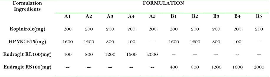
Table 1. Composition of different transdermal ropinirole films 15%v/w propylene glycol was used as plasticizer. 8% v/w limonene was used as penetration enhancer in all formulations.
Each patch (4.15cm2) contains 9mg of Ropinirole.
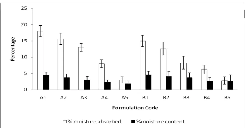
The physicochemical studies like moisture content, moisture uptake and mechanical properties etc. provide information regarding the stability of TDDS formulations. The results of moisture content and moisture absorption studies are shown in Fig. (1). The
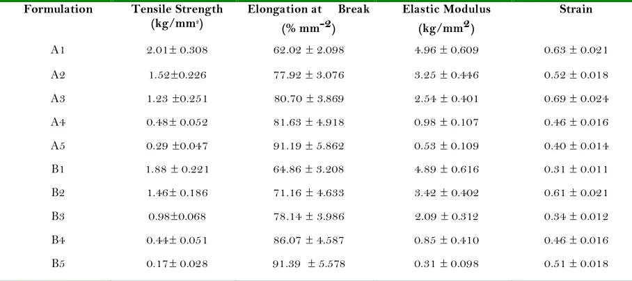
moisture content in the films ranged from 1.8±0.81 to 4.5±0.96% and 2.6±1.90 to 4.6±1.50% (for formulation A series and formulation B series respectively). The moisture absorption in the formulations is ranged from 3.4±0.96 to 18±1.74% and 2.8±1.12 to 15.1±1.75% (for formulation A series and formulation B series respectively). The results revealed that the moisture absorption and moisture content was found to increase with increasing ratio o f hydrophilic polymer (HPMC). The small moisture content in the formulations helps them to remain stable and from being a completely dried and brittle film. The tensile testing gives an indication of the strength and elasticity of the film, reflected by the parameters, tensile strength (TS) and elastic modulus (EM) and elongation at break (E/B). Another parameter strain has been used as an indicator of the overall mechanical quality of the film. A suitable transdermal film should have a relatively high TS, E/B and strain but low EM. The results of mechanical properties (tensile strength, elongation at break, elastic modulus and strain) are shown in Table2. Formulation A1 and B1 exhibited greater values of tensile strength and elastic modulus (2.01±0.308 kg/mm2 and 4.96±0.0.609 kg/mm2 for A1; 1.88±0.221 kg/mm2 4.89±0.616 kg/mm2 respectively). The results revealed that as the ratio of HPMC increased, the tensile strength and elastic modulus were found to be increased but elongation at break values decreased. An inverse relation was observed between tensile strength and elongation at break. These observations indicate that formulation A1 and B1 films were found to be strong, not brittle and flexible. In vitro dissolution studies were carried out for the
different formulations using USP dissolution apparatus to determine the drug content in the patches. The patches had uniform drug amounts present with the drug amounts in all the patches. An average of 9 mg of the drug was present in each patch. Very low standard deviation between and
within patches indicated homogenous distribution of the drug in the patches, suggesting that the technique of preparation was good. Figures. (2) And (3) show the release profiles of ropinirole from transdermal films. Formulations A1 and B1 exhibited greatest (89.92±6.002 and 87.54±2.58 % respectively) percentage of drug release values, which are significantly different compared to the lowest values observed with the formulations containing ERL 100 and ERS 100 (60.49 ±7.77 and 57.29 ±2.84 % respectively). In the present study it was observed that as the ratio of hydrophilic polymer (HPMC) increased in the formulations, the d r u g r e l e a s e r a t e increased s u b s t a n t i a l l y . The description of dissolution profiles by a model function has been attempted using d i f f e r e n t kinetics (zero order, first order and Higuchi square-root model). Higuchi square route seemed to be the most appropriate model describing kinetics from all films (correlation coef fi ci ent between 0.901 and 0.998). On the other hand n values (0.721≤ n≥1.012) indicated that amount of released drug was by non Fickian diffusion (30). The results of in vitro skin permeation of ropinirole from films are shown in Figures. (4) and (5). The formulations (area of 4.15cm2) A1 and B1 exhibited the greatest (1603.11 ±28.63 and 1507.45±59.54 µg/cm2
respectively) cumulative amount of drug permeation, which were significantly different compared to the lowest values observed with the formulations containing ERL 100 (Formulation A5) and ERS 100 (Formulation B5) (307.90 ± 16.81 and 237.50± 10.02 µg/cm2
respectively) in 24 hr. The permeation profiles of drug seem to follow zero order kinetics as it is evidenced
Formulation
Ingredients FORMULATION
A1 A2 A3 A4 A5 B1 B2 B3 B4 B5
Ropinirole(mg) 200 200 200 200 200 200 200 200 200 200
HPMC E15(mg) 1600 1200 800 400 -- 1600 1200 800 400
--Eudragit RL100(mg) 400 800 1200 1600 2000 -- -- -- --
JPRHC
Research Article
AJPRHC Volume 3 Issue 4
89-98
by correlation coefficients (0.918 to 0.986) better than first order (r2 = 0.646 to 0.798) and Higuchi’s
Table 2.Tensile Strength, Elongation at Break, Elastic Modulus and Strain values of Ropinirole Transdermal Films Values represent mean ± SD (n=3).
Formulation Tensile Strength (kg/mm2)
Elongation at Break (% mm-2)
Elastic Modulus (kg/mm2)
Strain
A1 2.01± 0.308 62.02 ± 2.098 4.96 ± 0.609 0.63 ± 0.021
A2 1.52±0.226 77.92 ± 3.076 3.25 ± 0.446 0.52 ± 0.018
A3 1.23 ±0.251 80.70 ± 3.869 2.54 ± 0.401 0.69 ± 0.024
A4 0.48± 0.052 81.63 ± 4.918 0.98 ± 0.107 0.46 ± 0.016
A5 0.29 ±0.047 91.19 ± 5.862 0.53 ± 0.109 0.40 ± 0.014
B1 1.88 ± 0.221 64.86 ± 3.208 4.89 ± 0.616 0.31 ± 0.011
B2 1.46± 0.186 71.16 ± 4.633 3.42 ± 0.402 0.61 ± 0.021
B3 0.98±0.068 78.14 ± 3.986 2.09 ± 0.312 0.34 ± 0.012
B4 0.44± 0.051 86.07 ± 4.587 0.85 ± 0.410 0.46 ± 0.016
B5 0.17± 0.028 91.39 ± 5.578 0.31 ± 0.098 0.51 ± 0.018
equation (r2 = 0.915 to 0.961). As the proportion HPMC increased in all the formulations, increased drug release and permeation in both series were observed. The required flux was obtained with formulation A1 (3.52 µg/cm2/hr) and B1 (3.41 µg/cm2/hr) (Table 3).
Because of these values as well as mechanical properties, the patches A1 and B1 were selected as optimum formulations with desired flux. Of these two A1 was selected as the best formulation. This formulation was investigated for in vivo drug release. As the concentration in the plasma that is anticipated to be attained with ropinirole transdermal patch is very low and requires a sophisticated analytical technique, instead, we used one of its pharmacological activity to assess its systemic presence. significant anxiolytic activity (27). We observed similar results after
administering 9 mg of the drug orally. Peak activity was shown at the end of 1 hour (Figure 6). With the transdermal patch anxiolytic activity was shown for upto 24 hours demonstrating the usefulness of the patch in attaining systemic levels of the drug. The primary skin irritation studies revealed that the patch caused any noticeable irritation on the rat skin. The stability studies indicated that the drug is stable in the short-term stability studies. TLC studies confirmed that there is no degradation product at the end of one month and as a reason a UV-Vis spectrometer can be conveniently used to determine the stability data. Thus, this study successfully developed a transdermal patch for ropinirole using its base form. Further elaborate studies on the formulation development and clinical studies have to be performed to bring this
JPRHC
Research Article
AJPRHC Volume 3 Issue 4
89-98
Table 3. : In vitroDrug Release,Ex vivoSkin Permeation, Transdermal Flux and Permeability Coefficient of Ropinirole Transdermal Films
Fig.1: Moisture absorption and moisture content of Ropinirole transdermal films, mean ± S.D (n=3)
Formulation Drug ReleasedCumulative %
Cumulative amount permeated
(µg/cm2)
Jss (µg/cm2/hr)
Kp (cmhr-1X10³)
A1 89.92±6.002 1603.07±28.95 3.52±0.81 0.137±0.089
A2 86.58±6.053 1244.93±27.78 2.85±0.56 0.114±0.076
A3 81.55±9.45 949.28±26.12 2.52±0.38 0.103±0.065
A4 71.44±6.21 753.53±35.38 1.85±0.49 0.075±0.043
A5 60.49±9.77 381.48±52.67 1.63±0.62 0.066±0.048
B1 87.54±2.58 1507±59.95 3.41±0.65 0.136±0.079
B2 82.64±4.6 1137.43±85.74 2.75±0.29 0.111±0.086
B3 76.79±5.05 881.79±93.38 2.62±0.18 0.107±0.067
B4 68.94±5.07 677.08±79.13 1.62±0.52 0.067±0.042
JPRHC
Research Article
AJPRHC Volume 3 Issue 4
89-98
Fig. 2: Release of ropinirole from Transdermal films(A series),mean ± SD (n=3)
JPRHC
Research Article
AJPRHC Volume 3 Issue 4
89-98
Fig. 5: Permeation of Ropinirole from transdermal films (B Series) through rat abdominal skin, mean ± S.D (n=3)
CONCLUSION
Matrix type transdermal therapeutic systems of ropinirole could be prepared with the required flux having suitable mechanical properties. Further work is recommended in support of its efficacy claims by long term pharmacokinetic and pharmacodynamic studies on human beings.
REFERENCES
1. Thomas JB, Finnies BC. The Transdermal Revolution. Drug Discovery Today. 2004; 9: 697-702.
2. Misra A. Transdermal drug delivery: present and future. The Pharma Review. 2004; 2:92-6.
3. Shah S. Transdermal Drug Delivery Technology Revisited: Recent Advances. Pharmaceutical Reviews. 2008; 6:19-25.
4. Robinson JR, Lee Vincent HL. Controlled Drug Delivery: Fundamentals & Applications. New York, USA: Marcel Dekker (1987).
5. Chien Yie W. Novel Drug Delivery Systems. New York: Marcel Dekker (1992).
6. Curran MP, Perry CM. Cabergoline: a review of its use in the treatment of Parkinson’s disease. Drugs. 2004; 64: 2125-41.
7. Reynolds NA, Wellington K, Easthope SE. Rotigotine: in Parkinson’s Disease. CNS Drugs. 2005; 19: 973-81.
B1
B2
B3
B4
B5
Time (h)
P
er
ce
n
ta
ge
t
im
e
sp
en
t
in
li
gh
t
ar
en
a
(%
)
JPRHC
Research Article
AJPRHC Volume 3 Issue 4
89-98
8. Kaestli LZ, Wasilewski-Rasca AF, Bonnabry P, Vogt-Ferrier N. Use of transdermal formulations in the elderly. Drugs Aging. 2008; 25: 269-80.
9. Nashatizadeh MM, Lyons KE, Pahwa R. A review of ropinirole prolonged release in Parkinson’s disease. Clin Inerv Aging. 2009; 4: 179-86.
10. Reichmann H. Transdermal delivery of dopamine receptor agonists. Parkinsonism Relat Disord. 2009; 4: Suppl 93-6.
11. Chandrasekhar NS, Shobha Rani RH. Physicochemical and pharmacokinetic parameters in drug selection and loading for transdermal drug deliver. Indian J Pharm Sci. 2008; 70: 94-6.
12. Luzardo-Alvarez A, Delgado-Charro MB, Blanco-Mendez J. Iontophoretic delivery of ropinirole hydrochloride: effect of current density and vehicle formulation. Pharm Res. 2001; 18: 1714-20.
13. Bhosale NR, Hardikar SR, Bhosale AV. Formulation and evaluation of transdermal patches of ropinirole. HCl. Res R Pharm Biol and Chem Sci. 2011; 2: 138-45. 14. Chen T, Chiang C. (1998). Transdermal administration
of ropinirole and analogs thereof. US Patent 5,807,570. Cygnus Inc., California.
15. Babu RJ, Pandit JK. Effect of Penetration enhancers on the release and skin permeation of bupranolol from reservoir-type transdermal delivery systems. Int J Pharm. 2005; 288: 325-34.
16. Gamal MM, El-Maghraby, Campbell M, Finnies BC. Mechanism of action of novel skin penetration enhancers; Phospholipid versus skin lipid liposomes. Int J Pharm. 2005; 305: 90-104.
17. Patel A. (1996). Transdermal formulation containing ropinirole. WIPO Patent Application WO/1996/039136. SmithKline Beecham, Pensylvania. 18. Das MK, Bhattacharya A, Ghosal SK. Transdermal
Delivery of Trazodone Hydrochloride from Acrylic Films Prepared from Aqueous Latex. Indian J Pharm Sci. 2006; 68: 1-32.
19. Panigrahi L, Pattnaik S, Ghosal SK. Permeation kinetics of diclofenac sodium from pseudolatex transdermal formulations through lipidized and delipidized mouse skin. Indian J Pharm Sci. 2005; 67: 124-7.
20. Gannu R, Yamsani VV, Yamsani SK, Palem CR, Yamsani MR. Optimization of hydrogels for transdermal delivery of lisinopril by box-behnken statistical design. AAPS PharmSciTech. 2009; 10:505-514.
21. Nagarjuna A, Rao SDV, Eswaraiah S, Mukkanti K, Suryanarayana MV. A liquid chromatographic method for quantification of ropinirole hydrochloride and its related impurities. Indian Drugs. 2006; 43: 813-20. 22. Susheel JV, Malathi S, Ravi TK. Analysis of ropinirole
in tablet dosage form. Indian J Pharm Sci. 2007; 69: 586-9.
23. Vavrova K, Lorencova K, Klimentova J, Novotny J, Holy A, Hrabalek A. Transdermal and Dermal delivery of adenovir: Effects of pH and permeation enhancers. Eur J Pharm Biopharm. 2008; 69: 597-604.
24. Sarpottdar PP, Gaskill J, Giannini RP. Effect of polyethylene glycol 400 on the penetration of drugs through human cadaver skin in vitro. J Pharm Sci. 1986; 75: 26-8.
25. Gupta R, Mukherjee B. Development and in vitro evaluation of diltiazem. hydrochloride transdermal patches based on povidone:ethylcellulose matrices. Drug Dev Ind Pharm. 2003; 29: 1-7.
26. Draize JH, Woodard G, Calvery HO. Methods for study of irritation and toxicity of substances applied topically to the skin and mucous membranes. J Pharmacol Exp Ther. 1944; 82:377-90.
27. Rogers DC, Costall B, Domeney AM, Gerrard PA, Greener M, Kelley ME, Hagan JJ, Hunter AJ. Anxiolytic profile of ropinirole in the rat, mouse and common mormoset. Psychopharmacology (Berl.). 2000; 151: 91-7.
28. Hiremath SB, Sohit A, Srinivas LD, Rashed MR. Effect of calcium and anxiolytic activity of diazepam and verapamil in rats. Indian J Pharmacol. 2010; 42:406-8. 29. Liltorp K, Larsen TG, Willumsen B, Holm R. Solid
state compatability studies with tablet excipients using non thermal methods. J Pharm Biomed Analysis. 2011: 55:424-8.
30. Peppas NA. Analysis of fickian and non fickian drug release from polymers. Pharm. Acta Helv. 1985; 60: 110-1.
ACKNOWLEDGEMENTS AND DECLARATION OF INTEREST
The authors would like to thank Hetero Drugs Ltd., Hyderabad, A.P, India for providing gift sample of ropinirole. The authors also like to thank principal and management of Vaagdevi College of Pharmacy for providing necessary facility useful in conduction of this work. The authors report no declarations of interest. AUTHORS AFFILIATIONS AND ADDRESS
FOR CORRESPONDENCE Aukunuru Jithan,
Mother Teresa College of Pharmacy, Hyderabad India-506001
Mobile: 0091(9849125290) Email: [email protected]
1Srikrupa Institute of Pharmaceutical Sciences, Siddipet, India 2 Vaageswari Institute of Pharmaceutical Sciences, Karimnagar,
India