CerS6
Is a Novel Transcriptional Target of p53 Protein
Activated by Non-genotoxic Stress
*
Received for publication, January 20, 2016, and in revised form, June 2, 2016 Published, JBC Papers in Press, June 14, 2016, DOI 10.1074/jbc.M116.716902
Baharan Fekry‡, Kristen A. Jeffries‡, Amin Esmaeilniakooshkghazi‡, Besim Ogretmen§1, Sergey A. Krupenko‡¶2, and Natalia I. Krupenko‡¶3
From the‡Nutrition Research Institute, University of North Carolina at Chapel Hill, Kannapolis, North Carolina 28081, the
§Department of Biochemistry and Hollings Cancer Center, Medical University of South Carolina, Charleston, South Carolina 29425,
and the¶Department of Nutrition, University of North Carolina at Chapel Hill, Chapel Hill, North Carolina 27599
Our previous study suggested that ceramide synthase 6 (CerS6), an enzyme in sphingolipid biosynthesis, is regulated by p53: CerS6 was elevated in several cell lines in response to tran-sient expression of p53 or in response to folate stress, which is known to activate p53. It was not clear, however, whetherCerS6
gene is a direct transcriptional target of p53 or whether this was an indirect effect through additional regulatory factors. In the present study, we have shown that theCerS6promoter is acti-vated by p53 in luciferase assays, whereas transcriptionally inac-tive R175H p53 mutant failed to induce the luciferase expres-sion from this promoter.In vitroimmunoprecipitation assays and gel shift analyses have further demonstrated that purified p53 binds within theCerS6promoter sequence spanning 91 bp upstream and 60 bp downstream of the transcription start site. The Promo 3.0.2 online tool for the prediction of transcription factor binding sites indicated the presence of numerous putative non-canonical p53 binding motifs in theCerS6promoter. Lucif-erase assays and gel shift analysis have identified a single motif upstream of the transcription start as a key p53 response ele-ment. Treatment of cells with Nutlin-3 or low concentrations of actinomycin D resulted in a strong elevation of CerS6 mRNA and protein, thus demonstrating that CerS6 is a component of the non-genotoxic p53-dependent cellular stress response. This study has shown that by direct transcriptional activation of
CerS6, p53 can regulate specific ceramide biosynthesis, which contributes to the pro-apoptotic cellular response.
The tumor suppressor p53 is a transcription factor com-monly activated upon certain types of cellular stress such as DNA damage, nutrient starvation, ribosomal stress, or onco-genic signaling (1– 4). The list of p53-regulated targets includes several hundred genes (5), and the overall cellular response to p53 activation is an integral function determined by differential activation of target genes (6). Not all p53-responsive genes are
direct transcriptional targets of p53, and indirect effects of the protein on transcription through other transcription factors or additional regulatory elements are common (7, 8). For example, one of the mechanisms multiplying the number of p53-respon-sive targets is the regulation of non-coding RNA transcription (9). A bioinformatics approach predicted that p53 is likely to regulate a handful of microRNAs, which in turn control expres-sion of hundreds of genes (10), a phenomenon dramatically expanding the p53 network. The number of genes directly reg-ulated by p53, however, could be much lower within a couple of hundred targets (11). Furthermore, although initially p53 was thought to act as the transcription activator, later it was dem-onstrated that it can also repress transcription through direct interaction with target genes as well as through indirect mech-anisms (1, 3, 8). The direct transcriptional regulation by p53 itself is a complex process that could be affected by p53 modi-fications, the interaction with p53 co-regulators, and coopera-tion with other transcripcoopera-tion factors (6).
Whether a given promoter is regulated by p53 depends on the amount of the protein, its modification state, the cofactors present at the promoter, or additional cooperating factors that enhance or repress the p53-induced transcription (12). These factors can also dictate which genes are targeted for transcrip-tional regulation (2, 7). However, the most important compo-nents defining the p53-directed transcriptional regulation are the p53 response elements on the DNA (13). The sequence-specific DNA binding activity of p53 is critical to its tumor suppressor activity in response to cellular and environmental stresses. Canonically, p53 binds as a tetramer to two decameric motifs, which have been defined by the consensus sequence RRRCWWGYYY with a separation of 0 –13 bp, where R ⫽ purine, Y⫽pyrimidine, and W⫽A or T (14). Further studies have identified numerous non-canonical DNA binding motifs for p53 that can differ significantly in their affinity to the pro-tein (2). For example, it has been demonstrated that a 10-base motif representing half of the canonical site can function as the p53 response element (15). Furthermore, most of the validated response elements have mismatches compared with the origi-nally defined consensus motif, with 10% of such elements hav-ing novel sequences not clearly related to the consensus motif (2). Of note, functional activities of such non-canonical response elements can vary significantly, with lower activity sites requiring much higher levels of p53 protein for the tran-scriptional regulation (2).
*This work was supported, in whole or in part, by National Institutes of Health Grant CA193782 (to N. I. K.). The authors declare that they have no conflicts of interest with the contents of this article. The content is solely the respon-sibility of the authors and does not necessarily represent the official views of the National Institutes of Health.
1Supported by the National Institutes of Health Grants CA88932, CA97165, and DE16572.
2Supported by the National Institutes of Health Grant DK54388.
3To whom correspondence should be addressed: Dept. of Nutrition and Nutrition Research Institute, UNC Chapel Hill, Kannapolis, NC 28081. Tel.: 704-250-5054; Fax: 704-250-5001; E-mail: [email protected].
Because p53 regulates transcription of so many genes involved in a variety of cellular processes, the protein has a very broad spectrum of its effect on cellular homeostasis. Pathways regulated by p53 include DNA repair, cell cycle arrest, apopto-sis, senescence, autophagy, angiogeneapopto-sis, and migration (1– 4). More recently, the p53-dependent regulation of cellular metab-olism has been also underscored (4). Thus, p53 regulates both glycolysis and oxidative phosphorylation shifting the energy production to the latter pathway (16, 17). Such regulation is achieved through the transcriptional repression of glucose transporters type 1, 3, and 4 and transcriptional activation of TIGAR,SCO2,GLS2, andParkin(18 –24). P53 also plays a key role in lipid metabolism activating fatty acid oxidation and inhibiting fatty acid synthesis (16), and it was further implicated in the regulation of sphingolipid metabolism (25). Sphingolip-ids play an important role in both the structural integrity of cellular membranes and as signaling molecules involved in the regulation of major cellular processes such as senescence, sur-vival, and death (26, 27). Our previous study suggested that one of the enzymes in sphingolipid biosynthesis, ceramide synthase 6 (CerS6),4is regulated by p53 (28). Thus, CerS6 was elevated in
several cell lines in response to transient expression of p53 and also in response to folate stress, which is known to activate p53 (29, 30). For example, CerS6 was strongly elevated in response to the antifolate methotrexate in a p53-dependent manner (31). It was not clear, though, whetherCerS6gene is a direct tran-scriptional target of p53 or this was an indirect effect through additional regulatory factors. In the present study, we have investigatedCerS6promoter as a direct transcriptional target of p53.
Results
The CerS6 Promoter Is Regulated by p53—We have studied the effect of p53 on theCerS6promoter regulation using a lucif-erase assay. The promoter has been previously cloned into pGL3 vector bearing firefly luciferase cDNA (32), and this con-struct was co-transfected with the p53-expressing vector in A549 cells. By itself, theCerS6promoter displayed a weak tran-scriptional activity (Fig. 1A). However, we have observed a sig-nificant (⬎10-fold) increase in the luciferase activity in cells transfected for the p53 expression compared with cells co-transfected with the “empty” (negative control) plasmid (Fig. 1A). When cells were co-transfected with the luciferase-ex-pressing reporter and the vector exluciferase-ex-pressing the transcription-ally deficient R175H p53 mutant, no increase in the luciferase activity was recorded (Fig. 1A). To eliminate potential impact from endogenous p53 protein onCerS6promoter, we have also performed luciferase assays in the p53-deficient A549 cell line. In this cell line p53 was silenced by the stable expression of the p53 shRNA, which completely prevented the protein expres-sion (28, 33). Similar to p53-proficient A549 cells, strong acti-vation of luciferase was seen in the p53-deficient cells upon co-expression with the p53-expressing vector but not the vec-tor expressing mutant p53 (Fig. 1B). Of note, levels of expressed wild type and mutant p53 were similar in each cell line (Fig. 1,A
andB,insets). Because the effect on theCerS6promoter was comparable in both cell lines, in further studies we used regular A549 cells, which allow the evaluation of stress stimuli on the CerS6 expression.
Previous study used four different length constructs of the CerS6 promoter to explore its regulation (32), and we tested these constructs for activation by p53. Luciferase assays have shown that all four promoter constructs resulted in a strong increase of luciferase activity when co-transfected with the p53 expression vector (Fig. 1C). Of note, the shortest construct was most active in the presence of p53 (Fig. 1C).
Identification of the p53 Binding Motif in the CerS6 Promoter—A previously published study of CerS6 promoter indicated its regulation by AP2, SP1/3, and CREB transcription factors but not by p53 (32). Our analysis of CerS6promoter 4The abbreviations used are: CerS6, ceramide synthase 6; RE, response
element.
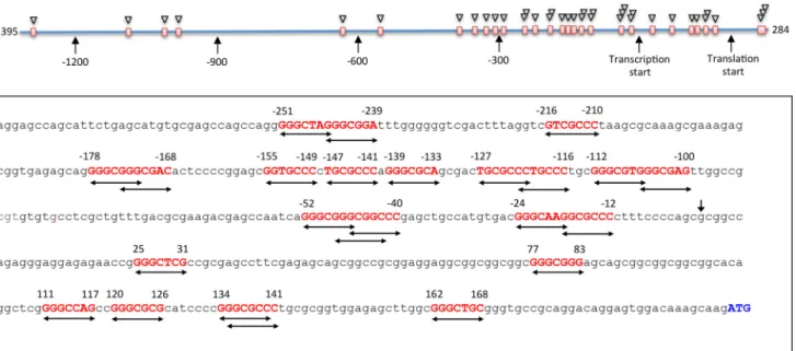
using several web-based tools for the prediction of transcrip-tion factor binding sites, including Patch 1, Match, Matrix Catch, Alibaba, and Transfac, did not produce hits correspond-ing to the p53 bindcorrespond-ing motifs. Thus, it appeared that this pro-moter does not have canonical p53-binding sites. Here we have applied Promo 3.0.2 online tool (34, 35) for additional analysis of the CerS6 promoter region, which included 1395 bp upstream of the transcription start and 284 bp of exon 1 (Fig. 2). Surprisingly, in contrast to other tools, Promo 3.0.2 has indi-cated multiple putative p53-binding sites in the CerS6 pro-moter (Fig. 2). Most of the hits (27 of the total of 32 sites) are located within of the proximal promoter (495 bp upstream of the transcription start) and 200 bp of exon 1. Of note, some of the predicted sites overlap within the same short contiguous sequence, suggesting that the real number of such sites is lower than the predicted number (Fig. 2).
Defining the Minimal CerS6 Promoter Responsive to p53—To decipher the mechanism of the activation ofCerS6promoter by p53, we created a set of truncated promoter constructs (Fig. 3). The originally cloned CerS6 promoter included 1395 bp upstream of the transcription start site and a part of exon 1 (32). The portion of exon 1 in the promoter consists of the entire non-translatable (bp 1–200) and a part of translatable (bp 201– 284) region. This promoter structure defined our strategy in the identification of the position of p53 response elements. By itself, the promoter has a weak transcriptional activity that is enhanced upon p53 expression by⬃6 –10-fold (Fig. 3). The removal of the translatable portion of exon 1 has improved the response to p53 by ⬃1.5-fold (Fig. 3). Whereas the precise mechanism of this effect is not clear, further promoter bashing was done with a construct lacking the translatable part of exon 1. Of note, the repressing effect of this translatable sequence on the promoter activity in the presence of p53 was observed for shorter promoter constructs as well; constructs III-V (Fig. 3) had⬃50% less activity if this portion of exon 1 was not removed
(data not shown). Our experiments demonstrated that the p53 response elements are likely positioned in theCerS6promoter region extended from⫺291 to 50 bp (Fig. 3); this construct revealed the strongest activation upon p53 expression, whereas further truncation of the promoter beyond this region impaired its activation by p53.
Purified p53 Binds the Essential CerS6 Promoter—The acti-vation ofCerS6 promoter by p53 in the luciferase assay can proceed indirectly through regulatory elements activated by p53. To investigate whether p53 directly binds to the essential CerS6promoter, we used the pulldown assay of the promoter fragment (from⫺204 to 60 relative to the transcription start site) on purified p53 protein. In these experiments the fragment corresponding to the above promoter region was excised from the plasmid using Sac1/HindIII endonucleases and purified from agarose gel after electrophoresis. The fragment was fur-ther incubated with full-length p53 protein and amplified by PCR with specific primers after the pulldown on p53-specific antibody and protein G-agarose (schematically depicted in Fig. 4A). Primers used for the amplification were fluorescently labeled, allowing the direct detection of PCR fragments (primer sequences are shown in Table 1). An intensive band corre-sponding to theCerS6promoter was observed after the pull-down (Fig. 4B) that indicated the direct interaction between p53 and the promoter. The specificity of this interaction was demonstrated in control experiments, which used either ALDH1L1 protein (36) instead of p53 and the corresponding antibody or just IgG in the absence of p53; no amplified pro-moter sequences were observed under these conditions (Fig. 4B,lanes 2 and3, correspondingly). These experiments also demonstrated that the interaction between p53 and the sequence corresponding to theCerS6promoter is very strong; we were able to pull it down with p53 and its specific antibody even in the absence of cross-linking (Fig. 4B,lane 4).
The interaction between essential CerS6 promoter and the p53 protein was further confirmed by the gel-shift assays (Fig. 5). Six fragments corresponding to different promoter size and generated by the PCR amplification were tested in these exper-iments (Fig. 5A). Fluorescently labeled primers (Table 1) used in these amplifications allowed the direct imaging of PCR prod-ucts. Gel-shift assays have shown a strong size-specific shift of the DNA in the presence of p53 for the four longest promoter constructs, 151 to 355 bp, whereas no such shift was observed
for the two shortest constructs (Fig. 5B). A high molecular mass band was also seen for all pulled-down samples in the presence of p53, indicating that this band was a result of nonspecific aggregation (Fig. 5B). Additional nonspecific bands were observed for the shortest construct (Fig. 5B). Overall, these experiments have demonstrated that the essential CerS6 pro-moter interacts with p53 and indicated that the p53-binding site(s) is located within the 151-bp region extending from the position ⫺91 to the position 60 relative to the transcription FIGURE 3.Activation of truncatedCerS6promoter constructs by p53.Theschematic on the leftdepicts promoter constructs used in this study (violet blocks correspond to the region upstream of the transcription start site;yellowblocks correspond to exon 1; thebrownblock in construct I indicates the translatable part of exon 1;numbersindicate position of corresponding base pairs). Luciferase activity (mean⫾S.D.) of three independent experiments (each in quadru-plicate) is shown.BP, basic plasmid (promoterless reporter).RLU, relative luminescence units.
FIGURE 5.Analysis of the binding of p53 toCerS6promoter using gel shift assay.A, schematic depicting DNA fragments used in these experiments. Fluorescent labeling of these fragments was achieved by the PCR amplification using primers with a covalently attached Cy5 fluorescent dye (Table 1). B, gel shift analysis of the interaction betweenCerS6promoter fragments (frompanel A) and purified p53 (50 nM).Arrowsindicate respective fragments and DNA shifted upon the p53 binding.C, gel shift analysis of the interaction between the 151-bp promoter fragment (frompanel A) and purified p53 (20 – 60 nM) in the presence or absence of p53-specific antibody (Ab). Thelast laneshows competition between the 151-bp fluorescent promoter fragment and non-fluorescent double-stranded DNA containing the canonical p53-binding motif from p21 promoter (shown in Table 2) for the p53 binding (60 nM).C,negative control, no p53.D, gel shift analysis of the interaction between the 151-bp promoter fragment (frompanel A) and increasing concentrations of purified p53.
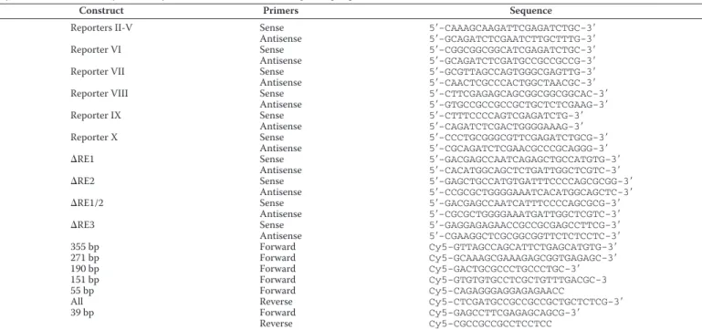
TABLE 1
Primers used for generation ofCerS6promoter constructs
Cy5 indicates fluorescent dye covalently attached to the 5⬘end of corresponding oligonucleotides.
Construct Primers Sequence
Reporters II-V Sense 5⬘-CAAAGCAAGATTCGAGATCTGC-3⬘
Antisense 5⬘-GCAGATCTCGAATCTTGCTTTG-3⬘
Reporter VI Sense 5⬘-CGGCGGCGGCATCGAGATCTGC-3⬘
Antisense 5⬘-GCAGATCTCGATGCCGCCGCCG-3⬘
Reporter VII Sense 5⬘-GCGTTAGCCAGTGGGCGAGTTG-3⬘
Antisense 5⬘-CAACTCGCCCACTGGCTAACGC-3⬘
Reporter VIII Sense 5⬘-CTTCGAGAGCAGCGGCGGCGGCAC-3⬘
Antisense 5⬘-GTGCCGCCGCCGCTGCTCTCGAAG-3⬘
Reporter IX Sense 5⬘-CTTTCCCCAGTCGAGATCTG-3⬘
Antisense 5⬘-CAGATCTCGACTGGGGAAAG-3⬘
Reporter X Sense 5⬘-CCCTGCGGGCGTTCGAGATCTGCG-3⬘
Antisense 5⬘-CGCAGATCTCGAACGCCCGCAGGG-3⬘
⌬RE1 Sense 5⬘-GACGAGCCAATCAGAGCTGCCATGTG-3⬘
Antisense 5⬘-CACATGGCAGCTCTGATTGGCTCGTC-3⬘
⌬RE2 Sense 5⬘-GAGCTGCCATGTGATTTCCCCAGCGCGG-3⬘
Antisense 5⬘-CCGCGCTGGGGAAATCACATGGCAGCTC-3⬘
⌬RE1/2 Sense 5⬘-GACGAGCCAATCATTTCCCCAGCGCG-3⬘
Antisense 5⬘-CGCGCTGGGGAAATGATTGGCTCGTC-3⬘
⌬RE3 Sense 5⬘-GAGGAGAGAACCGCCGCGAGCCTTCG-3⬘
Antisense 5⬘-CGAAGGCTCGCGGCGGTTCTCTCCTC-3⬘
355 bp Forward Cy5-GTTAGCCAGCATTCTGAGCATGTG-3⬘
271 bp Forward Cy5-GCAAAGCGAAAGAGCGGTGAGAGC-3⬘
190 bp Forward Cy5-GACTGCGCCCTGCCCTGC-3⬘
151 bp Forward Cy5-GTGTGTGCCTCGCTGTTTGACGC-3
55 bp Forward Cy5-CAGAGGGAGGAGAGAACC
All Reverse Cy5-CTCGATGCCGCCGCCGCTGCTCTCG-3⬘
39 bp Forward Cy5-GAGCCTTCGAGAGCAGCG-3⬘
start site. Gel shift experiments performed in the presence of the p53-specific antibody resulted in the DNA supershift, fur-ther confirming the p53-promoter interaction (Fig. 5C). This interaction was disrupted in the presence of DNA duplex con-taining a canonical p53-binding site from the p21 promoter (Fig. 5C). The interaction of the 151-bp promoter with p53 as a function of the protein concentration was further examined in the gel shift assay, which indicated that the binding constant for the interaction is⬃20 nM(Fig. 5D).
The p53 Response Elements in the CerS6 Promoter—Based on the results of the luciferase and gel-shift assays (above), we have further focused on a shorter part of the promoter, from the position⫺91 to the position 60. In this region three putative p53 binding motifs have been identified (Fig. 6A). To study whether the corresponding sequences are genuine p53 response elements (REs), we have created a set of promoter constructs that retained all three motifs or its different combi-nations (Fig. 6B) and tested them for the activation by p53 in luciferase assays. These assays have shown that putative p53 response element 2 is a key for the activation of CerS6 promoter by p53; the promoter construct retaining RE2 was as effective in
luciferase induction in response to p53 as the construct with all three response elements, RE1–3 (Fig. 6C). Correspondingly, the deletion of this putative response element decreased the acti-vation by⬃70% (Fig. 6C). Putative RE1 has shown some ability to activate the promoter, although to a lower extent than RE2 (Fig. 6C). In contrast, putative RE3 was not involved in the p53 response; its deletion did not affect the luciferase activation, and the construct retaining only RE3 was not induced by p53 (Fig. 6C). In agreement with the luciferase assay data, the gel- shiftassaydemonstratedtheinteractionbetweentheshortoligo-nucleotide bearing RE2 (Table 1) but not constructs bearing RE1 or RE3 (Fig. 6D). The interaction with RE2 was disrupted in the presence of DNA duplex containing a canonical p53-bind-ing site from the p21 promoter (Fig. 6E).
increase in CerS6 mRNA and protein (Fig. 7A). To assure that the elevation of CerS6 mRNA was associated with the p53 acti-vation and was not a side effect of the drug, we also treated A549 cells with Nutlin-3, a specific activator of the p53 pathway (38). In agreement with the Nutlin-3 function, significant elevation of the p53 protein was seen in cells exposed to 10M
concen-trations of the drug (Fig. 7B). Similar to actinomycin D, Nut-lin-3 has also evoked a strong increase in CerS6 mRNA, which was accompanied by the increase in the CerS6 protein levels (Fig. 7B).
Discussion
Ceramides, a group of sphingolipids, play an important role as components of cellular membranes (39 – 42) and as signaling molecules (26, 43). In humans, there are six ceramide synthases, CerS1– 6, which are involved in biosynthesis of ceramide mol-ecules with different acyl chain length (44, 45). Expression and activity of these enzymes are regulated at several levels, includ-ing transcription (46). The information on transcriptional reg-ulation of ceramide synthases, however, is scarce. The analysis ofCerS1andCerS6promoters indicated their potential regula-tion by several common transcripregula-tion factors, with Sp1 playing a key role in the activation of theCerS1promoter (32). It has been recently shown that two other ceramide synthase genes, CerS4andCerS5, are transcriptionally regulated by AP-1 in the estrogen receptor-dependent manner (47). Although altera-tions in biosynthesis and degradation of sphingolipids are asso-ciated with the p53 activation, genes encoding enzymes of the sphingolipid pathways, including ceramide synthases, are not known as transcriptional targets of p53. It has been reported, though, that p53 drivesde novoceramide synthesis by
activat-ing one of the ceramide synthases (48). The mechanism of such activation could be indirect, through the p53 effect on addi-tional transcription regulators or repressors. For example, through the regulation of microRNAs p53 can affect the expres-sion of a plethora of genes (10). In fact, such regulation has been shown for ceramide synthases: the alternative splice variant of CerS1is inhibited by microRNA-574-5p (32). Furthermore, it has been recently reported that the elevation of CerS6 expres-sion is associated with the reduction of microRNA-101 (49). Interestingly, this microRNA was predicted to have p53 response elements (10). The co-regulation of p53 and CerS6 and the role of microRNAs in this process, however, are not so clear. Thus, although CerS6 expression inversely correlates with miR-101, a positive correlation between the p53 accumu-lation and miR-101 has been recently reported (50). In this mechanism, miR-101 targets the proteasome maturation pro-tein POMP, leading to impaired proteasome assembly and activity. So far there were no reports, which would specifically study the transcriptional regulation ofCerSgenes by p53, but a recent meta-analysis indicated the presence of p53 regulatory elements in these genes, although corresponding target scores were rather low (8).
We previously demonstrated that CerS6 is elevated in response to p53 activation or upon transient transfection of p53 (28). The mechanism of such regulation was not clear as the p53 binding motifs were not found in theCerS6promoter region. Toward this end, indirect regulation of gene expression by p53, through the effect on other components of transcriptional machinery, is a common phenomenon (7, 8). In fact, much higher numbers of genes are indirect targets of p53 than its direct transcriptional targets (5). Interestingly, we have previ-ously identified a putative p53-binding site in theCerS6gene outside of the promoter region 3 kb downstream of the tran-scription start site (28) though mechanisms that would allow transcriptional regulation by utilizing such a remote site were not clear. An implication ofCerS6(LASS6) as a potential tran-scriptional target of p53 can be derived from the study, which used ChIP with the paired-end di-tag sequencing to create a global map of p53-binding sites in human genome (51). This high throughput screen, while not specifically identifying the CerS6gene as one of the p53 targets, indicated a probability for the presence of the p53-binding sites in proximity to the gene. Despite the fact that the canonical p53-binding sites were not present in CerS6 promoter, in our studies the promoter was strongly activated by p53 in luciferase reporter assays. This finding prompted us to further search for p53 binding motifs in theCerS6promoter region. The Promo 3.0.2 online tool for the prediction of transcription factor (TF) binding sites in DNA sequences (34, 35) has identified multiple non-canonical p53-binding sites upstream of the transcription start site and in the non-translatable portion of exon 1. Although typically minimal promoter includes the proximal sequence upstream of the tran-scription site, in the CerS6promoter the part of exon 1 was required for the full promoter activity. Of note, we have previ-ously observed similar dependence for ALDH1L1 promoter (52). Our study demonstrated that of numerous non-canonical p53-binding sites in theCerS6promoter; two upstream sites most closely located to the transcription start are important for FIGURE 7.Drugs activating p53 elevate levels of CerS6 mRNA and
the activation of the promoter by p53. This is in agreement with a general rule that the p53 response elements decrease their transactivation potential as they are distanced from the tran-scription start site (6, 11). Our data also indicate that one of these sites is a key response element of p53 whereas the other site might partially compensate for the absence of that key site. Originally regarded as the regulator of the genotoxic stress response, in recent years p53 has been established as a key player in metabolism regulation (4, 7, 16, 17). The identification of novel p53 transcriptional targets involved in cellular metab-olism provides a more integrative view of the effect of metabolic stress on homeostasis maintenance. Metabolic pathways regu-lated by p53 include glycolysis, oxidative phosphorylation, glu-coneogenesis, serine biosynthesis, and fatty acid synthesis and degradation (4, 7, 16, 17). Although some circuits in the meta-bolic network are regulated by p53 indirectly, several key genes involved in metabolic pathways are direct transcriptional tar-gets of p53, including those encoding for glucose transporters (23, 24), hexokinase 2 (53), mitochondrial glutaminase 2 (19, 20), carnitine palmitoyltransferase (54), pantothenate kinase-1 (55), phosphoglycerate dehydrogenase (56), and a component of the cystine/glutamate antiporter (57). Our study extends the p53 regulation to sphingolipid metabolism by demonstrating thatCerS6is abona fidep53 target gene.CerS6satisfies all four criteria for such genes (11): 1, it has p53 response elements in the promoter; 2, its promoter binds p53, which was demon-strated by several assays; 3, the promoter is activated by p53 in the luciferase assay; 4, stress stimuli activating the p53 response led to the elevation of CerS6. It is not clear at present whether p53 requires other co-regulators to activate theCerS6promoter or whether p53 modifications (for example, phosphorylation at different residues) affect the interaction with the response ele-ments in the promoter. Our in vitro experiments, however, indicated that non-modified p53 binds to theCerS6promoter. Interestingly, the transcriptional activation of CerS6 by p53 occurs in response to metabolic rather than genotoxic stress. Thus, our previous study demonstrated that CerS6 is elevated in a p53-dependent manner in response to folate stress enzyme ALDH1L1 (28); ALDH1L1, however, induces p53 through met-abolic alterations in the absence of DNA damage (33, 58). In line with this finding, the treatment of cancer cells with antifo-late methotrexate or foantifo-late withdrawal, two stimuli inducing alterations in folate, amino acid, and nucleotide metabolism, activated CerS6 in a p53-dependent fashion (28, 31). In the present study the transcriptional induction ofCerS6by p53 in the absence of genotoxic stress was further confirmed using two canonical activators of p53, actinomycin D and Nutlin-3, which do not induce DNA damage. Although actinomycin D at higher concentrations activates p53 through the induction of DNA damage (59), at low concentrations it mimics the effect of Nutlin-3 (37). In our experiments treatment of cells with Nut-lin-3 or low concentrations of actinomycin D resulted in a strong elevation of both CerS6 mRNA and protein, thus dem-onstrating that CerS6 is a component of the non-genotoxic p53-dependent cellular stress response. It is still not fully understood how the metabolic functions of p53 contribute to its tumor suppression activity. Our present study has shown that by transcriptional activation ofCerS6, the enzyme in
sph-ingolipid metabolism, p53 can activate ceramide biosynthesis, which contributes to the pro-apoptotic cellular response.
Experimental Procedures
Cell Culture and Reagents—Actinomycin D and other chem-icals were purchased from Sigma unless otherwise indicated. Nutlin-3 was obtained from Santa Cruz Biotechnology (Dallas, TX). Cell culture media and reagents were from Invitrogen Thermo Fisher (Waltham, MA). A549 cell line was from ATCC. Generation of Truncated CerS6 Promoter Constructs—Trun-cations of the promoter were carried out by the PCR-based site-directed mutagenesis usingPfuTurbo polymerase (Agilent Technologies, Santa Clara, CA) as we previously described (52). PCR primers (Table 1) were from Eurofins (Huntsville, AL). All constructs were confirmed by sequencing.
Luciferase Reporter Assay—Reporter constructs were trans-fected in A549 cells using Neon nucleofector (Invitrogen) according to the manufacturer’s manual. Luminescence was measured 48 h post-transfection using a Bright-Glo Luciferase assay system (Promega, Durham, NC). The pGL3-Basic plas-mid (promoter-less) was used in each experiment to determine the basal levels of luciferase expression. Each construct was tested in three independent transfection experiments. A dual luciferase reporter assay system (Promega) was used to normal-ize experiments for transfection efficiency (the pRL-TK plas-mid expressing Renilla Luciferase was co-transfected with each reporter vector at a ratio 1:10).
Expression and Purification of p53—The pCEP4-175 vector was a generous gift from Dr. Jennifer Pietenpol. Vector for the expression of wild type p53 was generated previously (33). Esch-erichia coliBL21(DE3) cells transformed with P53 expression construct were grown on an LB/ampicillin plate overnight. A single colony was inoculated into 5 ml of LB medium contain-ing 50g/ml ampicillin and grown overnight at 37 °C. Then 500 ml of LB medium with ampicillin were inoculated with the overnight culture and incubated at 37 °C untilA600reached 0.7
followed by the addition of IPTG (1 mMfinal concentration,
Fisher) and incubated additionally for 12 h at 25 °C. Cells were harvested by centrifugation (5000⫻g, 10 min), resuspended in 50 ml of 50 mMNaH2PO4buffer, pH 8.0, containing 300 mM
NaCl and incubated for 30 min with 1 mg/ml lysozyme at 4 °C. The suspension was chilled on ice, sonicated, and spun down at 10,000⫻gfor 40 min). Supernatant was loaded on a PrepEase nickel-nitrilotriacetic acid high yield agarose (USB Corp.) column, and the His-tagged protein was purified per manufacturer’s instructions. Protein preparations obtained after metal-affinity chromatography were further subjected to size-exclusion chroma-tography on Sephacryl S300 (GE Healthcare).
fluores-cently labeled primers (Table 1) and visualized using an Odys-sey Fc imager (LI-COR Biosciences) after agarose gel electro-phoresis. In the case of cross-linking, the p53-DNA complex was treated with 1% formaldehyde, and the reversal of cross-linking was done with 5MNaCl.
Gel Shift Assay—Experiments were performed with synthetic oligonucleotides or PCR-amplified fragments (Tables 1 and 2). DNA and p53 were incubated in the reaction mixture contain-ing 2l of 10⫻buffer (100 mMTris, 500 mMKCl, 10 mMDTT,
pH 7.5), 1l of poly(dI䡠dC) (1g/l in 10 mMTris-HCl, 1 mM
EDTA, pH 7.5), 2l of 25 mMDTT, 1l of the corresponding
DNA (20 pmol/l) in 1⫻Tris-EDTA buffer, 2l of purified p53 protein (5 ng/l in 20 mMTris-HCl buffer, pH 7.5), and 12l of
H2O. Reaction mixtures were incubated for 30 min at room
temperature. Samples of DNA with or without pure p53 added were resolved using 4% native PAGE prepared in 50 mM Tris-HCl buffer, pH 7.5, 0.38 M glycine, and 2 mM
EDTA). In titration experiments, increasing concentrations of p53 (2– 60 nM) were used. TheKdwas calculated accord-ing fluorescence intensity of shifted DNA bands. For super-shift experiments, DNA and p53 were incubated as above in the presence of 1 or 2g of p53-specific polyclonal antibody (Santa Cruz), and the mixture was resolved using 3– 8% gra-dient polyacrylamide gels. Synthetic primers were purchased from Eurofins. Longer promoter sequences were generated by PCR amplification using pGL3/CerS6 vector and primers shown in Table 1. The PCR mixture was resolved in 2% aga-rose gel, and the amplified fragment was purified from the gel using gel extraction kit (Qiagen).
Real Time PCR—Total RNA was purified using RNA Easy威 Mini kit (Qiagen). Reverse transcriptase reaction was per-formed with an oligo(dT)18primer using Advantage™
RT-for-PCR kit (Clontech). The resulting cDNA was used to measure CerS6 mRNA levels using MyiQ™ Single-Color Real-time PCR detection system (Bio-Rad) and the iQ5 optical system software (Bio-Rad). CerS6 RT-quantitative PCR primers were: 5⬘-GGG ATC TTA GCC TGG TTC TGG-3⬘(forward), and 5⬘-GCC TCC GTG TTC TTC AG-3⬘(reverse). -Actin mRNA levels were used to normalize samples.
Western Blotting Assays—Cell lysates for Western blotting analysis were prepared in a buffer containing 50 mMTris-HCl
(pH 8.0), 150 mMNaCl, 2 mMEDTA, 1% Triton X-100, 0.1%
SDS, 1 mMdithiothreitol, 1 mMPMSF, and protease inhibitor
mixture (Sigma). Samples were subjected to SDS-PAGE fol-lowed by immunoblot with the corresponding antibodies. CerS6 polyclonal antibodies (1:1000) were purchased from
Novus Biologicals (Littleton, CO). p53 was detected using monoclonal antibody (clone Pab421, 1:500). Actin was detected using a monoclonal antibody from Sigma (clone AC-15, 1:5000).
Author Contributions—B. F., K. A. J., and A. E. performed all the experiments, analyzed the data, and participated in the manuscript preparation. B. O. cloned the original CerS6 promoter and gave input on the overall promoter analysis. S. A. K. participated in the design of all experiments and data analysis, prepared the figures, and wrote the manuscript. N. I. K. conceived and coordinated the study, performed promoter analysis, participated in experimental design and data analysis, and wrote the paper. All authors reviewed the results and approved the final version of the manuscript.
References
1. Laptenko, O., and Prives, C. (2006) Transcriptional regulation by p53: one protein, many possibilities.Cell Death Differ13,951–961
2. Menendez, D., Inga, A., and Resnick, M. A. (2009) The expanding universe of p53 targets.Nat. Rev. Cancer9,724 –737
3. Rinn, J. L., and Huarte, M. (2011) To repress or not to repress: this is the guardian’s question.Trends Cell Biol.21,344 –353
4. Kruiswijk, F., Labuschagne, C. F., and Vousden, K. H. (2015) p53 in sur-vival, death, and metabolic health: a lifeguard with a license to kill.Nat. Rev. Mol. Cell Biol.16,393– 405
5. Neilsen, P. M., Noll, J. E., Suetani, R. J., Schulz, R. B., Al-Ejeh, F., Evdokiou, A., Lane, D. P., and Callen, D. F. (2011) Mutant p53 uses p63 as a molecular chaperone to alter gene expression and induce a pro-invasive secretome. Oncotarget2,1203–1217
6. Beckerman, R., and Prives, C. (2010) Transcriptional regulation by p53. Cold Spring Harb. Perspect. Biol.2,a000935
7. Goldstein, I., and Rotter, V. (2012) Regulation of lipid metabolism by p53: fighting two villains with one sword. Trends Endocrinol. Metab. 23,
567–575
8. Fischer, M., Steiner, L., and Engeland, K. (2014) The transcription factor p53: not a repressor, solely an activator.Cell Cycle13,3037–3058 9. Hermeking, H. (2007) p53 enters the microRNA world.Cancer Cell12,
414 – 418
10. Sinha, A. U., Kaimal, V., Chen, J., and Jegga, A. G. (2008) Dissecting mi-croregulation of a master regulatory network.BMC Genomics9,88 11. Riley, T., Sontag, E., Chen, P., and Levine, A. (2008) Transcriptional
con-trol of human p53-regulated genes.Nat. Rev. Mol. Cell Biol.9,402– 412 12. Vousden, K. H., and Prives, C. (2009) Blinded by the light: the growing
complexity of p53.Cell137,413– 431
13. Menendez, D., Inga, A., and Resnick, M. A. (2010) Potentiating the p53 network.Discov. Med.10,94 –100
14. el-Deiry, W. S., Kern, S. E., Pietenpol, J. A., Kinzler, K. W., and Vogelstein, B. (1992) Definition of a consensus binding site for p53.Nat. Genet.1,
45– 49
15. Menendez, D., Krysiak, O., Inga, A., Krysiak, B., Resnick, M. A., and Schönfelder, G. (2006) A SNP in the flt-1 promoter integrates the VEGF
TABLE 2
Oligonucleotide duplexes corresponding to putative p53 response elements in theCerS6promoter and the p53-binding motif in the p21 promoter (p21 RE)
Cy5 indicates fluorescent dye covalently attached to the 5⬘end of corresponding oligonucleotides.
Response element Duplex
CerS6 RE1 Cy5–5⬘-CGAAGACGAGCCAATCAGGGCGGGCGGCCCGAGCTGC-3⬘
3⬘-GCTTCTGCTCGGTTAGTCCCGCCCGCCGGGCTGGACG-5⬘
CerS6 RE2 Cy5–5⬘-CATGTGACGGGCAAGGCGGCCCCTTTCCCCAGCGC-3⬘
3⬘-GTACACTGCCCGTTCCGCCGGGGAAAGGGGTCGCG-5⬘
CerS6 RE3 Cy5–5⬘-GGCCAGAGGGAGGAGAGAACCGGGGCTCGCCGCGA-3⬘
3⬘-CCGGTCTCCCTCCTCTCTTGGCCCCGAGCGGCGCT-5⬘
p21 RE 5⬘-CGAGGAACATGTCCCAACATGTTGCTCGAG-3⬘
system into the p53 transcriptional network.Proc. Natl. Acad. Sci. U.S.A.
103,1406 –1411
16. Berkers, C. R., Maddocks, O. D., Cheung, E. C., Mor, I., and Vousden, K. H. (2013) Metabolic regulation by p53 family members.Cell Metab. 18,
617– 633
17. Wang, S. J., and Gu, W. (2014) To be, or not to be: functional dilemma of p53 metabolic regulation.Curr. Opin. Oncol.26,78 – 85
18. Matoba, S., Kang, J. G., Patino, W. D., Wragg, A., Boehm, M., Gavrilova, O., Hurley, P. J., Bunz, F., and Hwang, P. M. (2006) p53 regulates mito-chondrial respiration.Science312,1650 –1653
19. Suzuki, S., Tanaka, T., Poyurovsky, M. V., Nagano, H., Mayama, T., Oh-kubo, S., Lokshin, M., Hosokawa, H., Nakayama, T., Suzuki, Y., Sugano, S., Sato, E., Nagao, T., Yokote, K., Tatsuno, I., and Prives, C. (2010) Phos-phate-activated glutaminase (GLS2), a p53-inducible regulator of gluta-mine metabolism and reactive oxygen species.Proc. Natl. Acad. Sci. U.S.A.
107,7461–7466
20. Hu, W., Zhang, C., Wu, R., Sun, Y., Levine, A., and Feng, Z. (2010) Gluta-minase 2, a novel p53 target gene regulating energy metabolism and anti-oxidant function.Proc. Natl. Acad. Sci. U.S.A.107,7455–7460
21. Zhang, C., Lin, M., Wu, R., Wang, X., Yang, B., Levine, A. J., Hu, W., and Feng, Z. (2011) Parkin, a p53 target gene, mediates the role of p53 in glucose metabolism and the Warburg effect.Proc. Natl. Acad. Sci. U.S.A.
108,16259 –16264
22. Bensaad, K., Tsuruta, A., Selak, M. A., Vidal, M. N., Nakano, K., Bartrons, R., Gottlieb, E., and Vousden, K. H. (2006) TIGAR, a p53-inducible regu-lator of glycolysis and apoptosis.Cell126,107–120
23. Schwartzenberg-Bar-Yoseph, F., Armoni, M., and Karnieli, E. (2004) The tumor suppressor p53 down-regulates glucose transporters GLUT1 and GLUT4 gene expression.Cancer Res.64,2627–2633
24. Kawauchi, K., Araki, K., Tobiume, K., and Tanaka, N. (2008) p53 regulates glucose metabolism through an IKK-NF-B pathway and inhibits cell transformation.Nat. Cell Biol.10,611– 618
25. Heffernan-Stroud, L. A., and Obeid, L. M. (2011) p53 and regulation of bioactive sphingolipids.Adv Enzyme Regul.51,219 –228
26. Hannun, Y. A., and Obeid, L. M. (2008) Principles of bioactive lipid sig-nalling: lessons from sphingolipids.Nat. Rev. Mol. Cell Biol.9,139 –150 27. Maceyka, M., and Spiegel, S. (2014) Sphingolipid metabolites in
inflam-matory disease.Nature510,58 – 67
28. Hoeferlin, L. A., Fekry, B., Ogretmen, B., Krupenko, S. A., and Krupenko, N. I. (2013) Folate stress induces apoptosis via p53-dependentde novo ceramide synthesis and up-regulation of ceramide synthase 6.J. Biol. Chem.288,12880 –12890
29. Crott, J. W., Liu, Z., Keyes, M. K., Choi, S. W., Jang, H., Moyer, M. P., and Mason, J. B. (2008) Moderate folate depletion modulates the expression of selected genes involved in cell cycle, intracellular signaling, and folate uptake in human colonic epithelial cell lines. J. Nutr. Biochem. 19,
328 –335
30. Crott, J. W., Liu, Z., Choi, S. W., and Mason, J. B. (2007) Folate depletion in human lymphocytes up-regulates p53 expression despite marked induc-tion of strand breaks in exons 5– 8 of the gene.Mutat. Res.626,171–179 31. Fekry, B., Esmaeilniakooshkghazi, A., Krupenko, S. A., and Krupenko, N. I. (2016) Ceramide synthase 6 is a novel target of methotrexate mediating its antiproliferative effect in a p53-dependent manner. PLoS ONE 11,
e0146618
32. Meyers-Needham, M., Ponnusamy, S., Gencer, S., Jiang, W., Thomas, R. J., Senkal, C. E., and Ogretmen, B. (2012) Concerted functions of HDAC1 and microRNA-574-5p repress alternatively spliced ceramide synthase 1 expression in human cancer cells.EMBO Mol. Med.4,78 –92
33. Oleinik, N. V., Krupenko, N. I., Priest, D. G., and Krupenko, S. A. (2005) Cancer cells activate p53 in response to 10-formyltetrahydrofolate dehy-drogenase expression.Biochem. J.391,503–511
34. Messeguer, X., Escudero, R., Farré, D., Núñez, O., Martínez, J., and Albà, M. M. (2002) PROMO: detection of known transcription regulatory ele-ments using species-tailored searches.Bioinformatics18,333–334 35. Farré, D., Roset, R., Huerta, M., Adsuara, J. E., Roselló, L., Albà, M. M., and
Messeguer, X. (2003) Identification of patterns in biological sequences at the ALGGEN server: PROMO and MALGEN.Nucleic Acids Res. 31,
3651–3653
36. Krupenko, S. A., Horstman, D. A., Wagner, C., and Cook, R. J. (1995) Baculovirus expression and purification of rat 10-formyltetrahydrofolate dehydrogenase.Protein Expr. Purif.6,457– 464
37. Choong, M. L., Yang, H., Lee, M. A., and Lane, D. P. (2009) Specific acti-vation of the p53 pathway by low dose actinomycin D: a new route to p53 based cyclotherapy.Cell Cycle8,2810 –2818
38. Vassilev, L. T., Vu, B. T., Graves, B., Carvajal, D., Podlaski, F., Filipovic, Z., Kong, N., Kammlott, U., Lukacs, C., Klein, C., Fotouhi, N., and Liu, E. A. (2004)In vivoactivation of the p53 pathway by small-molecule antagonists of MDM2.Science303,844 – 848
39. Cremesti, A. E., Goni, F. M., and Kolesnick, R. (2002) Role of sphingomy-elinase and ceramide in modulating rafts: do biophysical properties deter-mine biologic outcome?FEBS Lett.531,47–53
40. Colombini, M. (2013) Membrane channels formed by ceramide.Handb. Exp. Pharmacol.215,109 –126
41. Castro, B. M., Prieto, M., and Silva, L. C. (2014) Ceramide: a simple sphingolipid with unique biophysical properties.Prog. Lipid Res.54,
53– 67
42. Futerman, A. H., and Hannun, Y. A. (2004) The complex life of simple sphingolipids.EMBO Rep.5,777–782
43. Saddoughi, S. A., and Ogretmen, B. (2013) Diverse functions of ceramide in cancer cell death and proliferation.Adv. Cancer Res.117,37–58 44. Stiban, J., Tidhar, R., and Futerman, A. H. (2010) Ceramide synthases:
roles in cell physiology and signaling.Adv. Exp. Med. Biol688,60 –71 45. Park, J. W., Park, W. J., and Futerman, A. H. (2014) Ceramide synthases as
potential targets for therapeutic intervention in human diseases.Biochim. Biophys. Acta1841,671– 681
46. Mullen, T. D., Hannun, Y. A., and Obeid, L. M. (2012) Ceramide synthases at the centre of sphingolipid metabolism and biology.Biochem. J.441,
789 – 802
47. Wegner, M. S., Wanger, R. A., Oertel, S., Brachtendorf, S., Hartmann, D., Schiffmann, S., Marschalek, R., Schreiber, Y., Ferreirós, N., Geisslinger, G., and Grösch, S. (2014) Ceramide synthases CerS4 and CerS5 are upregu-lated by 17beta-estradiol and GPER1 via AP-1 in human breast cancer cells.Biochem. Pharmacol.92,577–589
48. Panjarian, S., Kozhaya, L., Arayssi, S., Yehia, M., Bielawski, J., Bielawska, A., Usta, J., Hannun, Y. A., Obeid, L. M., and Dbaibo, G. S. (2008)De novo N-palmitoylsphingosine synthesis is the major biochemical mechanism of ceramide accumulation following p53 up-regulation. Prostaglandins Other Lipid. Mediat86,41– 48
49. Suzuki, M., Cao, K., Kato, S., Komizu, Y., Mizutani, N., Tanaka, K., Arima, C., Tai, M. C., Yanagisawa, K., Togawa, N., Shiraishi, T., Usami, N., Tani-guchi, T., Fukui, T., Yokoi, K.,et al.(2016) Targeting ceramide synthase 6-dependent metastasis-prone phenotype in lung cancer cells.J. Clin. In-vest.126,254 –265
50. Zhang, X., Schulz, R., Edmunds, S., Krüger, E., Markert, E., Gaedcke, J., Cormet-Boyaka, E., Ghadimi, M., Beissbarth, T., Levine, A. J., Moll, U. M., and Dobbelstein, M. (2015) MicroRNA-101 suppresses tumor cell prolif-eration by acting as an endogenous proteasome inhibitor via targeting the proteasome assembly factor POMP.Mol. Cell59,243–257
51. Wei, C. L., Wu, Q., Vega, V. B., Chiu, K. P., Ng, P., Zhang, T., Shahab, A., Yong, H. C., Fu, Y., Weng, Z., Liu, J., Zhao, X. D., Chew, J. L., Lee, Y. L., Kuznetsov, V. A.,et al.(2006) A global map of p53 transcription-factor binding sites in the human genome.Cell124,207–219
52. Oleinik, N. V., Krupenko, N. I., and Krupenko, S. A. (2011) Epigenetic silencing of ALDH1L1, a metabolic regulator of cellular proliferation, in cancers.Genes Cancer2,130 –139
53. Mathupala, S. P., Heese, C., and Pedersen, P. L. (1997) Glucose catabolism in cancer cells: the type II hexokinase promoter contains functionally ac-tive response elements for the tumor suppressor p53.J. Biol. Chem.272,
22776 –22780
54. Sanchez-Macedo, N., Feng, J., Faubert, B., Chang, N., Elia, A., Rushing, E. J., Tsuchihara, K., Bungard, D., Berger, S. L., Jones, R. G., Mak, T. W., and Zaugg, K. (2013) Depletion of the novel p53-target gene carnitine palmitoyltransferase 1C delays tumor growth in the neurofibromatosis type I tumor model.Cell Death Differ.20,659 – 668
activation of pantothenate kinase-1 gene.Cell Cycle12,753–761 56. Ou, Y., Wang, S. J., Jiang, L., Zheng, B., and Gu, W. (2015) p53
Protein-mediated regulation of phosphoglycerate dehydrogenase (PHGDH) is crucial for the apoptotic response upon serine starvation.J. Biol. Chem.
290,457– 466
57. Jiang, L., Kon, N., Li, T., Wang, S. J., Su, T., Hibshoosh, H., Baer, R., and Gu, W. (2015) Ferroptosis as a p53-mediated activity during tumour suppres-sion.Nature520,57– 62
58. Hoeferlin, L. A., Oleinik, N. V., Krupenko, N. I., and Krupenko, S. A. (2011) Activation of p21-dependent G1/G2arrest in the absence of DNA damage
as an antiapoptotic response to metabolic stress. Genes Cancer 2,
889 – 899
59. Tishler, R. B., Calderwood, S. K., Coleman, C. N., and Price, B. D. (1993) Increases in sequence specific DNA binding by p53 following treatment with chemotherapeutic and DNA damaging agents. Cancer Res. 53,