A COMPARISON OF CONTEMPORARY PORTABLE X-RAY SYSTEMS
Jessica Ruth Dillon
A thesis submitted to the faculty at the University of North Carolina at Chapel Hill in partial fulfillment of the requirements for the degree of Master of Science in the School of Dentistry
(Oral and Maxillofacial Radiology).
Chapel Hill 2020
ABSTRACT
Jessica Ruth Dillon: A Comparison of Contemporary Portable X-ray Systems (Under the direction of Brandon Johnson)
Using optically stimulated luminescent (OSL) dosimetry, full mouth series (FMX) effective dose (E) and operator dose were calculated for adult and child anthropomorphic phantoms using a wall-mounted source with circular collimation (Cir) and rectangular
collimation (RC), the NOMAD Pro 2TM handheld with Cir and RC, and the Xray2go handheld with Cir. To assess the impact of RC on exposure, an exploratory device positioning technique was evaluated. Technique errors and line pair resolution (LP) were also assessed. Dosimetry differences were analyzed using Analysis of Variance.
Adult handheld E with Cir was significantly lower than conventional Cir for both handheld devices (p<.0001). Child handheld E with Cir was similar to conventional Cir for the NOMADTM (p=.0984). RC E was significantly lower than all Cir (p<.0001). Operator groin dose was significantly higher than thyroid, chest and trigger hand for all handheld modalities
PREFACE
LCDR Jessica R. Dillon, Dental Corps, Navy Medicine Education, Training and Logistics Command, is a resident at the University of North Carolina at Chapel Hill Adams School of Dentistry, training in Oral and Maxillofacial Radiology.
The views expressed in this article reflect the results of research conducted by the author and do not necessarily reflect the official policy or position of the Department of the Navy, Department of Defense, nor the United States Government.
TABLE OF CONTENTS
LIST OF TABLES………..vi
LIST OF FIGURES………...…vii
LIST OF ABBREVIATIONS AND SYMBOLS……….viii
CHAPTER I: INTRODUCTION………1
CHAPTER II: REVIEW OF LITERATURE……….4
CHAPTER III: METHODS………..15
A. Dosimetry Component……….………..…15
B. Technical Performance and Image Quality Component………22
CHAPTER IV: RESULTS………33
A. Dosimetry Component………...33
B. Technical Performance and Image Quality Component………36
CHAPTER V: DISCUSSION………...49
APPENDIX A: ATOMmax Phantom Levels for Dosimeter Locations (Adult and Child)……..62
APPENDIX B: CIRS Phantom OSL Dosimeter Locations (Adult and Child)………63
APPENDIX C: Estimated Percent of Tissue Irradiated and OSL Locations for Adult and Child Phantom………65
APPENDIX D: 2007 Tissue Weighting Factors for Calculation of Effective Dose – ICRP 2007 Recommendations………...67
APPENDIX E: Criteria Used to Assess the Technical Quality of the Projections………...68
LIST OF TABLES TABLE
3.1 Specifications of X-ray Systems………31
3.2 Adult Intraoral Imaging Study Parameters………31
3.3 Child Intraoral Imaging Study Parameters………31
3.4 Dosimetry Acquisition Study Matrix……….32
4.1 Percent Reduction in Adult Effective Dose When Compared to the FOCUSTM Conventional Source with Circular Collimation………46
4.2 Percent Reduction in Surface Area Exposure Achieved by All Modalities………...46
4.3 Percent Reduction in Child Effective Dose When Compared to the FOCUSTM Conventional Source with Circular Collimation………47
4.4 Frequency and Percentage of All Technique Errors by Modality……….47
LIST OF FIGURES FIGURE
3.1 NOMADTM circular collimator………..26
3.2 NOMADTM rectangular collimator………26
3.3 Xray2go circular collimator………...27
3.4 FOCUSTM Circular collimator………...27
3.5 FOCUSTM Rectangular collimator……….…28
3.6 Optimized Exposure Parameters and Corresponding Images………28
3.7 Average Adult Tissue-Equivalent Phantom (ATOMmax Model 711HN - Cirs Inc., Norfolk, VA)………...29
3.8 Average Child Tissue-Equivalent Phantom (ATOMmax Model 706 HN - Cirs Inc., Norfolk,VA)……….………..29
3.9 OSL Dosimeters (Nanodot, Landauer, Inc., Glenwood, IL)………..30
3.10 Portable OSL reader (Microstarii, Landauer, Inc., Glenwood, IL)………30
4.1 Adult Effective Dose (µSv) by Modality………...…39
4.2 Adult Normalized Effective Dose (uSv) by Modality………...40
4.3 Child Effective Dose (µSv) by Modality………...41
4.4 Child Normalized Effective Dose (uSv) by Modality………...42
4.5 Operator Dose (µGy) To the Four Areas of Interest by Handheld Modality per Adult FMX Using the Exploratory Handheld Positioning……….43
4.6 Comparing Operator Dose (µGy) To the Four Areas of Interest by Handheld Modality per Adult FMX From the Exploratory Handheld Positioning with Manufacturer Recommended Positioning………44
LIST OF ABBREVIATIONS AND SYMBOLS
ADA – American Dental Association
ALARA – As Low As Reasonably Achievable ANOVA – Analysis of Variance
BWX – Bitewing projection
CBCT – Cone Beam Computed Tomography CC – Cone Centering
CIRS ATOM Max – Computerized Imaging Reference Systems, Inc. Diagnostic Head Phantom DXTTR – Dental X-ray Technique Training Replica
E – Effective Dose
FMX – Full Mouth Series
Adult: (18 intraoral projections = 14 periapicals and 4 bitewings) Child: (12: intraoral projections = 10 periapicals and 2 bitewings) H – Horizontal Angulation
ICRP – International Commission on Radiation Protection kVp – Kilovoltage Potential
LP – Line Pair mA – MilliAmperage mR – Milliroentgen
NCRP – National Commission on Radiation Protection OSLD – Optically Stimulated Luminescent Dosimeter P – Packet (sensor/receptor) placement
PA – Periapical Projection
PSP – Photostimulable Phosphor
RINN – American dental equipment manufacturer EPR – Electronic Patient Record
V – Vertical Angulation
XCP – Extension Cone Paralleling (Instrument) µ - micro
CHAPTER I INTRODUCTION
Within the last 30 years technological advancements in dental imaging including cone beam computed tomography, digital imaging and handheld x-ray devices have changed the landscape of dentistry and the way we practice for the better. However, these advancements do not come without risks or concern over the diagnostic quality of resultant images and patient and operator safety as there is substantial evidence for a cumulative dose-related response to ionizing radiation in the form of cancer developing years after initial exposure1,2. While dose reduction methods and image quality optimization for conventional wall-mounted x-ray sources are well-established, relatively less research has been done on handheld x-ray devices.
such as use during forensic exams or in the operating room. Anecdotally, it is also believed many operators in dental schools and private practice dental offices do not follow strict adherence to the manufacturer’s instructions for a variety of reasons. This study investigated dose using handheld devices in various positions/angulations.
Multiple handheld devices are available for purchase on the internet; however, not all are proven safe. Dentists in the United States are responsible for ensuring any handheld device they use has been cleared by the U.S Food and Drug Administration (FDA). Furthermore, the
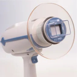
Conference of Radiation Control Program Directors (CRCPD) recommends that each handheld x-ray device be evaluated individually for safety purposes as the design characteristics vary significantly from one manufacturer to another.5 This study addressed this recommendation by evaluating an FDA cleared handheld device that is new to the market in the state of North Carolina, the Xray2go (Digital Doc LLC, El Dorado Hills, California).
to evaluate the impact of rectangular collimation with a handheld x-ray device on operator and simulated adult and child patient doses as well as radiographic image quality.
Therefore, the purpose of this study was to measure and compare adult and child effective dose (E) and operator dose from simulated full mouth series (FMX) using an
exploratory “real world” positioning technique. Additionally, technical performance and image quality were evaluated from two handheld ray devices and one conventional wall-mounted x-ray source using both circular and rectangular collimation. A primary goal was to evaluate the impact of rectangular collimation on operator dose and patient E as well as image quality. Specific research objectives were:
1. Measure adult and child effective dose from the NOMADTM Pro2 handheld device (circular & rectangular collimation), Xray2go handheld device (circular collimation) and KaVoTM FOCUSTM Instrumentarium conventional wall-mounted source (KaVo Kerr, Brea, CA) (circular and rectangular collimation).
2. Evaluate dose reduction from the NOMADTM Pro2 with the original equipment manufacturer (OEM) rectangular collimator attachment.
3. Evaluate operator dose from the NOMADTM Pro2 handheld device versus the Xray2go handheld device.
CHAPTER II
REVIEW OF LITERATURE
The use of handheld x-ray devices in dentistry has increased over the last 25 years and the development of and marketing for these devices is also growing6. Early handheld x-ray devices were designed for use by military dental personnel in deployment and field
device can be used for multiple operatories, decreasing the cost and maintenance of multiple mounted units, and there are no limitations from a support arm when using a wall-mounted source5. The use of handheld x-ray devices has also expanded in medicine,
veterinary work and security screenings11. Even so, their use for the masses is yet to be seen. Early studies regarding basic radiation safety and image quality can be found in the literature but there are still questions about handheld devices prevalent in colloquial conversations in dentistry.
All dentists use and rely on radiographs to some extent. Consequently, it is their responsibility to understand and minimize the risks of ionizing radiation in their practice and be able to accurately and with perspective explain these risks to their staff and patients. The International Commission on Radiological Protection (ICRP) determined the preferred unit for comparing risk from different radiographic examinations is effective dose12. This unit is used to estimate the risk to the population of low dose ionizing radiation to the probability of developing stochastic effects e.g., cancer formation. The National Council on Radiation Protection and Measurements (NCRP) Report No. 160 found exposure from medical computed tomography and nuclear medicine examinations contributes 36% of the estimated average effective dose of 6.2 millisieverts received per person in the United States annually. This report also showed a rapid surge in use of medical imaging since the early 1980s13.
Conversely, radiation exposure from diagnostic dental imaging has been considered a minor contributor of the estimated annual dose received per person. Occupational exposure to
in the maxillofacial region. Consequently, imaging from dental procedures contributes more to effective dose and the population’s radiation burden than previously thought14. As the use of CBCT, digital imaging and handheld x-ray devices is growing in dentistry, so must our commitment to maintain patient, occupational and public ionizing radiation exposure as low as reasonably achievable (ALARA) while maintaining high quality, diagnostic images.
Historically, the dental profession has made improvements to best practice techniques and armamentarium including faster receptors, digital imaging and collimation of the x-ray beam in order to minimize radiation doses and limit the release of radiation into the
environment. Nolan’s 1953 study was one of the first to focus on factors which can protect the patient from overexposure to radiation. He evaluated the effects of x-ray energy, focal
distance, amount of filtration, x-ray current, time of exposure, type of collimation and type of film and found patient exposure ranged from 35 to 315 R/full mouth examination and averaged 12.6 R/film15. A follow up study by Richards showed the effects of the faster Ektaspeed film compared with the very slow regular film of the 1920’s16. He reported a reduction in patient exposure to 170 mR/film and 143 mR/film with and without backscatter, respectively.
the latest guidelines18–20, the use of rectangular collimation is still not universally adopted despite consistent strong recommendations by the NCRP. Furthermore, there is even less information regarding the use of rectangular collimation with handheld x-ray devices.
While handheld x-ray devices have obvious similarities to conventional wall-mounted systems, a safe handheld device will also have important differences including its shielding design. NCRP Report No. 177 states traditional methods of shielding and source-to-operator distance are not applicable to handheld devices and that the instruction for the operator to “never hold the x-ray unit” is both unnecessary and impractical for properly designed handheld systems that have been cleared by the FDA1. That being said, an improperly designed
handheld device may present several safety issues including high radiation doses to patients and operators6.
Early studies on the safety and feasibility of handheld devices researched the first handheld device available on the market, the NOMADTM. A handheld image quality study in 2008 by Brooks et. al compared diagnostic quality of patient images obtained using a split-mouth design, the NOMADTM and a conventional wall-mounted x-ray source. They found image quality was similar and motion artifact was not significant21. The same year a clinical study by Goren and colleagues tested the NOMADTM for leakage radiation and patient and operator dose. Using thermoluminescent dosimetry (TLD) and F-speed film at .25 seconds for a lower anterior periapical and a molar bitewing, they found patient exposure ranged 14 – 27 mR/film and the estimated maximum annual operator exposure was 18 mR to the operator’s chest, 22.5 mR to the operator’s eye, and 45 mR to the operator’s finger. With this estimate it was concluded operator doses would be well below 500 mrem/year, the threshold that
also fell significantly below the maximum permissible radiation leakage of 100 mR/hour22 under 21 CFR 1020.30(k). It was concluded that the NOMADTM presented risks that are no greater than conventional wall-mounted systems. Of note, the device was operated with the beam parallel to the floor and the lead filled acrylic shield attached to the end of the x-ray tube. No mention of collimation was made but it can be assumed from photographs in the
manuscript that standard ubiquitous circular collimation was implemented.
A follow up study by Hermsen using the NOMADTM evaluated operators standing at various positions and angles outside of the recommended “zone of occupancy,” in order to resemble working conditions in a morgue setting. Using a Keithley Integrating Radiation Survey Meter and a Lucite cylinder phantom, they concluded the dose received by team members during a typical forensic deployment was at most .253 mSv and insignificant compared to the 50 mSv annual limit. They also showed that removing the lead infused acrylic ring resulted in radiation readings almost 10 times higher than measurements when the ring was in place23.
In 2009 Danforth et. al studied atypical device positioning using the NOMADTM at
applies to each diagnostic task at hand.
Following the optimistic findings of early studies using the NOMADTM handheld
device, the Conference of Radiation Control Program Directors came out with their QA Collectible on handheld dental x-ray units in 2010, cautioning dental providers that not all handheld devices are created equal. They particularly emphasized the use of a backscatter shield, higher tube current to reduce exposure time and potential motion, using high speed receptors and the use of an aiming ring to minimize cone cutting5. They also mention
optimizing design to reduce patient (and staff) dose by using smaller cylinder diameters, which refers to circular collimation.
effective dose were mostly a consequence of different geometries between the conventional wall-mounted system (300 mm source to collimator end length and square collimation) and the portable systems25. They also showed exposure to the operator’s hand was lowest when a protective shield was present. Their results support the use of long cone systems, rectangular collimation and a shield to reduce backscatter, however continued questions about safety are implied when they suggested the use of a tripod and/or an exposure switch cable.
Initial studies showed properly shielded handheld x-ray devices, which includes the use of a backscatter shield, are in accordance with national radioprotection criteria and image quality is comparable to wall-mounted units3,4. However, since the use of these devices is highly user dependent, additional research is warranted. Historically there had also been push-back from individual states in the United States and various international dental communities which limited their use and may have generated longstanding suspicion about whether handheld x-ray devices are safe.
It could be argued that operators of conventional wall-mounted x-ray systems should receive zero dose and therefore operators of handheld devices may need additional shielding in order to maintain the principle of ALARA. In 2012 Gray et. al surveyed dental facilities that used conventional wall mounted systems, then implemented the NOMADTM handheld system
while using radiation monitoring for their staff for both systems. They showed the average monthly dose for NOMADTM dosimeters was statistically significantly lower at 0.28 µSv
compared to the wall-mounted average dose of 7.86 µSv. These results were also in
reasonable agreement with data from multiple NCRP reports and a national dosimetry provider showing that the average dose received by dental staff was in fact higher than the NOMADTM
significantly less than wall mounted systems and that additional shielding efforts (e.g. wearing a lead apron) are not necessary for handheld devices3. It is important to note that the
aforementioned study only evaluated dose from the NOMADTM handheld device. McDiff and colleagues also supported the conclusion that no additional protection requirements are needed for properly trained operators of well-designed handheld x-ray units, which implies operating the handheld device parallel floor4. A study by Cho26 on two
handheld x-rays devices sold in Korea recommended the use of a backscatter shield, longer cone as well as lead gloves to decrease operator radiation dose. However, the authors state many handheld devices on the market in Korea do not have radioprotective shielding on the collimator cone, which affects operator dose at the level of the hand. Although calculating operator dose was not a major aim in this study, not reporting organ doses limits comparability to other studies on handheld devices.
In 2015 the European Academy of Dentomaxillofacial Radiology determined portable x-ray devices should be used in specific cases only9. Recent studies from international dental communities including the United Kingdom, Germany and Canada are addressing the concept of a restricted access “controlled area” (CA) with the use of handheld devices. In 2016 Makdissi et. al studied three test scenario positions of a handheld device relative to the operator (1. Close to the body and x-ray beam parallel to the ground 2. Away from the body and x-ray beam parallel to the ground and 3. Arms partially extended and x-ray beam
perpendicular to the ground). In their study the maximum dose to the operator was recorded for the left palm of the support hand when the device was held with the x-ray beam
levels, they still assume doses from wall mounted systems should be nil27.
In 2018 Rottke et. al aimed to show the expected distribution of scatter radiation from handheld devices and therefore evidence for regulations to reduce the size of the required controlled area. According to their study design and using theNOMADTM Pro2, they
concluded handheld x-ray systems cannot be rated disadvantageous to wall-mounted systems and that the actual CA was notably smaller than (German) regulations10. They recommend the use of rectangular collimation, but many operators refrained from using it and decided to use circular collimation in their study. Another limitation in their study design was the calculation of air kerma. Air kerma does not represent a measured body absorbed dose or a calculated effective dose which limits comparability to more current studies which use the radiation protection measurements of equivalent and effective dose.
Zenobio and colleagues evaluated the DIOX intraoral portable device for image quality and radioprotection criteria and found the device was safe for use and produced image quality equivalent to two conventional wall-mounted systems28. When simulating the exposure of a maxillary periapical radiograph, they showed the operator’s gonadal region without the acrylic shield received the highest estimated equivalent dose of 0.057 mSv/week which was almost 20 times lower than established allowable level. The regions of the salivary glands and oral mucosa were the organs with the highest absorbed dose in the patient. In 2019, Smith et. al also calculated air kerma measurements from five different handheld x-rays devices at numerous spatial positions and concluded there is potential for increased radiation risk to the operator, therefore handheld x-ray devices should only be used on a stand29.
regions (bitewing, upper molar, lower molar and upper anterior) in five mannequins were obtained using the NOMADTM Pro2handheld device and a wall-mounted unit. Their study design found aiming precision proved similar for both systems although individuals may perform better using one or the other modality30. They also mentioned that aiming precision is not a limiting factor for image quality. Of note, this study design did include rectangular collimation.
The literature on handheld x-ray devices varies greatly in study design and methodology. The majority of studies have been done using a model of the NOMADTM handheld device, and very limited research is found on the other available devices. Some studies only evaluate dosimetry or image quality while a few have attempted to evaluate both. In addition, there is no consistent trend for dosimetry methodology, with ionization chambers, personal dosimeters, and thermoluminescent (TLD) and optically stimulated luminescent (OSL) dosimeters found in handheld research. The use of ionization chambers can create unknown variables due to different sizes of chambers and positioning which could result in different air kerma measurements for the same conditions. The value of air kerma alone in modern studies is also questionable since the ICRP has already determined effective dose is the preferred unit for comparing different radiographic examinations12 and the discussion of dose without context is fairly meaningless, especially to non-radiology specialists. Variability with the threshold of detection for personal dosimeters also exists. The thresholds are 10 µSv and 100 µSv for optically stimulated luminescent and thermoluminscent dosimeters
respectively.
CHAPTER III METHODS
This study was designed to compare two handheld x-ray devices and one conventional wall-mounted x-ray source using both circular and rectangular collimator attachments that are currently being used in dental radiographic practice. When exposing radiographs, it is important to keep the dose as low as reasonably achievable (ALARA) while simultaneously producing a diagnostic image. Therefore, the design of this study included a dosimetry component and a technical component.
Part A: Dosimetry Component
1. Intraoral X-ray Systems and Collimator Attachments
Devices
1. NOMADTM Pro2 (handheld) 2. Xray2go (handheld)
3. FOCUSTM Instrumentarium (conventional wall-mounted) Modalities
• NOMADTM Pro2 with circular collimation
• NOMADTM Pro2 with rectangular collimation
• Xray2go with circular collimation
• FOCUSTM Instrumentarium with circular collimation
• FOCUSTM Instrumentarium with rectangular collimation
Dose associated with five modalities was measured. The NOMADTM Pro2, hereafter referred to as “NOMADTM,” circular technique utilized the 6 cm diameter original manufacturer
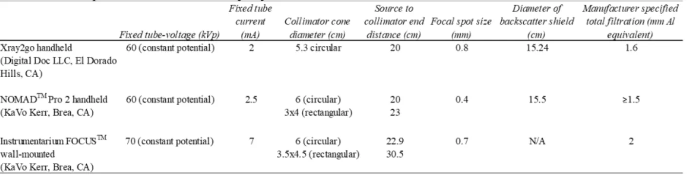
collimator attachment fitted over the circular collimator end resulting in a 23 cm source to collimator end distance (Figure 3.2). The Xray2go circular technique utilized the 5.3 cm diameter OEM circular collimator with a 20 cm source to collimator end distance (Figure 3.3). The FOCUSTM Instrumentarium, hereafter referred to as “FOCUSTM,” circular technique utilized
the 6 cm diameter OEM circular collimator with a 22.9 cm source to collimator end distance (Figure 3.4). The FOCUSTM rectangular technique utilized the 3.5 x 4.5 cm OEM rectangular
collimator with a 30.5 cm source to collimator end distance (Figure 3.5). Backscatter shields were in place for all handheld modalities. Technical specifications for allmodalities tested are presented in Table 3.1.
A subjective normalization procedure was performed due to the wide variation in technical specifications seen for all modalities. Exposure settings used in this study were optimized for each modality for acquisition with a Schick 33 direct digital complementary metal oxide semiconductor (CMOS) sensor (Dentsply, Sirona). Step wedge images were assessed and a consensus among expert observers established the lowest exposure time possible for each modality which still produced a diagnostically acceptable radiographic image. Optimized exposure parameters and corresponding images are found in Figure 3.6.
normalized effective doses were compared for all modalities. 2. Phantoms
Adult dosimetry was acquired using an average adult tissue-equivalent phantom
(ATOMmax Model 711HN – CIRS Inc., Norfolk, VA) (Figure 3.7). The phantom is sectioned in 25 mm thick axially oriented slices which permit access to specific tissues and anatomical locations of interest. Slices are modified to accept optically stimulated luminescent (OSL) dosimeter chips at these internal and external sites (Appendix A). During the imaging process, the phantom was oriented so that the section planes were approximately parallel to the floor. Dosimeters were positioned at 24 anatomical locations corresponding to tissues of interest seen in Appendix B.
Child dosimetry was acquired using a tissue equivalent phantom simulating the anatomy of a 10-year old child (ATOMmax Model 706 HN, CIRS Inc., Norfolk, VA) (Figure 3.8). The child phantom is divided into 25 mm thick axially oriented slices and dosimeters were positioned at 24 anatomical locations corresponding to tissues of interest (Appendix B).
3. Dosimeters and Reader
reading. Dosimeters used in this study were read with an OSL dosimeter reader (MicroStarii, Landauer, Inc., Glenwood, IL) (Figure 3.10). The reader was calibrated initially with a set of dosimeters supplied by the manufacturer that had been exposed to known amounts of energy from an 80 kVp x-ray source. Reader performance was checked before each use. Photon counts were converted to dose using an energy specific conversion factor reflecting the 60 and 70 kVp sources that were used throughout the study. All dosimetry was done by the investigator. 4. Adult Dosimetry Procedure
Eighteen projections simulating an adult full mouth series (FMX) and constituting one dosimeter run, were exposed using each modality on the adult ATOMmax phantom. Exposure parameters used were 70 kV/ 7mA and 0.04 seconds for anterior projections and .05 seconds for posterior projections for the FOCUSTM conventional wall-mounted unit with circular
To the best of the investigator’s ability, exposures were made with all modalities using the same distance from end of the collimator cone to the phantom which was meant to represent an actual separation that would occur if a beam-aiming device could be used. In addition, to the best of the investigator’s ability, exposures were made with all modalities using the same
projection position and vertical and horizontal angulations recommended for acquiring an FMX with a conventional wall-mounted source while keeping the phantom oriented so the section planes were approximately parallel to the floor. These angulations and exposure time parameters are listed in Table 3.2. This positioning of the handheld devices constitutes the exploratory technique in this study and is in contrast with both handheld device manufacturer’s
recommendations that the patient tilt their head to accommodate positioning the device so that the x-ray beam remains parallel to the floor and perpendicular to the operator. This study was specifically designed in order to evaluate the “real world” radiation dose for the patient and operator if the device is maneuvered the same way as a typical intraoral examination made with a conventional wall-mounted source with a patient seated upright in a chair. It is assumed this positioning technique would be most habitual and comfortable for the operator which should limit image re-takes.
5. Child Dosimetry Procedure
0.09 seconds for anterior projections and 0.12 seconds for posterior projections for the
NOMADTM handheld device with circular collimation and rectangular collimation, and 60 kV/ 2 mA and 0.08 seconds for anterior projections and 0.11 seconds for posterior projections for the Xray2go handheld device with circular collimation. For each dosimeter run, the simulated FMX was repeated 10 times (120 exposures) to provide a more reliable measure of energy in the dosimeters at the peripheries of the exposure areas. Dosimeter readings were then divided by 10 to determine the dose per single FMX. Each dosimetry run was repeated three times with the same modality using fresh sets of dosimeters to account for operator variability and the average dose of the three runs was calculated for each modality.
Similar to adult phantom acquisitions, child exposures were made with all modalities using the same end of collimator cone to phantom distance, projection position and vertical and horizontal angulations as described for the adult procedure using reduced exposure settings and 12 projections per FMX. Angulations and child exposure time parameters are listed in Table 3.3. The dosimetry acquisition matrix for the entire is found in Table 3.4.
6. Operator Dosimetry Procedure
Operator dosimetry was acquired using OSL dosimeter chips affixed to four locations of interest: thyroid, breast, groin area and handheld device trigger hand. One dosimeter was taped to each location on a lead apron worn by the operator and to the device trigger hand location for each dosimeter run during the previously described adult and child dosimetry procedures. Since simulated FMX exams were repeated 10 times per run, measured doses from the operator dosimeters were divided by 10 to achieve the average dose per FMX.
instructions (keeping the x-ray beam parallel to the floor and perpendicular to the operator). The adult phantom was tilted in order to accommodate this position. One simulated FMX run was completed using each handheld modality and the same exposure times used for the adult dosimetry procedure. Optically stimulated luminescent dosimeter chips were applied as previously mentioned above. Since simulated FMX exams were repeated 10 times per run, measured doses from the operator dosimeters were divided by 10 to achieve the average dose per FMX.
7. Dose Calculations and Adjustments
Effective dose was the primary outcome variable of this study. It is arrived at only by calculation and its value expresses the relative risk of human tissue detriment from ionizing radiation. Doses from OSL dosimeters at specific locations within the tissue or organ were averaged to express the average tissue-absorbed dose in micrograys (µGy). The products of these values and the estimated percentages of tissue or organ irradiated in the ATOMax phantom for an FMX and the radiation weighting factor for x-rays were used to calculate the equivalent dose (Appendix C). Effective dose, expressed in μSv, was calculated by using the equation E = Σ WT × HT and applying 2007 ICRP tissue weighting factors2, where effective dose (E) is the sum of the products of the tissue-weighting factor (WT), (Appendix D) and the equivalent doses2,14.
8. Investigator
9. Statistical Analysis
Dosimetry data was analyzed using Analysis of Variance (ANOVA) and Tukey Honestly Significant Difference (HSD) post-hoc analysis to assess differences among mean patient
effective doses and operator doses for all modalities.
Part B: Technical Performance and Image Quality
1. Investigator
The investigator was an oral and maxillofacial radiology resident and the sole operator for all exposures. The investigator was trained on each device according to the manufacturer’s instructions. The NOMADTM and the X-Ray2Go provide a training module that was viewed prior to use of the handheld devices and the recommended post-test was taken to achieve a perfect score.
2. Modalities
Images were produced with the FOCUSTM conventional wall-mounted source with circular and rectangular collimation, the NOMADTM handheld device with circular and
rectangular collimation and the X-Ray2Go handheld device with circular collimation, the only collimator shape available for this device. Technical performance, diagnostic acceptability and image quality were evaluated.
3. Receptors
All radiographs were exposed using a Schick 33 direct digital CMOS sensor. A size-2 sensor was used for all central, molar and bitewing exposures. A total of four projections (upper right molar periapical, molar bitewing, lower right molar periapical and maxillary central
These four projections were chosen to adequately represent the variation of images necessary to complete a full mouth series of radiographs.
4. Equipment
Handheld device exposures were made using the NOMADTM handheld device and the Xray2go handheld device. The NOMADTM was used with both circular and rectangular collimation. Xray2go exposures were made using circular collimation. Conventional wall-mounted source exposures were made using the FOCUSTM source with both circular and rectangular collimation. All exposures were made with the same optimized parameters for an adult for each modality that were used in Part A of the study.
Nine Dental X-ray Teaching and Training Replicas (DXTTRs) (DENTSPLY Rinn, Elgin, IL) were identified for use in the study. Each DXTTR is designed with natural teeth and human skulls. Selection of the DXTTRs was based on optimal, mechanical and operational conditions. 5: Technical Performance Procedure
The investigator exposed one four image series (upper right molar periapical, molar bitewing, lower right molar periapical and maxillary central periapical) using each of the five modalities on all nine DXTTRs (45 series total) utilizing the Extension Cone Paralleling (XCP) receptor-holding device (DENTSPLY Rinn, Elgin, IL). There was unlimited time to complete the images and the investigator treated the radiographs as if they were imaging a live patient. Images were assessed based on presence or absence of minor and major errors.
6. Evaluator Criteria and Image Assessment
All study images were blinded to the evaluator based on DXTTR and x-ray
device/collimator combination. The images were evaluated for the presence of horizontal angulation error, vertical angulation error, cone cut and motion based on predetermined criteria (Appendix E). Minor errors are represented by the presence of the error, but the intended
anatomic structures are displayed in the image. A major error in diagnostic quality was based on the absence of specified anatomic structures and would require a re-take.
7. Image Quality Procedure
A 16-group line pair per millimeter (LP) test tool with a range of resolutions from 5 to 20-line pairs per millimeter (Model 07-555, Nuclear Associates, Division of Victoreen, Carle Place, NY) was used to assess range of spatial resolution. One radiographic image was made with each modality using the line-pair phantom held in a jig to reduce the likelihood of motion artefacts and a 2.5 cm acrylic slab used to simulate actual source to object and object to image detector distances. Three experienced evaluators assessed all images. All study images were blinded to the evaluators based on x-ray device/collimator combination. Images were stored in the investigator’s Electronic Patient Record.
8: Statistical Analysis
A bivariate analysis and chi-square statistics were used to report number and location of errors by modality. Descriptive statistics were used to report average number of observed line pairs for all observers and the standard deviation per modality
9: IRB Study #:18-1496
CHAPTER III FIGURES
Figure 3.1: NOMADTM Circular Collimator
Figure 3.3: Xray2go Circular Collimator
Figure 3.5: FOCUSTM Rectangular Collimator
Figure 3.7: Average Adult Tissue-Equivalent Phantom (ATOMmax Model 711HN - CIRS Inc, Norfolk, VA)
Figure 3.9: OSL Dosimeters (Nanodot, Landauer, Inc., Glenwood, IL)
CHAPTER III TABLES
Table 3.1: Specifications of X-ray Systems
Table 3.2: Adult Intraoral Imaging Study Parameters
Table 3.3: Child Intraoral Imaging Study Parameters Image Type Area Vertical Horizontal Exposure time (sec) Xray2go
Exposure time (sec) NOMADTM Circular and
Rect. Collimation
Exposure time (sec) FOCUSTM Circular
Collimation
Exposure time (sec) FOCUSTM Rect.
Collimation No. of Images
PA maxillary Molar 25º 80º 0.12 0.13 0.05 0.08 2
PA maxillary Premolar 25º 75º 0.12 0.13 0.05 0.08 2
PA maxillary Canine-lateral 45º 25º 0.09 0.1 0.04 0.063 2
PA maxillary Centrals 45º 0º 0.09 0.1 0.04 0.063 1
PA mandibular Molar 0º 80º 0.12 0.13 0.05 0.08 2
PA mandibular Premolar -15º 75º 0.12 0.13 0.05 0.08 2
PA mandibular Canine-lateral -20º 25º 0.09 0.1 0.04 0.063 2
PA mandibular Centrals -20º 0º 0.09 0.1 0.04 0.063 1
BW Molar 10º 80º 0.12 0.13 0.05 0.08 2
BW Premolar 10º 75º 0.12 0.13 0.05 0.08 2
mAs/FMX 3.96 5.4 5.88 9.37 18 Total Images
Image Type Area Vertical Horizontal Exposure time (sec) Xray2go
Exposure time (sec) NOMADTM Circular and
Rect. Collimation
Exposure time (sec) FOCUSTM Circular
Collimation
Exposure time (sec) FOCUSTM Rect.
Collimation No. of Images
PA maxillary Molar 25º 80º 0.11 0.12 0.04 0.063 0
PA maxillary Premolar 25º 75º 0.11 0.12 0.04 0.063 2
PA maxillary Canine-lateral 45º 25º 0.08 0.09 0.032 0.05 2
PA maxillary Centrals 45º 0º 0.08 0.09 0.032 0.05 1
PA mandibular Molar 0º 80º 0.11 0.12 0.04 0.063 0
PA mandibular Premolar -15º 75º 0.11 0.12 0.04 0.063 2
PA mandibular Canine-lateral -20º 25º 0.08 0.09 0.032 0.05 2
PA mandibular Centrals -20º 0º 0.08 0.09 0.032 0.05 1
BW Molar 10º 80º 0.11 0.12 0.04 0.063 0
BW Premolar 10º 75º 0.11 0.12 0.04 0.063 2
CHAPTER IV RESULTS
Part A: Dosimetry Component Results
1. Adult Dosimetry Results
Figure 4.1 displays the average adult effective doses (µSv) from all five modalities. The lowest dose achieved was using the NOMADTM with the OEM rectangular collimator attachment (6.87 µSv). Mean (SD) FMX effective dose was statistically significantly less for handheld techniques with circular collimation than for the FOCUSTM conventional wall-mounted source with circular collimation (p<.0001). All effective doses using rectangular collimation were statistically significantly less than all circular collimation techniques (p<.0001). Statistically significant differences were found for all modality combinations except the NOMADTM with circular collimation and the Xray2Go (p=.8329).
Figure 4.2 displays the average normalized adult effective doses (µSv) from all five modalities. Normalized effective doses using circular collimation were 30.4 µSv for the conventional wall-mounted source and 26.4 µSv for the Xray2go and the NOMADTM. The lowest normalized effective dose was from the NOMADTM with rectangular collimation (8.6 µSv). The same statistical relationships seen with unadjusted effective doses were also seen with the normalized effective doses.
conventional wall-mounted source with circular collimation. Using the NOMADTM with the OEM rectangular collimator attachment, the effective dose was 74% less than the FOCUSTM conventional wall-mounted source with circular collimation. Table 4.2 shows the percent reductions in surface area exposure achieved by all modalities compared to conventional with circular collimation.
2. Child Dosimetry Results
Figure 4.3 displays the average child effective doses (µSv) from all five modalities. The lowest doses achieved were using the FOCUSTM conventional wall-mounted source with rectangular collimation (8.63 µSv) and the NOMADTM with the OEM rectangular collimator attachment (10.17 µSv) which were not statistically significantly different from each other. All effective doses using rectangular collimation were statistically significantly less than all circular collimation techniques (p<.0001). Statistically significant differences were not found between the Xray2Go and the NOMADTM with circular collimation, between the NOMADTM with circular collimation and the FOCUSTM conventional wall-mounted source with circular collimation, or between the NOMADTM with the OEM rectangular collimator attachment and the FOCUSTM conventional wall-mounted source with rectangular collimation.
Figure 4.4 displays the average normalized child effective doses (µSv) from all five modalities. Normalized effective doses using circular collimation were 29.4 µSv for the conventional wall-mounted source, 46.8 µSv for the Xray2go and 43.5 µSv for the
collimation.
Table 4.3 shows the percent reduction in effective dose that was achieved by each of the modalities when compared to the FOCUSTM conventional wall-mounted source. Using the NOMADTM handheld device and the OEM rectangular collimator attachment, the effective dose was 60% less than the FOCUSTM conventional wall-mounted source with circular collimation. The FOCUSTM conventional wall-mounted source with rectangular collimation was 66% less than the conventional wall-mounted unit with circular collimation.
3. Operator Dosimetry Results Adult Procedure
Figure 4.5 displays the average operator dose (µGy) per adult FMX to the four areas of interest for each of the handheld modalities using the exploratory positioning technique. Dose to the areas of the thyroid, chest and trigger hand were indistinguishable from ambient
background levels for all handheld modalities (p<.0001). Operator dose to the groin area using the handheld devices with circular collimation was 4.1 µGy for the XRay2go and 2.4 µGy for the NOMADTM. This was significantly higher than the dose to the thyroid, chest and trigger hand for all handheld modalities as well as the dose to the groin area using the NOMADTM with the OEM rectangular collimator attachment. Operator dose to the groin using the
NOMADTM with the OEM rectangular collimator attachment was 0.99 µGy. This was higher but not statistically significantly different than the dose to the areas of the thyroid, chest and trigger hand for all handheld modalities.
when compared to the exploratory positioning where the device position is variable throughout the FMX.
4. Operator Dosimetry Results Child Procedure
Figure 4.7 displays the average operator dose (µGy) per child FMX to the four areas of interest for each of the handheld modalities. Dose to the areas of the thyroid, chest and trigger hand were indistinguishable from ambient background levels for all handheld modalities (p<.0001). Operator dose to the groin area using the handheld devices with circular
collimation was 1.81 µGy for the Xray2go and 1.29 µGy for the NOMADTM. These doses were significantly higher than the thyroid, chest and trigger from all handheld modalities as well as the dose to the groin area using the NOMADTM with the OEM rectangular collimator attachment (p<.0001). Operator dose to the groin using the NOMADTM with the OEM
rectangular collimator attachment was 0.60 µGy. This was higher than the dose to the areas of the thyroid, chest and trigger hand for all handheld modalities but only statistically
significantly different from the thyroid and trigger hand using the NOMADTM with OEM rectangular collimation attachment and the chest from the NOMADTM using circular collimation.
Part B: Technical Performance and Image Quality Results
1. Technical Performance Results
Table 4.4 displays frequency and percentage of all technique errors (horizontal angulation error, vertical angulation error, cone cut or motion) by modality. There was not a statistically significant difference among modalities in terms of prevalence of error (p=.07) although less errors occurred using circular collimation modalities when compared to
handheld device with circular collimation and the greatest number of errors occurred with the conventional wall-mounted source with rectangular collimation. Of the rectangular
collimation modalities, the NOMADTM demonstrated less errors.
Table 4.5 displays the frequency and percentage of all technique errors by location in the mouth (upper right molar periapical, molar bitewing, lower right molar periapical or maxillary central periapical). There was a statistically significant difference among the locations in terms of proportion of error (p<.0001). Twice as many errors were observed for both the maxillary and mandibular periapical location compared to the bitewing location, and three times as many errors were observed for both the maxillary and mandibular periapical location compared to the anterior location. However, when controlling for modality, the statistically significant difference among the locations in terms of proportion of error was no longer seen.
The intra-rater agreement was almost perfect (.94). Because of sparseness of data, statistical analysis of each technical error (horizontal angulation error, vertical angulation error, cone cut or motion) was not possible.
2. Image Quality Results
Table 4.6 shows the line pair phantom test observation results. The average number of line pairs observed by three expert observers from the five unique modalities are displayed. The number of line pairs observed ranged from 12.7 (SD 1.5) for the Xray2go, 13.3 (2.1) for the conventional wall-mounted source with circular and rectangular collimation, and 13.7 (1.5) for the NOMADTM with circular and rectangular collimation. Coefficient of variation values range from 11-16% suggesting a small variation among interobserver agreement within
CHAPTER IV FIGURES
CHAPTER IV TABLES
Table 4.1: Percent Reduction in Adult Effective Dose When Compared to the FOCUSTM Conventional Source with Circular Collimation
Table 4.2: Percent Reduction in Surface Area Exposure Achieved by All Modalities
% Reduction in E from Conventional Circular
Modality
Dose
Conventional Circular
0%
NOMAD
TMCircular
34%
XRay2go
37%
Conventional Rectangular
53%
NOMAD
TMRectangular
74%
Modality % of ConventionalCircular
Conventional Rectangular 57%
NOMADTM Rectangular 64%
XRay2go 80%
XRay2go with penumbra 90%
Conventional Circular 100%
Table 4.3: Percent Reduction in Child Effective Dose When Compared to the FOCUSTM Conventional Source with Circular Collimation
Table 4.4: Frequency and Percentage of All Technique Errors by Modality
% Reduction in E from Conventional Circular
Modality
Dose
Conventional Circular
0%
NOMAD
TMCircular
-13%
XRay2go
-16%
Conventional Rectangular
66%
NOMAD
TMRectangular
60%
Modality No Yes Total
Conventional Circular 18 18 36
50% 50%
XRay2go 25 11 36
69.44% 30.56%
NOMADTM Rectangular 17 19 36
47.22% 52.78%
NOMADTM Circular 24 12 36
66.67% 33.33% Conventional Rectangular 15 21 36
41.67% 58.33%
Total 99 81 180
Any Error
Table 4.5: Frequency and Percentage of All Technique Errors by Location in the Mouth
Table 4.6: Average Number of Line Pairs Observed for Each Modality
Location No Yes Total
Anterior Centrals 38 7 45
84.44% 15.56%
Bitewing 30 15 45
66.67% 33.33%
Mandibular Periapical 16 29 45
35.56% 64.44%
Maxillary Periapical 15 30 45
33.33% 66.67%
Total 99 81 180
Frequency and Percentage of All Techniques Errors by Location
Any Error
Modality XRay2go Conventional Rectangular Conventional Circular NOMAD
TM
Circular NOMAD TM Rectangular
Line Pair Value (SD) 12.7 (1.5) 13.3 (1.5) 13.3 (2.1) 13.7 (1.5) 13.7 (1.5)
Coefficient of Variation 12% 11% 16% 11% 11%
CHAPTER V DISCUSSION
The NCRP Report No. 177 executive summary states in the last 15 years, three radiology innovations have found substantial application throughout general and specialty dentistry: digital imaging, cone-beam computed tomography and handheld intraoral imaging devices1. These developments can present a challenge to dentists who are responsible for not only learning the technology and correctly diagnosing and treating their patients, but also for developing and maintaining their own radiation safety and quality control programs.
Recommendations from sources such as the American Dental Association and the National Council on Radiation Protection and Measurements are extremely helpful for identifying best practice techniques to maximize radiation safety and optimize image quality when using handheld x-ray devices. Handheld x-ray devices that are cleared by the U.S. FDA are designed to shield the operator from source leakage radiation and backscatter radiation. A backscatter shield provides the operator with a zone of maximum protection that requires specific positioning of the device by the operator and tilting of the patient’s head in some circumstances, however, non-recommended use of handheld devices where positioning is
modeled after the typical conventional wall-mounted source positioning has been observed. This latter description of positioning is defined as the exploratory positioning in this study and is meant to address concerns that most operators do not adhere to strict manufacturer
persistently recommended by radiation safety bodies, such as rectangular collimation, are still not widely used in all dental offices and even less so with handheld x-ray devices.
Although dental schools and many practicing dentists are increasingly striving to practice evidence-based dentistry, the body of literature in regard to handheld intraoral x-ray devices are relatively sparse and the previous research done varies significantly in study design. Therefore, the purpose of this study was to assess the impact of rectangular collimation with a handheld intraoral x-ray device on estimated risk to the operator and patient. The 2007 ICRP
recommendations for calculating effective dose were used to estimate dose from a combination of modern imaging modalities. This study intended to present operator dose data that would represent the speculated “customary” use of handheld devices and also evaluated resultant technical performance and image quality.
The dosimetry component of the study evaluated two handheld intraoral x-ray devices and a conventional wall-mounted x-ray source using adult and child anthropomorphic phantoms. Five modalities were evaluated (one conventional wall-mounted unit with circular and
rectangular collimation, one handheld device with circular and rectangular collimation and a second handheld device using only circular collimation only). Handheld study exposures were acquired using exploratory device positionings designed to simulate the speculated use of handheld devices in every day practice versus strict adherence to the manufacturer’s guidelines, where the patient must tilt their head in order for the radiographer to keep the x-ray beam parallel to the floor and perpendicular to the operator.
operator dose from two handheld devices and the impact of customary versus recommended positioning of handheld devices on operator dose.
1. Patient Dosimetry
For adult phantom dosimetry, this study found that for an 18-image full-mouth
radiographic series using circular collimation, both FDA approved handheld devices resulted in effective doses that were significantly less than the conventional wall-mounted x-ray source. Previous research has also shown a reduced effective dose using handheld x-ray devices
compared to conventional wall-mounted sources31. The unadjusted effective doses achieved in this study using the handheld devices ranged from 16.7-17.4 µSv compared to 26.3 µSv from conventional circular, which is a difference of 34-37%. A non-biased quantitative normalization procedure was necessary to justify our subjective exposure optimization procedure. The average doses achieved from optimized posterior location exposure times for each modality were
measured. A more ideal technique would have been to use the full FMX exposure time for each modality in order to account for variable anterior and posterior exposure times and the collective nature of effective dose. However, the technique used in this study was a good approximation.
source to phantom distance with the handheld devices. It is thought that as operator fatigue sets in during handheld device use, the operator tends to hold the device closer to the body. This increased distance between the source and the phantom would increase the field of exposures while concomitantly decreasing the intensity of the x-ray beam resulting in a lower dose.
Operators of a handheld device are not able to take a “step back” to observe the exact position of the device in relation to the patient or the beam alignment ring. The results of this study show that this is likely to increase the variability in device positioning. In clinical practice, a beam-aiming device should be used, and this would help control for some variations in
positioning.
The results of this study can be compared to patient effective doses found from other radiographic examinations. Using the same methodology of calculating effective dose, Ludlow et. al showed the effective dose for an adult FMX with photo-stimulable phosphor (PSP)
detrimental effects among all modalities tested. These findings not only support the use of handheld intra-oral x-ray devices as a safe modality for patients but show that rectangular collimation should unequivocally be applied with handheld devices to achieve optimal reductions in dose and should not be reserved for conventional wall-mounted sources.
When discussing the child dosimetry results of this study it is important to note that the exposure parameters selected at the beginning of the study were subjectively optimized for each modality using an adult phantom. For child exposures, parameters were reduced by one
increment in available exposure times per x-ray device. This resulted in slight variations for time increment reductions due to differences in available unit parameters, yielding an average
reduction of 10-20% compared to adult exposures. Specifically, the wall-mounted source decrease in time (sec) per click ranged from 0.008-0.017 seconds whereas the handheld devices were consistently decreased by 0.01 seconds. Ideally, the reduction in exposure time from adult to child would have an equivalent effect on all modalities. The outcome in this study resulted in a relatively more reduced mAs per child FMX for the conventional wall-mounted modalities versus the handheld modalities seen with the non-normalized effective doses. This should be considered when comparing the different wall-mounted to handheld effective dose trends of the adult versus the child procedures. In the adult procedure, the effective doses from wall-mounted modalities were higher than the effective doses from the handheld modalities. In the child procedure, where a greater reduction in mAs from the adult exposure times occurred for the wall-mounted modalities, this trend was reversed. The effective doses from the wall-wall-mounted
The normalized child effective doses using circular collimation were 43.5 µSv for the NOMADTM and46.8 µSv for the Xray2go compared to 29.4 µSv for the wall-mounted source. This adjustment resulted in a 30% increase for Child E for handheld devices. While imaging geometry for all FMX locations were designed to be consistent for all modalities, the investigator noticed a tendency to unintentionally angulate the handheld devices slightly downward during acquisitions, unlike the wall-mounted source which allowed the operator to step back to view positioning. When evaluating the individual child tissue equivalent doses from all modalities, it was noted that the thyroid tissue received a large increase in dose from the handheld modalities which may be explained due to a tendency of a downward angulation during use. If an x-ray source is angulated downward, this would contribute to a saturation of radiosensitive tissue of a child more so than with an adult due to the thyroid tissue being located physically closer to the oral cavity in children, which would therefore increase effective dose. In addition, as described previously there was a greater reduction in exposure time for the child FMX that occurred with the conventional wall-mounted source versus the handheld devices. This difference in step-down time also correlates with the dose differences seen in the child dosimetry results.
Limited research has been completed on child dosimetry using handheld x-ray devices. A recent study by Bozic et. al concluded a child receives lower tissue equivalent doses for bitewing and anterior occlusal projections using a handheld device compared to a conventional wall-mounted unit34. More research is needed about handheld x-ray devices using exposure parameters specifically optimized for a child FMX.
conventional wall-mounted source with rectangular collimation achieved a lower dose compared to the NOMADTM handheld device using rectangular collimation, however there was no
statistically significant difference in dose between those modalities. The results of this study support the use of either an FDA approved handheld x-ray device or conventional wall-mounted source for intraoral imaging on children as long as rectangular collimation is used. Decreasing dose is especially important when discussing children because they are known to be more radiosensitive than adults33.
Although the effective doses in this study are meant to represent a “real world” scenario simulated by the investigator, due to limitations in phantom designs, the attenuation of x-rays from an intraoral imaging sensor could not be considered. The results of this study may then represent an overestimation of effective dose. Another limitation within the methodology was the optimization of exposure times based on subjective density. While this method represents how exposure parameters are selected in clinical practice, it could have created a bias in the resultant tube current-time product (mAs) values and therefore the effective doses achieved. 2. Operator Dosimetry
Relative to the annual occupational dose limits set by the NCRP35, the results of this study showed that all absorbed doses (µGy) to the operator using the exploratory handheld device positioning technique were minimal for both adult and child FMX exams. The per FMX exposure to the areas of the thyroid, chest and trigger hand were indistinguishable from
There was however, a statistically significantly higher operator dose to the groin area for all handheld modalities for both the adult and child full mouth examinations. The groin doses from lowest value to the highest were 0.60 µGy (NOMADTM with rectangular collimation for the child FMX) to 4.1 µGy (Xray2Go with circular collimation for the adult FMX). The dose per exposure for the groin was determined by averaging the FMX dose by number of exposures per FMX. This resulted in per exposure dose values for the groin region ranging from 0.05 µGy (NOMADTM with rectangular collimation per child exposure), to 0.23 µGy (Xray2Go with circular collimation per adult exposure). These findings were used to extrapolate potential annual occupational groin tissue dose in the same manner used by the NOMADTM User Manual36 and by Danforth et. al24 estimating 7200 radiographic exposure/year. This resulted in annual groin tissue doses ranging from 0.36 mGy (NOMADTM with rectangular collimation for child exposures) to 1.64 mGy (Xray2Go with circular collimation for adult exposures). The highest value in this annual dose range equates to the approximate surface dose from one direct NOMAD exposure using the manufacturer’s recommended settings for an adult bitewing radiograph with a digital sensor. From the tissue doses in this study, it would not be expected that an operator of handheld devices positioned at various angulation during the radiographic exam would receive an annual whole-body dose that exceeds the recommended threshold for monitoring of 1 mSv.
The lower dose seen in their study could be explained by the use of non-human tissue equivalent imaging subjects and varying study design and methods of calculating dose. Their results do show a similar relationship to this study where positioning of handheld devices other than the manner recommended by the manufacturer results in a higher exposure to the operator groin area. The results of the study by Zenobio et. al also showed operator exposure was greatest to the gonadal region and corroborate the results of the present study28.
The results of the current study show that even when positioning handheld x-ray devices in an unrecommended manner, which is anecdotally reported as a common method in private practice and dental schools, no modality tested would have a marked impact upon an operator’s allowable overall whole-body annual dose. However, this study showed when rectangular collimation is used with handheld devices, a statistically significant reduction in dose to the operator is seen and should be considered the standard of practice in order to achieve the principle of ALARA.
The results of the supplemental operator dosimetry procedure performed in this study, where the devices were operated according to the manufacturer’s recommended positioning, support publications from those manufacturers that this technique provides maximum protection to the operator. Doses to all areas measured were reduced compared to the exploratory
was performed in order to compare the results from our exploratory device positioning operator dosimetry results which used the same materials, methods and operator versus comparing to the results of other studies. This portion of the study however was not repeated to test for
reproducibility and as a result many possess limited reliability. A more robust comparison study could be completed in the future.
3. Technical Performance
When evaluating the total number of technical errors by modality, the highest number of errors resulted from exposures made with rectangular collimated modalities. However, the difference among all modalities in terms of prevalence of error was not statistically significant and device and type of collimation are therefore not a limiting factor for technical performance in this study. Both rectangular techniques resulted in an increased number of collimator
centering issues; however, all cone-cuts observed in this study were cosmetic or “minor” errors that would not warrant additional patient exposure from retakes due to unimaged anatomy. Three major errors were recorded that would indicate the need for remaking the radiograph. All three of these errors were the result of incorrect vertical angulation that did not capture root apices.
variable of multiple radiographers, the results may reflect the individual performance trends of the single operator. It is important to note that this study’s sole operator was trained on both devices according to the manufacturer’s recommendations and had the same amount of experience with each device. In addition, a beam positioning device (XCP-ORA) was used in this study and therefore horizontal and vertical angulation errors evaluated may be more of an indicator of receptor placement and not a true indicator of modality performance.
There were no instances of blur or image distortion due to operator/device motion observed for any modality in this study. Brooks et. al also showed motion to be a non-factor in image quality when using the NOMADTM 21. Although maxillary and mandibular periapical images were twice as likely to have a technical error, the majority of these errors were vertical angulation in the absence of a cone cut, which is not a primary indicator of modality
performance and instead that of receptor positioning. With the use of a beam alignment device in this study, the cone cuts observed were found to be the result of the restricted beam field of the rectangular collimator.
4. Image Quality
There was a very close agreement for observed resolution for all modalities
resolution. The NOMADTM handheld device has the smallest focal spot size of 0.4 mm and demonstrated the highest observed resolution of 13.7 LP/mm. The Xray2Go handheld device has the largest focal spot size of 0.8 mm and demonstrated the lowest observed resolution of 12.7 LP/mm. The conventional wall-mounted source has a focal spot size of 0.7 mm and an observed resolution of 13.3 LP/mm. Previous research has shown similar diagnostic quality using handheld devices compared to conventional wall-mounted sources21,25.
The procedure used to obtain the line pair images in this study was specifically designed to reduce possible motion from the handheld modalities. However, the results may change if the handheld devices were operated in a manner which simulates clinical practice. Motion of the actual handheld device would increase the effective focal spot size and therefore increase the geometric unsharpness of the image. Therefore, controlled results of this study may not be generalizable to actual clinical conditions. The dosimetry results of this study support safe clinical research to evaluate the technical performance and image quality of handheld devices in the future.
In conclusion, the NOMADTM and Xray2go handheld devices with circular collimation performed similarly in regard to dose for both adult and child patients and operators.
Additionally, there was not a significant difference in number of technique errors between the two modalities. A greater number of line pairs/mm were observed for the NOMADTM than the Xray2Go, which may be attributable to its smaller focal spot size.