HPV31 Utilizes the ATR-Chk1 Pathway to Maintain Elevated
RRM2 Levels and a Replication-Competent Environment in
Differentiating Keratinocytes
Daniel C. Anackerd, Heather L. Aloorc, Caitlin N. Sheparda, Gina M. Lenzia, Bryan A.
Johnsond, Baek Kima,b, and Cary A. Moodyc,d,*
aThe Center for Drug Discovery, Department of Pediatrics, Emory University School of Medicine,
Atlanta, Georgia, USA
bChildren’s Healthcare of Atlanta, USA
cLineberger Comprehensive Cancer Center and Department of Microbiology and Immunology
dUniversity of North Carolina at Chapel Hill, North Carolina, USA
Abstract
Productive replication of human papillomaviruses (HPV) is restricted to the uppermost layers of the differentiating epithelia. How HPV ensures an adequate supply of cellular substrates for viral DNA synthesis in a differentiating environment is unclear. Here, we demonstrate that HPV31 positive cells exhibit increased dNTP pools and levels of RRM2, a component of the
ribonucleotide reductase (RNR) complex, which is required for de novo synthesis of dNTPs. RRM2 depletion blocks productive replication, suggesting RRM2 provides dNTPs for viral DNA synthesis in differentiating cells. We demonstrate that HPV31 regulates RRM2 levels through expression of E7 and activation of the ATR-Chk1-E2F1 DNA damage response, which is essential to combat replication stress upon entry into S-phase, as well as for productive replication. Our findings suggest a novel way in which viral DNA synthesis is regulated through activation of ATR and Chk1 and highlight an intriguing new virus/host interaction utilized for viral replication.
Introduction
Human papillomaviruses (HPV) are small, double stranded DNA viruses that exhibit a strict tropism for epithelial cells. A subset of approximately 15 HPVs, termed high-risk (HR) (e.g. HPV16, 18, 31, 45), is the causative agent of cervical cancer, and is also associated with other genital malignancies, as well as an increasing number of head and neck cancers (zur Hausen, 2009). While the three licensed HPV vaccines show great efficacy and offer great promise in reducing the number of deaths due to cervical cancer, these vaccines are not
*Correspondent footnote: Address Correspondence to Cary Moody, [email protected].
Publisher's Disclaimer: This is a PDF file of an unedited manuscript that has been accepted for publication. As a service to our
customers we are providing this early version of the manuscript. The manuscript will undergo copyediting, typesetting, and review of
HHS Public Access
Author manuscript
Virology
. Author manuscript; available in PMC 2017 December 01.Published in final edited form as:
Virology. 2016 December ; 499: 383–396. doi:10.1016/j.virol.2016.09.028.
A
uthor Man
uscr
ipt
A
uthor Man
uscr
ipt
A
uthor Man
uscr
ipt
A
uthor Man
uscr
therapeutic. Identifying cellular pathways HPV commandeers to promote replication may identify potential therapeutic targets to limit viral replication and block disease progression.
The limited coding capacity of the HPV genome renders the virus reliant on cellular factors for viral replication. The viral life cycle is intimately linked to the differentiation status of the stratified epithelium and is characterized by three distinct phases of replication (Longworth and Laimins, 2004c). HPV infects the actively dividing basal cells of the stratified epithelium, where viral episomes undergo limited amplification to 50–100 copies per cell. Viral genomes are then maintained at a low copy number in these undifferentiated cells by replicating along with cellular DNA (Evans et al., 2003; Hoffmann et al., 2006). Upon epithelial differentiation, the productive phase of the viral life cycle is triggered, resulting in amplification of viral genomes to 1000s of copies per cell, late gene expression and virion production (Maglennon et al., 2011; Moody and Laimins, 2010). Paradoxically, these late viral events occur in cells that normally would have exited the cell cycle. Upon differentiation, HPV circumvents this problem largely through E7’s ability to target the tumor suppressor Rb for degradation, resulting in the release of active E2F transcription factors that promote unscheduled re-entry into the cell cycle (Cheng et al., 1995; Chien et al., 2000; Moody and Laimins, 2010). Traditionally, cell cycle re-entry upon differentiation has been thought to provide an S-phase environment conducive to rapid amplification of viral genomes. However, more recent studies indicate that productive replication occurs post-cellular DNA synthesis, in a prolonged G2-like phase (Banerjee et al., 2011; Wang et al., 2009). Therefore, HPV must have evolved means to provide cellular factors required for productive replication outside of S phase.
One of the most important resources HPV needs to acquire for productive replication is a sufficient supply of deoxyribonucleotide triphosphates (dNTPs), the building blocks for DNA synthesis. However, it is unclear how dNTP pools are maintained in a differentiating environment where infected cells are no longer dividing. The ribonucleotide reductase (RNR) enzyme complex is required for de novo synthesis of dNTPs (Nordlund and Reichard, 2006). RNR is the rate-limiting enzyme for dNTP synthesis, catalyzing the conversion of ribonucleoside diphosphates to their corresponding deoxyribonucleoside diphosphates (dNDP), providing balanced pools of dNTPs utilized for DNA replication, as well as repair. The active form of RNR is a tetramer composed of two identical large subunits of RRM1, and either two identical small subunits of RRM2, or an alternative subunit p53R2, which is p53-responsive (Nordlund and Reichard, 2006; Tanaka et al., 2000). In proliferating cells, the RRM1-RRM2 holoenzyme provides dNTPs for replication and repair in S-phase, while RRM1-p53R2 is active in G0/G1 where the consumption of dNTPs is minimal and restricted to mitochondrial DNA replication and repair.
RNR activity is tightly regulated in a cell-cycle dependent manner through multiple
mechanisms to coordinate the balance between dNTP synthesis and DNA replication. While transcription of both RRM1 and RRM2 occurs during S-phase (Engstrom et al., 1985), RRM1 levels are constant throughout the cell cycle due to its long half-life. In contrast, RRM2 levels fluctuate, reaching their highest level in S-phase (Bjorklund et al., 1990; Chabes and Thelander, 2000; Mann et al., 1988). The fluctuation in RRM2 is attributed to transcriptional regulation via the E2F1 transcription factor (Chabes et al., 2004; DeGregori
A
uthor Man
uscr
ipt
A
uthor Man
uscr
ipt
A
uthor Man
uscr
ipt
A
uthor Man
uscr
et al., 1995), as well as proteasome-mediated degradation by the anaphase promoting complex/Cdh1 in G1 (Chabes et al., 2003), and by SCFcyclinF in G2 (D’Angiolella et al., 2012). The S-phase specific elevation in RRM2 coincides with a significant increase in RNR activity (Bjorklund et al., 1990; Engstrom et al., 1985; Eriksson and Martin, 1981). Thus, RRM2 is considered limiting for RNR activity. Indeed, loss of RRM2 expression results in decreased dNTP levels (Aird et al., 2013; Taricani et al., 2014). RNR activity is important to genomic integrity, as well as cell viability, with an increase or imbalance of dNTP pools leading to mutagenesis (Hu and Chang, 2007), and decreased dNTP levels leading to impaired DNA replication and repair (Nordlund and Reichard, 2006). Previous studies demonstrated that RRM2 transcript and protein levels are increased in cervical cancer lines containing chromosomally integrated HPV16 and HPV18 genomes (Wang et al., 2014). HPV16 E7 was found to be sufficient to increase RRM2 transcript levels, however, whether RRM2 is increased in lines containing episomal HPV genomes and is important for viral replication has not been examined.
Recent studies have shown that levels of RRM2 can also be regulated through activation of the DNA damage kinase ATR (Ataxia telangiectasia and Rad3-related) and its downstream effector kinase Chk1 (Bertoli et al., 2013; D’Angiolella et al., 2012; Naruyama et al., 2008; Zhang et al., 2009). The importance of the DNA damage response (DDR) to HPV
replication is becoming increasingly clear (McKinney et al., 2015). Previous studies identified a role for the DDR kinase ATM (Ataxia Telangiectasia-Mutated) in productive replication of HPV31 (Moody and Laimins, 2009), which may facilitate viral DNA synthesis, at least in part, through the recruitment of DNA repair factors to viral replication centers (Chappell et al., 2015; Gillespie et al., 2012). While ATM responds primarily to double-strand DNA breaks, ATR is activated by single-strand DNA generated by DNA damage, as well as by stalled replication forks upon replication stress (Ciccia and Elledge, 2010; Zeman and Cimprich, 2014). ATR phosphorylates and activates Chk1, which is then released from chromatin to phosphorylate targets throughout the nucleus (Liu et al., 2000). Activation of ATR and Chk1 is critical for stabilization of stalled replication forks, and numerous studies suggest that the ATR-Chk1 pathway is essential for cancer cell survival in the face of replication stress (Buisson et al., 2015; Murga et al., 2011; Toledo et al., 2011). Recent studies by Bertoli et al demonstrated that the G1/S transcriptional program is rewired in response to replication stress in a Chk1-dependent manner, leading to high-level
expression of genes involved in DNA repair, as well as nucleotide synthesis, including RRM2 (Bertoli et al., 2013). In addition, several studies have demonstrated that the ATR-Chk1 pathway promotes RRM2 accumulation through stabilization of E2F1, in turn
providing dTNPs to prevent DNA damage and cell death (Bertoli et al., 2013; Buisson et al., 2015; Zhang et al., 2009).
HR HPVs are known to induce replication stress through expression of the E6 and E7 oncogenes (Bester et al., 2011; Spardy et al., 2008). E6/E7 expression drives uncontrolled S-phase entry, resulting in a disconnect between replication and supplies required for
replication, ultimately leading to DNA damage. Several studies have shown that the ATR-Chk1 pathway is active in HR HPV positive cells (Hong et al., 2015; Moody and Laimins, 2009; Reinson et al., 2013; Sakakibara et al., 2011), indicating that replication stress also occurs in the context of a viral infection. ATR-Chk1 activation can be induced independently
A
uthor Man
uscr
ipt
A
uthor Man
uscr
ipt
A
uthor Man
uscr
ipt
A
uthor Man
uscr
by E7 (Hong et al., 2015), as well as the viral helicase E1 (Reinson et al., 2013; Sakakibara et al., 2011), and inhibition of Chk1 activity decreases the stability of HPV genomes in undifferentiated cells (Edwards et al., 2013). In addition, the ATR-Chk1 pathway has recently been shown to be required for productive replication of HPV31 (Hong et al., 2015). However, how ATR and Chk1 activity contributes to viral replication is not known. The activation of ATR and Chk1 in both undifferentiated and differentiating HPV positive cells suggests that replication stress occurs throughout the viral life cycle. Activation of the ATR/ Chk1 pathway through HPV-induced replication stress may be required to establish an environment upon differentiation that is favorable for amplification of viral genome through stabilization of E2F1 and subsequent elevation of RRM2.
In this study, we demonstrate that HR HPV positive cells exhibit specific elevation of RRM2 levels in an E7-dependent manner, with little to no effect on the large subunit RRM1, or the alternative subunit p53R2. We have found that the increase in RRM2 corresponds with HPV31 positive episomal lines exhibiting higher levels of dNTPs compared to uninfected cells, which importantly, are maintained upon differentiation in an RRM2-dependent manner. Loss of RRM2 expression results in a decrease in viral copy number in
undifferentiated cells and a block in productive replication upon differentiation. We have found the levels of RRM2 in HPV31 positive cells are regulated through E7’s Rb binding domain and activation of the ATR-Chk1 DNA damage pathway through maintenance of the E2F1 transcription factor. Importantly, our studies connect activation of the ATR-Chk1 axis of the DDR to increased levels of E2F1 and RRM2 accumulation in HPV positive cells upon differentiation, providing further understanding of how HPV exploits the DDR to provide a replication-competent environment for completion of the viral life cycle.
Materials and Methods
Cell Culture
Human foreskin keratinocytes (HFKs) were collected from neonatal foreskin tissue as described previously (Ruesch et al., 1998) and were maintained in Dermalife keratinocyte growth media (KGM; Lifeline). HPV31 positive CIN612 9E cells were grown in E-media supplemented with 5ng/mL mouse epidermal growth factor (BD Biosciences) and co-cultured mitomycin C treated J2 3T3 fibroblasts, as described previously (Wilson and Laimins, 2005). Organotypic raft cultures were derived from HFKs and CIN612 cells as previously described (Anacker and Moody, 2012). Generation and maintenance of HFKs retrovirally transduced with pLXSN-HPV31 E6, pLXSN-31 E7, and pLXSN-31 E6/E7 in combination has been previously described (Hebner et al., 2006). Prior to harvesting DNA, protein or RNA, fibroblast feeder cells were removed from HPV positive cells using Versene (1 mM EDTA in phosphate-buffered saline). 293T cells were grown Dulbecco’s modified Eagle’s medium (DMEM; Life Technologies) supplemented with 10% bovine growth serum (BGS; ThermoFisher Scientific).
Plasmids and Inhibitors
The pLXSN-HPV31 E6, -HPV31 E7, and -HPV31 E6/E7 vectors have been previously described (Longworth and Laimins, 2004b). The pBR322 plasmid containing the wild-type
A
uthor Man
uscr
ipt
A
uthor Man
uscr
ipt
A
uthor Man
uscr
ipt
A
uthor Man
uscr
HPV31 genome and the E7 Rb ΔLHCYE genome was previously described (Hubert and Laimins, 2002; Longworth and Laimins, 2004b). The p1203 PML2d HPV16 plasmid containing the wild-type HPV16 genome was obtained from Addgene (plasmid no. 10869). VE-821 was obtained from Selekchem, and UCN-01 was obtained from EMD Millipore.
Generation of HPV16- and HPV31-Positive HFKs
HFKs stably maintaining HPV16 or HPV31 episomes were created as previously described (Wilson and Laimins, 2005). The pBR322-HPV31 plasmids containing the wild-type HPV31, HPV31 E7 ΔLHCYE mutant genomes have been previously described (Hubert and Laimins, 2002; Longworth and Laimins, 2004a). Briefly, the HPV31 E7ΔLHCYE construct contains an in frame deletion of the Rb binding domain of HPV31 E7. The HPV16 and HPV31 genomes were excised from the plasmid backbones using BamH1 and HindIII, respectively (New England Biolabs) and re-ligated using T4 DNA ligase (Life
Technologies). Primary HFKs were transfected with 1ug of the re-ligated genomes and 1ug PSV2-Neo using Fugene 6 according to manufactures instructions (Promega). Stable cell lines were generated through eight days of G418 selection (Sigma), and surviving populations were pooled and expanded for analysis.
Induction of Keratinocyte Differentiation
High calcium medium was used to induce epithelial differentiation as previously described (Moody et al., 2007). Sub-confluent cells were harvested as T0, and the remaining plates of cells were serum starved in basal keratinocyte growth medium (KGM; Lonza) with
supplements for 16 h. Cells were then incubated in keratinocyte basal medium (KBM; Lonza) without supplements but with 1.8 mM CaCl2 (Sigma). Cells were allowed to
differentiate for 24, 48, 72, or 96 h after addition of high calcium medium. DNA, RNA and protein were harvested at each time point, and viral genome amplification was measured by Southern blotting for each experiment to ensure activation of the productive phase of the viral life cycle.
Production of Lentivirus
Lentivirus was produced as previously described (Mighty and Laimins, 2011). Plasmids encoding shRNAs for RRM2 (TRCN0000038962 and TRCN0000038963) were obtained from the UNC Lentiviral Core (UNC-Chapel Hill), and a scramble non-target control shRNA in the pLKO background was obtained from Open Biosystems. Each shRNA plasmid (5ug) was co-transfected with 3.37ug Gag-Pol-Tet-Rev plasmid DNA and 1.66ug vesicular stomatitis virus G (VSV-G) plasmid DNA into 293T cells using polyethylenimine (PEI) to generate lentivirus particles. Supernatants containing lentivirus were harvested 72 h post-transfection, sterile filtered, and stored at −80°C until used. CIN612 9E cells were transduced with 5mL viral supernatant plus 4.8ug/mL hexadimethrine bromide (polybrene; Sigma-Aldrich) for three days prior to harvesting or differentiation in high calcium medium.
Western Blot Analysis
Whole cell lysates were harvested in radioimmunoprecipitation assay (RIPA) lysis buffer supplemented with Complete Mini and PhosSTOP tablets (Roche). Total protein levels were
A
uthor Man
uscr
ipt
A
uthor Man
uscr
ipt
A
uthor Man
uscr
ipt
A
uthor Man
uscr
determined via Bio-Rad protein assay. Equal protein amounts were electrophoresed on SDS-polyacrylamide gels and transferred to polyvinylidene difluoride (PVDF) membranes (Immobilon-P; Millipore). The following primary antibodies were used: RRM2, RRM1, p53R2, GAPDH, Involucrin (Santa Cruz); Chk1 (Abcam); phospho-Chk1 Ser345, E2F1 (Cell Signaling Technologies). Secondary antibodies used were: HRP conjugated anti-goat (Santa Cruz), HRP conjugated anti-rabbit (Cell Signaling Technologies), and HRP conjugated anti-mouse (GE Life Sciences). Clarity Western enhanced chemiluminescence (ECL) blotting substrate (Bio-Rad) was used to detect antibody binding.
Southern Blot Analysis
DNA isolation and Southern blotting were performed as previously described (Fehrmann et al., 2003). Briefly, cells were harvested in DNA lysis buffer consisting of 400mM NaCl, 10mM Tis pH 7.5 and 10mM EDTA. Cells were lysed by the addition of 30uL 20% SDS and subsequently treated with 15ul of 10mg/mL proteinase K overnight at 37°C. DNA was extracted by phenol chloroform extraction, followed by ethanol precipitation in the presence of sodium acetate. DNA was then digested with BamHI (New England Biolabs) (which does not cut the genome), or HindIII (New England Biolabs) (which cuts the genome once). DNAs were resolved on a 0.8% agarose gel for 15 h at 40 V, and transferred to a positively charged nylon membrane (Immobilon-Ny+; EMD Millipore). The DNA was fixed to the membrane via UV irradiation and then hybridized to a radioactive DNA probe consisting of 32P-labeled linearized HPV31 genome.
Measurement of Intracellular dNTPs
HFK, HFK-31 and CIN612 cells, as well as CIN612 cells transiently transduced with a scramble shRNA control (shScram) or shRNAs targeting RRM2 (shRRM2) were seeded in duplicate for each time point at 5 × 10⁵ in 10cm dishes. 72hr following seeding, cells were either harvested as a undifferentiated sample (T0) for dNTP analysis or exposed to high calcium medium for 72hr to induce differentiation. At the indicated time points, cells were lysed in ice cold 65% methanol, and vigorously vortexed for two minutes. Extracts were then incubated at 95 °C for three minutes, chilled for one minute on ice, then centrifuged for three minutes at 14,000 RPM. Supernatants were collected and dried in a speed vacuum. Samples were processed for the HIV-1 reverse transcriptase-based single nucleotide incorporation assay as previously described (Diamond et al., 2004).
Measurement of Ribonucleotide Synthesis
Ribonucleotide synthesis was measured as previously described (Yu et al., 2014). Briefly, HFK and CIN612 cells were seeded in triplicate at 3 × 10⁵ cells/well, and 1.5 × 10⁵ cells/ well in 6 well plates, respectively. Cells were cultured in E-media in the presence of mitomycin C-treated J2 3T3 fibroblasts. Forty-eight hours post-seeding, cells
(undifferentiated sample, T0) were labeled with 5.0 %Ci/mL D -[U-14C]-glucose in serum-free DMEM containing 200 %M glucose, without pyruvate, for two hours at 37°C. Cells were similarly labeled at 72 hours post-differentiation in high calcium medium. Total RNA was harvested from a separate sample in RNA STAT-60 and extracted according to the manufacturer’s instructions (Tel-Test). 14C incorporation was measured by scintillation counting in Ecoscint fluid (National Diagnostics) on a PerkinElmer Tri-Carb 2810 TR. The
A
uthor Man
uscr
ipt
A
uthor Man
uscr
ipt
A
uthor Man
uscr
ipt
A
uthor Man
uscr
level of 14C incorporation was normalized to total RNA present in the unlabeled control sample.
Quantitative Reverse Transcription PCR (RT-PCR)
Total RNA was isolated from HFKs, HFKs stably expressing HPV31 E6, E7, or E6/E7, as well as CIN612 cells using RNA STAT 60 (Tel-Test), followed by the removal of
contaminating DNA via treatment with RQ1 DNAse 1 according to manufacturer instructions (Promega). One microgram of RNA was reverse transcribed using iScript reverse transcription kit (Biorad). Fifty nanograms of cDNA was then analyzed in triplicate reactions using quantitative PCR with 375 nM primers and iTaq Universal SYBR Green Supermix (Biorad) in a total reaction volume of 10ul. Reactions were carried out on an ABI QuantStudio 6 Flex thermal cycler with a thermal profile of 10 min denaturation at 95°C, followed by 40 cycles of 95°C for 15 sec, 60°C for 30 sec, then 72°C for 30 sec. Data was analyzed using version 1.0 of the QuantStudio 6 and 7 Flex software. The gene-specific primer sequences utilized were as follows: RRM2 (Forward 5′
-CTGGCTCAAGAAACGAGGACTG-3′; Reverse 5′ -CTCTCCTCCGATGGTTTGTGTAC-′), RRM1 (Forward ′-AAAGGAAGAGCAGCGTGCCAGA-′; Reverse ′
-ACCTCATCCAGACCAGGACAC-′), E2F1 (Forward ′-ATGTTTTCCTGTGCCCTGAG-′; Reverse ′-ATCTGTGGTGAGGGATGAGG-′), GAPDH (Forward ′
-CTGTTGCTGTAGCCAAATTCGT -′; Reverse ′-ACCCACTCCACCTTTGAC -′). Relative transcript amounts were calculated using 2−ΔΔCT with GAPDH as the reference gene and normalized to uninfected HFK or pLXSN-vector control samples.
Immunohistochemistry
Sections from normal HFK and CIN612 organotypic raft cultures were stained by
immunofluorescence as described previously (Fehrmann et al., 2003). Cross sections of rafts were stained with a 1:50 dilution of RRM2 antibody (Santa Cruz), a 1:50 dilution of phospho-histone H2AX Ser139 (γH2AX) (Cell Signaling), a 1:100 dilution of Cyclin B1 (Santa Cruz), or a 1:50 dilution of keratin 10 (K10) (Santa Cruz). Cellular DNA was counterstained with DAPI, and the slides were mounted in Vectashield (Vector Laboratories). Images were captured using an Olympus BX61 microscope.
Results
RRM2 levels are increased in HPV episomal lines
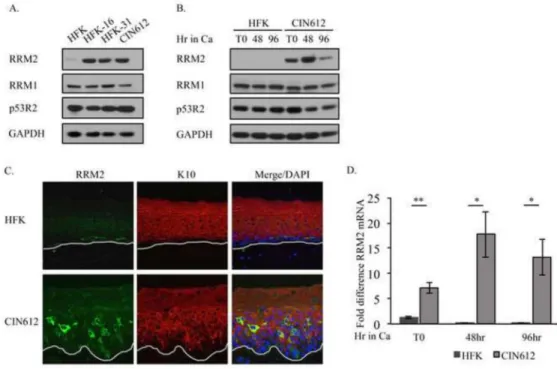
Previous studies demonstrated that RRM2 protein levels are increased in cervical cancer cells containing integrated viral genomes (Wang et al., 2014). To determine if RRM2 levels are also increased in lines harboring episomal copies of HPV, we compared the level of RRM2 protein in uninfected human foreskin keratinocytes (HFKs) to HPV31 positive CIN162 cells, which are derived from a CIN1 cervical lesion. We also examined RRM2 protein levels in HPV positive lines generated in the laboratory through transfection of HFKs with HPV31 (HFK-31) and HPV16 (HFK-16) genomes, followed by selection in neomycin. As shown in Figure 1A, RRM2 protein levels were substantially higher in CIN612 cells, as well as HFK-31 and HFK-16 cells compared to uninfected HFKs. In contrast, levels of RRM1 and p53R2 were similar between the HFKs and HPV positive
A
uthor Man
uscr
ipt
A
uthor Man
uscr
ipt
A
uthor Man
uscr
ipt
A
uthor Man
uscr
lines. To determine if elevated levels of RRM2 were maintained during the productive phase of the viral life cycle, we induced differentiation through growth in high calcium medium, which is commonly used to activate the productive phase of the viral life cycle. As shown in Figure 1B, we found that RRM2 levels remained elevated in CIN612 cells upon
differentiation, while in HFKs, RRM2 was undetectable. Similar results were observed for HFK-31 cells upon differentiation in methylcellulose, which also activates the productive phase of the viral life cycle (data not shown). Interestingly, while RRM1 levels were again maintained at similar between HFKs and CIN612 cells upon differentiation, p53R2 levels consistently decreased in CIN612 cells (Figure 1B). To confirm that RRM2 levels remain elevated upon differentiation in HPV31 positive cells, we performed immunohistochemistry (IHC) on cross sections of organotypic raft cultures derived from CIN612 cells as well as HFKs (Figure 1C). As shown in Figure 1C, high levels of RRM2 are present throughout the stratified epithelium of rafts derived from CIN612 cells, with numerous suprabasal cells exhibiting increased RRM2 staining. In contrast, HFK raft sections exhibited weak staining that was limited to the undifferentiated basal layer. These results indicate that HPV31 specifically increases levels of RRM2 and these levels are maintained in differentiating cells during the productive phase of the viral life cycle.
Since RRM2 levels are regulated in part at the level of transcription, we next wanted to determine if the increased RRM2 protein levels in HPV positive cells correlated with increased mRNA levels. As shown in Figure 1D, similar to the results observed by Western blot analysis, RRM2 transcript levels were significantly increased in CIN612 cells relative to HFKs prior to differentiation, as well as upon differentiation in high calcium medium. In contrast, RRM2 message levels decreased in HFKs upon differentiation (Figure 1D). These results suggest that HPV31 increases RRM2 protein levels, at least in part, at the level of transcription.
dNTP pools are elevated in HPV31-positive cells throughout the viral life cycle
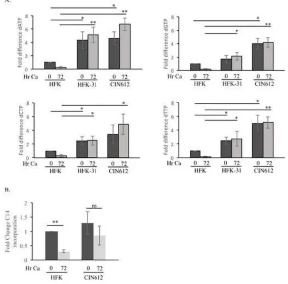
Levels of RRM2 are thought to regulate the activity of the RNR complex, with higher RRM2 correlating with increased reduction of ribonucleotides to deoxyribonucleotides (dNTPs) (Nordlund and Reichard, 2006). To determine if the increased RRM2 levels observed in HPV31 positive cells coincided with elevated nucleotides, we measured dNTP pools in HFKs, CIN612 cells, as well as HFK-31 cells. As shown in Figure 2A, intracellular pools of dATP, dCTP and dTTP were all markedly increased in undifferentiated CIN612 cells, as well as HFK-31 cells compared to the matched uninfected HFKs. dGTP levels were also increased, but to a lesser extent. Furthermore, all four dNTP pools were maintained at significantly elevated levels in HPV31 positive cells upon differentiation in high calcium medium compared to HFKs, where in contrast, individual dNTP pools were reduced by at least 70% (Figure 2A). The decrease in dNTP pools in HFKs mirrored the rapid decline in RRM2 protein levels (Figure 1B). These results suggest that HPV increases dNTP pools through elevated RRM2 levels and RNR activity.
We next wanted to determine if the increased dNTP pools observed in HPV positive cells were accompanied by an elevation in ribonucleotide precursors, which are synthesized through the pentose phosphate pathway (PPP) (Patra and Hay, 2014). For this, we examined
A
uthor Man
uscr
ipt
A
uthor Man
uscr
ipt
A
uthor Man
uscr
ipt
A
uthor Man
uscr
the incorporation of D-[U-14C]-glucose into total RNA as a measure of nucleotide
biosynthesis, as described previously (Yu et al., 2014). Interestingly, as shown in Figure 2B, despite higher dNTP pools, undifferentiated CIN612 cells exhibited a similar level of ribonucleotides to that of HFKs, which would suggest that nucleotide biosynthesis is not induced by HPV. However, due to higher levels of RRM2 in HPV31 positive cells, it is possible that ribonucleotides are reduced to dNTPs at an increased rate, resulting in ribonucleotide pools similar to that found in HFKs. Upon differentiation, ribonucleotides were maintained at an elevated though slightly reduced level in CIN612 cells (1.5-fold decrease), while significantly declining in the HFKs (3.3-fold decrease) (Figure 2B). Given that ribonucleotide levels are maintained in HPV31 positive cells upon differentiation, while decreasing in HFKs, it is possible that HPV maintains activation of the PPP to provide ribonucleotide precursors for the synthesis of dNTPs by the RNR complex.
RRM2 is necessary for HPV replication
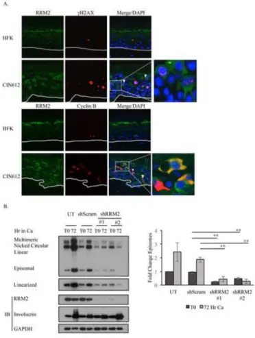
We have found that both RRM2 and dNTP levels are increased in HPV positive cells prior to and during cell differentiation. These results suggest that HPV may elevate RRM2 levels to drive increased RNR activity to provide dNTPs for viral replication. As shown in Figure 1C, staining of HPV31 positive organotypic raft cultures revealed a suprabasal population of cells that exhibited intense staining for RRM2. To determine if this increased staining could represent cells in which productive viral replication is occurring, we performed IHC for two cellular markers previously associated with viral genome amplification; the phosphorylated form of the histone variant H2AX (γH2AX), and cytoplasmic Cyclin B1. γH2AX localizes to HPV replication centers and has been used as a surrogate marker for viral genome amplification (Gillespie et al., 2012; Sakakibara et al., 2013). Previous studies by Wang et al demonstrated that HPV productively amplifies during a prolonged G2 phase, in cells with abundant cytoplasmic Cyclin B1 (Wang et al., 2009). As shown in Figure 3A, raft cultures derived from CIN612 cells exhibited numerous suprabasal cells that co-stained for RRM2 and γH2AX. Several of the RRM2/γH2AX positive cells exhibited γH2AX staining that resembles the large differentiation-dependent HPV31 replication foci detected by
fluorescence in situ hybridization (Moody and Laimins, 2009; Sakakibara et al., 2013). In contrast, rafts derived from HFKs again exhibited very weak staining for RRM2, with few γH2AX positive cells, as previously reported (Moody and Laimins, 2009). For the CIN612 rafts, we also observed numerous suprabasal cells co-staining for RRM2 and cytoplasmic Cyclin B1. In HFK rafts, however, cells staining positive for Cyclin B1 were restricted to the basal layer. Overall, these studies suggest that the increased levels of RRM2 observed upon differentiation may be necessary for HPV to productively replicate.
To determine if RRM2 is required for viral DNA synthesis, we utilized RRM2-specific small hairpin RNAs (shRNA) and examined the effect of transiently knocking down RRM2 levels on viral replication in both undifferentiated and differentiating CIN612 cells (Figure 3). As shown in Figure 3B, transient knockdown of RRM2 in CIN612 cells by two different shRNAs resulted in a significant decrease in HPV genome copy number in undifferentiated cells. In addition, decreased expression of RRM2 significantly affected the ability of viral genomes to amplify upon differentiation. Importantly, expression of the differentiation
A
uthor Man
uscr
ipt
A
uthor Man
uscr
ipt
A
uthor Man
uscr
ipt
A
uthor Man
uscr
specific marker Involucrin verified that RRM2 knockdown did not prevent epithelial differentiation (Figure 3B).
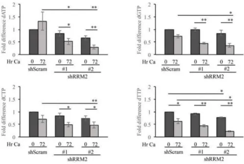
To determine if RRM2 contributes to viral replication through dNTP synthesis, we examined the effect of knocking down RRM2 expression on the maintenance of dNTP pools in CIN612 cells. Interestingly, as shown in Figure 4, transient RRM2 knockdown had only a modest effect on dNTP pools in undifferentiated cells. However, upon differentiation, dNTP levels were significantly reduced upon RRM2 knockdown. Overall, these results suggest that the increased levels of RRM2 observed in differentiating HPV positive cells are required for productive replication by providing an adequate supply of dNTPs for viral DNA synthesis.
RRM2 levels are increased in a manner dependent on E7’s Rb binding domain
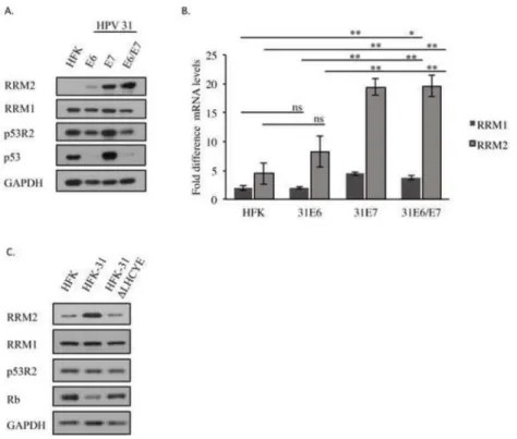
A previous study demonstrated that expression of HPV16 E7 alone is sufficient to increase RRM2 protein levels. These studies also suggested that E7’s ability to target Rb for
degradation may be required for increased RRM2 levels (Wang et al., 2014). To determine if HPV31 E7 is also capable of increasing RRM2 levels, we stably expressed HPV31 E6 or E7 alone, as well as E6 and E7 (E6/E7) in combination in HFKs using a retroviral vector. As shown in Figure 5A, while HPV31 E6 expression resulted in a slight increase in RRM2 protein levels compared to HFKs, expression of E7 resulted in a marked increase in RRM2 levels that was also maintained in cells expressing E6/E7 in combination. In contrast, minimal effect was observed on the levels of RRM1. While E7 expression alone did result in a slight increase in p53R2 (Figure 5A), likely reflecting E7’s ability to increase levels of p53 (Ruesch and Laimins, 1997), the increase was not maintained in cells expressing E6/E7 in combination, correlating with E6’s ability to target p53 for degradation (Scheffner et al., 1990).
To determine if the E7-dependent increase in RRM2 protein occurred at the level of transcription, we measured RRM2, as well as RRM1 mRNA levels. As shown in Figure 5B, levels of RRM2 transcripts were significantly increased (~4-fold) in HFKs expressing E7 alone, as well as E6/E7 in combination compared to control HFKs. Expression of E6 alone resulted in an ~1.5-fold increase in RRM2 transcript levels (Figure 5B), mirroring the minor increase in RRM2 protein levels observed. Although we did not observe a detectable effect of E7 or E6/E7 expression on the protein levels of RRM1, both E7 and E6/E7 expression resulted in an ~2-fold increase in RRM1 transcript levels compared to HFKs (Figure 5B). This reason for this discrepancy is currently unclear. Overall, these results suggest that RRM2 is upregulated by HPV31 E7 and that increased transcription contributes to the elevated RRM2 protein levels observed in HPV31 positive cells.
We next wanted to determine if E7’s ability to bind Rb is necessary for the elevated RRM2 levels we observed in HPV positive cells. To this end, we examined RRM2 protein levels in HFKs maintaining wild-type HPV31 genomes (HFK-31), or genomes containing a deletion in the E7 Rb binding domain (HFK-31 ΔLHCYE), which blocks E7’s ability to target Rb for degradation (Figure 5C). As shown in Figure 5C, loss of the E7 Rb binding domain resulted in a substantial decrease in the protein levels of RRM2 compared to HFK-31 cells. However, modest effects were observed for RRM1 and p53R2 protein levels. These results
A
uthor Man
uscr
ipt
A
uthor Man
uscr
ipt
A
uthor Man
uscr
ipt
A
uthor Man
uscr
demonstrate that E7 expression is required for the elevated levels of RRM2 in HPV31 positive cells and that this increase is dependent on E7’s ability to target Rb for degradation.
E2F1 is required for increased RRM2 expression in HPV31 positive cells
Previous studies demonstrated that RRM2 expression is regulated by the E2F1 transcription factor (Zhang et al., 2009). E7 is well known for its ability to target Rb and related pocket proteins p130 and p107 for degradation, resulting in deregulation of E2F transcription factors and entry into S-phase (Roman and Munger, 2013). As shown in Figure 6A, both HPV31 E7-, as well as E6/E7-expressing HFKs exhibited markedly increased levels of E2F1 protein compared to control HFKs, corresponding with the increased levels of RRM2 in these cells (Figure 5A). E6-expressing cells exhibited a modest increase in E2F1 protein levels (Figure 6A), mirroring the slight increase in RRM2 protein observed (Figure 5A). As shown in Figure 6B, CIN612 cells also exhibited greatly increased E2F1 protein levels compared to HFKS, and the high level of E2F1 was maintained upon differentiation in high calcium medium. Interestingly, we observed that CIN612 cells, but not uninfected HFKs, consistently exhibited a peak in E2F1 that corresponded with an accumulation of RRM2 at early times post-differentiation (24, 48hr) (Figure 1B, Figure 6B). In contrast, this increase was not observed for RRM1 or p53R2.
To determine if the increase in RRM2 in HPV31 positive cells was E2F1-dependent, we examined the effect of E2F1-specific small hairpin RNAs (shRNA) on RRM2 transcript and protein levels in CIN612 cells. As shown in Figure 6C, we observed that E2F1 knockdown resulted in a substantial decrease in RRM2 protein levels, with no detectable affect on RRM1 or p53R2. While the protein levels of RRM2 decreased dramatically in response to E2F1 knockdown, we observed only a two-fold reduction in transcript levels (Figure 6D). However, this difference may be due to the short half-life of the RRM2 protein (~3hr) (Eriksson et al., 1984). While we did not observe an effect on RRM1 protein levels upon E2F1 knockdown, we did observe a slight, but significant decrease in RRM1 mRNA levels (Figure 6D), which is not surprising as RRM1 is reported to be an E2F1 target gene (Pardee et al., 2004). The discrepancy observed between RRM1 protein and RNA levels upon E2F1 knockdown may reflect the long protein half-life of RRM1 protein (~15hr) (Chabes and Thelander, 2000; Engstrom et al., 1985). Our results suggest that E2F1 contributes to productive viral replication, at least in part, through driving expression of RRM2.
The increase in RRM2 expression in HPV31 positive cells is dependent on the ATR/Chk1 pathway
Increasing evidence supports a link between replication stress, activation of the ATR-Chk1-E2F1 pathway and the accumulation of RRM2. Increased RRM2 expression is thought to prevent DNA damage and maintain cell viability in response to replication stress by providing dNTPs for replication (Aird and Zhang, 2015; Buisson et al., 2015; Zhang et al., 2009). The ATR-Chk1 pathway is constitutively active in HPV positive cells (Hong et al., 2015; Moody and Laimins, 2009), suggesting that viral infection induces replication stress. Recent studies demonstrated that E7 alone is sufficient to induce Chk1 activation (Hong et al., 2015). These studies raise the possibility that HPV induces RRM2 accumulation through Chk1’s ability to increase E2F1 levels. As shown in Figure 7A, similar to the results of
A
uthor Man
uscr
ipt
A
uthor Man
uscr
ipt
A
uthor Man
uscr
ipt
A
uthor Man
uscr
Hong et al (Hong et al., 2015), we have found that Chk1 is activated (phosphorylated) to a higher extent in HPV31 positive CIN612 cells compared to HFKs, and that the levels of phosphorylated Chk1 remain elevated upon differentiation. In order to determine if HPV31 increases RRM2 levels through Chk1 activation, we used a chemical inhibitor of Chk1 activity, UCN-01. Previous studies utilizing this inhibitor demonstrated a link between Chk1 activation and RRM2 accumulation in response to replication stress upon S-phase entry (Zhang et al., 2009). In addition, recent studies demonstrated that UNC-01 inhibition of Chk1 activity blocks productive viral replication of HPV31 (Hong et al., 2015), and we have confirmed those results here (Figure 7B).
We first examined the effect of Chk1 inhibition on RRM2 and E2F1 protein levels in undifferentiated CIN612 cells. As shown in Figure 7C, we found that inhibition of Chk1 activity prior to differentiation resulted in decrease in both RRM2 (~2-fold) and E2F1 (~3-fold) protein levels, while having no detectable effect on RRM1 or p53R2. We next examined whether Chk1 activity is required for the accumulation of RRM2 and E2F1 at early times post-differentiation in HPV31 positive cells. For this, we exposed CIN612 cells to high calcium medium in the presence or absence of UNC-01 for 24, 48 and 72hr (Figure 7D). Again, while there was no detectable effect of Chk1 inhibition on protein levels of RRM1 or p53R2, the peak accumulation of RRM2 observed at 24hr post-differentiation was blocked, as was the accumulation of E2F1. Concomitant with the reduction in E2F1 protein at 24hr post-exposure to UNC-01, RRM2 message levels also decreased by ~2-fold at this time point (Figure 7E), suggesting that Chk1 increases RRM2 levels in an E2F1-dependent manner.
To further confirm the importance of the ATR-Chk1 signaling pathway in the upregulation of RRM2 in HPV positive cells, we examined the effect of a chemical inhibitor of ATR (VE-821) on the accumulation of RRM2, as well as E2F1 upon differentiation. As shown in Figure 7F, similar to the results observed upon Chk1 inhibition, we found that the
accumulation of both E2F1 and RRM2 was attenuated upon ATR inhibition. Overall, these results suggest that activation of the ATR/Chk1 pathway in HPV positive cells is required to provide an environment conducive to viral replication by activating E2F1-dependent RRM2 accumulation.
Discussion
RRM2 is considered the rate-limiting component of the RNR enzyme and is required for de novo synthesis of dNTPs, along with RRM1 (Nordlund and Reichard, 2006). Increased RRM2 levels are associated with greater RNR activity and this is reflected in our finding that along with higher RRM2 levels, HPV positive cells also have higher levels of dNTPs compared to uninfected HFKs. Importantly, high RRM2 and dNTP levels are maintained during the productive phase of the viral life cycle and are likely a necessary resource for the rapid amplification of viral genomes in a differentiating environment. In support of this, we have found that that knockdown of RRM2 expression in differentiating cells results in reduced dNTP pools and a block in productive replication. Interestingly, depletion of RRM2 in undifferentiated cells had minimal affect on dNTP levels. One possible explanation is that p53R2, which is present at high levels in undifferentiated HPV31 positive cells, functionally
A
uthor Man
uscr
ipt
A
uthor Man
uscr
ipt
A
uthor Man
uscr
ipt
A
uthor Man
uscr
compensates for the loss of RRM2, forming an active enzyme complex with RRM1 to maintain dNTP pools (Guittet et al., 2001). In contrast, upon differentiation p53R2 levels decrease, which would prevent the formation of a functional RNR complex in the absence of RRM2, resulting in decreased dNTP pools and inhibition of productive replication.
The finding that transient knockdown of RRM2 expression leads to a decrease in HPV31 copy number in undifferentiated cells, despite minimal effect on dNTP pools, was
surprising. However, previous studies with Epstein Barr virus demonstrated that inhibition of RRM2 function upon treatment with hydroxyurea, as well as use of RRM2 siRNAs both led to decrease in viral episomes in infected cells (Chodosh et al., 1998; Zhou et al., 2009). Zhou et al demonstrated that HU treatment, and RRM2 knockdown advanced the replication timing of EBV, in turn altering the chromatin organization of the virus and the stability of EBV episomes (Zhou et al., 2009). Whether RRM2 contributes to HPV replication through regulation of replication timing and effects on viral chromatin will be an interesting area of future investigation.
Under normal conditions in undifferentiated cells, the small size of the HPV genome, coupled with a low viral copy number, likely does not provide much of a drain on cellular resources, allowing the virus to replicate once per cell cycle along with cellular DNA. Upon differentiation, however, productive replication is thought to occur post-cellular DNA synthesis as cells transition from S- to G2-phase (Wang et al., 2009), resulting in
amplification from 50–100 copies per cell to 1000s of copies per cell. It is possible that the preceding synthesis of cellular DNA upon re-entry into the cell cycle may limit cellular substrates, requiring HPV to increase RRM2 levels to provide dNTPs necessary to facilitate amplification of viral genomes. This is supported by our findings that a subset of suprabasal cells in HPV31 positive raft cultures expresses high levels of RRM2 that co-stain with cellular markers of productive replication.
It is currently unclear whether the elevation in dNTP pools observed in HPV positive cells is also accompanied by increased ribonucleotide precursors synthesized through the pentose phosphate pathway, which branches from glycolysis at the first committed step (Patra and Hay, 2014). While we have found that undifferentiated HFK and HPV31 positive cells exhibit similar levels of ribonucleotides, this may simply reflect the increased reduction of ribonucleotides to deoxyribonucleotides in HPV31 positive cells as a result of increased RRM2 levels and RNR activity. In contrast, ribonucleotide levels remain elevated in HPV31 positive cells relative to HFKs upon differentiation, corresponding with maintenance of dNTP pools. Therefore, it is possible that HPV alters nucleotide metabolism at multiple levels to increase dNTP pools. Understanding the effect of HPV on the pentose phosphate pathway throughout the viral life cycle will be an important area of future research.
Our studies indicate that HPV increases levels of RRM2 through activation of the ATR-Chk1-E2F1 DNA damage response (DDR) pathway, with no detectable effect on RRM1 or p53R2 protein levels. These results mirror recent findings by Ricardo-Lax et al, in which hepatitis B virus was shown to increase dNTP pools in quiescent cells through increased RRM2 levels in a Chk1-E2F1-dependent manner (Ricardo-Lax et al., 2015). Activation of the ATR-Chk1 pathway is central to the cell’s response to replication stress, and this
A
uthor Man
uscr
ipt
A
uthor Man
uscr
ipt
A
uthor Man
uscr
ipt
A
uthor Man
uscr
pathway is constitutively active in HPV positive cells (Hong et al., 2015; Moody and Laimins, 2009). Importantly, recent studies demonstrated that Chk1 activity is required for productive replication of HPV31 (Hong et al., 2015), a finding we have confirmed in this study. The constitutive activation of the ATR-Chk1 pathway indicates that replication stress may be a constant occurrence in HPV-infected cells and present throughout the viral life cycle. We have found that ATR and Chk1 activity is required for the increased levels of E2F1 and RRM2 in undifferentiated HPV31 positive cells, as well as the accumulation of E2F1 and RRM2 at early times post-differentiation. The accumulation of E2F1 and RRM2 was found to range from 24–48hr upon growth in high calcium medium. One possibility is that the cell cycle distribution of the cells at the time of exposure to high calcium influences the timing of S-phase re-entry upon differentiation. Our finding that activation of the ATR-Chk1 pathway is required for elevated RRM2 levels in HPV31 positive cells suggests that HPV-induced cell cycle re-entry upon differentiation results in replication stress that elicits an E2F1 transcriptional response through activation of ATR and Chk1, culminating in increased RRM2 levels that provides dNTPs required for productive viral replication (Figure 8). This observation is reminiscent of studies by Buisson et al who demonstrated that S-phase entry induces replication stress due to a large demand on dNTPs at a time when RRM2 levels are still low (Buisson et al., 2015). This in turn activates the ATR-Chk1 pathway, leading to increased levels of RRM2 in an E2F1-dependent manner to provide dNTPs to maintain genomic stability and cell viability (Buisson et al., 2015). Importantly, our study indicates that HPV takes advantage of ATR/Chk1 activation upon S-phase re-entry to maintain a replication-competent environment in differentiating HPV positive cells.
The mechanism by which Chk1 leads to accumulation of E2F1, and in turn RRM2, in HPV positive cells is currently unclear, though several possibilities exist. Chk1 has been shown to phosphorylate E2F6, a negative regulator of E2F-responsive genes, upon replication stress, resulting in its removal from E2F-responsive promoters, allowing E2F1 to bind (Bertoli et al., 2013). Other studies have shown that E2F1 is phosphorylated in response to activation of the ATR-Chk1 pathway, leading to increased protein stability and transactivation potential, resulting in increased RRM2 levels (Buisson et al., 2015; Lin et al., 2001; Zhang et al., 2009). Future studies will focus on understanding the link between ATR-Chk1 signaling and increased E2F1 levels in HPV positive cells.
Increasing evidence supports the idea that oncogenes induce replication stress, especially those that promote uncontrolled S-phase entry (Bartkova et al., 2006; Di Micco et al., 2006; Halazonetis et al., 2008; Mallette et al., 2007). In regards to HPV, the expression of E7, and to a lesser extent E6, induces ATR/Chk1 activation (Hong et al., 2015; Spardy et al., 2008). In addition, studies by Bester et al demonstrated that expression of HPV16 E6/E7 results in perturbed replication, leading to replication stress and DNA damage attributed to E7’s ability to target Rb for degradation and promote cell cycle entry through deregulation of E2F transcription factors (Bester et al., 2011). In support of this, a previous study suggested that the HPV16 E7-dependent increase in RRM2 transcription occurs in an E2F-dependent manner and requires an intact Rb binding domain, however this was not directly shown (Wang et al., 2014). In our study, we have found that HPV31 E7 alone is also sufficient to increase levels of RRM2 and confirmed a role for the Rb binding domain in RRM2 regulation in infected cells. In addition, we have shown that the expression of HPV31 E7
A
uthor Man
uscr
ipt
A
uthor Man
uscr
ipt
A
uthor Man
uscr
ipt
A
uthor Man
uscr
alone is sufficient to increase the levels of E2F1. In unpublished studies, we have found that the E7 Rb binding domain is required for maintenance of ATR activity, as well as high levels of E2F1 (Johnson, B. and Moody, C., unpublished). While ATR/Chk1 activity has been shown to be required for productive replication (Hong et al., 2015), our study now identifies a function for this pathway in facilitating productive replication. Our results suggest that in HPV-infected cells, E7-induced S-phase entry upon differentiation results in replication stress that leads to increased levels of RRM2 in an ATR/Chk1-E2F1-dependent manner, in turn providing an environment conducive to productive viral replication.
While uncoordinated proliferation can lead to insufficient dNTPs that cause replication stress and promote genomic instability, elevated dNTP pools can also be highly detrimental, leading to DNA breaks, mutagenesis and even cell death (Aye et al., 2015). A recent survey identified RRM2 as being among the top 10% of overexpressed genes in 73 of 168 cancer analyses that involved multiple types of cancer, including cervical cancer (Aye et al., 2015). High levels of genomic instability are detected in HPV-associated pre-cancerous lesions (Korzeniewski et al., 2011). Our observation that high-risk HPV positive episomal lines exhibit markedly increased levels of RRM2 suggests that genomic instability could potentially arise in pre-cancerous lesions, at least in part, through increased RNR activity and elevated dNTPs. In addition to de novo dNTP synthesis, RRM2 overexpression is also associated with increased cellular invasiveness, angiogenesis, and proliferation in human cancer cells. Recent studies demonstrated that RRM2 overexpression in cervical cancer cells leads to production of reactive oxygen species (ROS) that enhances angiogenesis through HIF-1 alpha and VEGF production (Wang et al., 2014). The expression of angiogenic factors and increased microvessel density occur very early in the development of HPV-induced pre-cancerous lesions and cervical cancers (Smith-McCune et al., 1997; Smith-McCune and Weidner, 1994). Understanding if RRM2 overexpression is linked to increased mutagenesis, as well as angiogenesis in HPV-induced pre-cancerous lesions will provide important insight into the contribution of RRM2 in promoting carcinogenesis of HPV-associated lesions.
Acknowledgments
Funding Sources
This project was supported by NIH grant 1R01CA181581 and American Cancer Society grant A14-0113 (to C.A.M.), and NIH grants R01 GM104198 and R01 AI049781 (to B.K.).
References
Aird KM, Zhang G, Li H, Tu Z, Bitler BG, Garipov A, Wu H, Wei Z, Wagner SN, Herlyn M, Zhang R. Suppression of nucleotide metabolism underlies the establishment and maintenance of oncogene-induced senescence. Cell Rep. 2013; 3:1252–1265. [PubMed: 23562156]
Aird KM, Zhang R. Nucleotide metabolism, oncogene-induced senescence and cancer. Cancer Lett. 2015; 356:204–210. [PubMed: 24486217]
Anacker D, Moody C. Generation of organotypic raft cultures from primary human keratinocytes. J Vis Exp. 2012
Aye Y, Li M, Long MJ, Weiss RS. Ribonucleotide reductase and cancer: biological mechanisms and targeted therapies. Oncogene. 2015; 34:2011–2021. [PubMed: 24909171]
A
uthor Man
uscr
ipt
A
uthor Man
uscr
ipt
A
uthor Man
uscr
ipt
A
uthor Man
uscr
Banerjee NS, Wang HK, Broker TR, Chow LT. Human papillomavirus (HPV) E7 induces prolonged G2 following S phase reentry in differentiated human keratinocytes. J Biol Chem. 2011;
286:15473–15482. [PubMed: 21321122]
Bartkova J, Rezaei N, Liontos M, Karakaidos P, Kletsas D, Issaeva N, Vassiliou LV, Kolettas E, Niforou K, Zoumpourlis VC, Takaoka M, Nakagawa H, Tort F, Fugger K, Johansson F, Sehested M, Andersen CL, Dyrskjot L, Orntoft T, Lukas J, Kittas C, Helleday T, Halazonetis TD, Bartek J, Gorgoulis VG. Oncogene-induced senescence is part of the tumorigenesis barrier imposed by DNA damage checkpoints. Nature. 2006; 444:633–637. [PubMed: 17136093]
Bertoli C, Klier S, McGowan C, Wittenberg C, de Bruin RA. Chk1 inhibits E2F6 repressor function in response to replication stress to maintain cell-cycle transcription. Curr Biol. 2013; 23:1629–1637. [PubMed: 23954429]
Bester AC, Roniger M, Oren YS, Im MM, Sarni D, Chaoat M, Bensimon A, Zamir G, Shewach DS, Kerem B. Nucleotide deficiency promotes genomic instability in early stages of cancer
development. Cell. 2011; 145:435–446. [PubMed: 21529715]
Bjorklund S, Skog S, Tribukait B, Thelander L. S-phase-specific expression of mammalian
ribonucleotide reductase R1 and R2 subunit mRNAs. Biochemistry. 1990; 29:5452–5458. [PubMed: 1696835]
Buisson R, Boisvert JL, Benes CH, Zou L. Distinct but Concerted Roles of ATR, DNA-PK, and Chk1 in Countering Replication Stress during S Phase. Mol Cell. 2015; 59:1011–1024. [PubMed: 26365377]
Chabes A, Thelander L. Controlled protein degradation regulates ribonucleotide reductase activity in proliferating mammalian cells during the normal cell cycle and in response to DNA damage and replication blocks. J Biol Chem. 2000; 275:17747–17753. [PubMed: 10747958]
Chabes AL, Bjorklund S, Thelander L. S Phase-specific transcription of the mouse ribonucleotide reductase R2 gene requires both a proximal repressive E2F-binding site and an upstream promoter activating region. J Biol Chem. 2004; 279:10796–10807. [PubMed: 14688249]
Chabes AL, Pfleger CM, Kirschner MW, Thelander L. Mouse ribonucleotide reductase R2 protein: a new target for anaphase-promoting complex-Cdh1-mediated proteolysis. Proc Natl Acad Sci U S A. 2003; 100:3925–3929. [PubMed: 12655059]
Chappell WH, Gautam D, Ok ST, Johnson BA, Anacker DC, Moody CA. Homologous Recombination Repair Factors Rad51 and BRCA1 Are Necessary for Productive Replication of Human
Papillomavirus 31. J Virol. 2015; 90:2639–2652. [PubMed: 26699641]
Cheng S, Schmidt-Grimminger DC, Murant T, Broker TR, Chow LT. Differentiation-dependent up-regulation of the human papillomavirus E7 gene reactivates cellular DNA replication in suprabasal differentiated keratinocytes. Genes Dev. 1995; 9:2335–2349. [PubMed: 7557386]
Chien WM, Parker JN, Schmidt-Grimminger DC, Broker TR, Chow LT. Casein kinase II phosphorylation of the human papillomavirus-18 E7 protein is critical for promoting S-phase entry. Cell Growth Differ. 2000; 11:425–435. [PubMed: 10965847]
Chodosh J, Holder VP, Gan YJ, Belgaumi A, Sample J, Sixbey JW. Eradication of latent Epstein-Barr virus by hydroxyurea alters the growth-transformed cell phenotype. J Infect Dis. 1998; 177:1194– 1201. [PubMed: 9593003]
Ciccia A, Elledge SJ. The DNA damage response: making it safe to play with knives. Mol Cell. 2010; 40:179–204. [PubMed: 20965415]
D’Angiolella V, Donato V, Forrester FM, Jeong YT, Pellacani C, Kudo Y, Saraf A, Florens L, Washburn MP, Pagano M. Cyclin F-mediated degradation of ribonucleotide reductase M2 controls genome integrity and DNA repair. Cell. 2012; 149:1023–1034. [PubMed: 22632967]
DeGregori J, Kowalik T, Nevins JR. Cellular targets for activation by the E2F1 transcription factor include DNA synthesis- and G1/S-regulatory genes. Mol Cell Biol. 1995; 15:4215–4224. [PubMed: 7623816]
Di Micco R, Fumagalli M, Cicalese A, Piccinin S, Gasparini P, Luise C, Schurra C, Garre M, Nuciforo PG, Bensimon A, Maestro R, Pelicci PG, d’Adda di Fagagna F. Oncogene-induced senescence is a DNA damage response triggered by DNA hyper-replication. Nature. 2006; 444:638–642.
[PubMed: 17136094]
A
uthor Man
uscr
ipt
A
uthor Man
uscr
ipt
A
uthor Man
uscr
ipt
A
uthor Man
uscr
Diamond TL, Roshal M, Jamburuthugoda VK, Reynolds HM, Merriam AR, Lee KY, Balakrishnan M, Bambara RA, Planelles V, Dewhurst S, Kim B. Macrophage tropism of HIV-1 depends on efficient cellular dNTP utilization by reverse transcriptase. J Biol Chem. 2004; 279:51545–51553.
[PubMed: 15452123]
Edwards TG, Helmus MJ, Koeller K, Bashkin JK, Fisher C. Human papillomavirus episome stability is reduced by aphidicolin and controlled by DNA damage response pathways. J Virol. 2013; 87:3979–3989. [PubMed: 23365423]
Engstrom Y, Eriksson S, Jildevik I, Skog S, Thelander L, Tribukait B. Cell cycle-dependent expression of mammalian ribonucleotide reductase. Differential regulation of the two subunits. J Biol Chem. 1985; 260:9114–9116. [PubMed: 3894352]
Eriksson S, Graslund A, Skog S, Thelander L, Tribukait B. Cell cycle-dependent regulation of mammalian ribonucleotide reductase. The S phase-correlated increase in subunit M2 is regulated by de novo protein synthesis. J Biol Chem. 1984; 259:11695–11700. [PubMed: 6090444] Eriksson S, Martin DW Jr. Ribonucleotide reductase in cultured mouse lymphoma cells. Cell
cycle-dependent variation in the activity of subunit protein M2. J Biol Chem. 1981; 256:9436–9440. [PubMed: 6270086]
Evans MF, Aliesky HA, Cooper K. Optimization of biotinyl-tyramide-based in situ hybridization for sensitive background-free applications on formalin-fixed, paraffin-embedded tissue specimens. BMC Clin Pathol. 2003; 3:2. [PubMed: 12801424]
Fehrmann F, Klumpp DJ, Laimins LA. Human papillomavirus type 31 E5 protein supports cell cycle progression and activates late viral functions upon epithelial differentiation. J Virol. 2003; 77:2819–2831. [PubMed: 12584305]
Gillespie KA, Mehta KP, Laimins LA, Moody CA. Human papillomaviruses recruit cellular DNA repair and homologous recombination factors to viral replication centers. J Virol. 2012; 86:9520– 9526. [PubMed: 22740399]
Guittet O, Hakansson P, Voevodskaya N, Fridd S, Graslund A, Arakawa H, Nakamura Y, Thelander L. Mammalian p53R2 protein forms an active ribonucleotide reductase in vitro with the R1 protein, which is expressed both in resting cells in response to DNA damage and in proliferating cells. J Biol Chem. 2001; 276:40647–40651. [PubMed: 11517226]
Halazonetis TD, Gorgoulis VG, Bartek J. An oncogene-induced DNA damage model for cancer development. Science. 2008; 319:1352–1355. [PubMed: 18323444]
Hebner CM, Wilson R, Rader J, Bidder M, Laimins LA. Human papillomaviruses target the double-stranded RNA protein kinase pathway. J Gen Virol. 2006; 87:3183–3193. [PubMed: 17030851] Hoffmann R, Hirt B, Bechtold V, Beard P, Raj K. Different modes of human papillomavirus DNA
replication during maintenance. J Virol. 2006; 80:4431–4439. [PubMed: 16611903] Hong S, Cheng S, Iovane A, Laimins LA. STAT-5 Regulates Transcription of the Topoisomerase
IIbeta-Binding Protein 1 (TopBP1) Gene To Activate the ATR Pathway and Promote Human Papillomavirus Replication. MBio. 2015; 6:e02006–02015. [PubMed: 26695634]
Hu CM, Chang ZF. Mitotic control of dTTP pool: a necessity or coincidence? J Biomed Sci. 2007; 14:491–497. [PubMed: 17525869]
Hubert WG, Laimins LA. Human papillomavirus type 31 replication modes during the early phases of the viral life cycle depend on transcriptional and posttranscriptional regulation of E1 and E2 expression. J Virol. 2002; 76:2263–2273. [PubMed: 11836404]
Korzeniewski N, Spardy N, Duensing A, Duensing S. Genomic instability and cancer: lessons learned from human papillomaviruses. Cancer Lett. 2011; 305:113–122. [PubMed: 21075512]
Lin WC, Lin FT, Nevins JR. Selective induction of E2F1 in response to DNA damage, mediated by ATM-dependent phosphorylation. Genes Dev. 2001; 15:1833–1844. [PubMed: 11459832] Liu Q, Guntuku S, Cui XS, Matsuoka S, Cortez D, Tamai K, Luo G, Carattini-Rivera S, DeMayo F,
Bradley A, Donehower LA, Elledge SJ. Chk1 is an essential kinase that is regulated by Atr and required for the G(2)/M DNA damage checkpoint. Genes Dev. 2000; 14:1448–1459. [PubMed: 10859164]
Longworth MS, Laimins LA. The Binding of Histone Deacetylases and the Integrity of Zinc Finger-Like Motifs of the E7 Protein Are Essential for the Life Cycle of Human Papillomavirus Type 31. J Virol. 2004a:3533–3541. [PubMed: 15016876]
A
uthor Man
uscr
ipt
A
uthor Man
uscr
ipt
A
uthor Man
uscr
ipt
A
uthor Man
uscr
Longworth MS, Laimins LA. The binding of histone deacetylases and the integrity of zinc finger-like motifs of the E7 protein are essential for the life cycle of human papillomavirus type 31. J Virol. 2004b; 78:3533–3541. [PubMed: 15016876]
Longworth MS, Laimins LA. Pathogenesis of human papillomaviruses in differentiating epithelia. Microbiol Mol Biol Rev. 2004c; 68:362–372. [PubMed: 15187189]
Maglennon GA, McIntosh P, Doorbar J. Persistence of viral DNA in the epithelial basal layer suggests a model for papillomavirus latency following immune regression. Virology. 2011; 414:153–163. [PubMed: 21492895]
Mallette FA, Gaumont-Leclerc MF, Ferbeyre G. The DNA damage signaling pathway is a critical mediator of oncogene-induced senescence. Genes Dev. 2007; 21:43–48. [PubMed: 17210786] Mann GJ, Musgrove EA, Fox RM, Thelander L. Ribonucleotide reductase M1 subunit in cellular
proliferation, quiescence, and differentiation. Cancer Res. 1988; 48:5151–5156. [PubMed: 3044582]
McKinney CC, Hussmann KL, McBride AA. The Role of the DNA Damage Response throughout the Papillomavirus Life Cycle. Viruses. 2015; 7:2450–2469. [PubMed: 26008695]
Mighty KK, Laimins LA. p63 is necessary for the activation of human papillomavirus late viral functions upon epithelial differentiation. J Virol. 2011; 85:8863–8869. [PubMed: 21715473] Moody CA, Fradet-Turcotte A, Archambault J, Laimins LA. Human papillomaviruses activate
caspases upon epithelial differentiation to induce viral genome amplification. Proc Natl Acad Sci U S A. 2007; 104:19541–19546. [PubMed: 18048335]
Moody CA, Laimins LA. Human papillomaviruses activate the ATM DNA damage pathway for viral genome amplification upon differentiation. PLoS Pathog. 2009; 5:e1000605. [PubMed: 19798429] Moody CA, Laimins LA. Human papillomavirus oncoproteins: pathways to transformation. Nat Rev
Cancer. 2010; 10:550–560. [PubMed: 20592731]
Murga M, Campaner S, Lopez-Contreras AJ, Toledo LI, Soria R, Montana MF, D’Artista L, Schleker T, Guerra C, Garcia E, Barbacid M, Hidalgo M, Amati B, Fernandez-Capetillo O. Exploiting oncogene-induced replicative stress for the selective killing of Myc-driven tumors. Nat Struct Mol Biol. 2011; 18:1331–1335. [PubMed: 22120667]
Naruyama H, Shimada M, Niida H, Zineldeen DH, Hashimoto Y, Kohri K, Nakanishi M. Essential role of Chk1 in S phase progression through regulation of RNR2 expression. Biochem Biophys Res Commun. 2008; 374:79–83. [PubMed: 18616928]
Nordlund P, Reichard P. Ribonucleotide reductases. Annu Rev Biochem. 2006; 75:681–706. [PubMed: 16756507]
Pardee AB, Li CJ, Reddy GP. Regulation in S phase by E2F. Cell Cycle. 2004; 3:1091–1094. [PubMed: 15467444]
Patra KC, Hay N. The pentose phosphate pathway and cancer. Trends Biochem Sci. 2014; 39:347–354. [PubMed: 25037503]
Reinson T, Toots M, Kadaja M, Pipitch R, Allik M, Ustav E, Ustav M. Engagement of the ATR-dependent DNA damage response at the human papillomavirus 18 replication centers during the initial amplification. J Virol. 2013; 87:951–964. [PubMed: 23135710]
Ricardo-Lax I, Ramanan V, Michailidis E, Shamia T, Reuven N, Rice CM, Shlomai A, Shaul Y. Hepatitis B virus induces RNR-R2 expression via DNA damage response activation. J Hepatol. 2015; 63:789–796. [PubMed: 26026873]
Roman A, Munger K. The papillomavirus E7 proteins. Virology. 2013; 445:138–168. [PubMed: 23731972]
Ruesch MN, Laimins LA. Initiation of DNA synthesis by human papillomavirus E7 oncoproteins is resistant to p21-mediated inhibition of cyclin E-cdk2 activity. J Virol. 1997; 71:5570–5578. [PubMed: 9188631]
Ruesch MN, Stubenrauch F, Laimins LA. Activation of papillomavirus late gene transcription and genome amplification upon differentiation in semisolid medium is coincident with expression of involucrin and transglutaminase but not keratin-10. J Virol. 1998; 72:5016–5024. [PubMed: 9573271]
A
uthor Man
uscr
ipt
A
uthor Man
uscr
ipt
A
uthor Man
uscr
ipt
A
uthor Man
uscr
Sakakibara N, Chen D, Jang MK, Kang DW, Luecke HF, Wu SY, Chiang CM, McBride AA. Brd4 is displaced from HPV replication factories as they expand and amplify viral DNA. PLoS Pathog. 2013; 9:e1003777. [PubMed: 24278023]
Sakakibara N, Mitra R, McBride AA. The papillomavirus E1 helicase activates a cellular DNA damage response in viral replication foci. J Virol. 2011; 85:8981–8995. [PubMed: 21734054]
Scheffner M, Werness BA, Huibregtse JM, Levine AJ, Howley PM. The E6 oncoprotein encoded by human papillomavirus types 16 and 18 promotes the degradation of p53. Cell. 1990; 63:1129– 1136. [PubMed: 2175676]
Smith-McCune K, Zhu YH, Hanahan D, Arbeit J. Cross-species comparison of angiogenesis during the premalignant stages of squamous carcinogenesis in the human cervix and K14-HPV16 transgenic mice. Cancer Res. 1997; 57:1294–1300. [PubMed: 9102216]
Smith-McCune KK, Weidner N. Demonstration and characterization of the angiogenic properties of cervical dysplasia. Cancer Res. 1994; 54:800–804. [PubMed: 7508337]
Spardy N, Duensing A, Hoskins EE, Wells SI, Duensing S. HPV-16 E7 reveals a link between DNA replication stress, fanconi anemia D2 protein, and alternative lengthening of telomere-associated promyelocytic leukemia bodies. Cancer Res. 2008; 68:9954–9963. [PubMed: 19047177] Tanaka H, Arakawa H, Yamaguchi T, Shiraishi K, Fukuda S, Matsui K, Takei Y, Nakamura Y. A
ribonucleotide reductase gene involved in a p53-dependent cell-cycle checkpoint for DNA damage. Nature. 2000; 404:42–49. [PubMed: 10716435]
Taricani L, Shanahan F, Malinao MC, Beaumont M, Parry D. A functional approach reveals a genetic and physical interaction between ribonucleotide reductase and CHK1 in mammalian cells. PLoS One. 2014; 9:e111714. [PubMed: 25375241]
Toledo LI, Murga M, Zur R, Soria R, Rodriguez A, Martinez S, Oyarzabal J, Pastor J, Bischoff JR, Fernandez-Capetillo O. A cell-based screen identifies ATR inhibitors with synthetic lethal properties for cancer-associated mutations. Nat Struct Mol Biol. 2011; 18:721–727. [PubMed: 21552262]
Wang HK, Duffy AA, Broker TR, Chow LT. Robust production and passaging of infectious HPV in squamous epithelium of primary human keratinocytes. Genes Dev. 2009; 23:181–194. [PubMed: 19131434]
Wang N, Zhan T, Ke T, Huang X, Ke D, Wang Q, Li H. Increased expression of RRM2 by human papillomavirus E7 oncoprotein promotes angiogenesis in cervical cancer. Br J Cancer. 2014; 110:1034–1044. [PubMed: 24423925]
Wilson R, Laimins LA. Differentiation of HPV-containing cells using organotypic “raft” culture or methylcellulose. Methods Mol Med. 2005; 119:157–169. [PubMed: 16350403]
Yu Y, Maguire TG, Alwine JC. ChREBP, a glucose-responsive transcriptional factor, enhances glucose metabolism to support biosynthesis in human cytomegalovirus-infected cells. Proc Natl Acad Sci U S A. 2014; 111:1951–1956. [PubMed: 24449882]
Zeman MK, Cimprich KA. Causes and consequences of replication stress. Nat Cell Biol. 2014; 16:2– 9. [PubMed: 24366029]
Zhang YW, Jones TL, Martin SE, Caplen NJ, Pommier Y. Implication of checkpoint kinase-dependent up-regulation of ribonucleotide reductase R2 in DNA damage response. J Biol Chem. 2009; 284:18085–18095. [PubMed: 19416980]
Zhou J, Snyder AR, Lieberman PM. Epstein-Barr virus episome stability is coupled to a delay in replication timing. J Virol. 2009; 83:2154–2162. [PubMed: 19073720]
zur Hausen H. Papillomaviruses in the causation of human cancers - a brief historical account. Virology. 2009; 384:260–265. [PubMed: 19135222]
A
uthor Man
uscr
ipt
A
uthor Man
uscr
ipt
A
uthor Man
uscr
ipt
A
uthor Man
uscr
Research Highlights
• RRM2 is increased in HPV31 positive cells dependent on the Rb binding domain of E7
• RRM2 is required for maintenance of dNTP pools upon differentiation
• RRM2 is required for productive replication of HPV31
• RRM2 expression is regulated by the ATR/Chk1/E2F1 DNA damage response pathway