ACUTE EFFECTS OF WHOLE-BODY VIBRATION ON DYNAMIC POSTURAL CONTROL IN SUBJECTS WITH FUNCTIONAL ANKLE INSTABILITY
Daniel Lindorff Adelman
A thesis submitted to the faculty of the University of North Carolina at Chapel Hill in partial fulfillment of the requirements for the degree of Master of Arts Athletic Training Specialization in the Department of Exercise & Sport Science in the College of Arts & Sciences.
Chapel Hill 2013
Approved by:
Troy Blackburn PhD, ATC
Kevin Guskiewicz PhD, ATC Shiho Goto MS, ATC
© 2013
Ankle sprains are highly common in recreationally active individuals and can lead to functional ankle instability (FAI), characterized by repeated ankle sprains and functional deficits. Whole-body vibration (WBV) is a novel modality that influences neuromuscular function, and may be effective for rehabilitation of FAI. However, no study has investigated the effects of WBV on neuromuscular deficits associated with FAI. The objective of this study was to evaluate the acute effects of WBV on dynamic postural stability and muscle activity in individuals with FAI. We quantified dynamic postural stability as the time to stabilization (TTS) and measured preparatory and loading phase EMG of the gluteus medius, peroneus longus, rectus femoris, and tibialis anterior muscles of the involved leg. However, there were no significant effects of WBV on these measures. These findings suggest that acute WBV exposure may not be an effective method for rehabilitation for FAI.
Abstract
DANIEL ADELMAN: Acute Effects of Whole Body Vibration on Dynamic Postural Control in Subjects with Functional Ankle Instability
TABLE OF CONTENTS
LIST OF FIGURES ...VII LIST OF TABLES ... VIII LIST OF APPENDICES ... IX
CHAPTER I ...1
RESEARCH QUESTIONS AND HYPOTHESES: ...6
INDEPENDENT VARIABLES ...7
DEPENDENT VARIABLES ...7
ASSUMPTIONS ...7
DELIMITATIONS ...8
LIMITATIONS ...8
OPERATIONAL DEFINITIONS ...9
CHAPTER II ...10
INTRODUCTION ...10
EPIDEMIOLOGY ...10
FUNCTIONAL ANATOMY OF THE ANKLE ...11
ETIOLOGY ...15
DEFINITION ... 17
PREDISPOSING FACTORS ... 18
RESIDUAL SYMPTOMS AND DEFICITS ...21
WHOLE BODY VIBRATION ...28
HISTORY ... 28
PHYSIOLOGICAL EFFECTS ... 28
WBV IN ELDERLY POPULATION ... 29
WBV EFFECTS ON MUSCULAR STRENGTH ... 30
WBV EFFECTS ON BALANCE... 32
WBV EFFECTS ON EMG ... 34
GAPS IN WHOLE BODY VIBRATION LITERATURE ... 36
CURRENT REHABILITATION & PREVENTION TECHNIQUES ...37
SHIFT IN REHABILITATION PARADIGM... 38
REESTABLISHING NEUROMUSCULAR CONTROL ... 38
CURRENT STRATEGIES ... 40
GAPS IN REHABILITATION LITERATURE ... 43
TIME TO STABILIZATION ...43
DEFINITION ... 43
DYNAMIC STABILITY VERSUS STATIC POSTURAL SWAY ... 44
AREAS OF FURTHER INVESTIGATION ...45
CHAPTER III ...46
EXPERIMENTAL DESIGN ...46
SUBJECTS ...46
TIME TO STABILIZATION (TTS) ASSESSMENT ...51
WHOLE BODY VIBRATION ...52
TESTING PROCEDURES ...52
DATA REDUCTION...54
STATISTICAL ANALYSIS ...55
CHAPTER IV...56
RESULTS ...56
FIGURES ...57
TABLES ...67
CHAPTER V ...70
MANUSCRIPT ...70
APPENDICES ...84
LIST OF FIGURES
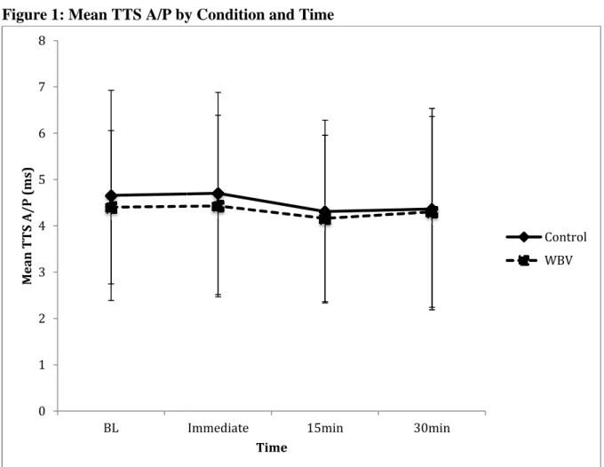
Figure 1: Mean TTS A/P by Condition and Time ... 57
Figure 2: Mean TTS M/L by Condition and Time ... 58
Figure 3: Gluteus Medius Preparatory EMG ... 59
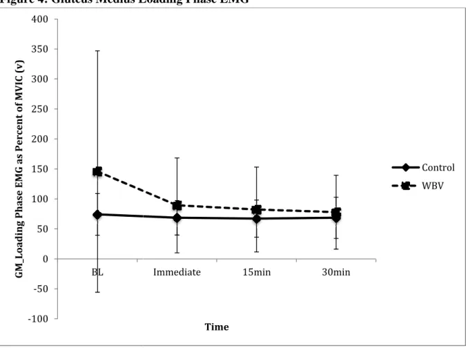
Figure 4: Gluteus Medius Loading Phase EMG ... 60
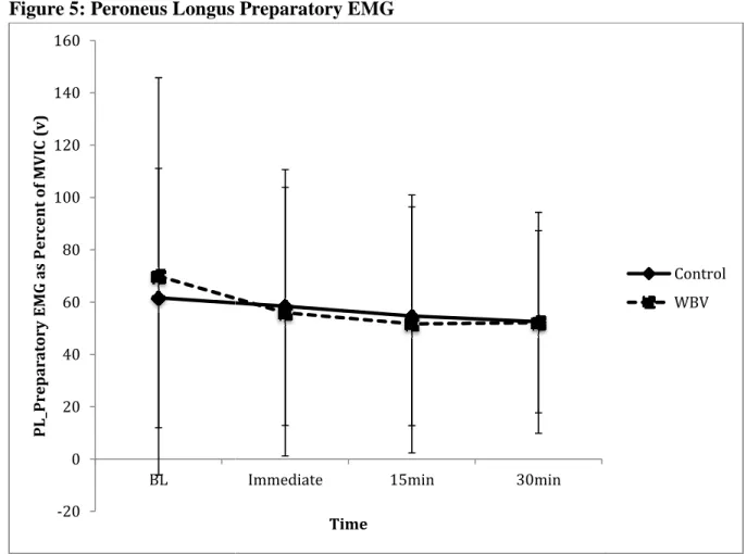
Figure 5: Peroneus Longus Preparatory EMG ... 61
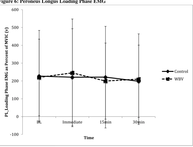
Figure 6: Peroneus Longus Loading Phase EMG ... 62
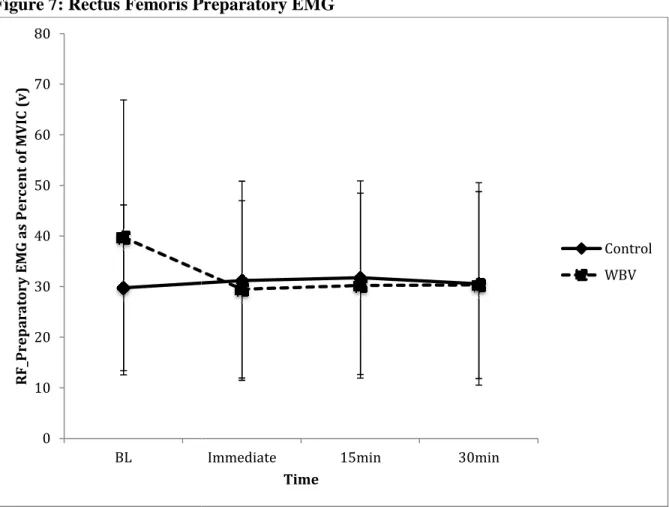
Figure 7: Rectus Femoris Preparatory EMG ... 63
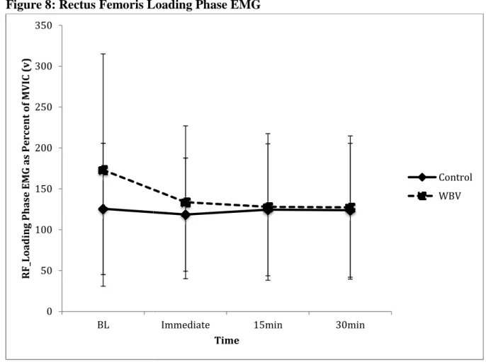
Figure 8: Rectus Femoris Loading Phase EMG ... 64
Figure 9: Tibialis Anterior Preparatory EMG ... 65
LIST OF TABLES
LIST OF APPENDICES
CHAPTER I INTRODUCTION
Ankle injuries are some of the most common injuries in recreational and athletic settings with more than 25,000 ankle sprains occurring daily in the United States (Mickel et al., 2006). Ankle sprains account for up to 44% of all injuries in the physically active population, with40-73% of these injuries being recurrent cases (Arnold, Wright, & Ross, 2011; Dizon & Reyes, 2010; Hughes & Rochester, 2008). Between 32-74% of these cases report chronic symptoms such as pain or weakness, and 32-47% report some level of functional ankle instability (Arnold et al., 2011).
management of the injury. (Garrick, 1977; Mickel et al., 2006; Yeung, Chan, So, & Yuan, 1994).
The neuromuscular deficits associated with FAI can be seen in numerous studies examining the effect of ankle injury on electromyography (EMG) of lower extremity musculature. Beckman and colleagues (1995) showed there were significant decreases in postural control and hip abductor muscle strength when measured following an ankle sprain. It is not clear however, if FAI caused the decreased hip musculature activation of FAI occurred due to this deficit. It was also shown that individuals with FAI displayed increased peroneal reaction time when exposed to a sudden inversion mechanism (Beckman &
Buchanan, 1995; Palmieri-Smith, Hopkins, & Brown, 2009). Injury to the peroneal muscle group could cause functional compensations further up the kinetic chain, changing gluteus medius activation patterns. The neuromuscular deficits associated with both the proximal and distal muscles of the kinetic chain make FAI a truly confounding condition leading to
multiple functional compensations.
Literature is inconsistent on the topic of ankle injury prevention because the forces and speed involved in acute ankle sprains are too great to be prevented from a mechanical or neural perspective (Konradsen, Voigt, & Hojsgaard, 1997). For example, the human body’s reflex response is too delayed, internal static support such as ligaments and muscles are too weak, and external taping is an ineffective means of preventing initial ankle injury (Dizon & Reyes, 2010; Hughes & Rochester, 2008; Refshauge, Kilbreath, & Raymond, 2000;
impairments, or altered arthrokinematics are common after injury. Neural deficits such as decreased proprioception, arthrogenic muscle inhibition (AMI), and neuromuscular control are equally as common following injury and can manifest as decreased postural control (Hertel, 2002). AMI is characterized by activation deficits of specific muscle groups due to damaged mechanoreceptors and reflex inhibition following injury (Palmieri-Smith et al., 2009). Literature suggests that neuromuscular training that restores both the mechanical and neural deficits can reduce the rate of ankle injury after previous ankle sprains (McGuine & Keene, 2006).
Because FAI impairs neuromuscular control, research supports a multifaceted rehabilitation program that incorporates the central, neuromuscular, and mechanical deficits associated with initial ankle injury (Garn & Newton, 1988; Goldie, Evans, & Bach, 1994; Rozzi et al., 1999). Gribble and colleagues (2004) state that static postural control tasks are often not functionally applicable in assessing neuromuscular control. Postural control can be classified as static (stationary) or dynamic with dynamic postural control requiring greater muscle recruitment, proprioceptive feedback, and afferent sensory integration (Gribble, Hertel, Denegar, & Buckley, 2004).
designed to challenge the postural control system and identify unstable landing patterns that could predispose an individual to recurrent ankle injury (Ross, Guskiewicz, & Yu, 2005). Literature has shown that individuals with FAI display a longer TTS than healthy subjects in the anterior/posterior (A/P) and medial/lateral (M/L) direction (Ross et al., 2005). Studies have also been conducted using an intervention period to try and improve TTS in individuals with FAI. Ross and Guskiewicz (2006) performed coordination training with stochastic resonance electrical stimulation intervention over a 6-week period in subjects with FAI, and found improved TTS after only 2 weeks. Therefore, TTS is an appropriate measure in evaluating the effectiveness of current rehabilitation programs in restoring neuromuscular control and dynamic postural stability.
Whole body vibration (WBV) is a novel exercise modality that could be used as an adjunct to dynamic stabilization training in treating FAI. WBV challenges multiple
components of the nervous and musculoskeletal system simultaneously due to the enhanced discharge of type Ia afferent receptors, muscle spindles, and golgi tendon organ (GTO) activity (Rittweger, 2010). WBV has been shown to elicit improvements in muscular
strength, muscular power, flexibility, and balance, which can aid in the rehabilitation of ankle injuries (Moezy, Olyaei, Hadian, Razi, & Faghihzadeh, 2008; Torvinen et al., 2002a).
activity from WBV could reduce AMI and neuromuscular deficits from injury (Cloak, Nevill, Clarke, Day, & Wyon, 2010; Rittweger, 2010). Last, the sensory stimulation effect of WBV on muscle and cutaneous receptors improves proprioception (Bogaerts, Verschueren,
Delecluse, Claessens, & Boonen, 2007). Improving joint stability via enhanced proprioception and muscular power with WBV has implications for improving
neuromuscular control and dynamic postural control (Meinyk et al., 2008; Moezy et al., 2008).
Changes in neuromuscular activation as a result of WBV alters the EMG signal in the lower extremity muscles, but the results of previous studies are unclear due to the varying parameters (Abercromby et al., 2007; Erskine, Smillie, Leiper, Ball, & Cardinale, 2007; Melnyk, Schloz, Schmitt, & Gollhofer, 2009; Rittweger, Mutschelknauss, & Felsenberg, 2003; Santilli et al., 2005; Torvinen et al., 2002a). Acute WBV exposure has been shown to enhance EMG amplitude due to the increased muscle spindle response and motor unit
activation similar to any muscle-loading scenario (Torvinen et al., 2002a). The exposure time to WBV could also have potential lasting neuromuscular effects. Cormie and colleagues (2006) found no significant changes in EMG activity after WBV exposure, but did find significant increase in power during a countermovement jump 30 minutes after WBV. Torvinen and colleagues (2002) also found a significant difference in stability index scores during static standing 2-minutes after a WBV intervention, and a nearly significant difference in stability index scores 60 minutes post WBV intervention.
FAI could be improved (Rozzi et al., 1999). Therefore, further research is warranted in examining the use of WBV as an adjunct to traditional therapy for FAI. The purpose of this study was to determine the acute effects of WBV on dynamic stabilization and lower extremity EMG in individuals with FAI.
RESEARCH QUESTIONS AND HYPOTHESES:
RQ1: Does whole body vibration (WBV) decrease time-to-stabilization (TTS) in
individuals with functional ankle instability (FAI)?
H1: Time-to-stabilization will decrease immediately post WBV, and remain
significantly decreased 30 minutes after intervention.
RQ2: Does WBV increase preparatory EMG of the peroneus longus, tibialis anterior,
rectus femoris, and gluteus medius muscles during TTS in individuals with FAI?
H2: Preparatory EMG relative to ground contact will be greater immediately post
WBV, and remain significantly improved 30 minutes after intervention.
RQ3: Does WBV increase mean EMG amplitude during the loading phase of the
peroneus longus, tibialis anterior, rectus femoris, and gluteus medius muscles during TTS in individuals with FAI?
H3: The mean EMG amplitude during the loading phase of all muscles will be greater
INDEPENDENT VARIABLES 1. Condition
a. Control (No WBV) b. WBV
2. Time
a. Baseline
b. Immediately Post c. 15 minutes Post d. 30 minutes Post DEPENDENT VARIABLES 1. Time-to-Stabilization
2. Electromyographic (EMG) Onset & Mean Amplitude of: a. Peroneus Longus
b. Tibialis Anterior c. Rectus Femoris d. Gluteus Medius ASSUMPTIONS
1. All participants were honest with their level of ankle stability and previous medical and injury history.
DELIMITATIONS
1. Subjects were a relatively homogenous sample (students at University of North Carolina Chapel Hill) of recreationally active individuals with FAI.
LIMITATIONS
1. Time-to-Stabilization measures may not have simulated functional dynamic stability requirements.
2. Only measured acute effects of coordination training with WBV up to 30 minutes, and could not make generalizations about long-term effects.
3. Time allotted for study did not allow longitudinal intervention period, only acute exposure to WBV.
4. Only measured treatment effect on individuals with FAI and cannot claim WBV would have same effects on a healthy population with goals of preventing initial ankle injury.
5. All participants were at different stages in the spectrum of FAI. Although FAI is a chronic condition, some subjects may have been coping with this syndrome for years, as opposed to an individual who only recently had been sustaining repeated ankle sprains and functional deficits.
6. Subjects performed testing barefoot without shoes and socks to ensure internal validity of the study. Athletic shoes are highly variable with differing levels of thickness and materials, which could create further variability between subjects.
OPERATIONAL DEFINITIONS
Functional Ankle Instability (FAI): A multifaceted syndrome of instability comprised of neuromuscular and mechanical instability that results in repetitive ankle sprains and prolonged symptoms of ankle injury including pain, weakness, and recurrent sensations of “giving way” (Arnold et al., 2011; Freeman, 1965; Freeman et al., 1965; Hertel, 2002; Wikstrom, Fournier, & McKeon, 2010).
Recreationally Active: Individuals who participate in 3 or more days of physical activity lasting for greater than 30 minutes a day.
CHAPTER II
REVIEW OF THE LITERATURE
INTRODUCTION
The purpose of this literature review was to provide evidence of the most current and relevant research available on the topic of FAI and WBV. This review was not meant to copy direct work from previous authors, but rather to guide the direction of this study and provide purpose based on the current gaps in research. Through an analysis of the ankle joint
complex and the multifaceted FAI condition, it is clear that a more complete rehabilitation program is necessary to safely and adequately restore normal function. The goal of
improving neuromuscular control by maximizing afferent input can potentially be
accomplished through the adjunct of WBV to current rehabilitation protocols. This literature review illustrates the need for a more successful management strategy of treating FAI, and one that hopefully can be addressed with WBV.
EPIDEMIOLOGY
are estimated at 1 per 10,000 persons per day, with 1 million people seeking medical attention each year (Fallat et al., 1998; Perlman et al., 1987).
The high prevalence of ankle injuries places a burden on time and financial resources. Complete rehabilitation has been reported to take 36-72 days with a cost of $300-$900 per patient, without considering surgical cases (Perlman et al., 1987). It has also been estimated that the evaluation and treatment of ankle injuries may amount to an annual aggregate cost of approximately 2 billion dollars (Fallat et al., 1998). From a time perspective, approximately 6% of people with repeated ankle injuries remain limited in their occupation and up to 15% report being limited for 9 months-6.5 years (Arnold et al., 2011; Schaap, Dekeizer, & Marti, 1989; Verhagen, Dekeizer, & Vandijk, 1995). Therefore, ankle injuries present a significant risk to health and quality of life. However, it is reasonable to expect that these trends will remain consistent or increase as more individuals engage in physical activity (Arnold et al., 2011).
FUNCTIONAL ANATOMY OF THE ANKLE
Ankle injuries, especially to the stabilization ligaments, are the most frequent injuries in physical activity. This is due primarily to the interaction of the bones and ligaments that provide static anatomical support to the ankle joint.
The ankle joint (aka talocrural) is comprised of four main bones including the tibia, fibula, talus, and calcaneus. Although the other tarsal bones are essential in overall
rounded in the distal third. The fibula is a long slender bone on the lateral aspect of the leg and has a minimal weight-bearing role (<10%). The primary purpose of the fibula is muscle attachment. Distally, the tibia and fibula form a syndesmosis joint, which is a fibrous articulation. The distal end of the tibia and fibula form the medial and lateral malleoli respectively. The fibula extends more distally than the tibia, which allows more boney stability on the lateral aspect. The malleoli serve as attachment sites for ligaments of the ankle. The talus is one of the largest tarsal bones and forms the link between the leg and foot. It has a large weight-bearing component and articulates with the calcaneus (inferiorly) and the medial and lateral malleoli. Lastly, the calcaneus forms the heel bone and is the site of many ligamentous attachments of the ankle joint, as well as the Achilles tendon (Prentice, 2009).
The ankle joint is also described as an ankle mortise, which is formed by the tibia (superior and medial), fibula (lateral), and the talus (inferior) called the talocrural joint. There are several degrees of freedom allowed at the ankle complex because of the mortise shape. The rearfoot, composed of the subtalar joint, consists of the articulation of the talus and calcaneus and allows for inversion, eversion, pronation, and supination to occur. The square shape of the talus also contributes to ankle stability because it is wider anteriorly than posteriorly. Therefore, in the dorsiflexed position, the ankle is in a close-packed position because the wider anterior aspect grips the narrow anterior portion of the malleoli.
the plantarflexion/dorsiflexion, inversion/eversion, and adduction/abduction movements allow for smooth congruent movement simultaneously in all three planes of motion.
The stabilizing ligaments of the ankle provide additional static support to the articulations of the bones. Connecting the tibia and fibula are the tibiofibular ligaments, which create a strong interosseus membrane in the syndesmosis joint. The oblique
arrangements of these fibers allows for diffusing of forces placed on the leg. The primary static restraint on the medial side is the triangular shaped deltoid ligament. The deltoid ligament is comprised of four parts including the anterior tibiotalar, tibionavicular,
tibiocalcaneal, and tibial talar part. The deltoid ligament attaches superiorly on the medial malleolus and inferiorly on the medial surface of the talus, the sustentaculum tali of the calcaneus, and the posterior margin of the navicular bone. The deltoid ligament resists eversion of the ankle and also helps support the longitudinal arch along with the
calcaneonavicular (aka spring) ligament. On the lateral aspect there are three main static supports including the anterior talofibular (ATF), posterior talofibular (PTF), and
calcaneofibular (CF) ligaments. The ATF and PTF limit anterior and posterior translation of the talus respectively and the CF limits calcaneal inversion. Due to the orientation of these ligaments, there is more range of motion (ROM) allowed in inversion compared to eversion. The arrangement of the medial and lateral ligament structures allows for dorsiflexion and plantarflexion of the talocrural joint and limits eversion and inversion at the subtalar joint. (Prentice, 2009).
anterior, extensor digitorum longus, and extensor hallicus longus all act to dorsiflex the ankle. The peroneus longus and brevis act to evert the ankle with the longus inserting on the 1st ray of the metatarsals and the peroneus brevis inserting on the base of the 5th metatarsal. The gastrocnemius and soleus muscles combine to form the triceps surae, which inserts on the calcaneus via the achilles tendon and acts to plantarflex the ankle. The tibialis posterior, flexor digitorum longus, and flexor hallicus longus pass along the medial aspect of the ankle and act to invert the ankle (Prentice, 2009). When contracted, the musculotendinous units generate tension, which leads to dynamic protection of joints. When considering dynamic stability, it is more important to consider the muscles eccentric action. The peroneus longus and brevis muscles concentrically create eversion, but eccentrically they control supination and inversion of the rearfoot and protect against lateral ankle sprains (Hertel, 2002).
Although static and dynamic structures act to control movement, only dynamic structures can initiate movement (Hertel, 2002).
Isolated motion rarely occurs within the individual joints of due to the complex approximation of joints and joint angles. The isolated motions include: plantarflexion and dorsiflexion in the sagittal plane, inversion and eversion in the frontal plane, and internal and external rotation in the transverse plane. Functional movement occurs around an oblique axis due to the orientation of the subtalar and talocrural joint. Pronation and supination are the culminating motions that occur as a result of the movement allowed at all three joints. Restricted motion in any of the three joints can lead to obligatory compensatoin other joints. Pronation in the open chain condition consists of dorsiflexion, eversion, and external
plantarflexion, eversion, and external rotation, while supination consists of dorsiflexion, inversion, and internal rotation. Together, these joints also contribute to static stability due to the bony congruency of the articulating surfaces, the static ligamentous support, and the musculotendinous structures that supply dynamic stability (Hertel, 2002).
ETIOLOGY
The unpredictable nature of physical activity leads to an inherent risk for injury. Ankle injuries in particular are increasingly common due to the complex movement that occurs at the joints during dynamic activities. Individuals who participate in a wide variety of tasks that require jumping and landing, cutting, decelerating, and changing directions are particularly vulnerable to these injuries. Basketball, volleyball, and soccer, are examples of sports that require aforementioned movements that can stress the ankle to its structural limitations, and can result in injury.
The mechanism of injury for most lateral ankle sprains may seem simple in nature but the exact causes are often more complex. Injury occurs, when the straining force on the ligaments exceeds the tensile stress of the tissues (Hertel, 2002). Hertel and colleagues (2002) suggests that lateral ankle sprains occur when the rearfoot undergoes excessive supination on an externally rotated tibia. Excessive inversion and internal rotation at the subtalar joint, coupled with external rotation of the tibia results in strain to the lateral ankle ligaments. Excessive plantarflexion can also increase the likelihood of injury creating an open-packed position of the talocrural joint (Hertel, 2002). In a study by McKay and
during basketball occurred during landing (45%) either on another players’ foot or on the court surface. Other mechanisms included sharp twist/turn (30%), collision (10%), and fall (5%) (McKay et al., 2001).
Wilkerson and colleagues discuss how the talus is the key structure when examining ankle pathomechanics (Wilkerson, 2002). The talus is the key linking structure between the leg and the foot, making the subtalar joint the point of integration for the proximal talocrural joint and the distal transverse tarsal and lateral tarsometatarsal joints. In weight bearing, these joints act as a torque converter, distributing ground reaction forces to the proximal structures. Rotation at one segment must be coupled by rotation at another. When the lateral border of the foot inverts, the transverse tarsal and subtalar joint lock in full inversion while the tibia externally rotates (Wilkerson, 2002). The combination of these motions, and the force by which they are produced, puts significant stress on the ATF and CF ligaments in particular, often pushing them to their anatomical limit, resulting in injury.
FUNCTIONAL ANKLE INSTABILITY
DEFINITION
The first definition of FAI was proposed by Freeman and colleagues (1965) and was described as an ankle that displays repeated sensations of “giving way” following initial injury (Freeman et al., 1965). Specific definitions of FAI have changed since 1965 but the basic principle still applies. Konradsen and colleagues (1990) added to Freeman’s definition by including recurrent ankle sprains and a sensation of joint weakness as contributing to the definition. (Konradsen & Ravn, 1990). The common trend throughout the classifications of FAI is some culmination of mechanical instability and articular deafferentation after injury to the lateral structures (Gutierrez et al., 2012). Distinguishing whether the injurious episode is acute or chronic also can lead to varying terminology and definitions. Chronic ankle
instability denotes the occurrence of repeated bouts of lateral ankle instability, resulting in numerous ankle sprains (Hertel, 2002).
PREDISPOSING FACTORS
Previous History of Injury
Fatigue
There are several other risk factors involved in ankle sprains. Gribble and colleagues (2004) discuss how fatigue can disrupt the afferent feedback of the muscle spindle discharge, which alters joint awareness (Gribble et al., 2004). In a group of 30 subjects (16 healthy, 14 chronically instable) Goldie and colleagues (2004) measured the effects of fatigue on a dynamic stabilization task known as the Star Excursion Balance Test (SEBT). Through a series of 5 fatiguing tasks including isokinetic ankle fatigue, isokinetic knee fatigue, isokinetic hip fatigue, lunging tasks, and a control group, subjects performed the star excursion task in the anterior, medial, and posterior direction. Results found a significant difference in maximum reaching distance between the group and side interaction (anterior p=.026, medial p=.022, posterior p=.013) indicating the detrimental effect of muscle fatigue contributing to joint stability (Gribble et al., 2004). Altering joint awareness changes the joint proprioception and kinesthetic properties, which may predispose it to injury.
Extrinsic Factors
Intrinsic Factors
McKay and colleagues (2001) noted that not stretching the posterior muscles of the gastrocnemius and achilles increased injury risk by 2.3 times (p=.03). Tight gastrocnemius muscle causes increased plantarflexion and supination at heel strike, making the joint unstable in the open-packed position (McKay et al., 2001). Other intrinsic factors such as foot type, muscle strength, and muscle reaction time could also be predisposing factors for initial ankle injury. However, research on these topics is controversial due to the different classifications of foot types and parameters to measure strength and reaction time (Beynnon et al., 2002).
Landing Mechanics
post-impact. The alterations are likely a result of increased stress placed on the static ankle structures and could result in repeated injury. Within the first 0-50ms post-impact, peak lateral force occurred approximately 13ms earlier in subjects with FAI. This caused subjects with FAI to bear 5-15% of their body mass with increased laterally directed forces, while control subjects bore the same forces medially. Garrett and colleagues (1999) discussed how reflex response would not be possible at this stage because the monosynaptic reflex latency of the ankle is 35-40ms, meaning the injurious force would occur before the protective reflex activation (Garrett, Kerr, & Caulfield, 1999). Caulfield and colleagues (2004) also discussed how subjects with FAI displayed a greater vertical component of ground reaction force (GRF). The rapid increase in vertical GRF reflects an inability to control the rate of weight distribution and force absorption capacity of the ankle and knee (Caulfield & Garrett, 2004). The tasks described above are directly related to the demands of sport and the importance of dynamic stability in preventing recurrent ankle injury.
RESIDUAL SYMPTOMS AND DEFICITS
occurrence of repetitive bouts of lateral ankle instability, resulting in numerous ankle sprains (Hertel, 2002). FAI is a culmination of these factors that manifests as specific deficiencies in proprioception, neuromuscular control, postural control, and neuromuscular strength.
Balance
Postural sway is defined as the deviation from the mean center of pressure (COP) of the foot (Tropp, Eckstrand, & Gillquist, 1984). Postural sway is objectively measured using a piezoelectric force plate measuring the magnitude and direction of the sway in GRF.
Symmetry is the ability to distribute forces evenly between two feet in an upright stance. Dynamic stability is the ability to transfer center of gravity (COG) around a supporting base (Goldie, Bach, & Evans, 1989; Guskiewicz & Perrin, 1996). Brown and Mynark (2007) examined the effects of ankle instability on dynamic stability measured with TTS. The study found that anterior/posterior TTS values were significantly different between the non-injured (0.71s ± 0.09) and injured groups (0.78s ± 0.12) (p=.04) (C. N. Brown & Mynark, 2007).
Joint Position Sense (JPS)
Following initial ankle injury, there are reductions in JPS and the ability to detect passive ankle motion (Garn & Newton, 1988; Lentell et al., 1995). JPS is defined as the ability to replicate joint movement or position in space (Garn & Newton, 1988; Gross, 1987). Garn and colleagues (1988) found evidence of diminished awareness of passive
plantarflexion following repeated ankle sprains (Garn & Newton, 1988). Even without a previous history of ankle injury, diminished JPS is seen as a predictor of ankle injury due to the inability to comprehend the joint position appropriately in space (Hertel, 2000). Lentell and colleagues (1995) found that subjects with unstable ankles had demonstrated significant deficits in passive inversion. Forty-two subjects with chronic ankle instability were tested for passive movement sense with a moveable-platform box. The study found that passive
could not detect changes in movement compared to the uninjured. A diminished awareness of passive movement may be responsible for the documented delays in muscle reflex activity (Lentell et al., 1995). Conversely, Gross (1987), in a study of 21 subjects, found that there was no significant difference in an individual’s ability to judge ankle position regardless of unilateral ankle injury. However, he did find that passive motion was significantly better at detecting position than active motion among subjects who never complained of ankle pain (Gross, 1987).
Delayed Peroneal Activation
Along with the altered JPS, FAI can cause delayed reaction time of the peroneal muscle group compared to an uninjured subject. The peroneal muscle groups play a crucial role in dynamic stability of the ankle because they are the first to contract in response to a sudden ankle inversion mechanism (Konradsen & Ravn, 1990; Konradsen et al., 1997; Palmieri-Smith et al., 2009). Konradsen and colleagues (1997) examined the peroneal
muscles reaction time to a sudden inversion mechanism and found that the involuntary reflex response was too slow to prevent injury to the static ligamentous structures (Konradsen et al., 1997). Ten volunteer subjects with mechanical ankle instability were tested in different standing and walking situations using an inversion trap-door model. The trap door was set to 30o degrees in the frontal plane to control the magnitude and onset of the inversion
mechanism. Kondradsen and colleagues (1997) found that peroneal EMG activity was
detected at 54ms following inversion (Konradsen et al., 1997). Subjects with previous history of ankle injury have shown significantly slower reaction times in the peroneus longus,
activation in a group of 15 unstable subjects compared to 15 stable subjects in a similar trapdoor study (Konradsen & Ravn, 1990; Konradsen et al., 1997). The increased delay of the peroneal reaction time with FAI, contributes to the continuing neural deficits associated with FAI.
Altered Hip Musculature
Numerous studies have examined how ankle injury has implications for altering hip strength and movement patterns up the kinetic chain (Beckman & Buchanan, 1995; Cathleen N. Brown et al., 2011; Bullocksaxton, 1994; Friel, McLean, Myers, & Caceres, 2006). Brown and colleagues (2011) examined changes in hip kinematics during a stop-jump landing task in 63 subjects with chronic ankle instability. They divided subjects into 3 groups: mechanical instability, FAI, and “copers”, defined as having repeated history of ankle sprains without instability, and found that subjects with distinct instability displayed significantly greater hip flexion at initial contact, maximum hip flexion, hip external rotation, and hip flexion
displacement during the landing task. They cited laxity of the lateral ankle as explanation for altered hip kinematics and altered sagittal plane motion causing increased compensation in the hip (Cathleen N. Brown et al., 2011).
that even though this study could not determine a cause and effect relationship, its findings supported the idea of the reflex chain of events that occur following injury (Bullocksaxton, 1994).
Hertel (2000) states that individuals with a previous ankle sprain will use a hip strategy for balance rather than an ankle strategy. The ankle strategy occurs when muscle contractions first fire at the ankle joint and cause a torque moving the body towards the stable surface. Conversely, the hip strategy occurs when hip flexion or extension is used to move towards the stable surface (Hertel, 2000). Brown and colleagues (2011) discuss how healthy adults can use an ankle strategy of stabilization where the center of gravity can be supported within the ankle joint. After injury however, individuals must utilize a more central hip strategy of stabilization, which explains the increased hip flexion and external rotation motion during the stop jump-landing task. Altering hip mechanics during a landing task has implication for injury else where along the kinetic chain including the knee, hip, and back (Cathleen N. Brown et al., 2011).
Muscular Strength
isometric, open chain force plate testing to measure the evertor strength and found that eversion strength was 88% of the normal side at 3 weeks post injury, and 96% of the
uninjured side 12 weeks post injury (p=.04) (Konradsen, Olesen, & Hansen, 1998). However, Ryan and colleagues (1994) reported significant weakness in inversion (22.7N) of the injured ankle but no significant strength deficits in eversion (26.6N) when measured via Cybex II dynamometer (Ryan, 1994).
The eccentric strength of the peroneals also plays an important role in dynamic stability. Palmieri-Smith and colleagues (2009) discuss how arthrogenic muscle inhibition (AMI) can inhibit muscle strength and function following ankle injury (Palmieri-Smith et al., 2009). The study involved testing 42 subjects (21 unstable, 21 stable) as they walked down a pathway with bilateral trapdoor capabilities to 30o inversion. The unstable group
Although strength deficits in the evertors are under debate, the overall presentation of FAI as a syndrome can manifest as ankle “weakness” (Konradsen & Ravn, 1990).
The contributing factors of FAI create a cycling effect that has led to further use of the term syndrome to describe ankle sprains. These compounding factors make FAI a truly debilitating condition that is difficult to address and manage in the effort to return an individual to normal function.
WHOLE BODY VIBRATION HISTORY
A recent exercise modality that has increased in popularity is whole body vibration (WBV) units. Sanders (1936) and Whedon and colleagues (1949) studied vibration training using an oscillating bed as means to counteract cardiovascular and musculoskeletal
deconditioning. Nazarov and Spirak (1985) were the first to apply vibration training to the athletic setting as an exercise modality in 1985 (Nazarov & Spivak, 1985). Since then, vibration training has emerged as a common rehabilitative and performance enhancement device among many clinicians in the health and exercise setting (Cloak et al., 2010).
PHYSIOLOGICAL EFFECTS
vibration training are still controversial and to the authors’ knowledge, there are only two comprehensive reviews on the topic of vibration training as an exercise modality (Rittweger, 2010; Sitja Rabert et al., 2012). The proposed benefits of whole body vibration training (WBVT) include: enhanced muscle spindle activation due to the alternating changes in muscle length and tension, joint-receptor activity due to the repetitive loading of the joints mimicking rapid landings, central nervous system (CNS) stimulation due to the enhanced mechanoreceptor activity, improved strength and power due to repeated muscle contractions, and improved balance and postural control due to a combination of all other factors
(Abercromby et al., 2007; Cloak et al., 2010; Pollock, Provan, Martin, & Newham, 2011; Rittweger, 2010; Torvinen et al., 2002b). Other physiologic changes include increases in: stimulation of skin receptors, skin and muscle profusion, neurotransmitter and hormone concentration, energy metabolism, intramuscular temperature, and improved bone health (Moezy et al., 2008; Rittweger, 2010; Schuhfried, Mittermaier, Jovanovic, Pieber, & Paternostro-Sluga, 2005). Bogaerts and colleagues (2007) discussed how the strong mechanical stimuli are transmitted to the body and stimulate the primary endings of the muscle spindles, which cause further sensory stimulation of other proprioceptors (Bogaerts et al., 2007). The exact physiologic mechanisms by which these other biologic responses such as changes in cell metabolism and intramuscular temperature will not be covered in this thesis, as it is not the primary goal.
WBV IN ELDERLY POPULATION
and WBV has been demonstrated as an effective adjunct to exercise programs aimed at reducing the predisposing factors associated with falls, improving osteoporosis through increased bone mineral density, and improving walking ability (Bogaerts et al., 2007; Kawanabe et al., 2007; Moezy et al., 2008). It is suggested that WBV applies a stimulus to the body, which is similar to those produced by traditional exercise, but without the
associated risk of falls. The alternating lengthening and shortening of muscles and tendons is thought to mimic the stresses of exercise training (Rittweger, 2010). Cheung and colleagues (2007) found that 75 elderly subjects improved balance after 3 months of WBV treatment. Subjects stood on the WBV platform operating at a frequency of 20Hz 3 minutes a day, 3 days a week. The whole body vibration groups significantly improved movement velocity (deg/s), maximum point excursion, and direction control measured via postural sway on a force plate. Cheung and colleagues (2007) attributed the balance improvements to increased recruitment of muscle fibers, muscular adaptation, and neuromuscular coordination with WBV (Cheung et al., 2007).
Bruyere and colleagues (2005) also used WBV and physical therapy to improve muscle function and potentially reduce the risk of falling. This study had a control and WBV group perform therapy including gait and balance training, weight transfer skills, and
strengthening exercises 3-times per week, only to find that the group with the adjunct of WBV significantly improved their muscular strength and balance (Bruyere et al., 2005).
WBV EFFECTS ON MUSCULAR STRENGTH
tonic vibration reflex (TVR), which is provoked by length changes in the muscles that
stimulate the muscle spindles primarily through type Ia afferents (Cormie, Deane, Triplett, & McBride, 2006; Mahieu et al., 2006). TVR is composed of motor unit activity that is
synchronized and unsynchronized within the vibration cycle (Martin & Park, 1997). It has also been proposed that the strength improvements are due to a lowering of the motor recruitment threshold during WBV, resulting in a more rapid activation of high-threshold motor units (e.g. fast twitch fibers), resulting in a greater summation of motor units (Delecluse, Roelants, & Verschueren, 2003; Mahieu et al., 2006; Rittweger, Beller, & Felsenberg, 2000).
Torvinen and colleagues (2002) found that there was a significant increase in vertical jump height after two months of WBV, and an increase in isometric lower extremity strength in a group of 23 young, nonathletic subjects (Torvinen et al., 2002b). Although scarce, the literature on WBV in sports medicine supports the use of WBV as a means of improving muscular strength and power and tissue extensibility (Issurin, Liebermann, & Tenenbaum, 1994; Mahieu et al., 2006; Rittweger, 2010). Mahieu and colleagues (2006) examined the improvements in muscular strength in a group of 33 competitive skiers. They found increases in high box jump test with the adjunct of WBV over a 6-week training period (p<.001, effect size 0.72). Because both groups displayed increased repetitions on the high box test, a comparison between groups revealed the WBV group increased scores by 13.53 ± 9.79cm, while the resistance training group increased high box test scores by 5.44 ± 7.66cm (p=.013, effect size .92) (Mahieu et al., 2006).
to increase norepinephrine levels and increase power output in the arm flexion movement (Bosco et al., 2000; Cormie et al., 2006). In a study of 9 men, Cormie and colleagues (2006) found acute increases in vertical jump height of the WBV group versus and sham treatment group immediately performing a non-fatiguing intervention. Jump height increased from 49.02 ± 7.58cm at baseline to 49.34 ± 7.17cm immediately post WBV intervention, whereas the sham group decreased jump height scores from 50.67 ± 7.13cm to 49.34 ± 6.9cm
immediately post (p<.05 between groups). They concluded that WBV would be a valuable adjunct to a warm-up because of the enhanced jump height associated with treatment (Cormie et al., 2006). Rittweger and colleagues (2000) conversely found that vertical jump height was lessened after acute exposure to WBV in a group of 37 subjects. However, the intervention that was used in this study consisted of a fatiguing task before measuring vertical jump, which would account for the diminished jump height (Rittweger et al., 2000).
WBV EFFECTS ON BALANCE
repetitive nature of the stimulus might cause change in the balance control strategies, causing overall improvements in postural stability. The study found significant differences between WBV and control groups overall stability indices, anterior/posterior stability indices, and medial/lateral stability indices. These changes may be due to the beneficial effects of WBV on muscle strength, improved synchronization of motor unit firing (indicating more efficient signal activation), and improved co-contraction of synergist muscles, which would improve postural control (Moezy et al., 2008). Cloake and colleagues (2010) also reported balance improvements in the anterior, medial, and anteromedial direction after a 6-week training intervention in a group of professional dancers with FAI during a SL stance. They
implemented a training protocol, which consisted of single leg (SL) balance task and the star excursion balance test (SEBT) twice a week, over a 6-week period. This study was in
contrast with other current research that measured balance for periods greater than 8-weeks with more than three training sessions per week. However, the author notes that the study sample had outstanding balancing ability prior to injury, which could also contribute to higher scores (Cloak et al., 2010).
There is tremendous variability in the literature in quantifying and measuring balance as well as improvements in balance. It is also difficult to make clinical applications of
colleagues (2011) in a study of 18 subjects, stated the lack of change in balance could be due to the relatively simple tasks performed with the adjunct of WBV, and that the effects of more challenging tasks is unknown (Pollock et al., 2011). Their subjects performed 5x1 minute bouts of WBV prior to measuring JPS, cutaneous sensation, and balance. Clinically, balance has a multifaceted meaning that should be considered when interpreting results.
WBV EFFECTS ON EMG
The most likely mechanism by which WBV elicits motor output changes is through the tonic-vibration reflex (TVR). TVR is provoked by the stimulation of type Ia afferent fibers and is composed of both synchronized and unsynchronized motor unit activity with the vibration cycle (Martin & Park, 1997). It has also been suggested that WBV elicits a type Ia-afferent-mediated myotatic (stretch) reflex, eliciting muscle contraction (Delecluse et al., 2003; Rittweger et al., 2000). These changes in motor output are detected through EMG recordings of specific muscles involved in the specific task. WBV causes increased stimulation of the muscle spindles and subsequent increased EMG activity.
Muscle tuning is defined as dampening that occurs in soft tissue as a response to direct mechanical stimulation (Wakeling, Nigg, & Rozitis, 2002). Similar to shock distribution during heel strike in walking and running, muscle tuning increases dampening coefficient so that surrounding soft tissue can reduce tissue resonance (Wakeling et al., 2002).
Prolonged exposure to WBV can elicit EMG measurements that would appear as though the muscle is fatigued (Rittweger et al., 2000). Rittweger and colleagues (2000), in a study of 37 subjects, found significant rapid and slow-onset fatigue with the exhaustive WBV exercises. Subjects underwent initial fatiguing test on the bicycle ergometer, followed by two subsequent fatiguing tests with WBV including squatting and vertical jumping. After the first WBV fatiguing tasks, researchers found a significant difference in the following variables compared the bicycle ergometer (BE): increased perceived exertion, decreased heart rate (171±16 BE, 122±28 WBV), specific oxygen uptake (44.8±7.9 BE, 21.3±4.0 WBV), lactate production (7.7±2.7 BE, 3.5±1.6 WBV), respiratory quotient (0.98±.05 BE, .90±.08 WBV), systolic blood pressure (148±18 BE, 132±16 WBV), and diastolic blood pressure (65±15 BE, 52±14 WBV). They stated that the WBV fatigue was not associated with cardiac
insufficiency but rather the neuromuscular system (Rittweger et al., 2000). Fatiguing of the TVR decreases voluntary force until 10-20 seconds after the end of vibration, affecting the ability to fire high threshold motor units. There is also a proposed fatiguing of the Ia afferent fibers themselves (Rittweger et al., 2000).
EMG recordings with WBV studies are taken with surface electrodes placed over the muscle belly of the desired muscle, with the ground electrode on a bony landmark.
GAPS IN WHOLE BODY VIBRATION LITERATURE
Because WBV is a relatively new modality that is not well supported by evidence in the sports medicine community, clinicians are hesitant to use these devices as adjuncts to their rehabilitation practices. However, emerging popularity of these devices cannot be ignored, and there are several gaps in the literature that should be investigated as the prevalence of these machines increases in the realm of exercise and sport science.
Perhaps the most apparent gap in WBV research is the population used in
randomized-controlled trials. There is a large amount of research conducted with an elderly population looking at the benefits of WBV on fall prevention and walking ability. More current research has moved to looking at the benefits of WBV on a healthy, younger
population, and even moving into an athletic population (Cormie et al., 2006; Mahieu et al., 2006; Torvinen et al., 2002b; Torvinen et al., 2003). The proposed improvements in muscular strength are useful for the athletic community, but research is lacking and inconclusive on precise benefits.
There is minimal research on the use of WBV in an injured athletic population. In a study of professional dancers with FAI, Cloake and colleagues (2010) stated the greatest difficulty was developing a training protocol for subjects due to the lack of objective
evidence in support of the use of WBV in FAI patients (Cloak et al., 2010). Therefore, there is a gap in the literature in applying these benefits to the injured athletic population.
Another gap in WBV research involves the parameters of treatment. In particular, most studies investigated the effects of WBV after a long-term intervention period (Cloak et al., 2010; Mahieu et al., 2006; Melnyk et al., 2009; Torvinen et al., 2002b; Torvinen et al., 2003). Few studies investigated the acute effects of WBV on muscle strength, muscle power, and postural stability. Rittweger and colleagues (2003) examined the acute neuromuscular effects of WBV after a period of exhaustive exercise (Rittweger et al., 2003). The associated fatigue effects of exhaustive exercise make it difficult to make claims about WBV as adjunct to therapy. Cormie and colleagues (2006) found acute increases in vertical jump height but could not make any correlation between changes in jump height and neuromuscular activity. While this study made claims about the acute effects of WBV, the intervention only
consisted of 30-second exposure to WBV (Cormie et al., 2006). To my knowledge, there is currently no study that has investigated the acute changes in neuromuscular control following a bout of rehabilitation exercise with WBV, particularly in regards to changes in EMG
activity.
CURRENT REHABILITATION & PREVENTION TECHNIQUES
SHIFT IN REHABILITATION PARADIGM
A rehabilitation program that focuses on the central and neuromuscular systems by emphasizing balance, coordination, and strength training has been recommended for improving ankle stability and postural stability in stable and unstable ankles (Bernier & Perrin, 1998; Eils & Rosenbaum, 2001; Freeman et al., 1965; Goldie et al., 1994; Michell, Ross, Blackburn, Hirth, & Guskiewicz, 2006; Ross & Guskiewicz, 2004; Rozzi et al., 1999; Tropp & Odenrick, 1988). The goal of rehabilitation is to include functional exercises that are directly transferable to the demands of activity and have the potential to reduce injury rate. Introducing a new adjunct to traditional shows how rehabilitation paradigms have shifted to introduce new movements and muscle activation patterns with the goal of improving postural stability.
REESTABLISHING NEUROMUSCULAR CONTROL
sustained an acute ankle sprain during their season. Researchers then compared the effect of each variable on the rate of ankle sprain and found that previous history of an ankle sprain was the leading risk factor, participation in the balance training program significantly reduced injury rate, indicating balance intervention programs are effective at reducing ankle sprains (McGuine & Keene, 2006).
Evaluating balance or postural stability is one method of assessing sensory deficits following injury (Mattacola & Dwyer, 2002). Balance is comprised of sensory stimulus from the visual, vestibular, and somatosensory systems. Rehabilitation exercises can be modified to challenge or isolate these specific systems. Balance is also a crucial component of all closed-kinetic chain activities because it incorporates sensory stimulus from the peripheries as well as visually and spatially (Hertel & Denegar, 1998). Decreased stability in the single leg standing task suggests altered proprioceptive response with a decreased efficacy of
producing protective reactive mechanisms. Because balance uses the same peripheral afferent mechanisms as joint proprioception, it is a good indicator of lower extremity dysfunction.
In a 4-week balance training program with 26 subjects (13 with FAI), Rozzi and colleagues (1999) implemented a series of balance training tasks to treat proprioceptive deficits and restore joint stability. Subjects underwent single leg standing and single leg standing on a Biodex Stability Surface and the results showed there was a significant
difference in pre-training (stability index score of 5.93 ± 3.65) versus post-training (Stability index score of 2.63 ± 1.92) in stability measures (p<.05), with lower scores representing greater stability (Rozzi et al., 1999).
multifaceted (Hertel & Denegar, 1998). Pain, swelling, and damage to joint
mechanoreceptors can impede reflex control as well as volitional muscle contractions. Therefore, the rehabilitation should address restoring volitional control, normal reflex patterns, and pattern-generated movements to improve neuromuscular control and restore complex pattern-generated movements that will mimic those required in sport (Hertel & Denegar, 1998).
Another method of assessing neuromuscular control is by measuring EMG for muscle receptor activity and reaction time (Hertel & Denegar, 1998). EMG can be used in the
rehabilitation setting as biofeedback, but it is also valuable in measuring latencies and neural deficits associated with injury. EMG measures allow the clinician to track whether the appropriate exercises are being used to optimize motor unit stimulation. By recording EMG activity of the hip musculature as well, the clinician can evaluate whether there are faulty movement patterns associated with proximal control as a contributing factor to injury (Beckman & Buchanan, 1995). While EMG does not directly represent neuromuscular control, it is a valuable measurement tool during rehabilitation to ensure that all exercises are having a meaningful effect on motor unit activation and amplitude.
CURRENT STRATEGIES
Mattacola and colleagues (2002) discussed current rehabilitation strategies for ankle injuries ranging from gentle ROM exercises to challenging balance tasks with provoked perturbations (Mattacola & Dwyer, 2002). Many of the exercises described can fit into multiple goals of rehabilitation, further emphasizing the multifaceted nature of
Restoration of full muscle strength is essential if normal function is to be regained. Strength training can be achieved with a wide variety of equipment including sport cord, theraband, free weights, weight machines, and isokinetic devices. In general non-weight bearing should progress to full weight bearing and then additional resistance should be followed. Double leg exercises are progressed to single leg exercises. Isometric contractions are progressed to concentric and eventually eccentric contractions. Hudson (2009) states that the local muscle groups for improvement following ankle injury are the gastrocnemius/soleus complex and the evertors (Hudson, 2009). Docherty and colleagues (1998) implemented a 6-week strength-training program with elastic tubing. The results showed that the 20 subjects with FAI improved the mean strength (N) of dorsiflexors from 33.3 ± 4.8 pretest to 50.6 ± 6.3 posttest (p<.0005), and evertors from 30.9 ± 6.5 pretest to 45.0 ± 4.9 posttest (p=.005). They also found improvements in JPS (degrees of error) in inversion from 6.8 ± 5.0 pretest to 2.8 ± 2.8 posttest (p=.009), and plantarflexion from 7.9 ± 6.0 to 1.4 ± 0.9 posttest (p=.027). Researchers stated the improvements in JPS were due to stimulation of afferent muscle spindles and reestablishing the neural connections from the static and dynamic gamma-efferent nerves (Docherty, Moore, & Arnold, 1998).
Proprioception improvements through afferent sensory organization have also been shown to rapidly improve dynamic stability, reduce the incidence of ankle sprain, and reduce injury recurrence (Hudson, 2009; Rasool & George, 2007; Rozzi et al., 1999). Eils and Rosenbaum (2001) studied 30 subjects with CAI and put them through a series of 12
different directions, single leg stance with resisted hip abduction of contralateral leg, double leg and single leg stance on inversion/eversion board, single leg stance on mini trampoline, single leg forefoot balance, uneven walking, balance on horizontal and vertically mobile platform, and maintain balance on computer controlled platform. Subjects held the balance positions for 45 seconds and had 30 seconds rest between exercises. The study found a significant difference in pretest and posttest groups in total sway, although individually, maximum sway in the medial-lateral was significant, and anterior-posterior sway was non-significant. Eils and Rosenbaum stated this was because medial-lateral sway occurs at the subtalar joint while anterior-posterior sway occurs at the tibiotalar joint (Eils & Rosenbaum, 2001).
Rehabilitation protocols that restore postural stability have also been effective at reducing ankle sprain incidence (McGuine & Keene, 2006). In a study of 30 healthy athletes, Rasool and George (2007) tested the effects of a 4-week intervention of the Star Excursion Balance Test (SEBT) on improving dynamic stability. The SEBT is a functional test that incorporates single leg standing on one leg and maximal reach with the other in the anterior, lateral, and posterior directions. They found overall improvements in reach distance by 11-36% after a 4-week intervention. The posterior reach direction improved from 97 ± 6cm at baseline to 112 ± 9cm and 121 ± 7cm at 2 and 4-weeks respectively (p<.001) (Rasool & George, 2007). Not only did this study show improvements in dynamic postural stability, these improvements were seen after only 4 weeks of intervention. Neuromuscular training and its effects on dynamic postural stability in an injured population have not been
GAPS IN REHABILITATION LITERATURE
Every rehabilitation program must be individualized to the patient. This concept in itself leads to difficulty making universal accusations about specific rehabilitation techniques (Mattacola & Dwyer, 2002). While current rehabilitation paradigms and protocols may progressively involve more complex tasks, the general principle is to maximize
neuromuscular improvements. The results of individual programs are so varied leading to uncertainty in the best treatment guidelines. Every perturbation, jump landing, or unstable surface is meant to challenge the neuromuscular system and strengthen the proprioceptive deficits that occurred as a result of injury. Depending on the resources available to them, a clinician may have a multitude of tools and instruments that could challenge the neural system. Therefore, the rehabilitation parameters of current studies vary significantly and rarely measure acute effects of treatment. Although restoring functional ability and return to play should be the ultimate goal of all rehabilitation, understanding the acute effects of treatment may be valuable to guide further appropriate treatments. There are currently no studies investigating the outcome of dynamic stability in an acute response to enhanced neuromuscular training. The purpose of this study is to further investigate specific protocols for restoring neuromuscular control and integrate current exercise modalities in the process.
TIME TO STABILIZATION DEFINITION
variation at the beginning of a SL jump landing resembles the GRF range of variation of a standardized SL stance (Ross et al., 2005). Anterior-posterior (A/P) and medial-lateral (M/L) TTS have been used to evaluate the dynamic stability of subjects with FAI after a SL jump landing (Ross & Guskiewicz, 2003, 2004; Ross et al., 2005). In calculating TTS the first step is to define the range of variation of a given GRF component. These values are defined as the smallest absolute range value of GRF component during the last 10 seconds of the single leg stance portion of a jump landing task (Ross & Guskiewicz, 2003).
The jump-landing task provides means to control for jump height and distance of each individual. Individuals are measured for maximum vertical-jump height at a distance of 70cm away from the device. Then the individual performs a double leg jump at 50% of the maximum jump height on to the center of a force plate. A measuring device is placed at 50% jump height. The individual is instructed to stick the landing on one leg and stabilize as quickly as possible and remain as motionless as possible for the duration of the test. Data collection begins at initial ground contact and lasts for 20seconds post. The jump landing data are collected on a force plate (Ross & Guskiewicz, 2003).
DYNAMIC STABILITY VERSUS STATIC POSTURAL SWAY
stability during a dynamic task. By definition, dynamic stabilization is an attempt to maintain a stable base of support while completing a prescribed movement (Gribble et al., 2004; Winter, Patla, & Frank, 1990). Essentially maintaining the center of gravity within the stable base of support during prescribed movement. The difficulty arises when the individual must stabilize responding an unanticipated movement such as reacting to a defender or object.
Compared to SL postural sway measures, TTS measures challenges the postural control system and allow clinicians to identify unstable landing patterns that could contribute to injury and be addressed through therapy (Ross et al., 2005). While landing strategies may be different for subjects with FAI, it is difficult to compare these strategies in laboratory and clinical setting. Correcting faulty landing strategies, that are identified with TTS, could have implications for improving ankle stabilization and preventing excessive stress at the ankle (Ross et al., 2005).
AREAS OF FURTHER INVESTIGATION
The literature on FAI and WBV is abundant but to date, only one study has investigated the effects of WBV on subjects with FAI. There limited research on FAI and advanced rehabilitation techniques although evidence suggests that challenging
CHAPTER III METHODOLOGY
EXPERIMENTAL DESIGN
This study utilized a randomized crossover design to evaluate the acute effects of WBV on dynamic postural control and EMG of the lower extremity musculature in subjects with FAI. Subjects completed two counterbalanced testing sessions separated by one week during which TTS and EMG of the gluteus medius (GM), rectus femoris (RF), peroneus longus (PL), and tibialis anterior (TA) were assessed prior to and immediately, 15 minutes, and 30 minutes following an intervention (Control of WBV). All testing procedures were conducted barefoot, and the order of testing sessions (Control of WBV) was counterbalanced.
SUBJECTS
A priori power analysis was performed based on an unpublished study by Ross et al.
Twenty-six individuals with confirmed FAI (12 females, 14 males; age = 20.4 ± 1.33, height = 172.48 ± 8.94cm, mass = 72.75 ± 11.37kg) were recruited to participate in the study. Two subjects failed to report for the 2nd (control) testing session making our total sample size for the control condition n = 24. Subjects ranged from 18 to 30 years of age and were
recreationally active as defined by participation in physical activity for at least 30 minutes, three times per week. Subjects included their primary mode of physical activity in the prescreening questionnaire to identify any individuals who participate in current balance oriented activity (i.e. yoga, ballet,). This was not a primary variable in the
inclusion/exclusion criteria but acted as a supplemental variable. Subjects were required to sign an informed consent form approved by the University of North Carolina Institutional Review Board.
All subjects displayed unilateral or bilateral FAI, defined as meeting each of the following self-reported criteria:
• Suffered at least two ankle sprains in the last 12 months that resulted in one or more of the following symptoms: swelling, ecchymosis, and decreased range of motion (Arnold et al., 2011; Freeman et al., 1965; Hertel, 2000, 2002).
• Experienced at least two sensations of “giving way” or feelings of instability in the last 12 months in their involved ankle.
dysfunction. Subjects were also excluded if they have contraindications for WBV including, cardiovascular, metabolic, or neuromuscular diseases, epilepsy, osteoporosis, osteoarthritis, menstrual irregularities, or pregnancy (Mahieu et al., 2006; Pollock et al., 2011; Torvinen et al., 2002b).
Subjects were required to answer the Cumberland Ankle Instability Tool (CAIT) as part of the online prescreening. The CAIT is a 9-item, 30-point tool for measuring the severity of ankle instability. Hiller and colleagues (2006) found the CAIT to be both a reliable and valid tool for assessing ankle instability (ICC=0.96). A score of 28 or higher indicates the individual is unlikely to have ankle instability while a score of 27 or lower indicates they likely have FAI. The CAIT not only quantifies the level of ankle instability but has also been cited as having the potential to predict future sprains in those with FAI (Hiller, Refshauge, Bundy, Herbert, & Kilbreath, 2006). Appendix 1 provides an example of the CAIT questionnaire.
(Hale & Hertel, 2005). Appendix 2 & 3 provide a representation of the FADI and FADI Sport questionnaire respectively.
EMG
A surface electromyography (EMG) system (Delsys Inc., Boston, MA; amplification factor=1000 (20-450Hz); CMRR at 60Hz > 80dB; input impedance > 1015//.02 Ω//pF) was used to capture activity of the peroneus longus, tibialis anterior, rectus femoris, and gluteus medius muscles during the TTS and MVIC measures with surface EMG electrodes (Delsys Inc., Boston, MA). Sampling frequency was 1000Hz.
The tibialis anterior and peroneus longus muscles were included in EMG analysis because previous literature has cited deficits in the lower extremity motor unit activity in subjects with FAI. Konradson and Ravn (1990) found significant EMG activity deficits and increased peroneus longus activation time following ankle injury. The peroneus longus and tibialis anterior are also the primary dynamic restraints to a lateral ankle sprain with the primary mechanism of plantarflexion and inversion (Melnyk et al., 2009). The rectus femoris and gluteus medius were included in this study because numerous studies have examined the effects of delayed muscular activation as either a cause or effect of ankle injury and
All subjects were fitted with surface EMG electrodes for the peroneus longus, tibialis anterior, rectus femoris, and gluteus medius of the affected side. Prior to electrode placement, the area of the electrodes were marked with a felt tip marker, shaved with an electric razor, lightly abraded, and cleaned with isopropyl alcohol. A reference electrode was positioned on the tibial tuberosity.
Electrode placement for the peroneus longus muscle belly was determined with the subject seated in 30 degrees knee flexion and the lower extremity externally rotated. First, the distance between the fibular head and lateral malleolus was recorded with a tape measure. The peroneus longus muscle belly was palpated during resisted isometric ankle eversion with resistance applied to the lateral side of the foot. The electrode was placed distal to the fibular head, 25% of the distance between the fibular head and the lateral malleolus over the greatest muscle bulk (Hermens HJ et al., 2000).
Electrode placement for the tibialis anterior muscle was determined with the subject seated in 30 degrees knee flexion and placed in dorsiflexion and inversion. First, the distance between the head of the fibula and the medial malleolus was recorded. The tibialis anterior muscle was palpated during resisted isometric ankle dorsiflexion and inversion with
resistance applied to the medial/dorsal foot. The electrode was placed at the proximal 1/3 of the line between the tip of the fibular head and the tip of the medial malleolus over the greatest muscle bulk (Hermens HJ et al., 2000).
ankle in the direction of knee flexion. The electrode was placed 50% from the ASIS to the superior pole of the patella over the area of greatest muscle bulk (Hermens HJ et al., 2000).
Electrode placement for the gluteus medius was determined with the subject in the side lying position on the unaffected side. First, the distance between the midpoint of the iliac crest and superior point of the greater trochanter was recorded with a tape measure. The gluteus medius was palpated during resisted hip abduction. The electrode was placed 50% from the iliac crest to the greater trochanter over the area of greatest muscle bulk (Hermens HJ et al., 2000).
Electrode placement was confirmed by observing muscle activity during isometric contraction of each muscle. All electrodes were secured with adhesive tape prior to testing to limit motion artifact.
TIME TO STABILIZATION (TTS) ASSESSMENT
Corporation, Columbus OH) and integrated with Motion Monitor Software system (Innovative Sports Training, Chicago, IL). Vertical ground reaction force (GRF) was sampled at a frequency of 1000Hz.
WHOLE BODY VIBRATION
A PowerPlate pro5 AIRdaptive WBV unit (PowerPlate® Pro 5, Power Plate International B.V., Badhoevendorp, The Netherlands) was used for the WBV intervention. All subjects performed 6 1-minute intervals of WBV. These parameters were based on the study by Tihanyi and colleagues (2007), who measured the effects of acute WBV exposure on muscular strength, and EMG activity post WBV treatment in recovering stroke patients. Tihanyi utilized an acute intervention of 6 1-minute intervals of WBV with 30Hz frequency and high amplitude, followed by a 2-minute rest period.
TESTING PROCEDURES
Subjects reported to the Neuromuscular Research Laboratory at the University of North Carolina Chapel Hill for a total of two sessions separated by 7 days throughout the study. The pretest screening was administered online to all interested subjects with FAI. Subjects answered the IRB approved questionnaire and determined if they meet the inclusion/exclusion criteria for the study.
Height, weight, and age were collected during the first testing session for