DEFINING THE CEREBRAL CAVERNOUS MALFORMATION PROTEIN SIGNALING NETWORK IN ENDOTHELIAL CELLS
Christopher Francis Dibble
A dissertation submitted to the faculty of the University of North Carolina at Chapel Hill in partial fulfillment of the requirements for the degree of Doctor of Philosophy in
the Department of Pharmacology
Chapel Hill 2013
Approved by:
Gary L. Johnson, Ph.D. Ellie Tzima, Ph.D.
David S. Rubenstein, M.D., Ph.D. David P. Siderovski, Ph.D.
ABSTRACT
CHRISTOPHER FRANCIS DIBBLE. Defining the cerebral cavernous malformation protein signaling network in endothelial cells
(Under the Direction of Gary L. Johnson, Ph.D.)
Cerebral cavernous malformations (CCM) are cerebrovascular lesions occurring with homozygous loss of function mutations of CCM1, -2, and -3. CCMs can cause neurological deficits, seizures, and fatal hemorrhagic stroke. Loss of function in any one of the three CCM proteins leads to defects in in vitro
ACKNOWLEDGEMENTS
First and foremost I want to thank my mentor Dr. Gary L. Johnson. I could not think of a better advisor and PI. I will not wax eloquent, but simply say that my
greatest goal as a scientist is to be like Gary. I also want to thank the members of the Johnson lab whom I've had the privilege to overlap with during my training. I count all of them among my friends, and have had fun with them both in and out of lab. I especially want to thank my fellow CCM crew, Bryan Richardson and Asya Borikova (and Lisa Crose before us), as well as Dr. Amy Abell, and the excellent Department of Pharmacology support staff. Additionally, thanks to Dr. Sompop Bencharit for support and advice.
I would also like to acknowledge the members of my dissertation committee, Drs. Angelique Whitehurst, Ellie Tzima, David Siderovski, David Rubenstein, and Gary Johnson. Every one of them was helpful, supportive, and they were always available to talk science.
I want to thank my funding sources, including the UNC MD/PhD program, the UNC Department of Pharmacology, the National Heart, Lung, and Blood Institute, and my parents.
Olympus Core, Drs. Tim O’Brien and Rich Superfine, and Drs. Denis Tsygankov and Tim Elston.
I want to extend sincere thanks to the members of the UNC MD/PhD program, especially Dr. Eugene Orringer, Dr. David Siderovski, and Alison Regan and Carol Herion. I would not be here without you. You took me into the program, believed in me, gave me the great advice to go to the Johnson lab, and have been constantly supportive. I also want to extend my thanks to the newer members of the MSTP leadership team, Drs. Kim Rathmell and Mohanish Deshmukh. I look forward to my final two years in the program.
Additionally I would like to acknowledge my first PI, Dr. Guido Ferrari at the Duke Center for AIDS Research. Guido took me in as a tech when I barely knew how to use a pipette, and dedicated himself to helping me get into medical school and encouraging me to pursue a PhD as well. I truly would not be here now without him.
TABLE OF CONTENTS
LIST OF TABLES ... xi
LIST OF FIGURES ... xii
LIST OF ABBREVIATIONS ... xiv
CHAPTERS I. Introduction ... 1
Epidemiology ... 1
Genetics ... 3
Pathology ... 6
Clinical course and treatment ... 7
The CCM proteins: structure, function, and signaling ... 10
CCM1 ... 10
CCM2 ... 12
CCM3 ... 15
The CCM regulatory network ... 17
RhoA and Rho kinase signaling ... 18
LIM kinase and cofilin ... 20
Endothelial precursor-derived endothelial cells ... 22
Endothelial progenitor cells and endothelial
progenitor-derived endothelial cells ... 24
The future of pharmacological treatments for CCM disease ... 25
Statins ... 26
Rho kinase inhibitors ... 28
Defining the molecular mechanism of CCM disease ... 29
II. Materials and Methods ... 36
Chapter 3 ... 36
Chapter 4 ... 38
Chapter 5 ... 42
Chapter 6 ... 43
Chapter 7 ... 50
III. Rho kinase inhibition rescues the endothelial cell cerebral cavernous malformation phenotype in vitro ... 52
Introduction ... 52
Results ... 53
Knockdown of CCM1, -2, or -3 induces RhoA overexpression and persistent RhoAactivity ... 53
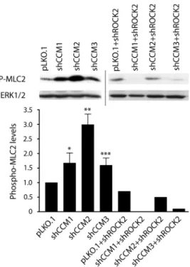
ROCK2 is required for increased phosphorylation of myosin light chain 2 in CCM1, -2 or -3 knockdown cells ... 55
Knockdown of CCM1, -2 or -3 inhibits endothelial cell vessel-like tube formation and invasion of extracellular matrix ... 56
changes required for vessel-like tube formation in
CCM1, -2 or -3 knockdown cells ... 57 Discussion ... 59 IV. The Cerebral Cavernous Malformation proteins regulate LIM
kinase-cofilin signaling in endothelial cells ... 70 Introduction ... 70 Results ... 73
RhoA-ROCK-dependent regulation of cofilin is dysregulated upon loss of the CCM proteins in
endothelial cells ... 73 Loss of CCM1, -2, or -3 protein results in increased
phospho-cofilin in vitro and in surgically resected
human CCM lesions ... 74 Pharmacological inhibition of ROCK decreases LIMK,
cofilin, and MLC2 hyperphosphorylation in mouse and
human CCM knockdown endothelial cells ... 75 LIMK and cofilin hyperphosphorylation is associated
with decreased tube formation and invasion, and LIMK1
knockdown is sufficient to rescue this phenotype ... 76 Discussion ... 78 V. Human endothelial progenitor-derived endothelial cells as a
model system for CCM ... 90 Introduction ... 90 Results ... 91
Characterization of wild type EP-ECs from healthy
Volunteers ... 91 CCM1, -2, and -3 knockdown results in angiogenesis
assay defects in EP-ECs ... 92 Loss of the CCM proteins dysregulates small GTPase
Discussion ... 94
VI. Defining the functional domain of Cerebral Cavernous Malformation 3 through its interactions with phosphatidylinositol- 3,4,5-trisphosphate ... 101
Introduction ... 101
Results ... 103
Threading analysis and homology modeling of CCM3 ... 103
Defining the PtdIns(3,4,5)P3 binding site of CCM3 ... 107
CCM2 interaction with ∆5KA, a CCM3 mutant lacking PtdIns(3,4,5)P3 binding residues ... 109
Secondary and tertiary structures of CCM3 ... 109
Cellular co-localization of CCM3 and membrane- bound constitutively-active phospholinositol-3-kinase ... 110
Discussion ... 111
VII. The molecular mechanism of CCM pathophysiology: E3 ubiquitin ligase dysregulation and accompanying dysregulation of the kinome ... 124
Introduction ... 124
Results ... 126
SMURF1 knockdown phenocopies CCM knockdown ... 126
Loss of CCM2 substantially affects the kinome ... 127
Discussion ... 129
VIII. Concluding remarks ... 137
LIST OF TABLES TABLE
4.1. Mutations in surgically resected CCM lesions stained
for phospho-cofilin ... 89 5.1. Real time PCR characterization of EP-ECs ... 100 6.1. Threading analysis using 3D-Jury ... 122 6.2. Determination of quaternary structure of wild type
LIST OF FIGURES FIGURE
1.1. Histology of a CCM lesion ... 32
1.2. Known structural features of the CCM proteins ... 33
1.3. Chemical representations of common Rho kinase inhibitors ... 34
1.4. Representation of regulation of the RhoA-ROCK signaling pathway by the CCM proteins ... 35
3.1. Quantitation of shRNA knockdown of CCM expression ... 62
3.2. CCM knockdown increases RhoA abundance and activity ... 63
3.3. Phospho-myosin light chain is increased with CCM knockdown ... 64
3.4. Invasion is decreased by CCM knockdown and rescued by ROCK inhibition ... 65
3.5. ROCK inhibitor Y-27632 and ROCK2 shRNA rescue tube formation in CCM knockdown endothelial cells ... 66
3.6. Tube formation in CCM knockdown cells is rescued by shROCK2 or Y-27632 treatment ... 67
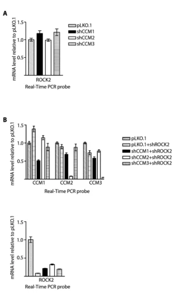
3.7. Expression levels of CCM1, -2, -3, and ROCK2 in single and double knockdown stable cell lines ... 68
3.8. CCM1, -2, and -3 knockdown endothelial cells generate filopodia but are unable to undergo cobblestone to cuboidal transition .. 69
4.1. Knockdown of CCM1, -2, or -3 leads to cytoskeletal dysregulation in human endothelial cells ... 82
4.2. Generation of stable shRNA knockdown endothelial cells ... 83
4.3. Phospho-cofilin is increased in CCM knockdown MEECs ... 84
4.4. Phospho-cofilin is increased in human CCM lesions ... 85 4.5. Pharmacological ROCK inhibition decreases
4.6. LIMK knockdown and ROCK inhibition rescue tube
formation in CCM1, -2, and -3 knockdown MEECs ... 87
4.7. LIMK1 knockdown rescues functional defects in CCM2 knockdown endothelial cells ... 88
5.1. Characterization of EP-ECs from wild type human donors ... 96
5.2. EP-ECs with high level of CCM knockdown fail to form tubes ... 97
5.3. Knockdown of CCM1, -2, or -3 results in decreased invasion ... 98
5.4. The biochemical signature of loss of the CCM proteins ... 99
6.1. CCM3 multiple sequence alignment and 3D model ... 115
6.2. Sequence alignment of the N and C-term bundles for CCM3 ... 116
6.3. Surface potential model for CCM3 ... 117
6.4. Phosphatidylinositol Membrane Lipid Array ... 118
6.5. Circular dichroism spectra of wild type and ∆5KA CCM3 ... 119
6.6. Immunofluorescence of wild type and ∆5KA CCM3 ... 120
6.7. Proposed CCM3 signaling model ... 121
7.1. SMURF1 knockdown phenocopies CCM2 knockdown in stress fibers ... 132
7.2. SMURF1 knockdown phenocopies loss of CCM2 in a tube formation assay ... 133
7.3. SMURF2 knockdown results in a loss of tube formation ... 134
7.4. CCM2 knockdown leads to substantial changes in the kinome ... 135
LIST OF ABBREVIATIONS AND SYMBOLS
AQUA: Advanced Quantitative Analysis bEND.3: Mouse brain Endothelial cell CCM: Cerebral cavernous malformations
CCM1: Cerebral Cavernous Malformation 1 gene
CCM1: Protein encoded by Cerebral Cavernous Malformation 1 gene, also known as KRIT1
CCM2: Cerebral Cavernous Malformation 2 gene
CCM2: Protein encoded by Cerebral Cavernous Malformation 2 gene; also known as OSM, Malcavernin
CCM2L: CCM2-Like protein
CCM3: Cerebral Cavernous Malformation 3 gene
CCM3: Protein encoded by Cerebral Cavernous Malformation 3 gene; also known as PDCD10
CFP: Cyan Fluorescent Protein DAB: 3,3’-Diaminobenzidine
DAPI: 4′,6′-diamidino-2-phenylindole EPC: Endothelial Progenitor Cell
EP-EC: Endothelial Progenitor-derived Endothelial Cell FAT: Focal Adhesion Targeting
FDA: Food and Drug Administration
GCKIII: Germinal Center Kinase 3
GDI: Guanine nucleotide Dissociation Inhibitor GEF: Guanine Nucleotide Exchange Factor GEMM: Genetically Engineered Mouse Model GFP: Green Fluorescent Protein
HEG1: Heart of Glass
HHD: Harmonin Homology Domain
HUVEC: Human Umbilical Vein Endothelial Cell ICAP1: Integrin Cytoplasmic Adapter Protein-1 IHC: Immunohistochemistry
IPS: Induced Pluripotent Stem cell KRIT1: Krev Interaction Trapped 1 LIMK: LIM Kinase
LOH: Loss of Heterozygosity
MEEC: Mouse embryonic endothelial cell
MEKK3: Mitogen-activated protein Kinase Kinase Kinase 3 MLC2: Myosin light chain 2
MLCK: Myosin Light Chain Kinase MLCP: Myosin Light Chain Phosphatase mM: millimolar
MRI: Magnetic Resonance Imaging mRNA: message RNA
MST4: Mammalian Ste20-like Kinase 4 NuDiX: Nucleoside Diphosphate linked to X OMIM: Online Mendelian Inheritance in Man
OSM: Osmosensing scaffold for MEKK3; also known as CCM2, malcavernin PECAM1: Platelet endothelial cell adhesion molecule 1
PCR: Polymerase chain reaction
PDCD10: Programmed cell death 10 protein PTB: Phosphotyrosine Binding
RNA: Ribonucleic Acid RNAi: RNA interference ROCK: Rho kinase
RT-PCR: Reverse Transcription Polymerase Chain Reaction
SDS-PAGE: Sodium Dodecyl Sulfate Polyacrylamide Gel Electrophoresis shRNA: short hairpin RNA
siRNA: small interfering RNA
SMURF1/2: Smad Ubiquitination Regulatory Factor 1/2 STK24/5: Serine/Threonine Kinase 24/25
VECadherin: Vascular endothelial cadherin VEGF: Vascular Endothelial Growth Factor
VEGFR2: Vascular Endothelial Growth Factor Receptor 2 YFP: Yellow Fluorescent Protein
µM: micromolar
I. Introduction
Cerebral Cavernous Malformation Disease: an overview
Cerebral cavernous malformations (CCM; OMIM 116860) are neurovascular lesions caused by the bi-allelic loss of the genes CCM1, CCM2, and CCM3. Lesions are characterized by the loss of normal vascular architecture and gradual
replacement by dilated, leaky capillaries (1). CCMs occur primarily in brain vasculature, but have been reported in numerous locations throughout the body, including the liver, spinal cord, and retina (2). It is unclear whether there is a biological reason for the substantially higher incidence of CCM in the brain, or whether it is a matter of increased detection, given that brain lesions are far more likely to cause symptoms. Patients suffer from a variety of symptoms, ranging from focal neurological deficits to hemorrhagic stroke. Unfortunately, therapeutic
strategies are limited to surgical resection or radio-ablation of lesions, procedures that involve substantial risk to the patient.
Epidemiology
with an average onset at 30 to 40 years of age, but approximately 25% of cases present in infancy or childhood (5-7). Development of a CCM lesion does not necessarily mean that patients become symptomatic, with some studies reporting that approximately 10-40% of patients with MRI confirmed lesions remain
asymptomatic (4, 8). There is a familial and sporadic form of CCM, with
approximately 10-40% of cases classified as familial in Caucasians and up to 50% familial in Hispanics (2, 9). However some studies suggest that a significant number of cases reported as sporadic are in fact familial. For example, a 1998 study by Labauge et al. showed that up to 75% of sporadic cases were familial (10). Mutations in CCM1 account for 60% of reported cases, with CCM2 and CCM3 mutations comprising approximately 18 and 10% of cases, respectively (11, 12). In up to 22% of CCM cases with multiple lesions no mutation can be detected in the CCM genes (13).
Genetics
CCM lesions develop upon homozygous inactivating mutations in CCM1, -2, or -3. To date more than 150 distinct CCM mutations have been published, including nonsense, splice-site, and frameshift mutations, with the common theme being loss of CCM protein function (11, 17-22). Many CCM mutations are thought to result in nonsense-mediated degradation of mRNA transcripts, leading to a total loss of protein expression. Interestingly, the only known CCM missense mutation not affecting splicing is located in the C-terminal end of the phosphotyrosine binding (PTB) domain of CCM2, and disrupts the interaction of CCM1 and -2 (23). Importantly, CCM lesions are thought to develop upon bi-allelic loss of the CCM genes in endothelial cells, not in vascular support cells or parenchyma. Gault et al. reported the first bi-allelic germline and sporadic mutation in a CCM lesion in a patient with CCM1 mutation (24). Laser capture micro-dissection in a large number of surgically resected lesions demonstrated that cells in the endothelial monolayer, and not the surrounding brain tissue, display loss of the CCM genes (25).
Subsequent work has affirmed this conclusion, with one exception. Louvi et al. recently demonstrated that conditional deletion of CCM3 in mouse astrocytes led to lesion formation. Given the integrated and complex signaling between astrocytes and endothelial cells these results seem plausible, however this work has not been recapitulated or demonstrated in human lesions (26).
The genetic mechanism of CCM lesion development remains controversial, but the leading theory is loss of heterozygosity (LOH) resulting from a 2-hit
most famous example being the loss of RB in the pediatric cancer retinoblastoma (27). According to this hypothesis, the first hit results from a germline mutation, and the second hit comes from somatic mutation in familial disease, with both hits
occurring somatically in sporadic CCM. There are several lines of evidence for the 2-hit hypothesis, including clinical observations from human patients and molecular genetics performed in surgically resected lesions, as well as observations from Genetically Engineered Mouse Models (GEMMs). The autosomal dominant pattern of inheritance and multiple lesions in familial patients, and the usual solitary lesions seen with sporadic cases, are consistent with a 2-hit mechanism. The best study to date on the genetic mechanism for CCM development was by Akers et al. using surgically resected CCM lesion tissue to identify bi-allelic mutations in familial patients. After sequencing the germline mutation, somatic mutations were identified through repeated cycles of amplification, subcloning, and sequencing (25). Bi-allelic germline and somatic mutations were found in the endothelium of lesions from CCM1, -2, and -3 familial patients, with only single mutations being found in
non-endothelial cells. Interestingly, not all non-endothelial cells in the CCM lesions had double hits, suggesting that a mosaicism of loss of heterozygosity is sufficient to cause CCM pathophysiology.
There is also compelling evidence for the 2-hit hypothesis in mouse models. Several mouse studies have shown that crossing ccm heterozygotes with a
developed lesions when crossed with p53 null mice (29). More recent work showed that CCM lesions develop spontaneously in ccm1 heterozygous mice when bred into a mismatch-repair deficient background, with either homozygous deletion of the tumor suppressor Trp53 or the mismatch-repair gene Msh2 (28). While these mouse models support the LOH hypothesis for lesion development, the conclusions that can be drawn are limited given that there is no evidence that p53 or Msh2 play a role in the natural history of CCM disease. The genetic mechanism of the second hit remains unknown, and is perhaps the most significant question remaining in the CCM field, besides discovering the molecular mechanism of disease. Multiple hypotheses have been proposed, including an epigenetic second hit,
uncharacterized cis-regulatory mechanisms, or stem cell or endothelial precursor cell mutation.
Significantly, LOH has been demonstrated in multiple vascular diseases related to CCM, including Cutaneomucosal Venous Malformations (MIM 600195), and Capillary Malformations (OMIM 163000) (30). Bi-allelic CCM loss of function is embryonic lethal in mice between E8.0 and E9.5, supporting the importance of the CCM genes in angiogenesis, and there are no known cases of humans born with homozygous germline CCM mutation (31-34). One other possible mechanism for a second hit would be trans-heterozygosity, (e.g. heterozygous for CCM1 and CCM2) from a second hit in another CCM gene. Trans heterozygosity has been
Furthermore, no cases have been reported of humans with trans-heterozygosity in CCM genes, although the possibility cannot be ruled out given that current clinical
CCM genetic testing techniques halts after finding the first mutation.
Finally, the discrepancy between genetic linkage data and CCM sequence analysis in patients has lead to the suggestion that there is a fourth, as of yet
unidentified CCM gene. For example, one study analyzed a panel of 29 familial CCM patients that had tested negative for CCM1 and -2 mutation, and was only able to identify CCM3 mutation in 3 of them (37). There are several possible explanations for this, including technical challenges in sequencing CCM3 mutations, but one interpretation is that there is a fourth gene that can cause CCM disease. Given that loss of CCM1, -2, and -3 gives identical phenotypes despite little common similarities in protein function, it definitely seems possible that future work discovers “CCM4.”
Pathology
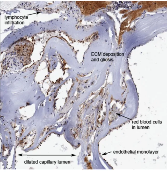
replacement of intervening neural parenchyma with gliosis and scarification (38). CCM lesions are often filled with slow flowing or thrombosed blood (40). Importantly, lesions lack the normal tight junctions between endothelial cells and astrocytes, although the mechanism of this loss is unclear (41). As a result of the repeated blood extravasation resulting from the loss of vascular integrity in these vessels, the lesions are also characterized by macrophage and lymphocyte infiltration into the proximal parenchyma, as well as striking hemosiderin deposition (38, 42). Lesions are thought to evolve through their lifetime, with early stage lesions generally displaying a mulberry-like pattern and later stage lesions showing greater vessel dilation with enhanced scarring and necrosis. Generally, lesion size and burden increase with age (2). The evolutive nature of lesions is also supported by the observed tendency for de novo development of CCMs in areas of surgical or radiological intervention (43, 44). Because of their disorganized architecture and highly friable nature, as well as the fact that the thin endothelial monolayer is the region of interest, performing assays such as PCR or immunohistochemistry (IHC) on pathological specimens is challenging. Given the dearth of CCM samples, increased biobanking and cataloging of surgically resected lesions is of great importance to advancing the field.
Clinical Course and treatment
The first clinical description of CCM was reported by the German
proportion of familial versus sporadic cases. Historically, 20% of cases were thought to be familial and 80% sporadic, but estimates vary because of the modest sample size in these clinical studies (9). However, recent genetic work on sporadic patients possessing multiple lesions has shown that the majority of these cases were in fact familial (2). In both cases bi-allelic loss of ccm1, -2, or -3 occurs in endothelial cells, but familial patients exhibit higher lesion burden, with one study showing over 50% of familial patients having multiple lesions, whereas sporadic cases displayed multiple lesions in approximately 12% of cases (4, 46). Symptoms resulting from CCMs are similar between familial and sporadic forms, but familial patients generally present between infancy and thirty years of age, whereas sporadic patients
generally present between forty and sixty years of age (9, 47). While CCM lesions can occur in many locations throughout the body, clinical manifestations are most common with CNS involvement. CCMs can cause a substantial variety of symptoms, including neurological deficits (e.g. dysphagia, hemiparesis, ataxia), epilepsy, and hemorrhagic stroke (48).
Despite the genetic advances in understanding CCM and the commercial availability of CCM gene sequencing, MRI remains the gold standard for diagnosis, as well as differentiating familial from sporadic CCM (49, 50). Blood flow through CCMs is very low; unlike in related vascular malformations such as AVM, CCMs are very difficult to detect by angiography, and are therefore referred to as occult (51). CCM lesions appear on MRI as a “popcorn” pattern and often display a dark
treatment (51).
The standard of care for CCM disease is observation until symptoms
necessitate intervention. The only medically treatable CCM symptoms are epilepsy and headache/migraine. However, anti-epileptics are ineffective for seizure control in half of CCM cases, and the headaches are only controllable through high dose narcotics; treatment regimens that have numerous side effects (53, 54). Intractable epilepsy, recurrent hemorrhage, and progressive neurological deficits are the primary indications for surgical resection of accessible lesions (39).
Unfortunately, significant subsets of CCM lesions are considered surgically inaccessible, and these often have poorer natural histories than surgically accessible lesions. Generally, lesions are considered surgically inaccessible if surrounded by eloquent tissue such as the brainstem, spinal cord, or thalamus. Surgically
inaccessible lesions are usually observed unless symptoms become overwhelming. Stereotactic radiosurgery has been touted as an alternative to traditional
microsurgery for lesion removal, but high complication rates remain (55). While CCM outcome data is limited, one retrospective study of 95 patients found that
radiosurgery was associated with a drop in annualized hemorrhage rate from 17 to 5 percent after 2 years. However, at the 5 year follow-up 16 percent of patients
previously noted, invasive intervention has been linked in some cases with lesion proliferation. Most importantly, lesions located in the brainstem or other eloquent areas will continue to present serious challenges for surgical therapy. Clearly, a better understanding of the CCM signaling network must be achieved, with the ultimate goal of producing a non-invasive pharmacological treatment for CCM disease.
The CCM proteins: structure, function, and signaling
CCM1
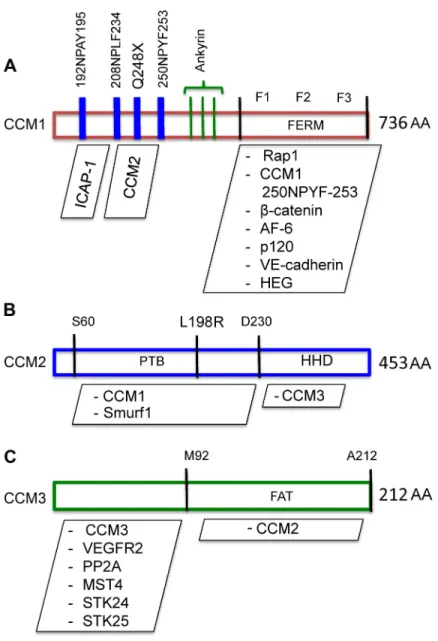
CCM1, also known as Krit1 (Krev interaction trapped protein 1) is a 16 exon, 736 amino acid protein that contains one band four point one ezrin/radixin/moesin (FERM) domain, three ankyrin repeat domains, and one Nucleoside Diphosphate linked to X (Nudix) domain (Fig. 1.2A) (57, 58). CCM1 was first identified in a yeast two-hybrid screen for binding partners of the small GTPase Rap1, and subsequently mapped to the Krit1 locus (57, 59, 60). CCM1, like CCM2 and -3, is a scaffold
protein lacking any known catalytic domains. A crystal structure of the interaction between the FERM domain and the C-terminus of the orphan membrane receptor Heart of Glass (HEG1) has been solved, but CCM1 has not been crystalized alone (61).
which interacts with an NPXY motif in the central region of CCM1 in a head-to-tail interaction, serving to maintain CCM1 in a closed conformation (63). This interaction is thought to regulate CCM1 activity by controlling access to the F2 and F3 FERM subdomains, which mediate binding with the small GTPase Rap1 (63). The CCM1-Rap1 interaction is of great interest because CCM1-Rap1 is a master regulator of vascular integrity and tight junction formation (64).
CCM1 possesses an N-terminal NPXY motif, which interacts with the PTB domain of Integrin cytoplasmic domain-associated protein 1 (1) (65, 66). ICAP-1 is a negative regulator of βICAP-1-integrin activity because it competes with Talin, an integrin-cytoskeleton linking protein, for integrin binding (67). Structural work in 2013 by Lui et al. demonstrated that indeed CCM1 functions as a molecular switch for β1-integrin activation by regulating Talin-ICAP-1 association (32). Lui et al. also
discovered the presence of a previously undescribed Nudix domain in a region formerly described as unstructured, although the functional significance remains unclear. Finally, while there are three ankyrin repeats present between the NPXY motifs and the FERM domain, no CCM1 ankyrin-mediated protein interactions have been found to date. However, ankyrin repeats commonly mediate protein-protein interactions, so it would not be surprising if their function is elucidated in CCM1 in the future (68).
the three CCM proteins exist in a ternary complex, with CCM1 and -3 bound to CCM2. CCM2 binding appears to regulate the subcellular localization of CCM1. Hilder et al. showed that CCM1 and -2 are primarily cytosplasmic when the wild type proteins are co-expressed, but that F217 mutation in CCM2 led to nuclear
localization of CCM1 (42). Furthermore, CCM1 localizes to the nucleus in the absence of CCM2, and CCM2 does not enter the nucleus without the presence of functional CCM1 (15). The biological function of CCM1’s nuclear translocation is currently unknown. No evidence has been found that CCM1 directly regulates
transcription. However, given that loss of CCM1 is associated with increased activity of the small GTPases RhoA and Rap1, it is possible that CCM1 may be affecting transcription through downstream regulation of transcription factors. Glading et al. demonstrated that loss of CCM1 led to accumulation of beta-catenin in the nucleus and a subsequent increase in beta-catenin-dependent transcription (70). Overall, CCM1 is perhaps the best-characterized CCM protein and appears to be the most commonly mutated as well. More work remains to be done on the CCM1-Rap1 interaction as its dysregulation likely plays a role in CCM pathophysiology.
CCM2
responses to hyper-osmotic shock (71). Concurrently, CCM2 was identified as the second human gene mutated in CCM by sequencing positional candidate genes based on earlier genetic mapping (19, 72). Like CCM1, no full crystal structure of CCM2 exists. However, a study in 2013 by Fisher et al. was able to crystalize the C-terminal domain (approximately 200 AA) of CCM2 to a resolution of 1.9 Ångstroms. The C-terminal domain of CCM2 is homologous to the N-terminal domain of
Harmonin; an adaptor protein linking actin to membrane bound proteins (73).
Mutations in Harmonin cause Usher Syndrome (OMIM 276900), a condition in which patients display sensory-neural deafness, visual impairment, and vestibular
dysfunction (74-76). The biological role of the HHD domain is thus far unknown, but reinforces the important scaffold or adaptor role CCM2 plays in cytoskeletal
regulation.
In contrast to the HHD domain, more is known about the function of the CCM2 PTB domain, with the Johnson lab undertaking the most comprehensive study of its role in 2007. Hilder et al. generated a point mutation (F217A) within the PTB domain, allowing for the definition of PTB-dependent binding partners for
CCM2. The mutation was correctly predicted to disrupt CCM1-CCM2 binding, based on the finding of a similar mutation in patients abrogating the CCM1-CCM2
complex in the cell (69).
Interestingly, recent work has discovered a CCM2 paralog, CCM2-like (CCM2L) that is expressed in endothelial cells during periods of cardiovascular development. CCM2L competitively inhibits CCM2 binding to CCM1 (but not CCM3), resulting in decreased angiogenesis in vitro and in vivo (77). The authors propose CCM2L as a molecular mechanism for coordinating vessel stability and growth in both development and postnatal vessel growth. The important role of CCM2L in endothelial cells, along with the mechanism of regulation suggests that the CCM signaling pathway is more important in development and vessel homeostasis than previously suspected.
Finally, and perhaps most significant, is the work done by Crose et al. characterizing the molecular interaction between CCM2 and the E3 ubiquitin ligase SMURF1 (SMAD ubiquitin regulatory factor 1). Using mouse brain microvascular cells, it was demonstrated that the PTB domain of CCM2 interacts with the Homologous to the E6-AP Carboxyl Terminus (HECT) domain of SMURF1 and serves to localize it to the membrane where it targets the small GTPase RhoA for degradation (78). This is highly significant because we hypothesize that
CCM3
CCM3, also known as PDCD10 (Programmed Cell Death 10), is a seven exon, 212 amino acid protein with a dimerization domain and Focal Adhesion Targeting-homology (FAT) domain (Fig. 1.2C). CCM3 was the most recent gene linked to CCM, and is also the smallest at 25 kilodaltons. CCM3 was identified by high-density microsatellite genotyping in a set of familial CCM patients testing negative for CCM1 or -2 mutation (21). Initial in vitro work suggested that CCM3 performed anti-apoptotic functions. For example, one study showed that introduction of recombinant CCM3 decreased natural cell death in fibroblast cell lines treated with apoptosis inducers, including TNF-α, staurosporin, and cyclohexamide (79). CCM3 is the only CCM protein for which a high-resolution crystal structure has been solved. Li et al. showed that CCM3 binds CCM2 via the FAT homology domain and that mutation of a conserved FAK-like hydrophobic pocket abrogates CCM2-CCM3 interaction (80). It was also demonstrated that the FAT homology domain interacts with paxillin, and the two proteins co-localize in several cell types (62). Furthermore, truncating mutations of the CCM3 FAT domain are seen in familial CCM disease, suggesting that the full presence of this domain is essential to the function of the CCM proteins and complex. CCM3 has an N-terminus dimerization domain, and has been shown to exist as a homodimer both in vitro and in vivo (80, 81).
Germinal Center Kinase III sub-family (GCKIII) of the Sterile 20 protein kinase family, including mammalian Sterile 20-like protein kinase 4, and Serine/Threonine-protein kinase 24 and 25 (STK24 and STK25). Work in zebrafish showed that both STK knockdown and low-level STK and CCM3 knockdown resulted in a CCM
cardiovascular phenotype (82). Furthermore, depletion of STK25 in Human Umbical Vein Endothelial Cells (HUVEC) leads to increased barrier permeability; a hallmark of CCM protein loss. The molecular interaction was later defined when it was
demonstrated that the N-terminus of CCM3 heterodimerizes with the GCKIII proteins in a mechanism similar to CCM3 homodimerization (83). Importantly, it was shown that the STKs directly activate the cytoskeletal scaffold protein Moesin, and that a loss of active Moesin results in increased RhoA activity (82). While further work is needed to clarify the role of the GCKIII kinases in CCM pathogenesis, it is clear that the STKs are important downstream effectors for CCM3.
Another molecular interaction of significance has been demonstrated between CCM3 and Vascular Endothelial Growth Factor 2 (VEGFR2). VEGFR2 expression is restricted to endothelial cells (both vascular and lymphatic) and is a master regulator of vascular endothelial function, such as proliferation, migration, survival,
permeability, vasculogenesis, and angiogenesis (84). He et al. generated a CCM3 conditional knockout mouse that demonstrated defects in VEGFR2 signaling, including decreased VEGFR abundance and decreased phosphorylation of
mutations affecting the C-terminal of CCM3 seen in familial CCM3 patients. The authors concluded that CCM3 regulates vascular development by modulating VEGFR2 signaling. The CCM3-VEGFR2 interaction is compelling because of VEGFR2’s importance in vascular regulation and the fact that clinically relevant CCM3 C-terminal mutants lead to destabilization of VEGFR2. However, this work has not been replicated thus far, and while previous groups have looked at VEGF signaling in the context of CCM mutation, nothing of significance was found. CCM3 remains the most enigmatic CCM protein, with a rapidly increasing literature on signaling, albeit with some disagreement between investigators on its role in the CCM pathway. Given the wealth of structural knowledge that has been elucidated recently, knowledge of CCM3 should continue to expand rapidly in the near future.
The CCM regulatory network
Virtually everything known about the CCM signaling network has been discovered within the last 10 years. While much more is known now than a decade ago, defining the molecular mechanism of CCM disease remains elusive.
the CCM proteins, with loss of CCM1, -2, and -3 resulting in similar increases in stress fibers, decreased capillary-like tube formation, and increased endothelial monolayer permeability. Hilder et al. showed that the CCM proteins form a ternary structure, suggesting that the full CCM complex may be required for normal
endothelial cell function. While this hypothesis is difficult to prove because of the lack of suitable fluorescent CCM antibodies, it is clear that the CCM proteins regulate one important common pathway in endothelial cells: RhoA and therefore Rho kinase (ROCK) activity.
RhoA and Rho kinase signaling
The similar biochemical and phenotypic abnormalities seen with loss of any of the 3 CCM proteins suggests that they coordinately regulate a common mechanism for maintaining vascular integrity. Recent work in our lab and by others has
implicated dysregulation of RhoA as a mechanism for loss of vascular integrity in CCM (32, 78, 86). RhoA is a 21 kilodalton small GTPase and first-identified member of the Rho family, which is in turn a subfamily within the Ras superfamily (87).
primary function ascribed to RhoA is regulation of actin and microtubule cytoskeletal dynamics, an action that occurs primarily through regulation of ROCK (90).
Importantly, normal RhoA activity is essential for proper endothelial cell function. RhoA activity is essential to the dynamic regulation of permeability required of
endothelial and smooth muscle cells, along with facilitating diapedesis of leukocytes. Furthermore, a number of vascular defects are associated with aberrant RhoA activity or loss. Expression of constitutively active RhoA decreases formation of capillary-like tubes on Matrigel, and GEMMs with knocked out RhoA or RhoA regulatory proteins exhibit embryonic lethality due to failed vascular patterning and defective sprouting angiogenesis, supporting the crucial role of RhoA in
angiogenesis (92). Overall, RhoA has been defined as a master regulator of the actin cytoskeleton and clearly plays a critical role in endothelial cell biology.
hyperactivation has been implicated in both hypertension and cancer metastasis (94, 95). ROCK affects actin through parallel pathways involving Myosin Light Chain Phosphatase (MLCP) and LIM kinase (LIMK), both important cytoskeletal regulators through their effectors Myosin Light Chain 2 (MLC2) and cofilin, respectively (96-98). Phosphorylation of MLC2 and cofilin favors actin assembly and stress fiber formation (99, 100). Phospho-MLC2 stays bound to actin, encouraging stress fiber formation and contraction (101). MLCP is a widely expressed phosphatase that
dephosphorylates myosin light chain (102). MLCP opposes the action of myosin light chain kinase (MLCK), but it appears that ROCK-mediated inhibition of MLCP is more physiologically important for regulating MLC2 phosphorylation than MLCK activity (103). Phosphorylation of MLC2 favors stress fiber formation and cell contraction, phenotypes seen in CCM knockdown cells in vitro. While ROCK is a direct regulator of actin organization in endothelial cells, it is also an important indirect cytoskeletal effector through regulation of LIM kinase (LIMK).
LIM kinase and cofilin
been established between the two (110). Interestingly, loss of function in LIMK is thought to cause Williams syndrome, a neurodevelopmental disorder associated with a number of vascular defects (111).
LIMKs are important regulators of actin dynamics through cofilin, an actin depolymerizing factor (112, 113). When phosphorylated by LIMK, cofilin’s actions on acting severing and monomer dissociation are decreased. ROCK phosphorylates LIMK at threonine 505 or 508, which in turn phosphorylates cofilin at serine 3 (114). Unphosphorylated cofilin severs actin filaments with uncapped ends, leading to an increase in the number of free filament ends (115, 116). Cofilin also enhances the rate of monomer dissociation from the pointed ends of actin (117, 118). Together, these actions lead to increased actin turnover and a more dynamic cytoskeleton. However, when phosphorylated by LIMK, cofilin is unable to bind and depolymerize actin (119). This leads to an increase in F-actin, a phenotype similar to that seen in endothelial cells lacking CCM proteins (120-122).
Previous work has characterized LIMK as being regulated by RhoA-ROCK in endothelial cells, and defined a role in maintaining vascular integrity in other systems (123, 124). In human endothelial cells stimulated with thrombin, ROCK
phosphorylates LIMK, leading to phosphorylation and inactivation of the
important regulator of vascular integrity in vivo. In one study, endotoxin stimulation led to increased RhoA and LIMK activation, followed by increased endothelial
permeability and subsequent mortality in wild type mice. LIMK-/- mice showed lower permeability and mortality in response to endotoxin treatment (124). In humans, several LIMK haplotypes have been linked to the rupture of intracerebral aneurisms in the Japanese population (125). Furthermore, LIMK has been shown to play an important role in modulating invasion and migration, characteristics important to endothelial cells for maintaining endothelial barriers (126-128).
LIMK hyperactivation resulting from loss of the CCM proteins raises the possibility of chemical inhibition of LIMK as a therapeutic target for CCM disease. The LIMK-cofilin pathway is further downstream of RhoA and ROCK, and
furthermore cofilin is the only known target for LIMK (111). Unlike ROCK, no commercially available inhibitors of LIMK exist. However, like ROCK, LIMK hyperactivation has been implicated in a number of important diseases such as cancer. Therefore it seems likely that LIMK inhibitors will be brought to market in the near future, and if so they should definitely be tested in the context of CCM.
However, it is currently difficult to test pre-clinical efficacy of pharmacological
inhibitors because of a lack of availability of human endothelial cells with CCM gene mutations.
Endothelial precursor-derived endothelial cells
Rationale for development of a novel human primary cell system
biologically relevant human cell system to work with. Availability of human lesions is limited, although the patient advocacy group The Angioma Alliance maintains a growing bio-bank of frozen lesions. Even though the number of CCM mouse models has increased greatly in the last 5 years, problems remain. Several of the GEMMs are ccm heterozygotes crossed on a mismatch repair background, which does result in reliable lesion generation but may not be a truly physiologically relevant model. Furthermore, while mouse models are unquestionably useful for advancing the CCM field, human and mouse pathophysiology can be substantially different at times, as evidenced by the numerous examples in other fields of pharmacological therapies in mice that were ineffective in humans (129). Similarly, while important work,
especially in vascular development, has been done on CCM orthologues in
zebrafish, biological relevance to the human disease is questionable. For example, much work has been done to characterize the interaction between the CCM1 and -2 orthologues santa and valentine and the orphan receptor HEG1 in zebrafish, with investigators showing that loss of HEG1 phenocopies loss of the CCM proteins, and HEG1 couples to CCM1 to regulate endothelial cell junctions (33, 130). However, no HEG1 homologue exists in humans, and no CCM3 orthologue exists in Danio rerio. Current in vitro work relies mainly on HUVECs, along with other mouse and human cell lines such as human microvascular endothelial cells and mouse embryonic endothelial cells (MEECs). Techniques such as RNAi and fluorescent protein
optimizing a method of obtaining endothelial cells from CCM patients would be of great benefit to the field. One way of achieving this goal is by deriving endothelial cells from CCM patients, and this can be accomplished by isolating endothelial progenitor cells (EPCs) from peripheral blood.
Endothelial progenitor cells and endothelial progenitor-derived endothelial cells
EPCs were first described by Asahara et al. as a CD34 positive subset of peripheral blood mononucleocytes (PBMC) that expressed VEGFR2, CD31, and endothelial constitutively nitric oxide synthetase, all markers specific to endothelial cells, in addition to the stemness markers CD34 and AC133 (131, 132). Some EPCs circulate, but the majority reside in the bone marrow, and they contribute significantly to the repair and formation of new blood vessels in adults (133). Further work
showed that EPCs can differentiate into vascular endothelium in response to both growth factors and vascular injury, leading to the concept that vasculogenesis and angiogenesis may occur simultaneously in postnatal life (134).
Importantly, EPCs will differentiate into endothelial cells when cultured on fibronectin for 7 days in media with the appropriate mix of endothelial cytokines. We term these outgrowth cells endothelial progenitor-derived endothelial cells (EP-ECs). EP-ECs have great potential in CCM research because they are endothelial cells that can be isolated from peripheral blood of CCM patients. These endothelial cells would be heterozygous and therefore would better reflect the biology of CCM
expression of either the remaining wild type allele or the mutant. In fact, EP-ECs have recently been used to investigate the mechanism of corticosteroid therapy in infantile hemangioma (OMIM 602089), a vascular disorder related to CCM. Using EP-ECs derived from hemangioma patients with confirmed genetic mutations, Greenberger et al. demonstrated that the corticosteroid dexamethasone causes lesion regression by inhibiting the proliferation potential of EP-ECs (135). Patient derived EP-ECs could be a powerful new system for studying endothelial biology and vascular disease. EP-ECs could even be combined with induced pluripotent stem cell techniques to create a library of human CCM gene mutations. Certainly, EP-ECs would be an ideal system for studying drug response and pharmacological therapy for CCM. However, the use of EP-ECs is not without caveats. Our
experience has been that they cannot always be successfully isolated from donors, and investigators report differing rates of success in achieving EP-EC outgrowth. While not published, discussion with EP-EC experts and our own personal
experience suggests that age and sex of the donor may play a substantial role in the amount of circulating EPCs and their proliferative potential. Certainly, further
characterization is warranted, given the potential of EP-ECs in CCM and cardiovascular investigation in general.
The future of pharmacological treatments for CCM Disease
anticonvulsants. When symptoms become intolerable, the only options are surgery or radio-ablation. However, these treatments are associated with a number of serious complications, and there is some evidence that invasive intervention can trigger further lesion evolution. Furthermore, some CCM lesions, including the ones most likely to exhibit severe symptoms, are located in surgically inaccessible areas such as the brainstem or pons. Therefore, discovery of a pharmacological treatment for CCM is of paramount importance.
The increase in the understanding of CCM signaling pathways in the last decade has made the discovery or synthesis of a CCM therapeutic increasingly possible. Specifically, attention is focused on two therapeutic avenues: small
molecule ROCK inhibitors, or off-label use of currently Food and Drug Administration (FDA) approved drugs.
Statins
The advantages of using drugs already approved by the FDA are that basic safety and dosing information in humans is known, and lengthy and expensive pre-clinical workup is not required. The most promising class of FDA approved drugs proposed to treat CCM are statins, in particular Simvastatin, because of their potential as RhoA activity inhibitors. Statins have been used to treat
(136). However, statins are also associated with a number of beneficial pleotropic effects, including improved endothelial relaxation, reduced arteriosclerosis
progression, improved hypertension control, and increased cardiac ejection fraction (137). It is thought that most of the pleotropic effects from statin treatment come from indirect inhibition of small GTPases such as RhoA and Ras (138). Mevalonate can be converted into the isoprenoid residues farnesylpyrophosphate and
geranylgeranylpyrosphosphate, which can be covalently attached to intracellular proteins through prenylation (139). Small GTPases are particularly dependent on prenylation for localization and activity, and it has been demonstrated that statins can inhibit angiogenesis by inhibiting RhoA geranylgeranylation and therefore
preventing proper RhoA localization (140). This finding is important for the CCM field given that loss of the CCM proteins results in RhoA hyperactivation. Given the well-documented cardiovascular protective effects of statins, and their good safety profile, statins have been discussed as a possible CCM treatment for several years. Significantly, in 2009 Whitehead et al. demonstrated that permeability barrier
function could be rescued in heterozygous CCM2 mice with Simvastatin treatment (32).
start for CCM patients. Another way to study statin effectiveness on CCM would be to conduct a retrospective cohort study of CCM patients taking statins. The feasibility would be limited by the number of overlapping hypercholesterolemia and CCM patients, and the availability and accessibility of medical records. However, given that approximately 1 in 200 of the general population has a CCM mutation, and approximately 1 in 4 Americans over 45 are thought to take statins regularly, it is possible that significant data could be gleaned from such a study (136).
Rho kinase inhibitors
A second promising therapeutic strategy for treatment of CCM is small
molecule inhibition of ROCK (Fig. 1.3). As discussed previously, loss of CCM1, -2, or -3 results in increased ROCK activity in cultured endothelial cells, mouse models, and human lesions (32, 78, 86, 142). Furthermore, both RNAi knockdown and
chemical inhibition of ROCK have been demonstrated to rescue the CCM phenotype in vitro (86, 142). Most significantly, a recent study in a CCM mouse model
demonstrated that dosing with a small molecule ROCK inhibitor reduced CCM lesion size and overall lesion burden (143). Previous mouse work had shown that ROCK inhibition rescued vascular leak in ccm1 and -2 heterozygous mice (142). In some ways, ROCK is a more attractive target than RhoA because it is more specific. RhoA, and lipid prenylation in general, are extremely important in most cells, whereas ROCK is a single arm of the RhoA pathway. Additionally, ROCK2
development of small molecule inhibitors for ROCK by both the pharmaceutical industry and academia because of the applications to both cardiovascular disease and cancer. CCM patients may benefit from the applicability of ROCK inhibition to these diseases as increasingly potent and specific ROCK inhibitors are developed and brought to market.
Currently no ROCK inhibitors are approved for use in humans in the United States. However, one compound, Fasudil, has been used in Japan since 1995 to treat cerebral vasospasm, and is well tolerated in humans and mice (145). Cerebral vasospasm is an intense vasoconstriction in brain arteries that occurs following brain hemorrhage, most commonly after sub-arachnoid hemorrhage. In the United States, calcium channel blockers such as Nimodipine and Nicardipine are used instead of Fasudil (146). Studies have shown that Fasudil is effective in reducing vasospasm from 57 to 37% with subsequent reduced mortality and improved outcomes (147). Importantly, this data demonstrates safety and efficacy in humans, as well as the ability of Fasudil to cross the blood brain barrier and act on cerebral blood vessels.
Defining the molecular mechanism of CCM disease
outcomes, despite their heterogeneous functions in the cell. We propose that the CCM proteins regulate RhoA activity and that the common pathway of CCM disease is hyperactivation of the RhoA/ROCK signaling network (Fig. 1.4). The phenotypes of CCM knockdown endothelial cells, along with the cytoskeletal
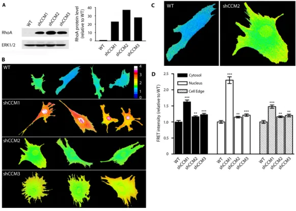
signaling dysregulation observed, are consistent with RhoA hyperactivity. We have demonstrated increases in total RhoA and RhoA activity through Western blotting, a RhoA biosensor, and active RhoA pulldowns. Furthermore, we have shown that the ROCK targets MLC2 and LIMK are hyperphosphorylated with loss of the CCM proteins, with deleterious consequences on the ability of the actin cytoskeleton to undergo the profound changes required of endothelial cells. Both
hyperphosphorylation and phenotype defects are reversed by chemical inhibition of ROCK or knockdown of ROCK or LIMK, indicating the importance of RhoA-ROCK signaling to CCM. Yet the molecular mechanism whereby loss of the CCM proteins results in RhoA hyperactivation remains elusive.
The similar increase in RhoA-ROCK activity observed with loss of CCM1, -2, and -3, along with the binding data indicating that the three proteins exist as a
ternary complex in the cell, initially lead us to hypothesize that all three functioned on a common molecular pathway to regulate RhoA activation. Crose et al.
demonstrated that CCM2’s PTB domain interacts with the HECT domain of SMURF1, and CCM2 acts to localize SMURF1 to the cell membrane to regulate localized degradation of RhoA (78). Recent work has lead to the understanding that ubiquitin-mediated proteosomal degradation plays an important role in
Crose’s data lead us to hypothesize that CCM1 and -3 likely regulated RhoA activity through E3 ligases, either SMURF1 or -2 (148). However, CCM1 and -3 do not possess a PTB domain like CCM2, and we have been unable to show that they bind endogenous SMURF1 or -2. Therefore, we propose that the CCM proteins
regulate RhoA activity through separate molecular pathways. We hypothesize that CCM2 regulates RhoA through spatio-temporal regulation of SMURF1, that CCM1 regulates subcellular localization of CCM2 and thus SMURF1
degradation of RhoA, and CCM3 regulates RhoA through a currently unknown mechanism, although likely by influencing CCM2 through it’s interactions with the CCM complex.
It is clear that the CCM proteins regulate a complex network of proteins in endothelial cells. A new technique pioneered by the Johnson lab, Multiplexed inhibitor beads coupled with mass spectrometry (MIB-MS) allows us to look in an unbiased way for the first time at the effects of loss of the CCM proteins on the entire kinome of the cell. MIB-MS has demonstrated that the CCM proteins, likely through regulation of small GTPase activity, affect a surprisingly high number of kinases, including several critical to angiogenesis and transcription, such as the angiopoetin receptor (TIE2) and Transforming growth factor beta-receptor (TGFβR). This windfall of data about the CCM regulatory network needs further investigation and
Figure 1.1: Histopathology of a CCM lesion. Note loss of normal vascular
Figure 1.4: Representation of regulation of the RhoA-ROCK signaling pathway by the CCM proteins. (A) In the presence of all three CCM proteins, SMURF1 is localized to the plasma membrane where it serves to degrade RhoA, therefore regulating ROCK signaling and balancing cytoskeletal dynamics between
breakdown and assembly. (B) With loss of CCM1, -2, or -3, degradation of RhoA is dysregulated and total and active RhoA increases, leading to an increase in
II. Materials and Methods
Chapter III
Establishment of knockdown cell lines
Lentiviral gene-specific shRNAs in pLKO.1 were obtained from the UNC-CH Lenti-shRNA Core Facility. Infection was accomplished using Lipofectamine 2000
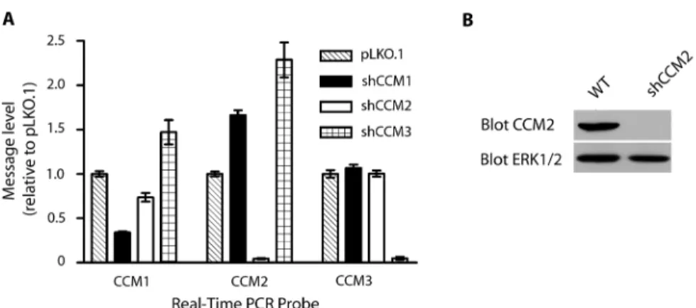
(Invitrogen) and cells were selected for resistance with 4µg/mL puromycin. Stable knockdown was achieved and measured by real-time PCR with CCM specific primers. Experiments were conducted between 1 and 2 weeks after infection to ensure stable protein knockdown. Multiple shRNAs were used for the same protein to control for off target effects.
RhoA biosensor
Tube formation assay and live cell imaging
7.0 x10^4 cells were incubated for 15 hours on Matrigel (BD Biosciences) and stained with rhodamine-phalloidin as previously described (78). Imaging was
performed on either a Pathway (BD Biosciences) or Cellomics ArrayScan (Thermo Scientific). For live cell imaging, 6 fields of cells were imaged via transmitted light microscopy every 10 minutes for 15 hours. Cellomics Arrayscan software was used to quantitate mean tube area.
Statistical significance
Where indicated, statistical significance was calculated using the two-tailed Student’s t-test. Data represented the mean of at least 2 experiments.
Invasion assays
5.0x10⁴ cells in 0.05% FBS/DMEM were seeded in the top chamber of a Biocoat Matrigel invasion chamber (BD Biosciences) with 1.5% FBS/DMEM in the bottom chamber. At 8 hours cells were fixed, permeabilized and stained with rhodamine-phalloidin. For each membrane, five fields of cells were counted. For assays with Y-27632, cells were treated with 10µM Y-27632 (Calbiochem) for 5 hours prior to and during invasion assays.
Image analysis
the Johnson lab performed image analysis for the tube formation assays.
Western blotting
100 µg of protein were separated by Western blot and blotted with anti-RhoA (Santa Cruz, 26C4) or anti-phospho-MLC2 (Cell Signaling, Ser19) antibody or mouse monoclonal anti-CCM2 antibody (Johnson lab, UNC 48.8.5).
Chapter IV
Mutation sequence analysis of human samples
Lesions were acquired through the kind of help of Dr. Amy Akers and the Angioma Alliance’s CCM DNA and Tissue Bank (www.angiomaalliance.org). Lesions were resected from clinically and radiographically confirmed familial CCM patients. The sequence of CCM gene mutations were determined by screening peripheral blood mononucleocyte DNA with a panel of CCM PCR probes. This is the standard method for genetic diagnosis of CCM in patients.
Immunohistochemistry
Immunohistochemistry was performed with the assistance of the Dr. Nana Feinberg and the UNC Translational Pathology Laboratory. Tissue slide preparation: Cell pellets were resuspended at 50°C in 2% low melting agarose (#BP-165-25, Fisher Scientific) in PBS buffer and transferred to plastic molds. Pellets were allowed to harden at 4°C and then pushed out from the mold to the tissue processing
tissues.
Chromogenic detection: immunohistochemistry was performed in a Bond Autostainer (Leica Microsystems). Antigen retrieval for both phospho-cofilin and CD31 antibodies was performed for 30 min at 100 ºC in Bond-Epitope Retrieval solution. Slides were incubated in anti-phospho-cofilin or anti-CD31 primary
antibodies for 1hour. Rabbit monoclonal phospho-cofilin, Ser3, 77G2, (Cell Signaling Technology) was used at 1:300 and mouse monoclonal CD31-1A10-CE-S (Leica Microsystems) at 1:200. Antibody detection was performed using the Bond Polymer Refined Detection System.
Fluorescence detection: Tissue slides were first incubated with anti-CD31 antibody (1:200) to create the endothelial cell mask. CD31 was detected by HRP linked Bond Polymer. After CD31 staining, anti-phospho-cofilin antibody (1:300) was applied. Goat anti-mouse-HRP (Envision+) was used as a secondary antibody and Cy5-tyramide (PerkinElmer) as a fluorescent tag. The stained slides were mounted with ProLong Gold antifade reagent (Molecular Probes) containing 4,6-diamidino-2-phenylindole (DAPI) to define nuclei.
Aperio FL/AQUA image analysis: Aperio FL (Aperio) with integrated HistoRx AQUA technology (HistoRx) was used to scan slides at 20x through DAPI, CY3 and CY5 channels. Scanned images were analyzed through spectrum using the AQUA traditional algorithm according to AQUAnalysis™ Aperio Edition (Rev. 1.0,
Cell Culture and shRNA
bEND.3 cells were purchased from ATCC. Lentiviral gene-specific shRNAs in a pLKO.1 system were generated by the University of North Carolina-Chapel Hill Lenti-shRNA Core Facility. Infection was performed according to the RNAi
consortium protocol. bEND.3 cells were cultured in 10% FBS/DMEM, Mouse
Embryonic Endothelial Cells (MEEC) in 3% FBS/DMEM, and Human Umbilical Vein Endothelial Cells (HUVEC) in EGM2 media (Lonza). 4µg/mL puromycin was used to maintain shRNA selection. HUVECs were used up to passage 4.
Immunofluorescence
Glass coverslips were coated for 1 hour at 37 ºC with a solution of 1:50 Growth Factor Reduced Matrigel (BD Biosciences) in PBS. 5.0x10^3 bEND.3 cells were seeded in a 6-well plate containing Matrigel-coated glass coverslips and incubated for 24 hours. Cells were then fixed in 3% Paraformaldehyde,
permeabilized, and stained with Alexafluor 488-Phalloidin, anti-phospho-cofilin with an Alexafluor 555 secondary, and Hoechst dye. Images were taken from 5
images, creating binary masks for each cell by manually defining the cell boundaries using ImageJ (NIH) and then automatically extracting geometric features using Cellprofiler software (Broad Institute of Harvard and MIT, Cambridge, MA).
Functional assays
Tube formation: 7.5 × 10^4 cells were incubated for 15 hours on Matrigel (BD Biosciences), fixed, permeabilized, and stained with rhodamine-phalloidin and Hoechst. Imaging was performed on a Cellomics ArrayScan (Thermo Scientific) or a Pathway (BD Biosciences). Mean tube area was quantitated with Cellomics
Arrayscan software.
Matrigel invasion: 4.0x10^3 MEECs were plated in serum-free media on a Matrigel-coated invasion chamber (BD Biosciences) with 1.5% FBS containing media and incubated for 8 hours. Wells were fixed, permeabilized, and stained with rhodamine phalloidin and Hoechst. Images were taken from 5 representative fields per sample on a confocal microscope (Zeiss) and quantitation was achieved by counting nuclei on an ImageJ macro.
Western blotting
50µg of total protein cell lysates were separated on an SDS-PAGE gel. Membranes were probed with anti-RhoA (Santa Cruz), anti-LIMK1, anti-phospho-LIMK1/2, anti-cofilin, and anti-phospho-cofilin antibodies (Cell Signaling
densitometry for all blots. For drug treatment experiments, cells were incubated with the indicated concentrations of Y-27632 or Fasudil (EMD4 Biosciences) for 45 minutes before collection.
Statistical significance
Statistical significance was calculated using a paired two-tailed Student's t test.
Chapter V PBMC isolation
Peripheral blood draws were performed using sodium citrate as an anti-coagulant. The use of human material was approved by the UNC Institutional
Review Board (IRB# 10-1595). 80mL of blood was diluted 1:1 with Hank’s Balanced Salt Solution (HBSS) (Gibco) containing 1mM EDTA and 0.5% BSA. The
blood/HBSS solution was gently layered in a 1:1 ration on Histopaque-1077 (Sigma) in polypropylene centrifuge tubes. The tubes were then centrifuged at 400 x G for 30 minutes at room temperature. After centrifuging, the upper plasma layer was
aspirated and the distinct opaque layer at the plasma-Histopaque boundary containing PBMCs was transferred to new tubes and brought up to 50mL using EGM-2 (Lonza) supplemented with 50mL FBS and 5mL antibiotic/antimycotic (Gibco) (termed EGM-2C). The tubes were then centrifuged at 400 x G for 15 minutes at room temperature. The supernatant was aspirated, and every 2
G at room temperature. The media was aspirated, then the pellets were
re-suspended in 50mL EGM-2C and centrifuged at 200 x G for 10 minutes in order to remove platelets. The pellets were then re-suspended and cell count was
determined.
Establishing and passaging EP-ECs
Approximately 1 hour before PBMCs are isolated, a six well plate was coated with 50ug/mL type I collagen and 0.02N acetic acid solution. The plate was washed 2x with HBSS and 20 x 10^6 PBMC were plated in each well in 2mL of EGM-2C and placed in an incubator at 37 °C with 5% CO2. After 72 hours supernatant was slowly aspirated and 2mL fresh media was added. Media was changed like this every day for 7 days, after which it was changed every second day. Colonies of cells with cobblestone morphology appeared in approximately 4 weeks, and these EP-ECs were passaged when the colonies expanded to cover 50-70% of the dish. EP-ECs were cultured so as to maintain high confluency and were subsequently passaged when they reached 95% confluency.
Chapter VI
Generating a three-dimensional model of CCM3 using multiple templates
with normalized RAPDF scores better than -55 (described below) were within 3.5 Ångstrom root mean squared deviation of the corresponding experimental PDB structure, indicating correct overall topology. Additionally, the refinement method consistently improved initial models, in some cases resulting in models within the accuracy of the corresponding experimental structures themselves.
Initial models
The initial comparative modeling templates were identified using the
secondary structure enhanced profile-profile threading alignment (LOMETS) and the four part iterative threading assembly refinement protocol of I-TASSER (151, 152). Ten different protein structure prediction servers were sampled, each producing at least five models. All those conforming, at least in part, to the selected templates (1kil, 1s35, 1sum, 1txd, 2boq, 2i0m, 2of3) were used in further analysis, along with five models produced by PROTINFO (153) from the two full length templates (1sum, 2i0m).
Iterations of ENCAD energy minimization and SCWRL3.0 sidechain
optimization were applied to increase the sampled conformational space between models, such that variation and coverage were sufficient for clustering analysis. With the resultant model set, an iterative density calculation was applied, which cycles between a cluster density calculation and removal of outliers. Centroids for the five largest sub-clusters were then taken as the five input models for refinement.
From the five initial models, a set of consensus interatomic distances was derived as all atom-atom pair distances that occur within a 0.5 Ångstrom window for at least four of the five models. RAPDF was used to score the consensus distances. Here the philosophy was that the probabilities derived from a Bayesian analysis of distances observed in a structurally non-redundant database of experimentally derived protein models versus random are likely to be useful to build models similar to the native state protein conformation (154). A batch-by-batch method compiled the final distance set, starting with the consensus distances having the highest RAPDF scores, resulting in a single interatomic distance for each possible residue pair in the protein. Each distance was weighted for importance in model building by the RAPDF score and whether the distance was observed in four or all of the five input models. Finally, three constraint sets were built using different maximal distance cutoffs (12 Å, 16 Å, 20 Å).
Model building and final selection
The three constraint sets were each used in fifty rounds of CYANA restrained torsion angle dynamics simulations, for which a Ramachandran plot-like distribution of torsion angles observed in the non-redundant structure database was used to prescribe probabilities for torsion angles (155). Each round produced twenty all-atom models, with a total of three thousand conformations created. Half of the
iterative density calculation was again applied to cyclically remove outliers and re-cluster, and finally to select the centroid for each of the five largest clusters. A new set of interatomic distances were obtained from the resulting five models, and used in a second round of consensus modeling that produced the final tertiary structure predictions.
Homodimer interface site prediction
Protein-protein interface sites were identified by applying the optimal docking area method (http://www.molsoft.com/oda), which segregates surface patches and applies atomic desolvation calculations parameterized with octanol/water transfer experiments adjusted to protein-protein interactions (156).
Examination of phospholipid and CCM2 binding using recombinant CCM3 proteins
Production of recombinant CCM3 protein was described previously (69). Full-length murine CCM3 was amplified using PCR from a mouse fibroblast cDNA library and cloned into pMCSG7-His. Recombinant murine 6xHis-CCM3 was expressed in BL21 cells and purified by nickel affinity chromatography. Three mutants were generated using site-directed mutagenesis (QuikChange, Stragetagene) including two K-to-A mutations (∆2KA:K169A, and K172A), three K-to-A mutations
0.1% ovalbumin in TBS-T for one hour then incubated with 1 µg/ml of recombinant protein for two hours. After washing unbound protein using TBS-T, bound protein was detected by immunoblotting with an anti-His antibody (Santa Cruz
Biotechnology).
Pull-down experiments
For pull-down experiments, vectors encoding FLAG-tagged CCM1 and CCM2 were transfected into HEK293 cells using Lipofectamine (Invitrogen). Twenty-four hours after transfection, cells were harvested in a non-ionic detergent containing lysis buffer, and total protein concentration was determined by the Bradford method. 10 µg His-tag recombinant wild type and mutant ∆5KA of CCM3 were bound to CNBr-activated sepharose beads (GE Biosciences) and incubated with 500 µg of cell lysate for 16 hours at 4ºC. Beads were collected by centrifugation and washed 3x with lysis buffer. Washed beads were then mixed with 30 µL of 2x SDS-PAGE buffer and analyzed on a 10% polyacrylamide gel. FLAG-tagged and His-tag proteins were detected by immunoblotting as previously described (69).
Size-exclusion chromatography-multi angle laser light scattering (SEC-MALS)