RESEARCH ARTICLE
The Prostaglandin E
2
-EP3 Receptor Axis
Regulates
Anaplasma
phagocytophilum-Mediated NLRC4 Inflammasome Activation
Xiaowei Wang1, Dana K. Shaw1, Holly L. Hammond1, Fayyaz S. Sutterwala2,
Manira Rayamajhi3, Kari Ann Shirey1, Darren J. Perkins1, Joseph V. Bonventre4, Thangam S. Velayutham5, Sean M. Evans6, Kyle G. Rodino6, Lauren VieBrock6, Karen M. Scanlon1,
Nicholas H. Carbonetti1, Jason A. Carlyon6, Edward A. Miao3, Jere W. McBride5, Michail Kotsyfakis7, Joao H. F. Pedra1*
1Department of Microbiology and Immunology, University of Maryland School of Medicine, Baltimore, Maryland, United States of America,2Division of Infectious Diseases, Department of Medicine, Cedars-Sinai Medical Center, Los Angeles, California, United States of America,3Department of Microbiology and Immunology, Lineberger Comprehensive Cancer Center, University of North Carolina at Chapel Hill, Chapel Hill, North Carolina, United States of America,4Renal Division, Brigham and Women’s Hospital, Department of Medicine, Harvard Medical School, Boston, Massachusetts, United States of America,5Department of Pathology, University of Texas Medical Branch, Galveston, Texas, United States of America,6Department of Microbiology and Immunology, Virginia Commonwealth University School of Medicine, Richmond, Virginia, United States of America,7Institute of Parasitology, Biology Centre, Czech Academy of Sciences, Budweis, Czech Republic
Abstract
Rickettsial agents are sensed by pattern recognition receptors but lack pathogen-associated molecular patterns commonly observed in facultative intracellular bacteria. Due to these molecular features, the orderRickettsialescan be used to uncover broader principles of bac-terial immunity. Here, we used the bacteriumAnaplasma phagocytophilum, the agent of human granulocytic anaplasmosis, to reveal a novel microbial surveillance system. Mecha-nistically, we discovered that uponA.phagocytophiluminfection, cytosolic phospholipase A2
cleaves arachidonic acid from phospholipids, which is converted to the eicosanoid prosta-glandin E2(PGE2) via cyclooxygenase 2 (COX2) and the membrane associated
prostaglan-din E synthase-1 (mPGES-1). PGE2-EP3 receptor signaling leads to activation of the NLRC4
inflammasome and secretion of interleukin (IL)-1βand IL-18. Importantly, the receptor-inter-acting serine/threonine-protein kinase 2 (RIPK2) was identified as a major regulator of the immune response againstA.phagocytophilum. Accordingly, mice lacking COX2 were more susceptible toA.phagocytophilum, had a defect in IL-18 secretion and exhibited splenomeg-aly and damage to the splenic architecture. Remarkably,Salmonella-induced NLRC4 inflam-masome activation was not affected by either chemical inhibition or genetic ablation of genes associated with PGE2biosynthesis and signaling. This divergence in immune circuitry was
due to reduced levels of the PGE2-EP3 receptor duringSalmonellainfection when compared
toA.phagocytophilum. Collectively, we reveal the existence of a functionally distinct NLRC4 inflammasome illustrated by the rickettsial agentA.phagocytophilum.
a11111
OPEN ACCESS
Citation:Wang X, Shaw DK, Hammond HL, Sutterwala FS, Rayamajhi M, Shirey KA, et al. (2016) The Prostaglandin E2-EP3 Receptor Axis Regulates
Anaplasma phagocytophilum-Mediated NLRC4 Inflammasome Activation. PLoS Pathog 12(8): e1005803. doi:10.1371/journal.ppat.1005803
Editor:Dario S. Zamboni, University of São Paulo FMRP/USP, BRAZIL
Received:January 27, 2016
Accepted:July 11, 2016
Published:August 2, 2016
Copyright:© 2016 Wang et al. This is an open access article distributed under the terms of the
Creative Commons Attribution License, which permits unrestricted use, distribution, and reproduction in any medium, provided the original author and source are credited.
Data Availability Statement:High-throughput sequencing data have been deposited at the Gene Expression Omnibus database (www.ncbi.nlm.nih. gov/geo) from the National Center for Biotechnology Information (GSE63647).
Author Summary
Elimination of bacteria is orchestrated by the immune system. Intracellular bacteria are generally recognized by cytosolic molecules named Nod-like receptors (NLRs). One such protein scaffold that senses needle-like structures and globular proteins, namely, the bacte-rial type III secretion (T3SS) and flagellin, is the NLRC4 inflammasome. The NLRC4 inflammasome induces caspase-1 autoproteolysis and secretion of the pro-inflammatory cytokines interleukin (IL)-1βand IL-18. Here, we show that the obligate intracellular rick-ettsial pathogenAnaplasma phagocytophilum, which does not have a T3SS or flagellin-coding genes, induces a distinct NLRC4 inflammasome circuitry through the eicosanoid prostaglandin E2and the EP3 receptor. Conceptually, these findings establish the existence
of a distinct microbial surveillance system where the NLRC4 inflammasome senses an obligate intracellular pathogen of public health relevance. Therefore, we propose that rick-ettsial agents can be used to uncover broader principles of immune surveillance given their unique life style to survive inside the mammalian host and lack of pathogen associated molecular patterns commonly present in most facultative intracellular bacteria.
Introduction
Rickettsial diseases are arthropod-borne illnesses caused by obligate intracellular bacteria grouped in the orderRickettsiales[1,2]. They include: (i) rickettsioses due to bacteria of the genusRickettsia, including the spotted fever and the typhus group; (ii) scrub typhus due to
Orientia tsutsugamushi;and (iii) ehrlichioses and anaplasmosis due to bacteria within the
fam-ily Anaplasmataceae [1,2]. Some aspects of rickettsial recognition by the immune system have been described [1,2]. For instance,Rickettsiaspp. have a structurally distinct form of lipopoly-saccharide (LPS) that appears identifiable by Toll-like receptor (TLR)4 [2–5], whereas the TLR2-MyD88 (Myeloid Differentiation Primary Response Protein 88) axis plays a critical role in host defense against ehrlichial infection [6,7]. However, how these organisms are sensed by pattern recognition receptors (PRRs) remains mostly undefined.Bona fide pathogen-associ-ated molecular patterns (PAMPs) are conspicuously absent in some of these microbes when compared to classically-defined bacterial pathogens [2,8–10]. As an example,Anaplasmaand
Ehrlichiaspp. are considered Gram-negative bacteria, but are unable to synthesize LPS or
pep-tidoglycans [8,9,11]. Additionally,O.tsutsugamushidoes not carry genes in its genome for producing lipid A and has no LPS [10,12].
Counterintuitively, three independent groups have demonstrated that the NOD (Nucleo-tide-Binding Oligomerization Domain Protein)-RIPK2 (Receptor-Interacting Serine/Threo-nine-Protein Kinase 2) pathway, which recognizes peptidoglycans [13], were important to combatEhrlichia,AnaplasmaandOrientiaspp. infection [6,14,15]. Furthermore, the non-canonical caspase-11 inflammasome, the molecular scaffold that senses LPS in the cytosol and regulates inflammatory cell death or pyroptosis [16], was shown to mediateEhrlichia-induced immunopathology [17]. Nonetheless,Ehrlichiaspp. do not carry genes for the biosynthesis of LPS in their genomes [11], and are neither cytosolic bacteria nor do they trigger pyroptosis [8]. Mice deficient in NLRC4 [NOD-like receptor (NLR) containing a caspase activating and recruitment domain (CARD) 4], the adaptor molecule that is engaged by NAIP (Neuronal apo-ptosis inhibitory protein) receptors upon recognition of the bacterial type III secretion system (T3SS) and flagellin [18–24], are also susceptible toA.phagocytophilum[25]. Importantly,A.
phagocytophilumis aflagellated and does not have a T3SS [9,26].
Anaplasma phagocytophilumInfection and NLRC4 Inflammasome Activation
data collection and analysis, decision to publish, or preparation of the manuscript.
These findings suggest that the life style of rickettsial agents induces a mode of immune rec-ognition, which can be exploited for the discovery of unique pathogen-sensing systems. Previ-ously, we discovered that mice deficient inNlrc4andCaspase-1/11are susceptible toA.
phagocytophiluminfection [25]. We also reported thatA.phagocytophilumcauses NLRC4
inflammasome activation and caspase-1 autoproteolysis through the phospholipid-binding protein Annexin A2 [27,28]. The mechanistic delineation of how the NLRC4 inflammasome was induced remained elusive. In this article, we show a novel mode of NLRC4 inflammasome circuitry that is dependent on the eicosanoid prostaglandin E2(PGE2). UponA.
phagocytophi-luminfection, cytosolic phospholipase A2(cPLA2) cleaves arachidonic acid from
phospholip-ids, which is converted to PGE2via cyclooxygenase 2 (COX2) and membrane associated
prostaglandin E synthase-1 (mPGES-1), the terminal enzyme that catalyzes the isomerization of prostaglandin H2(PGH2) to PGE2[29,30]. PGE2-EP3 receptor signaling then leads to
NLRC4 inflammasome assembly, which induces the release of IL-1βand IL-18. Consistent with our previous reports where mice deficient in RIPK2 are susceptible toA.phagocytophilum infection [14], we identified RIPK2 as a major regulator of the innate immune response against
A.phagocytophilum.Ripk2-/-immune cells exhibited a defect in activation for the nuclear factor
(NF)-κB and the NLRC4 inflammasome pathways. Altogether, we define the existence of a functionally distinct NLRC4 inflammasome upon microbial infection.
Results
A
.
phagocytophilum
infection stimulates eicosanoid biosynthesis
A.phagocytophilumtransiently infects bone-marrow derived macrophages (BMDMs) [27,28]
and clinical features in animal models and infected patients suggest classical macrophage activation [31–34]. To determine which genes are important for host immunity, we infected macrophages withA.phagocytophilum. Deep sequencing analysis [deposited at the Gene Expression Omnibus database (GSE63647)] indicated that the transcription of genes that encode for phospholipase A2(pla2g12a,pla2g5 and pla2g2e), COX2 (ptgs2) and PGE synthase
(ptges) was increased uponA.phagocytophiluminfection (Fig 1A). These genes are critical for prostanoid biosynthesis (Fig 1B) [35] and correlated with elevated enzymatic activities of cyto-solic phospholipase A2(cPLA2), COX1 and COX2 (Fig 1C–1E), which led to increased levels
of arachidonic acid (AA), PGE2, prostaglandin D2(PGD2) and thromboxane A2(TBXA2) (Fig
1F–1I) uponA.phagocytophiluminfection.
cPLA
2promotes activation of the
A
.
phagocytophilum
-induced NLRC4
inflammasome
Eicosanoids have been associated with NLRC4 inflammasome activation [36] and phospholipase A2 releases arachidonic acid from phospholipids for eicosanoid biosynthesis (Fig 1B) [35]. There-fore, we examined whether cPLA2was regulating theA.phagocytophilum-induced NLRC4
inflam-masome. Pharmacological inhibition of cPLA2, but not other phospholipases [e.g., soluble
phospholipase A2(sPLA2), phospholipase C (PLC) and phospholipase D (PLD)] reduced the levels
of PGE2, PGD2and TBXA2uponA.phagocytophiluminfection of macrophages (Fig 2A–2C). We
also observed lower levels of IL-1β, IL-18 and caspase-1 activation upon bacterial stimulation of immune cells (Fig 2D, 2E and 2G). Similar results were obtained with macrophages deficient in cPLA2at low and highA.phagocytophilummultiplicity of infection (MOI) (Fig 3A–3F and 3H),
indicating that pharmacological inhibition of cPLA2does not lead to off-target effects and the
results obtained occurred independently of bacterial numbers. Importantly, secretion of IL-6 and translation of IL-1βand IL-18 by macrophages, which are not regulated by the inflammasome,
remained unaffected during pre-treatment of macrophages with pharmacological inhibitors or in the absence of cPLA2(Fig 2F and 2GandFig 3G and 3H).
Surprisingly, chemical inhibition or genetic ablation of cPLA2did not affect caspase-1
autoproteolysis and cytokine secretion when macrophages were infected withSalmonella(S1 Fig), a pathogen that stimulates the NLRC4 inflammasome through the T3SS and flagellin [18–24]. Altogether, these results revealed that although bothA.phagocytophilumand
Salmo-nellatrigger formation of the NLRC4 inflammasome, the signaling cascades that enable its
acti-vation appeared fundamentally different.
PGE
2stimulates assembly of the
A
.
phagocytophilum
-induced NLRC4
inflammasome
To gain better insights into theA.phagocytophilum-induced NLRC4 inflammasome pathway, we pre-treated macrophages with the pan-COX inhibitor indomethacin [37]. Pre-treatment of cells with indomethacin followed byA.phagocytophiluminfection decreased the release of PGE2, PGD2, TBXA2, secretion of IL-1βand IL-18, NLRC4 oligomerization and caspase-1
acti-vation, but not IL-6 secretion by macrophages (Fig 4). To the contrary, pharmacological inhibi-tion of lipoxygenase enzymes, 12/15-LOX (PD146176) or 5-LOX (AA861), did not affect any of the parameters measured (Fig 4). Next, we pre-treated cells with celecoxib, a highly selective COX2 inhibitor [38], followed byA.phagocytophiluminfection. Pre-treatment of wildtype Fig 1. A. phagocytophilum infection induces eicosanoid biosynthesis.(A) Heat map of deep sequencing analysis showing the expression of eicosanoid metabolism genes in murine BMDMs (1.5×10⁷cells) infected withA.phagocytophilum(MOI50) for 18 hours. (B) Schematics of eicosanoid metabolism in murine macrophages. Eicosanoid biosynthesis occurs after the release of arachidonic acid from cell membranes by phospholipase A2 (e.g., cPLA2). Arachidonic acid is converted to thromboxane and prostaglandins by cyclooxygenases (COX1/2), whereas 12‐
HETE (12‐hydroxyeicosatetraenoic acid), 15‐HETE, 5‐HETE and leukotrienes are synthesized by lipoxygenases (12/15‐LOX and 5-LOX). (C-I) 1.5×10⁷wildtype (WT) BMDMs were stimulated withA.phagocytophilum(MOI25) overnight. Cells were scraped followed by sonication. Enzymatic activities of (C) cPLA2, (D) COX1 and (E) COX2 were measured. Levels of (F) arachidonic acid (AA), (G) PGE2, (H) PGD2, and (I)
TBXA2in the supernatants of WT BMDMs infected withA.phagocytophilum(MOI50) were detected. Student’s t test.*P<0.05. (-)
non-stimulated.
doi:10.1371/journal.ppat.1005803.g001
macrophages with celecoxib or, alternatively,A.phagocytophiluminfection of COX2 (Ptgs2)-deficient macrophages blunted the release of prostanoids, IL-1βand IL-18, but not IL-6 secre-tion (Fig 5A–5FandFig 6A–6F).A.phagocytophiluminfection of COX2 (Ptgs2)-deficient mac-rophages and celecoxib inhibition of COX2 also decreased NLRC4 oligomerization and caspase-1 activation uponA.phagocytophiluminfection (Fig 5G and 5HandFig 6G and 6H). As expected, no effect was observed for TLR4-deficient macrophages (Fig 6), asA.
phagocyto-philumdoes not carry genes for the biosynthesis of LPS in its genome [9]. Strikingly,
Salmo-nellainfection or nigericin stimulation of the NLRP3 inflammasome in COX2 (Ptgs2)-deficient
macrophages had no effect on the release of IL-1β, IL-18, IL-6, inflammasome oligomerization or caspase-1 activation (S2B–S2F Fig). Secretion of PGE2served as positive control for this
experiment (S2A Fig).
A
.
phagocytophilum
-induced NLRC4 inflammasome activation is
coupled to the PGE
2-EP3 receptor
The enzymatic activity of COX2 leads to the biosynthesis of prostanoids [38]. To determine which prostanoid affected theA.phagocytophilum-induced NLRC4 inflammasome, we Fig 2. Chemical inhibition of cPLA2affects theA.phagocytophilum-induced NLRC4 inflammasome.
Wildtype (WT) BMDMs (1 x10⁶cells) pre-treated for 30 minutes with pharmacological inhibitors of secreted PLA2(sPLA2) (LY315920–10μM), cPLA2(AACOCF3–10μM), phospholipase C (PLC) (U73122- 10μM) and
phospholipase D (PLD) (FIPI–0.3μM) and infected withA.phagocytophilum(MOI50) for 18 hours. (A) PGE2,
(B) PGD2, (C) TBXA2,(D) IL-1β, (E) IL-18 and (F) IL-6 levels were measured by ELISA in the cell culture
supernatants. (G) SDS-PAGE immunoblot (IB) of caspase-1 p20. pro-IL-1βand pro-IL-18 detected in cell lysates. One way ANOVA-Tukey.*P<0.05. NS–not significant. (-), non-stimulated.
doi:10.1371/journal.ppat.1005803.g002
performed a multi-pronged approach that included pharmacological inhibition,“add-back” assays and gene-targeted deletion of the membrane associated prostaglandin E synthase-1 (mPGES-1), the terminal enzyme that catalyzes the isomerization of PGH2to PGE2[29,30].
First, we observed that addition of PGE2in macrophages deficient forptgs2(COX2) restored
caspase-1 function and IL-1βand IL-18 secretion uponA.phagocytophiluminfection (Fig 7A– 7C). Conversely, the prostanoids PGD2and TBXA2did not elicit the activation of the NLRC4
inflammasome in the presence ofA.phagocytophilum(Fig 7A–7C). Second, specific pharmaco-logical inhibition of the terminal PGE2synthase enzyme, mPGES1 [29,30], led to reduced
cas-pase-1 activation and IL-1βand IL-18 secretion uponA.phagocytophiluminfection in a dose-dependent manner (Fig 7D–7H). Third, PGE2“add-back”assays restored the phenotype in
mPGES1-/-macrophages duringA.phagocytophiluminfection (Fig 7I–7M). Importantly,
secre-tion of IL-6 and translasecre-tion of IL-1βand IL-18 by macrophages, which are not regulated by the inflammasome, remained unaffected during pharmacological inhibition,“add-back”and gene-targeted deletion assays (Fig 7C, 7H and 7MandS3 Fig). Collectively, we provide convincing evidence that PGE2is the sole eicosanoid that induces the activation of the NLRC4
inflamma-some uponA.phagocytophiluminfection.
Fig 3. cPLA2regulates theA.phagocytophilum-induced NLRC4 inflammasome.BMDMs (1 x10⁶cells)
from wildtype (WT) or cPLA2-deficient mice were infected withA.phagocytophilum(MOI 10/50) for 18 hours.
(A) cPLA2enzymatic activity was measured. Release of (B) PGE2, (C) PGD2, (D) TBXA2,(E) IL-1β, (F) IL-18
and (G) IL-6 were measured by ELISA in the cell culture supernatants. (H) SDS-PAGE immunoblot (IB) of caspase-1 (p20) in the supernatants. pro-IL-1βand pro-IL-18 were detected in cell lysates. Student’s t test. *P<0.05. NS–not significant. (-) non-stimulated.
doi:10.1371/journal.ppat.1005803.g003
RIPK2 elicits NLRC4 inflammasome activity during
A
.
phagocytophilum
infection
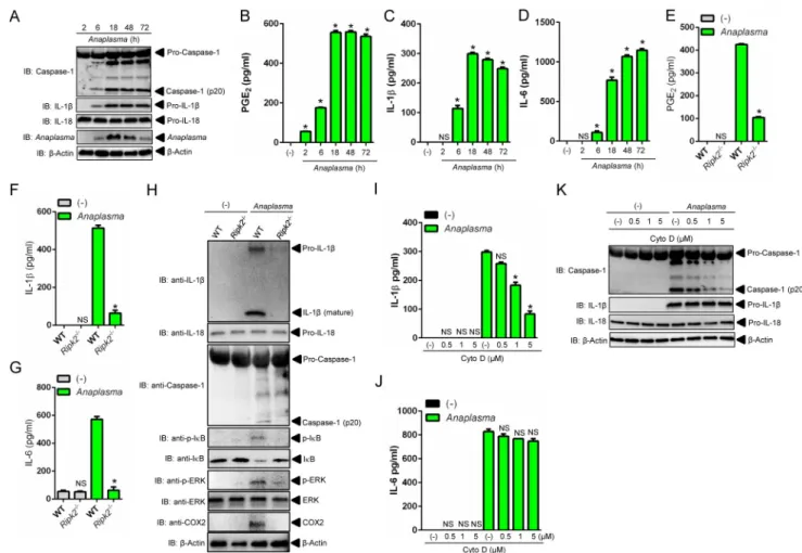
Next, we performed a kinetics experiment in macrophages to better characterizeA.
phagocyto-philuminfection in the context of NLRC4 inflammasome biology. As previously shown,A.
phagocytophilumwas undetectable inside macrophages at 2-hours post-infection [27]. A small
number of bacteria was observed at 6 hours, followed by an increased load at 18 hours and reduction at 48 hours, which led to almost complete elimination after 72 hours of infection in macrophages (Fig 8A) [27]. Consistently, PGE2secretion, caspase-1 activation and IL-1βand
IL-18 secretion but not IL-6, peaked at 18 hours, the same time point where the greatest num-ber ofA.phagocytophilumwas detected inside macrophages (Fig 8A–8DandS4A Fig).
A.phagocytophilumdoes not synthesize LPS or peptidoglycans [8,9,11]. Therefore, one
interesting immunological question pertains to the host molecule that induces NF-κB activa-tion upon infecactiva-tion. We reasoned that RIPK2 could be this master regulator. This hypothesis Fig 4. The COX pathway mediates theA.phagocytophilum-induced NLRC4 inflammasome.Wildtype (WT) BMDMs (1 x10⁶cells) were pre-treated with indomethacin (100 nM), AA861 (1μg/ml) and PD146176 (1μg/ml) for
2 hours followed byA.phagocytophiluminfection (MOI50) for 18 hours. The levels of (A) PGE2, (B) PGD2, (C)
TBXA2,(D) IL-1β, (E) IL-18 and (F) IL-6 were measured by ELISA in the cell culture supernatants. (G) NLRC4
detection by native gel/immunoblotting (IB) and (H) SDS-PAGE caspase-1 western blot indicating
autoproteolysis (p20). Pro-IL-1βand IL-18 were used as loading controls. One-way ANOVA-Tukey*P<.05; NS, not significant. (-) non-stimulated.
doi:10.1371/journal.ppat.1005803.g004
rested on four findings. First, RIPK2 activates NF-κB signaling and mitogen activated protein (MAP) kinases upon infection [13]. Second,A.phagocytophiluminteracts with the host endo-plasmic reticulum (ER) [39], which may exert RIPK2 activity in the absence of peptidoglycans due to cellular stress [40]. Third, COX2 expression is regulated through a signaling cascade that converges at the MAP kinase and the NF-κB pathways [41]. Fourth, mice deficient in RIPK2 are susceptible toA.phagocytophiluminfection and secrete reduced levels of IL-18 in the peripheral blood [14]. Accordingly,ripk2-/-macrophages exhibited a defect in NF-κB and MAP kinase signaling, which led to decreased translation of COX2, pro-IL-1βand IL-6 secre-tion (Fig 8G and 8H). RIPK2 activity also affected PGE2release and caspase-1 autoproteolysis
uponA.phagocytophiluminfection, as indicated by reduced levels of PGE2,IL-1β, IL-18 and
caspase-1 activation in cell culture supernatants ofripk2-/-macrophages (Fig 8E, 8F and 8H
andS4B Fig). Finally,A.phagocytophiluminternalization was important for PGE2release and
NLRC4 inflammasome activation, as demonstrated in our experiments with cytochalasin D, a Fig 5. COX2 regulates theA.phagocytophilum-induced NLRC4 inflammasome.Wildtype (WT) BMDMs (1 x10⁶cells) were pre-treated with celecoxib (0.1μM to 10μM) for 2 hours followed byA.phagocytophiluminfection
(MOI50) for 18 hours. The levels of (A) PGE2, (B) PGD2, (C) TBXA2,(D) IL-1β, (E) IL-18 and (F) IL-6 were
measured by ELISA in the cell culture supernatants. (G) SDS-PAGE/Western blot (IB) indicating caspase-1 autoproteolysis (p20). (H) NLRC4 inflammasome oligomer detection in the supernatants by native gel/ immunoblotting (IB). One-way ANOVA-Tukey*P<.05; NS, not significant. (-) non-stimulated.
doi:10.1371/journal.ppat.1005803.g005
potent mycotoxin that inhibits actin polymerization (Fig 8I–8KandS4C and S4D Fig). Collec-tively, we identified RIPK2 as a major regulator of the innate immune response againstA. phagocytophilum.
The PGE
2-EP3 receptor regulates activation of the NLRC4
inflammasome upon
A
.
phagocytophilum
infection
We then blunted the PGE2signaling cascade with chemical antagonists that bind covalently to
the four PGE2receptor subtypes (EP1-EP4) [30] and compared our findings withSalmonella.
We observed that inhibition of the PGE2-EP3 receptor significantly decreased IL-1βand IL-18
release, and caspase-1 activation, but not IL-6 secretion uponA.phagocytophiluminfection (Fig 9C–9EandS5 Fig). The EP3 receptor for PGE2is sensitive to pertussis toxin (PT) [42].
Macrophages pre-treated with PT and then stimulated withA.phagocytophilumalso resulted in inhibition of the NLRC4 inflammasome (S5A–S5D Fig). Importantly, the catalytically inac-tive pertussis toxin (PT), with a two amino acid substitution (9K129G) [43], did not block PGE2signaling uponA.phagocytophilumcolonization (S5A–S5D Fig). Next, we took
Fig 6. COX2 modulates NLRC4 inflammasome activity uponA.phagocytophiluminfection.BMDMs (1 x10⁶
cells) from wildtype (WT),Tlr4- and COX2 (Ptgs2)-deficient mice were infected withA.phagocytophilum(MOI 10/ 50) for 18 hours. The levels of (A) PGE2, (B) PGD2, (C) TBXA2,(D) IL-1β, (E) IL-18 and (F) IL-6 were measured by
ELISA in the cell culture supernatants. (G) Caspase-1 (p20) native gel and (H) immunoblotting (IB).β-Actin, pro-IL-1βand pro-IL-18 detected in lysates. One way ANOVA-Tukey.*P<0.05. NS–not significant. (-) non-stimulated.
doi:10.1371/journal.ppat.1005803.g006
advantage of theep3-/-mice and showed that in the absence of the EP3 receptor molecule,A.
phagocytophilumdid not induce caspase-1 activation and IL-1βand IL-18 secretion by
macro-phages (Fig 9F–9I). Conversely, lack of the PGE2-EP3 receptor did not affect the NLRC4
inflammasome induced bySalmonella(Fig 9J–9MandS6 Fig).
PGE2exerts its actions by acting on G-protein-coupled receptors (GPCRs). PGE2binds to
the EP3 receptor, which inhibits the membrane associated adenylyl cyclase via Gαi [44]. This signaling relay decreases cytosolic cyclic AMP (cAMP) production, as adenylyl cyclase cata-lyzes the conversion of adenosine triphosphate (ATP) to cAMP[44] (S8 Fig). We validated Fig 7. PGE2activates theA.phagocytophilum-induced NLRC4 inflammasome.(A-C)Ptgs2-/-BMDMs (1 x10⁶cells) were infected withA.
phagocytophilum(MOI50) for 4 hours followed by addition of PGE2(10μM), PGD2(10μM) or TBXA2(10μM) for 18 hours. (A) Levels of IL-1β
and (B) IL-18 in supernatants were measured by ELISA. (C) Caspase-1 autoproteolysis was measured in supernatants of infected cells. pro-IL-1β, pro-IL-18 andβ-actin were detected in cell lysates with SDS-PAGE immunoblot (IB). (D-H) Wildtype (WT) BMDMs (1 x10⁶cells) were
pre-treated with the specific inhibitor of mPGES1 (CAY10526–1μM) for 30 minutes followed byA.phagocytophiluminfection (MOI50) for 18 hours. The levels of (D) PGE2, (E) PGD2, (F) IL-1βand (G) IL-18 in the culture supernatants were measured by ELISA. (H) Caspase-1 p20
autoproteolysis in culture supernatants. pro-IL-1β, pro-IL-18 andβ-actin were detected in cell lysates with a SDS-PAGE immunoblot. (I-J) WT andmPGES1-/-BMDMs (1 x10⁶cells) were infected withA.phagocytophilum(MOI50) for 18 hours. The levels of (I) PGE
2and (J) PGD2were
measured in culture supernatants by ELISA. (K-M)mPGES1-/-BMDMs (1 x10⁶cells) were infected withA.phagocytophilumfor 4 hours
followed by addition of PGE2at indicated concentrations for 18 hours. (K) IL-1βand (L) IL-18 levels in cell culture supernatants were measured
by ELISA. (M) Caspase-1 autoproteolysis in cell culture supernatants. pro-IL-1β, pro-IL-18 andβ-actin in cell lysates were detected with SDS-PAGE immunoblot (IB). One-way ANOVA-Tukey and Student’s t test;*P<.05; NS, not significant. (-) non-stimulated.
doi:10.1371/journal.ppat.1005803.g007
these observations with sulprostone, an EP3 agonist and positive control in our assays (S7A Fig). Consistently,A.phagocytophilumcolonization of macrophages led to reduced production of cAMP (S7A Fig). Moreover, pharmacological blockade of the PGE2-EP3 receptor via the
EP3 antagonist or PT hindered the inhibition of cAMP byA.phagocytophilum(S7A Fig). Next, we showed that membrane, but not soluble, adenylyl cyclase modulated theA. phago-cytophilum-induced NLRC4 inflammasome. Forskolin, a selective inhibitor of the membrane-associated adenylyl cyclase [45], inhibited IL-1β, IL-18 and caspase-1 autoproteolysis duringA.
phagocytophiluminfection of macrophages (S7B–S7E Fig). On the other hand, pre-treatment
of macrophages with KH7, a specific pharmacological inhibitor of soluble adenylyl cyclase [45], did not affect NLRC4 inflammasome function duringA.phagocytophiluminfection (S7F–S7I Fig). Altogether, these findings: (i) indicated that the PGE2-EP3 axis is critical for the
Fig 8. NLRC4 inflammasome activation is dependent onA.phagocytophiluminternalization and RIPK2 function in macrophages.
(A-D) Wildtype (WT) BMDMs (1 x10⁶cells) were infected withA.phagocytophilum(MOI 50). Cell culture supernatants and lysates were collected at indicated time points post-infection. (A) Caspase-1 autoproteolysis was detected in cell culture supernatants, whereas the presence ofA.phagocytophilum, pro-IL-1β, pro-IL-18 andβ-actin are shown in cell lysates with SDS-PAGE immunoblots (IB). The levels of (B) PGE2, (C) IL-1βand (D) IL-6 were measured in cell culture supernatants by ELISA. (E-G) BMDMs from WT andRipk2-/-mice were infected
withA.phagocytophilum(MOI50) (1 x10⁶cells) for 18 hours. The levels of (E) PGE2, (F) IL-1βand (G) IL-6 were measured in cell culture
supernatants by ELISA. (H) Caspase-1 autoproteolysis was measured in cell culture supernatants, whereas pro-IL-1β, pro-IL-18, p-IκB, IκB, p-ERK, p-ERK, COX2 andβ-actin were detected in cell lysates by immunoblotting (IB). (I-K) WT BMDMs (1 x10⁶cells) were pre-treated with
indicated concentrations of cytochalasin D for 30 minutes followed byA.phagocytophiluminfection (MOI50) for 18 hours. The levels of (I) IL-1β and (J) IL-6 in cell culture supernatants were measured by ELISA. (K) Caspase-1 autoproteolysis was detected in cell culture supernatants. pro-IL-1β, pro-IL-18 andβ-actin were detected in cell lysates with SDS-PAGE immunoblot (IB). One-way ANOVA-Tukey and Student’s t test. *P<.05; NS, not significant. (-) non-stimulated.
doi:10.1371/journal.ppat.1005803.g008
NLRC4 inflammasome elicited byA.phagocytophilum; and (ii) explained whySalmonellais unable to trigger a similar pathway when compared toA.phagocytophilum. This was likely due to reduced expression of the EP3 receptor duringSalmonellainfection of macrophages (Fig 9A and 9B).
Mice deficient in COX2 are susceptible to
A
.
phagocytophilum
To prove that the results obtainedin vitrocould also be observedin vivo, we then infected mice deficient in COX2 (Ptgs2) withA.phagocytophilum.Ptgs2-deficient animals were more suscep-tible toA.phagocytophiluminfection (Fig 10A) and exhibited reduced levels of IL-18 in the peripheral blood when compared to the wildtype mice (Fig 10B). As previously seen, no detect-able levels of IL-1βwere observed in the blood ofA.phagocytophilum-infected mice [25]. Fig 9. The PGE2-EP3 axis regulates activation of the NLRC4 inflammasome uponA.phagocytophiluminfection.(A-B) Wildtype (WT)
BMDMs (1 x10⁶cells) were infected withA.phagocytophilum(MOI50) for 18 hours orSalmonella(MOI25) for 1 hour. RNA and protein levels of
the PGE2-EP3 receptor was measured by qRT-PCR and western blot (IB).β-actin was also detected in cell lysates. (C-E) WT BMDMs (1 x10⁶
cells) was pre-treated with the EP3 antagonist (L-798106) for 30 minutes at indicated concentrations followed byA.phagocytophilumcolonization (MOI50) for 18 hours. The levels of (C) IL-1βand (D) IL-6 were measured in cell culture supernatants by ELISA. (E) Caspase-1 autoproteolysis in cell culture supernatants. pro-IL-1β, pro-IL-18 andβ-actin were detected in cell lysates by SDS-PAGE immunoblots (IB). (F-H) WT andEp3
-/-BMDMs (1 x10⁶cells) were infected withA.phagocytophilum(MOI 10/50) for 18 hours. The levels of (F) IL-1β, (G) IL-18 and (H) IL-6 in culture
supernatants were measured by ELISAs. (I) Caspase-1 autoproteolysis in cell culture supernatants. pro-IL-1β, pro-IL-18 andβ-actin were detected in cell lysates by SDS-PAGE immunoblots (IB). (J-M) Naïve or LPS-primed (50 ng/ml) WT andEp3-/-BMDMs (1 x10⁶cells) were infected withSalmonella(MOI25) for 1 hour. The levels of (J) IL-1β, (K) IL-18 and (L) IL-6 in cultured supernatants were measured by ELISA. (M) Caspase-1 autoproteolysis in cell culture supernatants. pro-IL-Caspase-1β, pro-IL-18 andβ-actin were detected in cell lysates by SDS-PAGE immunoblots (IB). Student’s t test and ANOVA-Tukey.*P<.05; NS, not significant. (-) non-stimulated.
doi:10.1371/journal.ppat.1005803.g009
These findings agreed with our prior publications, showing that IL-18 release mediated by RIPK2 and the NLRC4 inflammasome regulates interferon (IFN)-γproduction by CD4+T cells uponA.phagocytophiluminfection [14,25]. COX2 (Ptgs2)-deficient mice infected withA.
phagocytophilumalso revealed lower levels of PGE2, PGD2, TBXA2and splenomegaly (Fig
10C–10G). COX2 (Ptgs2)-deficient animals had increased cellular infiltration in the red pulp and damage to the splenic architecture uponA.phagocytophiluminfection (Fig 10H). In sum, these results showed that COX2 is critically important forA.phagocytophiluminfectionin vivo.
Discussion
The NLRC4 inflammasome is currently thought to only recognize components of the bacterial T3SS and flagellin [18–24]. Other inflammasomes, however, such as the NLRP3 scaffold, sense a wide-range of molecular structures leading to caspase-1 activation and cytokine secretion [16]. We hypothesized that an alternative signaling cascade for the NLRC4 inflammasome Fig 10. COX2 restrictsA.phagocytophiluminfectionin vivo.A.phagocytophiluminfection of WT (n = 20) and COX2 (ptgs2)-/-(n = 10) mice. Bacterial load in the (A) peripheral blood of infected mice at day 15. (B) IL-18, (C) PGE2,
(D) PGD2and (E) TBXA2release in the serum of infected animals. (F-G) Splenomegaly for COX2 (ptgs2-/-) mice
infected withA.phagocytophilum. (H) Splenic architecture depicting the red (RP) and the white (WP) pulp duringA. phagocytophiluminfection. One-way ANOVA-Tukey; Student t test;*P<0.05. NS–not significant. (-) non-stimulated.
doi:10.1371/journal.ppat.1005803.g010
must exist because mice deficient in NLRC4 are susceptible toA.phagocytophiluminfection [25], an obligate intracellular rickettsial bacterium that does not have a T3SS and flagellin [11]. Furthermore,Annexin a2-deficient mice were more susceptible toA.phagocytophilum infec-tion and showed splenomegaly, thrombocytopenia and monopenia [28]. Macrophages defi-cient in Annexin A2, a phospholipid-binding protein, secreted significantly smaller amounts of IL-1βand IL-18 and had a defect in NLRC4 inflammasome oligomerization and caspase-1 acti-vation [28]. In contrast,Annexin a2-/-macrophages released IL-1β, IL-18, and IL-6 at wild-type levels when infected withSalmonella, a canonical NLRC4 agonist [28].
We provide unequivocal evidence that two distinct signaling pathways occur for NLRC4 inflammasome activation within the cell: one termed classical (i.e., stimulated bySalmonella) and another referred to as alternative (i.e. described here, responding toA.phagocytophilum). Given how inflammasome biology intersects with a growing number of disciplines, we reason that these findings are conceptually valuable because we reveal that eicosanoid receptors in immune cells activate diverging signaling cascades. For instance, bothA.phagocytophilumand
Salmonellalead to PGE2production by macrophages. However,Salmonellais unable to
acti-vate the eicosanoid-dependent NLRC4 inflammasome pathway because it does not induce PGE2-EP3 receptor expression.
PGE2is likely acting in an autocrine/paracrine manner to drive NLRC4 inflammasome
acti-vation uponA.phagocytophiluminfection. This is based on the evidence thatA. phagocytophi-luminfection upregulates the EP3 receptor, which is known to elicit PGE2signaling in a
cell-intrinsic manner [30,35]. Alternatively, PGE2may also affect the function of“bystander”cells
in a paracrine manner given that our exogenous PGE2“add-back”assays restored NLRC4
inflammasome activity inA.phagocytophilum-infected cells.
Can rickettsial agents be used to uncover broader principles of immune sensing? The answer to this question may have to deal with the biology of these organisms. Rickettsial agents differ greatly in terms of how they invade and replicate within the mammalian host when com-pared to other bacteria commonly used to study microbial immunity. Their obligate intracellu-lar life style, coupled to the intense selective pressure to survive both in the arthropod vector and the mammalian host [1,2] suggests that these microbes have to employ extreme measures to conceal themselves from the immune system. This reasoning may explain whyA.
phagocyto-philumtriggers such a distinct pathogen-recognition mechanism when compared to other
bacteria.
In summary, we discovered a novel mode of NLRC4 inflammasome activation triggered by the rickettsial bacteriumA.phagocytophilum. We revealed that some microbial pathogens lack-ing the T3SS and flagellin activate the NLRC4 inflammasome. We also illustrated how this pro-tein scaffold distinguishes bacterial infection within the cell. Altogether, our findings suggest that there are broader yet-to-be discovered principles of microbial sensing in the context of NLRC4 inflammasome biology.
Materials and Methods
Mice and bacteria
Breeding and experiments were performed in strict compliance with guidelines set forth by the National Institutes of Health (Office of Laboratory Animal Welfare [OLAW] assurance num-ber A3200-01). Procedures were approved by the Institutional Biosafety (IBC:00002247) and Animal Care and Use (IACUC:0413017 and 0216015) committees at the University of Mary-land, Baltimore.Ripk2-/-(007017), C57BL/6 (000664) andPtgs2-/-(COX2) mice (008101) were purchased from Jackson Laboratories. Femurs frommPGES1-/-[29] andEp3-/-[46] mice were a gift from Leslie Crofford and Richard Breyer at Vanderbilt University School of Medicine.
Tlr4-/-andcPla2-/-mice were previously described [47,48]. Mice were gender matched and at least 6–10 weeks of age. BMDMs were generated, as previously described [27]. Culturing for
theA.phagocytophilumstrain HZ and calculations were described elsewhere [27].Salmonella
strain SL1344 was a gift from Dr. Stefanie Vogel at the University of Maryland, Baltimore School of Medicine.Salmonellawas grown in HS media at 37°C and enumerated, as previously described [49]. Cell cultures were tested and determined to beMycoplasma-negative through a commercially available PCR kit (Southern Biotech -13100-01).
Chemical reagents
LPS (50ng/ml) was purchased from InvivoGen. Nigericin (10μM), indomethacin (100 nM) and celecoxib (0.1μM to 10μM) were purchased from Sigma-Aldrich. AA861 (1μg/ml) and PD146176 (1μg/ml) were purchased from BioMol International. CAY10526 (10010088), KH7 (13243), Forskolin (11018), Cytochalasin D (11330), PGE2(14010), PGD2(12010) and U46619
(thromboxane A2 analogue, 16450) were purchased from Cayman Chemicals. The PGE2
receptor antagonists EP1–1μM (SC51089), EP2–5μM (AH6809) and EP4–5μM (ONO-AE3-208) were purchased from Cayman Chemical, whereas the antagonist for the PGE2EP3–10μM
(L-798106) and the PGE2EP3 receptor agonist (sulprostone—3μM) was purchased from
Sigma. The inhibitors for the phospholipases cPLA2 (AACOCF3), sPLA2 (LY315920), PLC (U73122) and PLD (FIPI) were purchased from Tocris Bioscience. Pertussis toxin (PT) and the catalytically inactive pertussis toxin (PT) with a two amino acid substitution (9K129G) were described previously [43,50].
Bacterial infection of macrophages
1×10⁶BMDMs were seeded into 24-well plate in 300μl of media containing 5% fetal bovine serum (FBS) overnight prior to the challenge by eitherA.phagocytophilum(MOI 10 and 50) or
Salmonella(MOI 25) for 1 hour. 50ng/ml of LPS was used for cell priming at 37°C and 5% CO2
for 30 minutes duringSalmonellainfection. LPS-primed cells were washed twice extensively followed by the addition of bacteria. In inhibition assays, 1×10⁶WT and genotype-deficient BMDMs were pre-treated with pharmacological inhibitors at indicated time and concentra-tions followed by the stimulation withA.phagocytophilum(MOI 10 and 50) overnight or
Sal-monella(MOI 25) for 1 hour. For thePtgs2-/-andmPGES1-/-“add-back”experiments, 1×10⁶
WT and deficient cells were infected withA.phagocytophilum(MOI 50) for 4 hours followed by the addition of the respective eicosanoid at indicated concentrations for 18 hours. After infection, cultured supernatants and cell lysates collected from each well were used for ELISA and immunoblot assays.
Native polyacrylamide gel electrophoresis
Equal amounts of supernatants were mixed with the native sample buffer (62.5 mM Tris-HCl, 40% glycerol, 0.01% bromophenol blue, pH 6.8), loaded into 4–15% Mini-PROTEAN TGX Precast Gels and run at 200 volts for 2 hours in 1×Tris/Glycine native running buffer (25 mM Tris, 192 mM glycine, pH8.3). NativeMark Unstained Protein Standard (Invitrogen) was visu-alized with Gel Code Blue Safe Protein Stain solution (Thermo Scientific).
Immunoblotting
Cell lysates were prepared in radioimmunoprecipitation (RIPA) lysis buffer (Boston Biopro-ducts) with Halt Protease Inhibitor Cocktail (Thermo Scientific) and PhosSTOP (Roche Applied Science). 4–15% Mini-PROTEAN TGX precast gels were run at 200 volts for 30
minutes in the 1×Tris-Glycine-SDS running buffer (Boston Bioproducts). Transfer was per-formed using the Bio-Rad Trans-Blot Turbo with either polyvinylidene fluoride (PVDF) or nitrocellulose membranes Rad). Membranes were blocked in 5% skim milk or BSA (Bio-Rad). Western blot antibodies for caspase-1 (1:1000, Millipore 06–503 or 06-503-I, 1:1,000, Proteintech 22915-1-AP; 1:2000 Genentech 4175, cell line 4B4.2.1, or 1:1000, AdiPoGen Inter-national AG-20B-0042), NLRC4 (1:1000, Millipore, 06–1125), IL-1β(1:1000 R&D Systems and Cell Signaling, AF401-NA and 12426S), IL-18 (1:1000, MBL JM-5180-100),β-actin (1:1000, Sigma A2103), COX2 (1:1000, Cell Signaling 12282), phospho-IκB-α(1:1000, Cell Signaling 9246s), p-ERK (1:400, Cell Signaling 4370), ERK (1:1000, Cell Signaling 9102), IκB-α(1:1000, Cell Signaling 4812), PTGER3 (1:1,000, Abcam ab117998), anti-mouse horseradish peroxidase (HRP), anti-goat HRP, anti-rabbit HRP (1:5000, Abcam ab97046, ab97110 and ab97051, respectively), anti-rat HRP (1:5000 Abcam and Santa Cruz Biotechnology, ab97057 and sc-2006) were used. A rabbit polyclonal antibody raised againstA.phagocytophilum[51] was kindly provided by Erol Fikrig at Yale University School of Medicine (1:2,000). Enhanced chemiluminescence (ECL) western blotting substrate and Super Signal West Pico Chemilumi-nescent substrate were used (Thermo Scientific). Restore Western Blot Stripping Buffer was used for the stripping of antibodies on the blots (Thermo Scientific).
ELISA
IL-1βand IL-6 were measured with the BD OptEIA Set (BD Biosciences). IL-18 capture (1:1,000, D047-3) and detection antibodies (1:2,000, D048-6) were purchased from MBL. PGE2
was measured with the ELISA kit (Enzo Life Sciences). PGD2was measured with the ELISA kit
(Cayman Chemicals). Thromboxane A2was measured with the Mouse Thromboxane A2
ELISA Kit (Abbexa).
Quantitative RT-PCR
Quantitate RT-PCR was performed using the Power SYBR Green PCR Master Mix (Invitro-gen) in an ABI 7500 real-time PCR instrument. Primer sequences forA.phagocytophilumwere as follows: 16S-F (5’-CAGCCACACTGGAACTGAGA-3’) and 16S-R (5’-CCCTAAGGC CTTCCTCACTC-3’). Gene expression was normalized by using the primersβ-actin-F (5’-AC GCAGAGGGAAATCGTGCGTGAC-3’) andβ-actin-R (5’-ACGCGGGAGGAAGAGGATG CGGCAGTG-3’). The absolute quantification method was used. For the PGE2-EP3 receptor
quantification, PureLink RNA Mini Kit (Invitrogen) and the Verso cDNA synthesis Kit (Thermo Scientific) were used. Gene expression was normalized by using the primers GAPDH-F (5’-TGATGACATCAAGAAGGTGGTGAAG-3’) and GAPDH-R (5’-TCCTT GGAGGCCATGTGGGCCAT-3’). Primer sequences for the EP3 receptor were as follows: EP3-F (5’-GGTTCCTGTGAAGGACTGAAGAC-3’) and EP3-R (5’-AAGGTTCTGAGGCTG GAGATA-3’). The relative quantification method (fold changes) was used.
Enzymatic assays
15×106wildtype cells were stimulated withA.phagocytophilum(MOI 25) overnight. Cells were scraped followed by sonication. COX1/2 enzymatic assays were performed with COX activity assay kit (Cayman Chemicals), whereas cPLA2activity was measured following instructions by
the manufacturer (Abnova). Arachidonic acid levels were measured according to the instruc-tions of the ELISA kit (MyBiosource). cAMP was measured by using the cyclic AMP XP Assay Kit (Cell Signaling Technology).
Illumina sequencing and bioinformatics
BMDMs were grown into 6-well culture plates at 7×10⁶per well. Cells were stimulated withA. phagocytophilum. Uninfected BMDMs were used as controls and the experiment was per-formed in triplicate. Total RNA was isolated with the PureLink RNA Mini Kit (Invitrogen). Illumina Sequencing was performed at the University of Maryland, Baltimore. Briefly, Illumina RNAseq libraries were prepared with the TruSeq RNA Sample Prep kit (Illumina, San Diego, CA). The indexed libraries were pooled and sequenced using the HiSeq platform (Illumina) for the mouse samples in order to generate 101 base pair reads. The reads were further trimmed due to low quality at the trailing 3' end. These trimmed paired end reads were populated into 2 separate FASTQ format files and the quality of the reads was tested using the FastQC toolkit to ensure quality of the sequencing reads.
The RNA sequencing reads were used as input for the TopHat read alignment tool to be aligned to the mouse genomic reference sequence (Ensembl GRCm38 version) for each of the samples. The reference genomic sequences for the GRCm38 genome build were downloaded from the Ensembl resources. The output from TopHat was obtained as BAM format files. In the alignment phase, we allowed up to two mismatches per 30 base pair segment and removed reads that aligned to more than 20 genomic locations. The BAM alignment files obtained from the TopHat alignment tool was analyzed to generate the alignment statistics for each sample, namely, the total number of reads, the number of mapped reads and the percent of mapped reads.
For the differential gene expression analysis, the alignment BAM files from TopHat were further utilized to compute gene expression levels and test each gene for differential expression. The mouse gene set reference annotation (version GRCm38) in GTF format was downloaded from the Ensembl resources. The number of reads that mapped to each gene described in the Ensembl annotation was calculated using the python package HTSeq-an alignment read count tool. The read count represented the expression of the gene. Differential gene expression analy-sis was conducted using the DESeq R package (available from Bioconductor). The DESeq anal-ysis resulted in the determination of differentially expressed genes. DESeq utilized the read counts provided by the HTSeq read count tool. The read counts for each sample were normal-ized for sequencing depth and distortion caused by highly differentially expressed genes. The negative binomial model was used to test the significance of differential expression between two genotypes. The differentially expressed genes were deemed significant if the FDR (False Discovery Rate) was less than 0.01, the gene expression was above the 45th percentile and gene showed greater than 2-fold change difference (over expressed or under expressed) between conditions. Principal component analysis and other clustering methods were used to visualize the clustering of the replicates across samples. Heat maps were generated to illustrate the genes showing significant differences between multiple comparisons of the control and other infec-tion and/or treatment condiinfec-tions.
In vivo
infection
C57BL/6 (n = 20) and COX2 (Ptgs2)-/-(n = 10) mice were infected by intraperitoneal injection
withA.phagocytophilumstrain HZ (1×10⁷cells). Blood samples were collected at days 0, 5 and
10 for the IL-18 ELISA. Spleens were removed, normalized to the body weight, and compared to those of non-infected mice. Spleens were fixed at day 15 post-infection with 10% neutral buffered formalin and embedded in paraffin wax. Sections (5μm) were obtained and stained with hematoxylin and eosin. Measurement ofA.phagocytophilumload was done at day 15 post-infection in the peripheral blood of infected animals using quantitative RT-PCR, as described above.
Statistical analysis
All experiments in this study were performed with at least 2–5 replicates. All data were expressed as means ± standard errors of the means (SEM). The differences between groups were examined by either unpaired Student'sttest or one-way analysis of variance (ANOVA). All statistical calculations and graphs were made by using GraphPad Prism version 6.0. P<0.05 was considered statistically significant.
Supporting Information
S1 Fig. TheSalmonella-induced NLRC4 inflammasome is not affected by inhibition of phospholipases.Wildtype (WT) BMDMs (1 x10⁶cells) were pre-treated for 30 minutes with inhibitors of secreted PLA2(sPLA2) (LY315920–10μM), cPLA2(AACOCF3–10μM),
phospho-lipase C (PLC) (U73122–10μM) and phospholipase D (PLD) (FIPI–0.3μM). Cells were then primed with LPS (50ng/ml) and infected withSalmonella(MOI25) for 1 hour. (A) IL-1β, (B) IL-18 and (C) IL-6 were measured in cell culture supernatants by ELISA. (D) SDS-PAGE immunoblot (IB) of caspase-1 p20. (E-G) BMDMs from wildtype (WT) or cPLA2-deficient
mice (1 x10⁶cells) were infected withSalmonella(MOI25) for 1 hour. Levels of (E) IL-1βand (F) IL-6 were measured in cell culture supernatants by ELISA. (G) SDS-PAGE followed by immunoblot (IB) of caspase-1 p20 in the supernatants. pro-IL-1βand pro-IL-18 detected in lysates. ANOVA-Tukey.P<0.05. NS–not significant. (-), non-stimulated.
(TIF)
S2 Fig. COX2 does not influence canonical inflammasome activation.BMDMs from wild-type (WT) and COX2 (Ptgs2)-deficient mice (1 x10⁶cells) primed with LPS (50ng/ml) for 1 hour and infected withSalmonella(MOI25–1 hour) or stimulated with nigericin (10μM–18 hours). (A) PGE2,(B) IL-1β,(C) IL-18 and (D) IL-6 release in cell culture supernatants was
measured by ELISA. (E) Caspase-1 native gel immunoblotting (IB). (F) SDS-PAGE/Western blot indicating caspase-1 autoproteolysis (p20). Student’s t test.P<0.05.β-actin and pro-IL-18 used as loading controls.
(TIF)
S3 Fig. PGE2does not affect IL-6 secretion duringA.phagocytophiluminfection.(A)
Ptgs2-/-BMDMs (1 x10⁶cells) were infected withA.phagocytophilumfor 4 hours followed by
addition of PGE2(10μM), PGD2(10μM) or TBXA2(10μM) for 18 hours. IL-6 was measured
in the cell culture supernatants by ELISA. (B) Wildtype (WT) BMDMs (1 x10⁶cells) were pre-treated with the mPGES1 inhibitor CAY10526 at indicated concentrations for 30 minutes fol-lowed byA.phagocytophiluminfection (MOI50) for 18 hours. IL-6 was measured in the cell culture supernatants by ELISA. (C)mPGES1-/-BMDMs (1 x10⁶cells) were infected withA.
phagocytophilumfor 4 hours followed by addition of PGE2(10μM). IL-6 was measured in the
cell culture supernatants by ELISA. One-way ANOVA-Tukey. NS, not significant. (-) non-stimulated.
(TIF)
S4 Fig. PGE2and IL-18 secretion is dependent onA.phagocytophiluminternalization and
RIPK2 function in macrophages.(A) Wildtype (WT) BMDMs (1 x10⁶cells) were infected
withA.phagocytophilum(MOI50). Cell culture supernatants were collected at indicated time
points post-infection. The levels of (A) IL-18 was measured in cell culture supernatants by ELISA. (B) BMDMs from wildtype (WT) andRipk2-/-mice were infected withA.
phagocyto-philum(MOI50) (1 x10⁶cells) for 18 hours. The levels of IL-18 were measured in cell culture
supernatants by ELISA. (C-D) WT BMDMs (1 x10⁶cells) were pre-treated with indicated
concentrations of cytochalasin D for 30 minutes followed byA.phagocytophiluminfection (MOI50) for 18 hours. The levels of (C) PGE2and (D) IL-18 in cell culture supernatants was
measured by ELISA. One-way ANOVA-Tukey; Student’s t test.P<.05. NS, not significant. (-) non-stimulated.
(TIF)
S5 Fig. The EP3 receptor modulates NLRC4 inflammasome activity uponA.
phagocytophi-luminfection.Wildtype (WT) BMDMs (1 x10⁶cells) were pre-treated for 30 minutes with antagonists of PGE2receptors: (1)–(naïve); (2) (EP1–1μM) (SC51089); (3) (EP2–5μM)
(AH6809); (4) (EP3–10μM) (L-798106); (5) (EP4–5μM) (ONO-AE3-208); (6) active (PT– 0.1μg/ml) and (7) catalytically inactive (PT–0.1μg/ml) pertussis toxin and stimulated with (A-D)A.phagocytophilum(MOI50) for 18 hours. The levels of (A) IL-1β, (B) IL-18 and (C) IL-6 release in cell culture supernatants were measured by ELISA. (D) Caspase-1 autoproteoly-sis immunoblotting (IB). pro-IL-1βand pro-IL-18 were detected in cell lysates. (E-I) WT BMDMs (1 x10⁶cells) were pre-treated for 30 minutes with the EP3 antagonist or pertussis toxin at indicated concentrations for 30 minutes followed byA.phagocytophiluminfection (MOI50) for 18 hours. (E, G) IL-18; (F) IL-1βand (H) IL-6 release in cell culture supernatants were measured by ELISA. (I) Caspase-1 autoproteolysis immunoblotting (IB). pro-IL-1βand pro-IL-18 were detected in cell lysates. ANOVA-Tukey.P<0.05. NS–not significant. (-), non-stimulated.
(TIF)
S6 Fig. The EP3 receptor does not regulate the activity of the canonical NAIP/NLRC4 inflammasome induced bySalmonellainfection.WT BMDMs (1 x10⁶cells) primed with LPS (50ng/ml) were pre-treated for 30 minutes with antagonists of PGE2receptors: (1)–
(naïve); (2) (EP1–1μM) (SC51089); (3) (EP2–5μM) (AH6809); (4) (EP3–10μM) (L-798106); (5) (EP4–5μM) (ONO-AE3-208); (6) active (PT–0.1μg/ml) and (7) catalytically inactive (PT– 0.1μg/ml) pertussis toxin and stimulated with (A-C)Salmonella(MOI25) for 1 hour. The lev-els of (A) IL-1βand (B) IL-18 release in cell culture supernatants were measured by ELISA. (C) Caspase-1 autoproteolysis immunoblotting (IB). pro-IL-1βand pro-IL-18 were detected in cell lysates. One way ANOVA-Tukey; NS–not significant. (-) non-stimulated.
(TIF)
S7 Fig. Membrane-associated adenylyl cyclase modulates theA.phagocytophilum-induced NLRC4 inflammasome.(A) Wildtype (WT) BMDMs (1 x10⁶cells) were pre-treated with the EP3 agonist sulprostone (3μM), the EP3 antagonist L-798106 (10μM), or active pertussis toxin (PT–0.1μg/ml) for 30 minutes followed byA.phagocytophilum(MOI50) infection for 18 hours. cAMP levels were measured. (B-I) WT BMDMs (1 x10⁶cells) were pre-treated with the selective (B-E) membrane (Forskolin) or (F-I) soluble (KH7) adenylyl cyclase inhibitors at indicated concentrations for 30 min followed byA.phagocytophilumcolonization (MOI50) for 18 hours. The levels of (B, F) IL-1β, (C, G) IL-18 and (D, H) IL-6 in the cell culture superna-tants were measured by ELISA. (E, I) Caspase-1 autoproteolysis was detected with SDS-PAGE immunoblot (IB). Pro-IL-1βand pro-IL-18 were detected in cell lysates. One-way ANOVA-Tukey.P<0.05. NS–not significant. (-) non-stimulated.
(TIF)
S8 Fig. Schematic representation of theA.phagocytophilum-induced NLRC4 inflamma-some.A.phagocytophiluminfection and formation of the occupied vacuole (ApV) leads to dis-ruption and molecular rearrangements within the cell [28]. (1) Cytosolic phospholipase A2 (cPLA2) releases (2) arachidonic acid from phosphatidylinositol 4,5-bisphosphate [PI(4,5)P2],
the major polyphosphoinositide phospholipid present in the inner leaflet of the plasma
membrane [52]. (3) Cyclooxygenase 2 (COX2) and microsomal PGE synthase-1 (mPGES1) [29] convert hydrolyzed arachidonic acid to prostanglandin E2(PGE2). PGE2exerts its actions
by acting on G-protein-coupled receptors (GPCRs). PGE2binds to the EP3 receptor, which
inhibits the membrane associated adenylyl cyclase (AC) via Gαi (4). This signaling relay decreases cytosolic cyclic AMP (cAMP) production. Lower levels of cAMP induce the activa-tion of the NLRC4 inflammasome (5). Receptor-interacting serine/threonine-protein kinase 2 (RIPK2) stimulates the production of pro-IL-1βvia nuclear factor (NF)-κB signaling (6). RIPK2 also triggers formation of the NLRC4 inflammasome oligomer through COX2 up-regu-lation (7) via mitogen-activated protein kinase (MAPK) signaling [41]. Caspase-1 cleaves pro-IL-1βand pro-IL-18 leading to the release of mature cytokines (8).
(TIF)
Acknowledgments
The authors acknowledge Vishva Dixit (Genentech) for providing the anti-caspase-1 antibody and Erol Fikrig (Yale University School of Medicine) for the anti-A.phagocytophilumantibody; Stefanie Vogel (University of Maryland, Baltimore School of Medicine) for providing strains of Salmonella; Leslie Crofford (Vanderbilt University School of Medicine) and Richard Breyer (Vanderbilt University School of Medicine) for providing mice deficient in the enzyme mPGES1 and the EP3 receptor, respectively; Erin McClure (University of Maryland, Baltimore School of Medicine) for the graphic design; the Core facilities at the University of Maryland, Baltimore for services related to Illumina sequencing, informatics and pathology; Sarah Davis (Vanderbilt University School of Medicine), Elizabeth M. Johnson (Vanderbilt University School of Medicine), Hal Neely (University of Maryland, Baltimore School of Medicine), Mar-tin Flajnik (University of Maryland, Baltimore School of Medicine), Eileen O’Leary (Massachu-setts General Hospital and Harvard Medical School) and Vickie Knepper-Adrian (University of Iowa School of Medicine) for technical assistance.
Author Contributions
Conceived and designed the experiments: JHFP XW. Performed the experiments: XW DKS HLH MR TSV LV SME KGR KMS. Analyzed the data: JHFP XW DKS MK. Contributed reagents/materials/analysis tools: FSS NHC JAC KAS DJP JWM EAM JVB MK. Wrote the paper: JHFP XW.
References
1. Rikihisa Y.Anaplasma phagocytophilumandEhrlichia chaffeensis: subversive manipulators of host cells. Nat Rev Microbiol. 2010; 8(5):328–39. doi:10.1038/nrmicro2318PMID:20372158
2. Walker DH, Ismail N. Emerging and re-emerging rickettsioses: endothelial cell infection and early dis-ease events. Nat Rev Microbiol. 2008; 6(5):375–86. Epub 2008/04/17. doi:10.1038/nrmicro1866 PMID:18414502
3. Jordan JM, Woods ME, Soong L, Walker DH.Rickettsiaestimulate dendritic cells through toll-like receptor 4, leading to enhanced NK cell activationin vivo. J Infect Dis. 2009; 199(2):236–42. doi:10. 1086/595833PMID:19072551
4. Jordan JM, Woods ME, Olano J, Walker DH. The absence of Toll-like receptor 4 signaling in C3H/HeJ mice predisposes them to overwhelming rickettsial infection and decreased protective Th1 responses. Infect Immun. 2008; 76(8):3717–24. doi:10.1128/IAI.00311-08PMID:18490467
5. Bechelli J, Smalley C, Zhao X, Judy B, Valdes P, Walker DH, et al. MyD88 mediates instructive signal-ing in dendritic cells and protective inflammatory response dursignal-ing rickettsial infection. Infect Immun. 2016; 84(4):883–93. doi:10.1128/IAI.01361-15PMID:26755162
6. Chattoraj P, Yang Q, Khandai A, Al-Hendy O, Ismail N. TLR2 and Nod2 mediate resistance or suscepti-bility to fatal intracellularEhrlichiainfection in murine models of ehrlichiosis. PLoS One. 2013; 8(3): e58514. doi:10.1371/journal.pone.0058514PMID:23526993
7. Koh YS, Koo JE, Biswas A, Kobayashi KS. MyD88-dependent signaling contributes to host defense against ehrlichial infection. PLoS One. 2010; 5(7):e11758. doi:10.1371/journal.pone.0011758PMID: 20668698
8. Rikihisa Y. Molecular pathogenesis ofEhrlichia chaffeensisinfection. Annu Rev Microbiol. 2015; 69:283–304. doi:10.1146/annurev-micro-091014-104411PMID:26488275
9. Severo MS, Stephens KD, Kotsyfakis M, Pedra JH.Anaplasma phagocytophilum: deceptively simple or simply deceptive? Future Microbiol. 2012; 7(6):719–31. doi:10.2217/fmb.12.45PMID:22702526
10. Ge Y, Rikihisa Y. Subversion of host cell signaling byOrientia tsutsugamushi. Microbes Infect. 2011; 13 (7):638–48. doi:10.1016/j.micinf.2011.03.003PMID:21458586
11. Dunning Hotopp JC, Lin M, Madupu R, Crabtree J, Angiuoli SV, Eisen JA, et al. Comparative genomics of emerging human ehrlichiosis agents. PLoS Genet. 2006; 2(2):e21. PMID:16482227
12. Amano K, Tamura A, Ohashi N, Urakami H, Kaya S, Fukushi K. Deficiency of peptidoglycan and lipo-polysaccharide components inRickettsia tsutsugamushi. Infect Immun. 1987; 55(9):2290–2. PMID: 3114150
13. Ting JP, Duncan JA, Lei Y. How the noninflammasome NLRs function in the innate immune system. Science. 2010; 327(5963):286–90. doi:10.1126/science.1184004PMID:20075243
14. Sukumaran B, Ogura Y, Pedra JH, Kobayashi KS, Flavell RA, Fikrig E. Receptor interacting protein-2 contributes to host defense againstAnaplasma phagocytophiluminfection. FEMS Immunol Med Micro-biol. 2012; 66(2):211–9. doi:10.1111/j.1574-695X.2012.01001.xPMID:22747758
15. Cho KA, Jun YH, Suh JW, Kang JS, Choi HJ, Woo SY.Orientia tsutsugamushiinduced endothelial cell activation via the NOD1-IL-32 pathway. Microb Pathog. 2010; 49(3):95–104. doi:10.1016/j.micpath. 2010.05.001PMID:20470879
16. Guo H, Callaway JB, Ting JP. Inflammasomes: mechanism of action, role in disease, and therapeutics. Nat Med. 2015.
17. Yang Q, Stevenson HL, Scott MJ, Ismail N. Type I interferon contributes to noncanonical inflamma-some activation, mediates immunopathology, and impairs protective immunity during fatal infection with lipopolysaccharide-negative ehrlichiae. Am J Pathol. 2015; 185(2):446–61. doi:10.1016/j.ajpath. 2014.10.005PMID:25481711
18. Kofoed EM, Vance RE. Innate immune recognition of bacterial ligands by NAIPs determines inflamma-some specificity. Nature. 2011; 477(7366):592–5. doi:10.1038/nature10394PMID:21874021
19. Zhao Y, Yang J, Shi J, Gong YN, Lu Q, Xu H, et al. The NLRC4 inflammasome receptors for bacterial flagellin and type III secretion apparatus. Nature. 2011; 477(7366):596–600. doi:10.1038/nature10510 PMID:21918512
20. Hu Z, Zhou Q, Zhang C, Fan S, Cheng W, Zhao Y, et al. Structural and biochemical basis for induced self-propagation of NLRC4. Science. 2015; 350(6259):399–404. doi:10.1126/science.aac5489PMID: 26449475
21. Zhang L, Chen S, Ruan J, Wu J, Tong AB, Yin Q, et al. Cryo-EM structure of the activated NAIP2-NLRC4 inflammasome reveals nucleated polymerization. Science. 2015; 350(6259):404–9. doi:10. 1126/science.aac5789PMID:26449474
22. Diebolder CA, Halff EF, Koster AJ, Huizinga EG, Koning RI. Cryoelectron tomography of the NAIP5/ NLRC4 inflammasome: implications for NLR activation. Structure. 2015; 23(12):2349–57 doi:10.1016/ j.str.2015.10.001PMID:26585513
23. Zhao Y, Shi J, Shi X, Wang Y, Wang F, Shao F. Genetic functions of the NAIP family of inflammasome receptors for bacterial ligands in mice. J Exp Med. 2016; 213(5):647–56. doi:10.1084/jem.20160006 PMID:27114610
24. Rauch I, Tenthorey JL, Nichols RD, Al Moussawi K, Kang JJ, Kang C, et al. NAIP proteins are required for cytosolic detection of specific bacterial ligandsin vivo. J Exp Med. 2016; 213(5):657–65. doi:10. 1084/jem.20151809PMID:27045008
25. Pedra JH, Sutterwala FS, Sukumaran B, Ogura Y, Qian F, Montgomery RR, et al. ASC/PYCARD and caspase-1 regulate the IL-18/IFN-γaxis duringAnaplasma phagocytophiluminfection. J Immunol. 2007; 179(7):4783–91. PMID:17878377
26. Rikihisa Y, Lin M, Niu H. Type IV secretion in the obligatory intracellular bacterium Anaplasma phago-cytophilum. Cell Microbiol. 2010; 12(9):1213–21. doi:10.1111/j.1462-5822.2010.01500.xPMID: 20670295
27. Chen G, Wang X, Severo MS, Sakhon OS, Sohail M, Brown LJ, et al. The tick salivary protein Sialosta-tin L2 inhibits caspase-1-mediated inflammation duringAnaplasma phagocytophiluminfection. Infect Immun. 2014; 82(6):2553–64 doi:10.1128/IAI.01679-14PMID:24686067
28. Wang X, Shaw DK, Sakhon OS, Snyder GA, Sundberg EJ, Santambrogio L, et al. The tick protein Sia-lostatin L2 binds to Annexin A2 and inhibits NLRC4-mediated inflammasome activation. Infect Immun. 2016; 84(6):1796–805. doi:10.1128/IAI.01526-15PMID:27045038
29. Trebino CE, Stock JL, Gibbons CP, Naiman BM, Wachtmann TS, Umland JP, et al. Impaired inflamma-tory and pain responses in mice lacking an inducible prostaglandin E synthase. Proc Natl Acad Sci U S A. 2003; 100(15):9044–9. PMID:12835414
30. Kawahara K, Hohjoh H, Inazumi T, Tsuchiya S, Sugimoto Y. Prostaglandin E2-induced inflammation:
Relevance of prostaglandin E receptors. Biochim Biophys Acta. 2015; 1851(4):414–21. doi:10.1016/j. bbalip.2014.07.008PMID:25038274
31. Dumler JS, Barat NC, Barat CE, Bakken JS. Human granulocytic anaplasmosis and macrophage acti-vation. Clin Infect Dis. 2007; 45(2):199–204. PMID:17578779
32. Dumler JS. The biological basis of severe outcomes inAnaplasma phagocytophiluminfection. FEMS Immunol Med Microbiol. 2012; 64(1):13–20. doi:10.1111/j.1574-695X.2011.00909.xPMID:22098465
33. Lepidi H, Bunnell JE, Martin ME, Madigan JE, Stuen S, Dumler JS. Comparative pathology, and immu-nohistology associated with clinical illness afterEhrlichia phagocytophila-group infections. Am J Trop Med Hyg. 2000; 62(1):29–37. PMID:10761721
34. Choi KS, Scorpio DG, Dumler JS.Anaplasma phagocytophilumligation to toll-like receptor (TLR) 2, but not to TLR4, activates macrophages for nuclear factor-kappa B translocation. J Infect Dis. 2004; 189 (10):1921–5. PMID:15122530
35. Dennis EA, Norris PC. Eicosanoid storm in infection and inflammation. Nat Rev Immunol. 2015; 15 (8):511–23. doi:10.1038/nri3859PMID:26139350
36. von Moltke J, Trinidad NJ, Moayeri M, Kintzer AF, Wang SB, van Rooijen N, et al. Rapid induction of inflammatory lipid mediators by the inflammasomein vivo. Nature. 2012; 490(7418):107–11. doi:10. 1038/nature11351PMID:22902502
37. Shirey KA, Lai W, Pletneva LM, Karp CL, Divanovic S, Blanco JC, et al. Role of the lipoxygenase path-way in RSV-induced alternatively activated macrophages leading to resolution of lung pathology. Mucosal Immunol. 2014; 7(3):549–57. doi:10.1038/mi.2013.71PMID:24064666
38. Simmons DL, Botting RM, Hla T. Cyclooxygenase isozymes: the biology of prostaglandin synthesis and inhibition. Pharmacol Rev. 2004; 56(3):387–437. PMID:15317910
39. Truchan HK, Cockburn CL, Hebert KS, Magunda F, Noh SM, Carlyon JA. The pathogen-occupied vac-uoles ofAnaplasma phagocytophilumandAnaplasma marginaleinteract with the endoplasmic reticu-lum. Front Cell Infect Microbiol. 2016; 6:22. doi:10.3389/fcimb.2016.00022PMID:26973816
40. Keestra-Gounder AM, Byndloss MX, Seyffert N, Young BM, Chavez-Arroyo A, Tsai AY, et al. NOD1 and NOD2 signalling links ER stress with inflammation. Nature. 2016; 532(7599):394–7. doi:10.1038/ nature17631PMID:27007849
41. Taxman DJ, Lei Y, Zhang S, Holley-Guthrie E, Offenbacher S, Ting JP. ASC-dependent RIP2 kinase regulates reduced PGE2production in chronic periodontitis. J Dent Res. 2012; 91(9):877–82. doi:10.
1177/0022034512454541PMID:22828789
42. Tsuchiya H, Oka T, Nakamura K, Ichikawa A, Saper CB, Sugimoto Y. Prostaglandin E2attenuates
pre-optic expression of GABAA receptors via EP3 receptors. J Biol Chem. 2008; 283(16):11064–71. doi: 10.1074/jbc.M801359200PMID:18292084
43. Pizza M, Covacci A, Bartoloni A, Perugini M, Nencioni L, De Magistris MT, et al. Mutants of pertussis toxin suitable for vaccine development. Science. 1989; 246(4929):497–500. PMID:2683073
44. Morimoto K, Shirata N, Taketomi Y, Tsuchiya S, Segi-Nishida E, Inazumi T, et al. Prostaglandin E2
-EP3 signaling induces inflammatory swelling by mast cell activation. J Immunol. 2014; 192(3):1130–7. doi:10.4049/jimmunol.1300290PMID:24342806
45. Sokolowska M, Chen LY, Liu Y, Martinez-Anton A, Qi HY, Logun C, et al. Prostaglandin E2inhibits
NLRP3 inflammasome activation through EP4 Receptor and intracellular cyclic AMP in human macro-phages. J Immunol. 2015; 194(11):5472–87. doi:10.4049/jimmunol.1401343PMID:25917098
46. Ceddia RP, Lee D, Maulis MF, Carboneau BA, Threadgill DW, Poffenberger G, et al. The PGE2EP3
receptor regulates diet-induced adiposity in male mice. Endocrinology. 2016; 157(1):220–32. doi:10. 1210/en.2015-1693PMID:26485614
47. Perkins DJ, Polumuri SK, Pennini ME, Lai W, Xie P, Vogel SN. Reprogramming of murine macro-phages through TLR2 confers viral resistance via TRAF3-mediated, enhanced interferon production. PLoS Pathog. 2013; 9(7):e1003479. doi:10.1371/journal.ppat.1003479PMID:23853595
48. Bonventre JV, Huang Z, Taheri MR, O'Leary E, Li E, Moskowitz MA, et al. Reduced fertility and post-ischaemic brain injury in mice deficient in cytosolic phospholipase A2. Nature. 1997; 390(6660):622–5. PMID:9403693
49. Tennant SM, Wang JY, Galen JE, Simon R, Pasetti MF, Gat O, et al. Engineering and preclinical evalu-ation of attenuated nontyphoidalSalmonellastrains serving as live oral vaccines and as reagent strains. Infect Immun. 2011; 79(10):4175–85. doi:10.1128/IAI.05278-11PMID:21807911
50. Carbonetti NH, Artamonova GV, Mays RM, Worthington ZE. Pertussis toxin plays an early role in respi-ratory tract colonization byBordetella pertussis. Infect Immun. 2003; 71(11):6358–66. PMID:14573656
51. Ijdo JW, Sun W, Zhang Y, Magnarelli LA, Fikrig E. Cloning of the gene encoding the 44-kilodalton anti-gen of the aanti-gent of human granulocytic ehrlichiosis and characterization of the humoral response. Infect Immun. 1998; 66(7):3264–9. PMID:9632594
52. Lucas KK, Dennis EA. The ABC's of group IV cytosolic phospholipase A2. Biochim Biophys Acta. 2004; 1636(2–3):213–8. PMID:15164769