*For correspondence: [email protected]
Present address:†Discovery Chemistry, Genentech Inc, South San Francisco, United States; ‡Department of Cell Biology, Neurobiology, and Anatomy, Medical College of Wisconsin, Milwaukee, United States
Competing interests:The authors declare that no competing interests exist.
Funding:See page 14
Received:20 November 2019 Accepted:26 February 2020 Published:02 March 2020
Reviewing editor: Nir Ben-Tal, Tel Aviv University, Israel
Copyright Patel et al. This article is distributed under the terms of theCreative Commons Attribution License,which permits unrestricted use and redistribution provided that the original author and source are credited.
Structure-based discovery of potent and
selective melatonin receptor agonists
Nilkanth Patel
1, Xi Ping Huang
2,3, Jessica M Grandner
1†, Linda C Johansson
1,
Benjamin Stauch
1, John D McCorvy
2,3‡, Yongfeng Liu
2,3, Bryan Roth
2,3,4,
Vsevolod Katritch
1*
1
Department of Biological Sciences and Department of Chemistry, Bridge Institute,
USC Michelson Center for Convergent Biosciences, University of Southern
California, Los Angeles, United States;
2Department of Pharmacology, University of
North Carolina Chapel Hill Medical School, Chapel Hill, United States;
3National
Institute of Mental Health Psychoactive Drug Screening Program, Department of
Pharmacology, University of North Carolina Chapel Hill Medical School, Chapel Hill,
United States;
4Division of Chemical Biology and Medicinal Chemistry, University of
North Carolina Chapel Hill Medical School, Chapel Hill, United States
Abstract
Melatonin receptors MT1and MT2are involved in synchronizing circadian rhythms andare important targets for treating sleep and mood disorders, type-2 diabetes and cancer. Here, we performed large scale structure-based virtual screening for new ligand chemotypes using recently solved high-resolution 3D crystal structures of agonist-bound MT receptors. Experimental testing of 62 screening candidates yielded the discovery of 10 new agonist chemotypes with sub-micromolar potency at MT receptors, with compound21reaching EC50of 0.36 nM. Six of these
molecules displayed selectivity for MT2over MT1. Moreover, two most potent agonists, including 21and a close derivative of melatonin,28, had dramatically reduced arrestin recruitment at MT2,
while compound37was devoid of Gisignaling at MT1, implying biased signaling. This study
validates the suitability of the agonist-bound orthosteric pocket in the MT receptor structures for the structure-based discovery of selective agonists.
Introduction
The type 1A and 1B melatonin receptors (MT1and MT2) are G protein-coupled receptors (GPCRs)
that respond to the neurohormone melatonin (N-acetyl-5-methoxytryptamine) (Pe´vet, 2016; Reppert et al., 1994). Melatonin is found in all mammals, including humans, where it regulates sleep and helps to synchronize the circadian rhythm with natural light-dark cycles (Brzezinski, 1997; Xie et al., 2017). Chemically, melatonin is synthesized from serotonin in the pineal gland of the brain during darkness (Ganguly et al., 2002). Both MT1and MT2share canonical
helical 7-transmembrane (7-TM) topology (Johansson et al., 2019;Stauch et al., 2019), although they are differentially expressed and implicated in diverse biological functions and pathologies (Dubocovich and Markowska, 2005). While exogenous melatonin has been commonly used for the treatment of insomnia and jetlag, more effective and long-lasting MT agonists such as ramelteon have been approved for primary chronic insomnia treatment, because of their low side-effect profile as compared to other sleeping aids such as benzodiazepines (Hardeland et al., 2011;Erman et al., 2006). Other MT agonists such as tasimelteon and agomelatine, are used for non-24-hour sleep-wake disorders in blind individuals and as an atypical anti-depressant for major depressive disorders, respectively (Lavedan et al., 2015;de Bodinat et al., 2010). Recent studies also suggest MT recep-tors play an essential role in learning, memory, and neuroprotection (Liu et al., 2016) and illustrate the potential utility of partial and selective MT2receptor agonists as antinociceptive drugs
Canul et al., 2015). Moreover, MT2single nucleotide polymorphisms (SNPs) are implicated in type-2
diabetes (Karamitri et al., 2018) (T2D), emphasizing the importance of MT receptors in a wide vari-ety of functions relevant to human health and the quality of life (Karamitri and Jockers, 2019).
Although MT1and MT2receptors have distinctive in vivo functions, most of the currently available
drugs non-selectively activate both MT1and MT2receptors (Zlotos et al., 2014).Recent studies on
melatonin receptors using partially selective MT2ligands and gene knockout approaches have shed
light on difference in the biology of melatonin receptor subtypes. For example, the MT2receptor
regulates non-rapid eye movement (NREM) while MT1mediates rapid eye movement (REM) phases
of the vigilance state in sleep architecture (Comai et al., 2013;Fisher and Sugden, 2009;Liu et al., 2016). The discovery of novel and selective MT ligands may, therefore, lead to useful tool com-pounds for better pharmacological dissection of the melatonin system, and accelerate the develop-ment of alternatives to existing drugs (Jockers et al., 2016;Zlotos, 2012).
Recently, the three-dimensional structures of MT1and MT2were determined using an X-ray
free-electron laser (XFEL), providing atomic-level details of receptor-ligand interactions (Johansson et al., 2019;Stauch et al., 2019). Although both receptors were resolved in complexes with agonists – agomelatine, 2-phenylmelatonin, 2-iodomelatonin and ramelteon, thermostabilizing mutations that were necessary for crystallization rendered these receptors functionally inactive. Therefore, the accuracy of these agonist-bound inactive structures in reproducing the active-state conformation of the orthosteric pocket, and their utility in the prospective discovery of new agonists requires further validation.
Here, we utilized the MT structural information to perform a large scale virtual ligand screen (VLS) on both MT1and MT2receptors, using libraries of 8.4 million available-for-purchase fragment-like
and lead-like compounds (Sterling and Irwin, 2015). Subsequent experimental testing of 62 com-pounds selected from the top scoring molecules led to the discovery of ten new agonist chemotypes with sub-micromolar potencies, with one of them, compound21, displaying sub-nM agonist potency (EC50= 0.36 nM) in G-protein assays. Six of these hits, including the most potent one, demonstrated
selectivity for MT2, while five hits were partial agonists at MT2. Moreover, the two most potent MT2
compounds,21and a close derivative of melatonin,28, show reduced arrestin signaling, thus result-ing in substantial bias towards G-protein signalresult-ing. Our results demonstrate that structure-based VLS approach can yield novel, highly potent and selective ligand chemotypes with potential utility as chemical probes with distinct properties and candidate leads for the treatment of circadian rhythm related sleep and mood disorders.
Results
Benchmarking receptor models
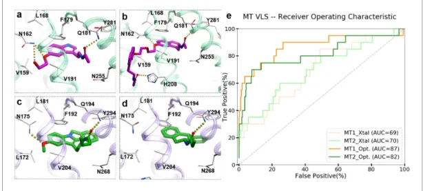
To evaluate the ability of the structure-based receptor models to recognize high-affinity melatonin receptor ligands, we performed extensive docking of a subset of known ligands of MT1 and MT2
receptors (Figure 1—figure supplement 1) into (i) the unmodified 3D structures obtained from X-ray crystallography (MT1_Xtal, MT2_Xtal), as well as (ii) into the receptor models where the
ligand-binding pocket was optimized by conformational modeling (MT1_Opt, MT2_Opt). Analysis of the
docking poses for the known MT ligands in both crystal structures and optimized MT receptor mod-els showed favorable binding scores with docking poses consistent with the orientation and binding modes of crystallized ligands (Figure 1a–d). The major interactions include aromatic stacking of the heterocyclic core with ECL2 hydrophobic residue F179/192ECL2(the residue numbers for MT1and
MT2listed for UniProt (Bateman et al., 2017) ids: P48039 and P49286, respectively, followed by
superscripted Ballesteros – Weinstein numbering schemeBallesteros and Weinstein, 1995), as well as hydrogen bonding interactions with N162/1754.60 and Q181/194ECL2 (Johansson et al., 2019;Stauch et al., 2019) . The performance of each model was then evaluated as the area under the corresponding receiver operator characteristic (ROC) curve (AUC), benchmarking the ability of these models to correctly detect ligands among decoys. The AUC values for the optimized models of MT receptors showed substantial improvement over AUC values for MT crystal structures (MT1_Opt = 87 vs. MT1_Xtal = 69; and MT2_Opt = 82 vs. MT2_Xtal = 70) (seeFigure 1e). Overall,
these results validated the improved VLS performance of the optimized models of MT1and MT2
receptors, which were then used for large-scale prospective VLS.
Prospective VLS and candidate selection
A library of 8.4 million commercially available compounds was docked into the optimized MT1_Opt
and MT2_Opt structural models (see Materials and methods), and for every compound, docking
scores and binding interactions were predicted. The top 5000 scoring compounds were selected from these VLS runs for each receptor, which were further evaluated by redocking into both MT1
and MT2 receptors with increased computational sampling. The initial hit list contained 700
com-pounds predicted to bind to both receptors. To evaluate these top docking solutions, we created additional models of MT receptors by restoring thermostabilizing mutations (Figure 1—figure sup-plement 2) in the proximity of the orthosteric site to wild-type residues (A1043.29G and W2516.48F in MT1; W2646.48F in MT2), and performed further conformational optimizations. We determined that
the impact of these mutations on the docking results was negligible. The dock scores for selected MT ligands were better than the standard ICM VLS cutoff 32.0 kJ/mol, which is better than or com-parable to the docking score of melatonin ( 29.3) and other high affinity MT receptor ligands (Johansson et al., 2019).
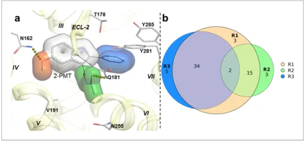
To capture chemotype diversity, we selected the top 500 compounds for each receptor using chemical clustering in combination with docking scores. A final set of 62 compounds (23 from only MT1VLS; 25 from only MT2VLS; 14 from both MT1and MT2VLS) were selected for purchase based
on a multidimensional composite criterion accounting for compound novelty, chemical diversity, well-defined interaction patterns with binding site residues N162/1754.60and/or Q181/194ECL2, and
interaction similarities with ligands observed in the crystal structures (See Figure 2; Supplementary file 1Table S1).
Most of the compounds represented new chemotypes with Tanimoto chemical distance values >0.22 (Abagyan et al., 2016), separating them from known high-affinity MT ligands available in CHEMBL24 (Gaulton et al., 2017). We also chose a close analog of melatonin – com-pound28(Tanimoto distance = 0.05), which to our knowledge, has not yet been characterized as a ligand for MT receptors (Gaulton et al., 2017;Kim et al., 2019). Compound28served as an addi-tional positive control, which also helped us to evaluate the effect of a single chemical group substi-tution in melatonin on the binding and function at the MT receptors.
Figure 1.Predicted binding modes of selected known MT ligands. (a) 5-HEAT and (b) S-26131 docked into MT1_Opt model; whereas (c) IIK-7 and (d) 4P-PDOT docked into MT2_Opt model. (e) ROC plots for MT receptor crystal structures and optimized models.
The online version of this article includes the following figure supplement(s) for figure 1:
Figure supplement 1.Chemical structures of known (a) MT1-selective and (b) MT2-selective ligands, used in the benchmark.
Figure supplement 2.MT1crystal structure in complex with 2-PMT (PDB id: 6ME3) displaying mutated residues near orthosteric ligand binding pocket.
Experimental hit identification and validation
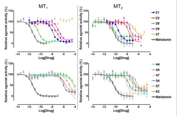
The selected 62 candidate compounds from VLS were tested experimentally for binding and func-tional profiles in both MT receptors. Eleven compounds (Table 1; Figure 3) demonstrated
sub-Figure 2.Structural features in selected hit candidate compounds. (a) 2-phenylmelatonin in complex with MT1 receptor with the topology of chemical features shown as colored spheres indicating R1 (orange) = 5-methoxy, R2 (green) = alkylamido chain, and R3 (blue) = 2-phenyl substitutions, (b) Venn diagram summarizing the topologically equivalent chemical features in selected 62 candidate compounds from MT1and MT2VLS.
The online version of this article includes the following figure supplement(s) for figure 2:
Figure supplement 1.Histogram of predicted logP values of known high-affinity MT ligands (Nref= 515) from ChEMBL and selected MT ligands (Nsel= 30).
Table 1.Hit compounds from VLS with Gi/omediated potency EC50<1mM for at least one MT receptor.
MT1 MT2 MT2/MT1
Compound pKi±SEM* pEC50±SEM EC50(nM) Emax†±SEM LE‡c pKi±SEM pEC50±SEM EC50(nM) Emax±SEM LE Selectivity§ Tanimoto¶
21 6.31±0.11 7.91±0.05 12.0 93.8±2.5 0.69 6.91±0.05 9.44±0.08 0.36 86.1±3.2 0.83 30.6 0.50
23 5.42±0.03 7.16±0.09 57.5 96.9±5.3 0.56 5.56±0.13 7.69±0.08 20.42 91.7±3.0 0.60 2.7 0.22
28 7.78±0.10 10.39±0.04 0.04 95.3±2.6 0.86 7.63±0.08 10.35±0.10 0.04 69.44.0 0.85 0.7 0.05
29 5.22±0.07 6.83±0.06 144.5 87.5±4.5 0.53 5.61±0.05 7.46±0.10 34.67 69.48.0 0.58 3.3 0.43
37 5.07±0.13 ND >30000 ND ND 5.45±0.10 6.85±0.19 141.25 61.19.1 0.53 >1000.0 0.57
44 4.19±0.36 3.33±0.36 57544.0 72.8±4.7 0.33 4.95±0.30 6.58±0.13 263.03 88.9±6.3 0.51 267.2 0.59
45 4.54±0.15 5.06±0.12 8709.6 90.6±14.3 0.44 5.26±0.19 6.37±0.13 426.58 75.0±7.4 0.56 16.9 0.59
47 4.58±0.07 5.25±0.16 2344.2 112.4±5.2 0.46 5.91±0.12 7.99±0.10 10.23 91.7±3.0 0.66 186.9 0.60
54 5.03±0.06 6.06±0.07 741.3 82.8±4.3 0.54 5.56±0.10 7.74±0.10 18.20 75.0±3.7 0.68 36.9 0.43
57 4.84±0.03 5.72±0.11 1778.3 87.5±9.1 0.47 5.37±0.04 6.88±0.15 131.83 66.78.3 0.57 10.3 0.53
62 4.32±0.11 4.39±0.42 42658.0 54.1±10.0 0.36 5.49±0.33 7.28±0.14 52.48 58.34.8 0.60 875.9 0.64
Melatonin 9.06±0.14 11.38±0.06 0.004 100.0±5.6 0.93 9.27±0.14 10.30±0.14 0.05 100.0±5.6 0.84 0.1 0.00
Standard error of the mean, N = 3.
†Activation compared to melatonin.
Ligand efficiency (based on EC50).
§Selectivity in folds (calculated as: Antilog (log(E
max/EC50) MT2- log (Emax/EC50) MT1)). MT1selectivity is shown as underlined values. ¶Tanimoto distance from closest MT receptor ligands in ChEMBL database with pAct >6. Hits with EC
50<100 nM are displayed inbold, and with
Emax<70% in italic.
micromolar potencies in Gi/o mediated signaling assays (18% hit rate). Ten of these eleven
com-pounds also showed binding affinities Ki<10mM in a competition binding assay (Figure 3—figure
supplement 1). The melatonin derivative28identified by VLS was as potent as melatonin itself in MT2(EC50= 0.04 nM) and had the same potency (EC50 = 0.04 nM) at MT1. The most potent new
chemotype,21,displayed an EC50 = 0.36 nM for MT2, with a 30-fold selectivity over MT1receptor
(MT1EC50= 12 nM). Overall, seven hits had EC50<100 nM for at least one of the melatonin
recep-tors. Similar to other low molecular weight MT ligands, most of the hits identified belong to a library of fragment-like compounds with molecular weights less than 250 Da, and have exceptionally high ligand efficiency (LE), far exceeding the~0.3 value considered as a standard for a promising lead. For example, compound21(Mol. Wt. = 224 Da) had the highest LE values of 0.83 kcal/mol per non-hydrogen atom for MT2 and 0.69 kcal/mol per non-hydrogen atom for MT1 receptors
(Hopkins et al., 2004; Hopkins et al., 2014). The excellent LE of these molecules allows the potential for further optimization of their drug-like properties.
Chemical and conformational diversity of hits
Most of the hit compounds, as shown inChemical structure 1, are novel and display diverse chemo-types distinct from known high-affinity MT ligands (ChEMBL, pAct >6.0), with Tanimoto distance exceeding 0.4 for all but two ligands (28and23). While the majority of known MT agonists reported in ChEMBL have either substituted indene or naphthalene core, only two of the eleven hits reported here have fused heterocycles and several others have two substituted aromatic rings connected by a flexible chain. Most compounds have diverse substitutions at positions topologically equivalent to the 5-methoxy, acetylamido and C2 position of melatonin (Figure 2). Two compounds–21and 29–
Figure 3.Functional characterization of selected VLS hits for agonist activity at MT1and MT2receptors in Gi/o-mediated cAMP production inhibition assays. Results were normalized to the Emaxvalue (%) of receptor activation by melatonin. These VLS hits showed no activity at control HEK293 T cells without transiently transfected MT1or MT2receptors (results not shown).
The online version of this article includes the following figure supplement(s) for figure 3:
Figure supplement 1.Radioligand3H-Melatonin competition binding data for selected hit compounds. Figure supplement 2.Tango assay measuring agonist-inducedb-arrestin recruitment by MT1receptor. Figure supplement 3.Tango assay measuring agonist-inducedb-arrestin recruitment by MT2receptor.
have a pyrimidine scaffold, whereas four compounds–23,37, 44, and57– have a methoxyphenyl group in place of the 5-methoxy indole scaffold in melatonin. Another interesting core is the cyclo-pentyl-fused thienopyridine of compound 45. Only 2 compounds, 28 and 54, have substituted indoles similar to melatonin.
The predicted binding poses of the selected hit compounds in their docking models of MT recep-tors are shown inFigure 4. Nine out of eleven hits have methoxy or a similar group predicted to make hydrogen bonding interactions with N162/1754.60, which was found to be a critical residue for
receptor activation, despite playing a limited role in ligand affinity or structural stability of the recep-tor (Stauch et al., 2019).
Furthermore, seven of the hits were predicted to form hydrogen bonding interactions with Q181/ 194ECL2similar to alkylamide tail of melatonin. Five hits were predicted to occupy a significant space
in the pocket flanked by TMs-II, III, and VII forming hydrophobic interactions, especially with residues Y281/2947.38and Y285/2987.43, as previously found in the MT receptor structures (Johansson et al.,
2019;Stauch et al., 2019). These hydrophobic interactions are similar to those formed by the phe-nyl moiety of 2-phephe-nylmelatonin. Both types of hydrogen bonding and hydrophobic interactions were found to be critical for a ligand’s steric fit into the MT receptor binding pocket and are the pri-mary determinants of ligand affinity.
Structural basis of subtype selectivity of the hits
Six of the identified hits were found to be at least 30 fold more potent at MT2compared to MT1in
the Gi/o-mediated cAMP inhibition assays. Among the hits reported here with novel scaffolds,
com-pound21has the highest potency for both MT2(EC50= 0.36 nM) and MT1(EC50= 12 nM).
Com-pound21was predicted to bind both the MT receptors in a similar orientation by forming hydrogen bonding interactions with N162/1754.60and Q181/194ECL2with its methoxy anchor and acetylamido
tail, respectively. These interactions had been reported to be critical for ligand affinity and potency at the MT receptors (Johansson et al., 2019;Stauch et al., 2019).
Other compounds also possess remarkable MT2selectivity. For example, compound 47is
187-fold selective towards MT2(EC50= 10 nM for MT2, and 2.34mM for MT1, respectively). The pyrrole
ring mimics the indole ring of melatonin, the amide group forms hydrogen bonding with N162/
Chemical structure 1.Chemical structures of hit compounds with EC50<1mM at the MT receptors.
1754.60and the chlorophenyl group forms hydrophobic interactions with ECL2 and TM-II, III, and VII
residues (Figure 4). Despite the lack of polar interactions with Q181/194ECL2, the compound displays sub-micromolar potency for MT2. Similarly, compound 45 also lacks a substitution topologically
equivalent to acetylamido tail of melatonin (R2 feature) and yet has a sub-micromolar potency and 17-fold selectivity for MT2(EC50= 427 nM). In contrast, compound44was predicted to form
interac-tions with Q181/194ECL2, but it lacks an R3 equivalent substitution, which still makes it 267-fold selective for MT2(EC50= 263 nM). These findings suggest that either R2 or R3 could be sufficient in
maintaining the potency and selectivity at MT2.
Functional selectivity of the hit ligands
All the discovered hits show activity as agonists in Gi/o-protein signaling assays at both MT1and MT2
receptors. At the same time, some compounds show functional profiles notably distinct from full and balanced agonism, especially at MT2. Thus, four of the hits, 28, 29,57, and62 had their efficacy
(Emax) reduced to less than 70% in MT2, and are therefore considered partial agonists
(Audinot et al., 2003). The identified hits were also evaluated for theirb-arrestin recruitment (Fig-ure 3—fig(Fig-ure supplements 2and3), with the comparative analysis of G-protein andb-arrestin activ-ity shown inFigure 5. In the case of the MT1receptor, there are no significant deviations from the
overall balanced G-protein/Arrestin signaling profiles for most compounds (Figure 5a). One excep-tion is compound37, which completely lacks G-protein signaling, though it still binds to MT1and
displays substantial b-arrestin activity. In the case of MT2, however, there are several compounds
that show a marked reduction inb-arrestin signaling compared to G-protein, especially compounds
21and28, which show bias factors of 15.5 and 33.9, respectively (Figure 5b). These results suggest that MT ligands may show rather distinct functional bias profiles in G-protein and b-arrestin
Figure 4.Predicted binding poses for top six new chemotypes discovered with VLS. (a)21, (b)23, (c)62, (d)29, (e) 47and (f)54inthe MT2receptor (purple). The center panel shows a canonical 7-TM receptor structure of MT2 receptor (blue helices; part of TM-V is not displayed for clarity) in complex with 2-phenylmelatonin shown as green spheres (PDB id: 6ME6).
signaling, as observed in many other GPCRs (Kenakin, 2019; Roth, 2019), though the biological importance of this bias in MT remains to be investigated (Cecon et al., 2018).
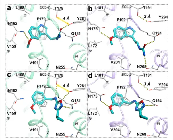
To gain more insights into these variations in ligand activity at MT receptors, we analyzed confor-mational differences among ligand-receptor pairs for these compounds. As the hit compounds are fragment–like and may attain multiple energetically-favorable poses at the orthosteric site upon docking, the specific conformational features driving partial agonism remain unclear. However, anal-ysis of compounds28and37with the most pronounced bias to G-protein in MT2andb-arrestin in
MT1, respectively, suggests possible explanations for these phenomena.
Compound28is very similar to melatonin, except the amide is replaced by a urea. This substitu-tion renders compound28as a partial agonist at MT2(Emax= 69.4%) while largely maintaining full
agonism at MT1(Emax= 95.3%). Docking predictions suggest that compound28assumes an
orienta-tion in the binding pocket similar to 2-phenylmelatonin with subtle differences, as shown inFigure 6. In MT2orthosteric site, the urea of compound28forms hydrogen bonding interactions with the side
chains of polar residues Q194ECL2and N2686.52owing to its additional nitrogen. Such interactions, however, are energetically unfavorable in the case of MT1 with possible steric clashes
(Johansson et al., 2019;Stauch et al., 2019). Instead, the interactions of acetylamido group of mel-atonin with Q181ECL2are replaced by a hydrogen bond between Y2817.38and oxygen from the urea
in compound 28. These interactions become favorable in MT1 as the residue Y2817.38 is rotated
towards TM-VI placing it 4 A˚ away from T178ECL2. In the case of MT2, however, residues Y2947.38–
T191ECL2are 3 A˚ apart forming an intermolecular hydrogen bond with Y2817.38oriented away from TM-VI allowing favorable orientation of Q194ECL2to form a hydrogen bond with compound28.
A similar pattern of ligand-receptor interaction is observed from the docking of the most selective compound37into MT receptors. Compound37has a distinct and much bulkier substitution with a 3-cyclopropyl-1,2,4-oxadiazol group (R2 feature). In MT1, this oxadiazol group is predicted to form
hydrogen bonds with Q181ECL2, T178ECL2and Y2817.38, while the cyclopropyl group is predicted to
fit in the sub pocket formed by ECL2, TM-V, and VII. These interaction pattern changes in MT2,
where the prominent hydrogen bonding between side chains of T191ECL2and Y2947.38is formed, precluding hydrogen bonding of these residues to compound37. Moreover, the methoxy group (R1 feature) of the compound, which maintains a hydrogen bond with N1754.60 in MT
2 is lost with
interaction is found to be critical in receptor activation (Stauch et al., 2019), and loss of this interac-tion is likely to explain lack of activity of compound37in Gi-mediated signaling at MT1. This
interac-tion difference, however, does not seem to affect theb-arrestin mediated signaling by compound
37in MT1. This peculiar feature of37is supported by our mutational studies, where N162Q
muta-tion in MT1actually increased potency of 37 slightly (2-fold), while Y281F, as expected, reduced
potency by over 10x (Figure 6—figure supplement 1). For comparison, 28drastically (>100 fold) reduced potency in both MT1mutants N162Q and Y281F. Taken together, these results support a
key role of N162/1754.60anchoring interactions in G
i-mediated receptor activation, and also suggest
a distinct role of residues Y281/2947.38in governing ligand bias at MT receptors. Further analysis, including mutation and SAR studies of compound37derivatives are needed for comprehensive vali-dation of this hypothetical mechanism in future studies.
Off-target profiling
To verify the ligand selectivity, the lead compounds21,28, and37, were subjected to binding pro-filing at a panel of 47 common drug targets (including many GPCRs and neurotransmitter transport-ers). At a final concentration of 10mM, they did not show any substantial binding at these targets, except for compound28that displayed over 50% inhibition at three 5-HT receptors (Figure 6—fig-ure supplement 2), with binding affinity of 851 nM (pKi = 6.07 ± 0.11) for 5-HT1A, 1525 nM Figure 6.Predicted binding poses for compounds28(a, b), and37(c, d) in MT1(light green) and MT2(lavender) receptors, respectively. The red dotted lines with arrows indicate a missing hydrogen bond between residues T178 and Y281 in MT1receptor, while the yellow dots show hydrogen bonding interactions.
The online version of this article includes the following figure supplement(s) for figure 6:
Figure supplement 1.Tango functional assays with MT1 mutants for melatonin.
Figure supplement 2.Binding activities of Compounds 21, 28 and 37 at a set of 47 potential off-targets.
(pKi= 5.82±0.02) for 5-HT1D, and 286 nM (pKi= 6.54±0.03) for 5-HT7A. Assessment of functional
activity of 28 in Gs-mediated cAMP production shows that it is a weak partial agonist with
EC50 = 1819 nM, as compared to 0.04 nM at both MT1and MT2receptors. These results
demon-strated that the lead compounds 21, 28, and 37 act specifically at MT receptors and show high selectivity for MT1or MT2over many common drug targets.
Discussion
The discovery of potent and selective MT ligands with novel chemotypes holds promise for the development of next-generation drugs to treat circadian rhythm and mood disorders, pain, insom-nia, type-2 diabetes, and cancer (Karamitri and Jockers, 2019;Liu et al., 2016). Herein, we utilized the recently solved 3D structures of the melatonin receptors, in complex with the agonist 2-phenyl-melatonin, (Johansson et al., 2019; Stauch et al., 2019) to perform prospective virtual ligand screening of large fragment-like compound libraries. This approach resulted in the discovery of ten new chemotypes of potent agonists, both full and partial, for MT1and MT2. The number of
sub-micromolar hits and potency of the best among them is one of the highest reported for a VLS cam-paign in class A GPCRs (Lyu et al., 2019) and in-line with another VLS screen for MT receptors, pub-lished while this study was in revision (Stein et al., 2020). This is remarkable, considering that most GPCR structures have a limited capacity to distinguish agonists vs. antagonists (Costanzi and Vilar, 2012;Weiss et al., 2018) and prospective VLS campaigns often result in antagonists even when an agonist-bound VLS model is used (Lyu et al., 2019;Roth, 2019).
There are several factors, related to both the VLS procedure and the intrinsic properties of the MT receptors, that likely contributed to the high hit rate and agonistic potency of the hits in our study. Thus, the high quality of the initial crystal structure, further improved by ligand-guided optimi-zation of the pocket for VLS, has been critical for the success of our previous VLS campaigns, and likely played a similar role here (Katritch et al., 2010;Lane et al., 2013;Zheng et al., 2017). At the same time, some intrinsic properties of MT receptors also likely facilitated successful VLS for ago-nists. As we mentioned above, endogenous ligand melatonin itself has unusually high picomolar potency at MT receptors (~4 pM at MT1and 50 pM at MT2, seeTable 1). Melatonin and most other
high-potency ligands are small (<250 DA) and yet they still fully occupy the very small, enclosed MT pocket. Chemical space of such size-limited fragment-like libraries is much smaller than the usual drug-like space, and can be more exhaustively searched, likely resulting in higher hit rates. More-over, most known MT receptor ligands show agonist activity, while antagonists of similar potency are notoriously hard to find, suggesting that agonists may be intrinsically preferred ligands for MT receptors (Jockers et al., 2016).
Two of the hit compounds,37and62, are MT2-selective partial agonists, which may have a
desir-able profile for eliciting antinociceptive effects mediated by melatonin receptors, and may be poten-tially useful in developing novel analgesics for pain management with reduced side effects (Lo´pez-Canul et al., 2015). Of note, while some of the newly discovered hits are selective for MT2,none of
the hits in this study had substantial MT1selectivity. This may be explained by the lack of a bulky
chemical group at the R1 position, which are known to confer strong MT1selectivity, e.g. in bitopic
CTL 01–05-B-A05 ligand that stretches out of the pocket via narrow side channel (Stauch et al., 2019). Design of such bitopic ligands with MT1selective chemotypes would need to explore larger
compounds (MW >500), which were not considered in the current VLS screen.
This study represents a successful application of structure-based VLS to identify agonists with novel chemotypes, sub-nanomolar potencies, and a high degree of receptor subtype selectivity for a class A GPCR (Lyu et al., 2019;Wang et al., 2017). This study also represents a successful imple-mentation of molecular modeling and structure-based virtual screening, aimed at the melatonin receptors, facilitated by the availability of high-quality structures capturing vital ligand-receptor interactions (Alkozi et al., 2018;Johansson et al., 2019;Stauch et al., 2019). Prevalence of ago-nists in the hit set suggests the importance of activated, agonist-bound conformations of the orthos-teric pocket models for successful agonist screening. Note that, even though the receptors were thermostabilized by 9 and 8 point mutations (MT1/MT2, respectively) to aid crystallization rendering
the receptor conformations inactive on the intracellular side, the agonist-bound orthosteric pockets remain relevant for structure-based drug discovery applications. Our benchmarking also corrobo-rated the important role of ligand guided receptor optimization (LiBERO) (Katritch et al., 2012) in
improving the outcomes of a structure-based VLS, similar to some of our previous VLS campaigns (Lane et al., 2013;Zheng et al., 2017). Another critical aspect of this successful VLS is the discovery of novel chemotypes with reliable docking poses. With our screening library assembled to be frag-ment-like with regards to molecular weights, our hits are diverse and amenable to chemical optimi-zation to improve their pharmacological profiles. Thus, our results also illustrate the utility of fragment-like compounds in the early stages of drug discovery.
The chemical diversity, selectivity, high potency and agonist activities of the identified hits serve as a valuable starting point for the development of tool compounds to explore the biology of mela-tonin receptors. With the potential for selective modulation over the melamela-tonin receptor subtype-mediated biology, these novel chemotypes could provide new leads for the development of next-generation treatments for insomnia, pain, sleep and mood-related disorders, type 2 diabetes, and cancer.
Materials and methods
Key resources table
Reagent type (species) or resource Designation Source or reference Identifiers Additional information Cell line (Homo sapiens) HTLA cells (HEKT based) PMID:25895059 Transfected construct (Homo sapiens)
MTNR1A PMID:25895059 AddGene #66443
Recombinant DNA reagent, PCR primers MTNR1A N162Q Forward
This paper CTGCCGTCCTGCCGcaaCTGAGGGCAGGCAC
Recombinant DNA reagent, PCR primers
MTNR1A N162Q Reverse
This paper GTGCCTGCCCTCAGttgCGGCAGGACGGCAG
Recombinant DNA reagent, PCR primers
MTNR1A Y281F Forward
This paper GTTCGTAGCGAGCTtCTACATGGCTTAC
Recombinant DNA reagent, PCR primers
MTNR1A Y281F Reverse
This paper GTAAGCCATGTAGaAGCTCGCTACGAAC
Commercial assay or kit
BrightGlo Reagent
Promega.com Cat # E2610
Chemical compound, drug Hit compounds Enamine, Molport, Chembridge
See 62 compounds listed inSupplementary file 1 Software, algorithm ICM-Pro, V3.8–7 Molsof.com cell line (Homo sapiens)
HEK293 T ATCC CRL-11268
transfected construct (Homo sapiens)
Human MT1 PMID:31019306
Chemical compound, drug
Luciferin Goldbio.com Cat#: LUCNA-1G
Receptor model preparation and optimization
X-ray crystal structures of MT1(Stauch et al., 2019) and MT2(Johansson et al., 2019) receptors in
complex with 2-phenylmelatonin (PDB IDsBerman et al., 2000: 6ME3, 6ME6) were used to gener-ate virtual screening models. Both structures were converted from PDB coordingener-ates to ICM objects using the object conversion protocol implemented in ICM-Pro (Abagyan et al., 2016). This process
includes the addition of hydrogens and assignments of secondary structures, the energetically favor-able protonation states to His, Asn and Gln side chains, and of formal charges to the ligand in a complex with the receptor, followed by local minimization of polar hydrogens using energy minimi-zation protocols in ICM-Pro. The orthosteric ligand-binding pocket was further optimized with energy-based global optimization in ICM using Biased Probability Monte-Carlo (BPMC), where the orthosteric ligand and amino acid side chains within 5 A˚ radius were kept flexible and co-optimized (Abagyan and Totrov, 1994), as described in LiBERO protocol (Katritch et al., 2012) and its previ-ous applications (Lane et al., 2013;Zheng et al., 2017). To validate the models, a set of 20 known MT receptor ligands were selected from ChEMBL database (Gaulton et al., 2017) along with 780 MT receptor decoys selected for each MT1and MT2 receptor from GPCR decoy database (GDD)
(Gatica and Cavasotto, 2012) and docked into crystal structures, and optimized ligand models of MT receptors. Following the previously described ligand guided receptor binding pocket optimiza-tion protocol, the Receiver Operator Characteristic curves (ROC) were plotted based on the True Positive Rates (TPR) and False Positive Rates (FPR) (Katritch et al., 2012) to evaluate the model opti-mization. The AUC values were calculated as the areas under these ROC curves and used as a model selection criteria for prospective VLS runs. The RMSD values of ligand binding pocket side chain heavy atoms for MT1and MT2were 0.51 A˚ and 0.76 A˚, respectively, compared to their
correspond-ing crystal structures.
To perform additional evaluation of screening results with the thermostabilizing mutants in the proximity of the orthosteric site, as displayed inFigure 1—figure supplement 2, were restored to wild-type (WT) residues. The Phe residue at F251/2646.48located 4.3 A˚ from the ligand was mutated to Trp, followed by local minimization of side-chain conformations using energy-based sampling and minimization protocols (Abagyan and Totrov, 1994). Similarly, A1043.29, located 5.2 A˚ was also restored to Gly in the MT1receptor model. Docking to this model suggests that these stabilizing
mutations do not substantially impact the binding of known ligands and selected hit candidates into the orthosteric pocket.
Screening library
We selected a subset of commercially available (in-stock and on-demand) fragment-like compounds from the ZINC database with physicochemical properties similar to already reported melatonin receptor ligands (Gaulton et al., 2017;Sterling and Irwin, 2015). We considered compounds with molecular weight250 Da and logP values ranging 1 to 5 based on the logP data of high-affinity MT ligands (Figure 2—figure supplement 1). The initial dataset comprised of ~10 million com-pounds was converted from SMILES to 3D format, and formal charges were assigned. This set was further reduced to 8.4 million compounds after applying additional filters for net charges (between 1 to 1) and removing compounds with highly reactive functional groups and promiscuous PAINS chemotypes (‘molPAINS’ and ‘bad groups’ in ICM-Pro v.3.8–6) (Baell and Holloway, 2010).
Virtual ligand screening
The VLS of 8.4 million compounds library for MT1and MT2models were performed using the VLS
protocol in ICM-Pro (Abagyan et al., 1994). The receptor energy potential maps were calculated using a fine potential grid (0.5 A˚). Several energy terms, including van der Waals, hydrophobic, elec-trostatic and hydrogen bonding interactions were considered for map calculations. Full torsional flex-ibility of ligands was allowed, and their internal conformational strain was considered while the receptor atoms were assigned rigid for docking. The docking was performed using BPMC conforma-tional sampling and energy minimization protocol implemented in ICM-Pro for scoring and finding the best docking solutions at the default effort level 1. These top compounds were further docked into corresponding MT receptor models with an increased sampling effort value of 3. Each VLS run for the 8.4 million compound library used 32,000 CPU core hours on 3 Linux workstations with a total of 192 CPU cores. The chemical similarity of selected compounds was calculated using Tanimoto chemical distance function ‘Distance(chem1 chem2)’, available in Molsoft’s ICM-Pro (Totrov, 2008). The fingerprints in this function use a combination of ECFP and linear fingerprints as described in ICM-Pro manual (http://www.molsoft.com/icm/fingerprints.html).
Binding and functional assays
Radioligand binding assays
All compounds for in vitro testing were purchased from Enamine, Molport, and Chembridge in stock libraries, with verified identity and guaranteed purity of >95% (37 compounds) or >90% (25 com-pounds), seeSupplementary file 2for compound QC data).
The Radioligand binding assays were conducted by the NIMH Psychoactive Drug Screening Pro-gram (PDSP). The NIMH PDSP is directed by Bryan L. Roth, MD, PhD, at the University of North Car-olina at Chapel Hill, North CarCar-olina, and Program Officer Jamie Driscoll at NIMH, Bethesda, MD. Binding assay procedures and protocols are also available online at http://pdspdb.unc.edu/ pdspWeb/?site=assays. All the radioligand binding assays were performed using membranes pre-pared from transiently transfected HEK293T cells (purchased from ATCC, CRL-11268, authenticated by the supplier using morphology, growth characteristics, and STR profiling and certified myco-plasma-free) and in standard binding buffer (50 mM Tris, 10 mM MgCl2, 0.1 mM EDTA, 0.1% BSA,
0.01% ascorbic acid, pH 7.4) as recently reported (Stauch et al., 2019). [3H]melatonin (PerkinElmer,
specific activity = 77.4–84.7 Ci/mmol) is used as the radioligand. Competitive binding assays were performed with various concentrations of test compounds (100 fM to 10mM), [3H]melatonin (0.2–1.7 nM), and MT1or MT2membranes in a total volume of 150mL. Assay plates were sealed and
incu-bated for 4 hr at 37˚C in a humidified incubator until harvesting. Plates were harvested using vacuum filtration onto 0.3% polyethyleneimine pre-soaked 96-well Filtermat A (PerkinElmer) and washed three times with cold wash buffer (50 mM Tris, pH 7.4). Filters were dried and melted with a scintilla-tion cocktail (Meltilex, PerkinElmer). Radioactivity was counted using a Wallac TriLux Microbeta counter (PerkinElmer). Results were analyzed using GraphPad Prism 7.0.
Signaling assays
G
sand G
i/o-cAMP assays
GloSensor cAMP assays were conducted according to the recently published procedure (Stauch et al., 2019) with minor modifications. Briefly, HEK293 T cells (as above) were transiently co-transfected with receptor (MT1or MT2) and GloSensor cAMP (Promega) plasmids overnight, plated
in Poly-L-Lysine coated 384-well white clear bottom plates in DMEM + 1% dialyzed FBS. Cells were used for assays at a minimum of 6 hr after plating. Culture medium was first removed and cells were stimulated with drugs in assay buffer (1x HBSS, 20 mM HEPES, 1 mg/ml BSA, 0.1 mg/ml ascorbic acid, pH 7.4) for 15 min at room temperature (this and all the following steps), followed by addition of a mixture of isoproterenol (final of 100 nM) and luciferin (final of 1 mM) for Gi/o-cAMP production
inhibition assays and luciferin (final of 1 mM) for Gs-cAMP production assays. The plates were
counted for luminescence after 25 min in a luminescence plate reader. Results were analyzed using GraphPad Prism 7.0.
Tango assays
Tango arrestin recruitment assays were carried out according to the previously published procedure Kroeze et al. (2015). In brief, HTLA cells were transiently transfected with receptor TANGO DNA constructs overnight in DMEM with 10% FBS. Transfected cells were then plated into poly-L-Lys coated 384-well plates using DMEM supplemented with 1% dialyzed FBS. After 6 hr incubation, drug dilutions, prepared in DMEM with 1% dFBS, were added for incubation overnight (16–20 hr). Medium and drug solutions were removed, Bright-Glo reagent (20 uL/well) was added for lumines-cence counting 20 min later. Results were analyzed in GraphPad Prism 7.0.
Bias factors were estimated according to the published procedure Kenakin et al. (2012) with modifications. Briefly, normalized and pooled results were analyzed by fitting the Black and Leff operational model in Prism 7.0 to obtain Log(t/KA) values for each pathway (Tango and Gi-cAMP).
Within each signaling pathway, a difference of Log(t/KA),DLog(t/KA), between a test compound and
selected reference (melatonin in this case) was calculated. For a testing compound, the difference of
Acknowledgements
We would like to thank the High-Performance Computing Center at the University of Southern Cali-fornia, and the Google Cloud Platform (for higher education and research) for providing computa-tional resources for virtual screening.
Additional information
Funding
Funder Grant reference number Author
National Institute of Diabetes and Digestive and Kidney Dis-eases
U24DK116195 Bryan Roth
National Institute of Mental Health
RO1MH112205 Bryan Roth
BioXFEL Science and Technol-ogy Center
1231306 Benjamin Stauch
Swedish Research Council LT000046/2014-L Linda C Johansson
EMBO ALTF 677-2014 Benjamin Stauch
The funders had no role in study design, data collection and interpretation, or the decision to submit the work for publication.
Author contributions
Nilkanth Patel, Data curation, Software, Formal analysis, Validation, Visualization, Methodology, Writing - original draft, Writing - review and editing; Xi Ping Huang, Resources, Data curation, For-mal analysis, Validation, Investigation, Visualization, Writing - review and editing; Jessica M Grand-ner, Conceptualization, Data curation, Methodology, Writing - review and editing; Linda C Johansson, Validation, Visualization, Writing - review and editing; Benjamin Stauch, Validation, Writ-ing - review and editWrit-ing; John D McCorvy, Data curation, Formal analysis, WritWrit-ing - review and edit-ing; Yongfeng Liu, Validation, Investigation; Bryan Roth, Resources, Supervision, Funding acquisition, Writing - review and editing; Vsevolod Katritch, Conceptualization, Resources, Data curation, Soft-ware, Supervision, Funding acquisition, Validation, Methodology, Writing - original draft, Project administration, Writing - review and editing
Author ORCIDs
Nilkanth Patel https://orcid.org/0000-0002-9856-3041
Xi Ping Huang http://orcid.org/0000-0002-2585-653X
Jessica M Grandner https://orcid.org/0000-0001-5068-8665
Linda C Johansson https://orcid.org/0000-0003-4776-5142
Benjamin Stauch http://orcid.org/0000-0001-7626-2021
John D McCorvy https://orcid.org/0000-0001-7555-9413
Vsevolod Katritch https://orcid.org/0000-0003-3883-4505
Decision letter and Author response
Decision letterhttps://doi.org/10.7554/eLife.53779.sa1 Author responsehttps://doi.org/10.7554/eLife.53779.sa2
Additional files
Supplementary files.Supplementary file 1. Selected 62 compounds from VLS that were acquired and tested
experimet-nally. The table lists vendors invormation, and Tanimo distances to the closest known MT ligands in ChEMBL database.
.Supplementary file 2. Detailed QC data for the selected 62 compounds.
.Transparent reporting form
Data availability
All chemical structures of candidate hits and chemical quality control information is deposited as supplementary information. All data generated and analysed during this study is included in the main manuscript or supplementary files.
References
Abagyan R, Totrov M, Kuznetsov D. 1994. ICM?A new method for protein modeling and design: Applications to docking and structure prediction from the distorted native conformation.Journal of Computational Chemistry 15:488–506.DOI: https://doi.org/10.1002/jcc.540150503
Abagyan R, Orry A, Raush E, Totrov M. 2016.ICM Manual. 3.8. La Jolla, CA, MolSoft LLC.
Abagyan R, Totrov M. 1994. Biased Probability Monte Carlo Conformational Searches and Electrostatic Calculations for Peptides and Proteins.Journal of Molecular Biology235:983–1002.DOI: https://doi.org/10. 1006/jmbi.1994.1052
Alkozi HA, Sa´nchez Montero JM, Doadrio AL, Pintor J. 2018. Docking studies for melatonin receptors.Expert Opinion on Drug Discovery13:241–248.DOI: https://doi.org/10.1080/17460441.2018.1419184
Audinot V, Mailliet F, Lahaye-Brasseur C, Bonnaud A, Le Gall A, Amosse´ C, Dromaint S, Rodriguez M, Nagel N, Galizzi J-P, Malpaux B, Guillaumet G, Lesieur D, Lefoulon F, Renard P, Delagrange P, Boutin JA. 2003. New selective ligands of human cloned melatonin MT1 and MT2 receptors.Naunyn-Schmiedeberg’s Archives of Pharmacology367:553–561.DOI: https://doi.org/10.1007/s00210-003-0751-2
Baell JB, Holloway GA. 2010. New substructure filters for removal of pan assay interference compounds (PAINS) from screening libraries and for their exclusion in bioassays.Journal of Medicinal Chemistry53:2719–2740. DOI: https://doi.org/10.1021/jm901137j,PMID: 20131845
Ballesteros JA, Weinstein H. 1995. Integrated methods for the construction of Three-Dimensional models and computational probing of Structure-Function relations in G Protein-Coupled receptors.Methods Neurosci25: 366–428.DOI: https://doi.org/10.1016/S1043-9471(05)80049-7
Bateman A, Martin MJ, O’Donovan C, Magrane M, Alpi E, Antunes R, Bely B, Bingley M, Bonilla C, Britto R, Bursteinas B, Bye-AJee H, Cowley A, Da Silva A, De Giorgi M, Dogan T, Fazzini F, Castro LG, Figueira L, Garmiri P, et al. 2017. UniProt: the universal protein knowledgebase.Nucleic Acids Research45:D158–D169. DOI: https://doi.org/10.1093/nar/gkw1099,PMID: 27899622
Berman HM, Westbrook J, Feng Z, Gilliland G, Bhat TN, Weissig H, Shindyalov IN, Bourne PE. 2000. The protein data bank.Nucleic Acids Research28:235–242.DOI: https://doi.org/10.1093/nar/28.1.235,PMID: 10592235 Brzezinski A. 1997. Melatonin in humans.New England Journal of Medicine336:186–195.DOI: https://doi.org/
10.1056/NEJM199701163360306,PMID: 8988899
Cecon E, Oishi A, Jockers R. 2018. Melatonin receptors: molecular pharmacology and signalling in the context of system Bias.British Journal of Pharmacology175:3263–3280.DOI: https://doi.org/10.1111/bph.13950,PMID: 2 8707298
Comai S, Ochoa-Sanchez R, Gobbi G. 2013. Sleep–wake characterization of double MT1/MT2 receptor knockout mice and comparison with MT1 and MT2 receptor knockout mice.Behavioural Brain Research243:231–238. DOI: https://doi.org/10.1016/j.bbr.2013.01.008
Costanzi S, Vilar S. 2012. In Silico screening for agonists and blockers of theb(2) adrenergic receptor: implications of inactive and activated state structures.Journal of Computational Chemistry33:561–572. DOI: https://doi.org/10.1002/jcc.22893,PMID: 22170280
de Bodinat C, Guardiola-Lemaitre B, Mocae¨r E, Renard P, Mun˜oz C, Millan MJ. 2010. Agomelatine, the first melatonergic antidepressant: discovery, characterization and development.Nature Reviews Drug Discovery9: 628–642.DOI: https://doi.org/10.1038/nrd3140,PMID: 20577266
Dubocovich ML, Markowska M. 2005. Functional MT1 and MT2 melatonin receptors in mammals.Endocrine27: 101–110.DOI: https://doi.org/10.1385/ENDO:27:2:101,PMID: 16217123
Erman M, Seiden D, Zammit G, Sainati S, Zhang J. 2006. An efficacy, safety, and dose-response study of ramelteon in patients with chronic primary insomnia.Sleep Medicine7:17–24.DOI: https://doi.org/10.1016/j. sleep.2005.09.004,PMID: 16309958
Fisher SP, Sugden D. 2009. Sleep-promoting action of IIK7, a selective MT2 melatonin receptor agonist in the rat.Neuroscience Letters457:93–96.DOI: https://doi.org/10.1016/j.neulet.2009.04.005,PMID: 19429170 Ganguly S, Coon SL, Klein DC. 2002. Control of melatonin synthesis in the mammalian pineal gland: the critical
role of serotonin acetylation.Cell and Tissue Research309:127–137.DOI: https://doi.org/10.1007/s00441-002-0579-y,PMID: 12111543
Gatica EA, Cavasotto CN. 2012. Ligand and decoy sets for docking to G protein-coupled receptors.Journal of Chemical Information and Modeling52:1–6.DOI: https://doi.org/10.1021/ci200412p,PMID: 22168315 Gaulton A, Hersey A, Nowotka M, Bento AP, Chambers J, Mendez D, Mutowo P, Atkinson F, Bellis LJ,
2017. The ChEMBL database in 2017.Nucleic Acids Research45:D945–D954.DOI: https://doi.org/10.1093/ nar/gkw1074,PMID: 27899562
Hardeland R, Cardinali DP, Srinivasan V, Spence DW, Brown GM, Pandi-Perumal SR. 2011. Melatonin–a pleiotropic, orchestrating regulator molecule.Progress in Neurobiology93:350–384.DOI: https://doi.org/10. 1016/j.pneurobio.2010.12.004,PMID: 21193011
Hopkins AL, Groom CR, Alex A. 2004. Ligand efficiency: a useful metric for lead selection.Drug Discovery Today 9:430–431.DOI: https://doi.org/10.1016/S1359-6446(04)03069-7,PMID: 15109945
Hopkins AL, Keseru¨ GM, Leeson PD, Rees DC, Reynolds CH. 2014. The role of ligand efficiency metrics in drug discovery.Nature Reviews Drug Discovery13:105–121.DOI: https://doi.org/10.1038/nrd4163,PMID: 244 81311
Jockers R, Delagrange P, Dubocovich ML, Markus RP, Renault N, Tosini G, Cecon E, Zlotos DP. 2016. Update on melatonin receptors: iuphar review 20.British Journal of Pharmacology173:2702–2725.DOI: https://doi.org/ 10.1111/bph.13536,PMID: 27314810
Johansson LC, Stauch B, McCorvy JD, Han GW, Patel N, Huang XP, Batyuk A, Gati C, Slocum ST, Li C, Grandner JM, Hao S, Olsen RHJ, Tribo AR, Zaare S, Zhu L, Zatsepin NA, Weierstall U, Yous S, Stevens RC, et al. 2019. XFEL structures of the human MT2melatonin receptor reveal the basis of subtype selectivity.Nature569:289– 292.DOI: https://doi.org/10.1038/s41586-019-1144-0,PMID: 31019305
Karamitri A, Plouffe B, Bonnefond A, Chen M, Gallion J, Guillaume JL, Hegron A, Boissel M, Canouil M, Langenberg C, Wareham NJ, Le Gouill C, Lukasheva V, Lichtarge O, Froguel P, Bouvier M, Jockers R. 2018. Type 2 diabetes–associated variants of the MT 2 melatonin receptor affect distinct modes of signaling.Science Signaling11:eaan6622.DOI: https://doi.org/10.1126/scisignal.aan6622,PMID: 30154102
Karamitri A, Jockers R. 2019. Melatonin in type 2 diabetes mellitus and obesity.Nature Reviews Endocrinology 15:105–125.DOI: https://doi.org/10.1038/s41574-018-0130-1,PMID: 30531911
Katritch V, Jaakola VP, Lane JR, Lin J, Ijzerman AP, Yeager M, Kufareva I, Stevens RC, Abagyan R. 2010. Structure-based discovery of novel chemotypes for Adenosine A(2A) receptor antagonists.Journal of Medicinal Chemistry53:1799–1809.DOI: https://doi.org/10.1021/jm901647p,PMID: 20095623
Katritch V, Rueda M, Abagyan R. 2012. Ligand-Guided Receptor Optimization. In: Orry A. J. W, Abagyan R (Eds).Homology Modeling: Methods and Protocols. Totowa, NJ: Humana Press. p. 189–205.DOI: https://doi. org/10.1007/978-1-61779-588-6
Kenakin T, Watson C, Muniz-Medina V, Christopoulos A, Novick S. 2012. A simple method for quantifying functional selectivity and agonist Bias.ACS Chemical Neuroscience3:193–203.DOI: https://doi.org/10.1021/ cn200111m,PMID: 22860188
Kenakin T. 2019. Biased receptor signaling in drug discovery.Pharmacological Reviews71:267–315. DOI: https://doi.org/10.1124/pr.118.016790,PMID: 30914442
Kim S, Chen J, Cheng T, Gindulyte A, He J, He S, Li Q, Shoemaker BA, Thiessen PA, Yu B, Zaslavsky L, Zhang J, Bolton EE. 2019. PubChem 2019 update: improved access to chemical data.Nucleic Acids Research47:D1102– D1109.DOI: https://doi.org/10.1093/nar/gky1033,PMID: 30371825
Kroeze WK, Sassano MF, Huang XP, Lansu K, McCorvy JD, Gigue`re PM, Sciaky N, Roth BL. 2015. PRESTO-Tango as an open-source resource for interrogation of the druggable human GPCRome.Nature Structural & Molecular Biology22:362–369.DOI: https://doi.org/10.1038/nsmb.3014,PMID: 25895059
Lane JR, Chubukov P, Liu W, Canals M, Cherezov V, Abagyan R, Stevens RC, Katritch V. 2013. Structure-based ligand discovery targeting orthosteric and allosteric pockets of dopamine receptors.Molecular Pharmacology 84:794–807.DOI: https://doi.org/10.1124/mol.113.088054,PMID: 24021214
Lavedan C, Forsberg M, Gentile AJ. 2015. Tasimelteon: a selective and unique receptor binding profile. Neuropharmacology91:142–147.DOI: https://doi.org/10.1016/j.neuropharm.2014.12.004,PMID: 25534555 Liu J, Clough SJ, Hutchinson AJ, Adamah-Biassi EB, Popovska-Gorevski M, Dubocovich ML. 2016. MT1 and MT2
melatonin receptors: a therapeutic perspective.Annual Review of Pharmacology and Toxicology56:361–383. DOI: https://doi.org/10.1146/annurev-pharmtox-010814-124742,PMID: 26514204
Lo´pez-Canul M, Comai S, Domı´nguez-Lo´pez S, Granados-Soto V, Gobbi G. 2015. Antinociceptive properties of selective MT(2) melatonin receptor partial agonists.European Journal of Pharmacology764:424–432. DOI: https://doi.org/10.1016/j.ejphar.2015.07.010,PMID: 26162699
Lyu J, Wang S, Balius TE, Singh I, Levit A, Moroz YS, O’Meara MJ, Che T, Algaa E, Tolmachova K, Tolmachev AA, Shoichet BK, Roth BL, Irwin JJ. 2019. Ultra-large library docking for discovering new chemotypes.Nature 566:224–229.DOI: https://doi.org/10.1038/s41586-019-0917-9,PMID: 30728502
Pe´vet P. 2016. Melatonin receptors as therapeutic targets in the suprachiasmatic nucleus.Expert Opinion on Therapeutic Targets20:1209–1218.DOI: https://doi.org/10.1080/14728222.2016.1179284,PMID: 27082492 Reppert SM, Weaver DR, Ebisawa T. 1994. Cloning and characterization of a mammalian melatonin receptor that
mediates reproductive and circadian responses.Neuron13:1177–1185.DOI: https://doi.org/10.1016/0896-6273(94)90055-8,PMID: 7946354
Roth BL. 2019. Molecular pharmacology of metabotropic receptors targeted by neuropsychiatric drugs.Nature Structural & Molecular Biology26:535–544.DOI: https://doi.org/10.1038/s41594-019-0252-8,PMID: 31270468 Stauch B, Johansson LC, McCorvy JD, Patel N, Han GW, Huang XP, Gati C, Batyuk A, Slocum ST, Ishchenko A,
Brehm W, White TA, Michaelian N, Madsen C, Zhu L, Grant TD, Grandner JM, Shiriaeva A, Olsen RHJ, Tribo AR, et al. 2019. Structural basis of ligand recognition at the human MT1melatonin receptor.Nature569:284– 288.DOI: https://doi.org/10.1038/s41586-019-1141-3,PMID: 31019306
discovery of melatonin receptor ligands to modulate circadian rhythms.Nature149:2027.DOI: https://doi. org/10.1038/s41586-020-2027-0
Sterling T, Irwin JJ. 2015. ZINC 15–ligand discovery for everyone.Journal of Chemical Information and Modeling 55:2324–2337.DOI: https://doi.org/10.1021/acs.jcim.5b00559,PMID: 26479676
Totrov M. 2008. Atomic property fields: generalized 3D pharmacophoric potential for automated ligand superposition, pharmacophore elucidation and 3D QSAR.Chemical Biology & Drug Design71:15–27. DOI: https://doi.org/10.1111/j.1747-0285.2007.00605.x,PMID: 18069986
Wang S, Wacker D, Levit A, Che T, Betz RM, McCorvy JD, Venkatakrishnan AJ, Huang XP, Dror RO, Shoichet BK, Roth BL. 2017. D4dopamine receptor high-resolution structures enable the discovery of selective agonists. Science358:381–386.DOI: https://doi.org/10.1126/science.aan5468,PMID: 29051383
Weiss DR, Karpiak J, Huang XP, Sassano MF, Lyu J, Roth BL, Shoichet BK. 2018. Selectivity challenges in docking screens for GPCR targets and antitargets.Journal of Medicinal Chemistry61:6830–6845.DOI: https://doi.org/ 10.1021/acs.jmedchem.8b00718,PMID: 29990431
Xie Z, Chen F, Li WA, Geng X, Li C, Meng X, Feng Y, Liu W, Yu F. 2017. A review of sleep disorders and melatonin.Neurological Research39:559–565.DOI: https://doi.org/10.1080/01616412.2017.1315864,PMID: 2 8460563
Zheng Z, Huang X-P, Mangano TJ, Zou R, Chen X, Zaidi S, Roth BL, Stevens RC, Katritch V. 2017. Structure based discovery of new antagonist and biased agonist chemotypes for the kappa opioid receptor.Journal of Medicinal Chemistry7:b00109.DOI: https://doi.org/10.1021/acs.jmedchem.7b00109
Zlotos DP. 2012. Recent progress in the development of agonists and antagonists for melatonin receptors. Current Medicinal Chemistry19:3532–3549.DOI: https://doi.org/10.2174/092986712801323153,PMID: 226 80635
Zlotos DP, Jockers R, Cecon E, Rivara S, Witt-Enderby PA. 2014. MT1 and MT2 melatonin receptors: ligands, models, oligomers, and therapeutic potential.Journal of Medicinal Chemistry57:3161–3185.DOI: https://doi. org/10.1021/jm401343c,PMID: 24228714