The Development of a Novel Series of Cationic Porphyrins as Disinfectants for Use in Public Health
Aaron Jamal Young
A dissertation submitted to the faculty of the University of North Carolina at Chapel Hill in partial fulfillment of the requirements for the degree of Doctor of Philosophy in the Department of Environmental Sciences and Engineering
Chapel Hill 2011
ii
Abstract
Aaron Jamal Young: The Development of a Novel Series of Cationic Porphyrins as Disinfectants for Use in Public Health.
(Under the direction of Dr. Louise M. Ball)
In the United States, alone, an estimated 4 million to 33 million cases of gastrointestinal illness resulting from contaminated water supplies occur
annually. There is a need for the exploration of new types of disinfectants for water treatment with different mechanisms of action that can be used along with or in place of currently used disinfectants to further improve modern drinking water treatment.
Photodynamic inactivation (PDI) of pathogens is a unique approach to water treatment. In general, PDI consists of a chromophore that absorbs energy from light, and ultimately uses that energy to inactivate pathogens via singlet oxygen. Cationic porphyrins are one group of chromophores that have proven to be effective in the inactivation of viral, bacterial, fungal and parasitic pathogens. It is believed that the positive charge on cationic photosensitizers (PS) help them to better associate with the predominantly negatively charged surfaces on
pathogens most resistant to chemical disinfection.
iii
iv
Acknowledgements
v
Table of Contents
List of Tables………..viii
List of Figures………....x
List of Abbreviations………...xii
Chapter I. Introduction……….1
Review of Literature……….1
Specific Aims………...11
References………..14
II. Porphyrin Synthesis and Determination of Optimal Chain Length for Inactivation……….………..19
Background……….19
Materials………..21
Methods………22
Results……….28
Discussion………37
References………..43
III. Porphyrin Photodegradation and Toxicity ………45
vi
Materials and Methods………..48
Results……….49
Discussion………56
References………..59
IV. Porphyrin Binding, ROS Production, and Subsequent Inactivation……….62
Background……….62
Materials and Methods………..64
Results……….68
Discussion………75
References………..78
V. Porphyrin Activation at Wavelengths of Minor Absorbance………80
Background……….80
Materials and Methods……….……….81
Results……….82
Discussion………88
References………..91
VI. Conclusion……….92
Review of Specific Aims………92
Future implications……….97
vii
Appendix 2. Additional 1H NMR and MS spectra of porphyrins
before and after exposure to light..………..…………103 Appendix 3. Sample calculations for loss of water during
the stability experiment in Chapter 3……….………109 Appendix 4. Actual values from porphyrin binding in Chapter 4………...111 Appendix 5. A sample HPLC chromatogram from
viii
List of Tables
Table 2.1 The percent yield for each reaction in the porphyrin synthesis………29
Table 2.2 Porphyrin inactivation of undiluted E. coli………...…….…...34
Table 2.3 1µM porphyrin reduction of diluted E. coli ………36
Table 2.4 MS2 reductions with and without light……….…..36
Table 2.5 MS2 reductions using the commercially available TMPyP………37
Table 2.6 Comparisons of C3PyP and free chlorine inactivation of E. coli……...40
Table 2.7 Comparisons of C3PyP and free chlorine inactivation of MS2……...40
Table 3.1 The MS peaks of the four porphyrins and their photoproducts ………54
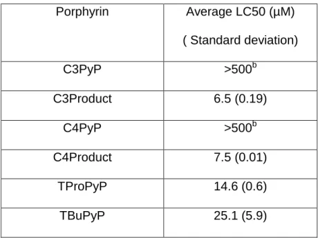
Table 3.2 The LC50s for each of the porphyrins and their photoproducts ..…….56
Table 4.1 Inactivation of E. coli by fixed and flexible cationic porphyrins………..69
Table 4.2 Inactivation of Salmonella by fixed and flexible cationic porphyrins….69 Table 4.3 Porphyrins’ binding to E. coli………..70
Table 4.4 Porphyrins binding to Salmonella ……….70
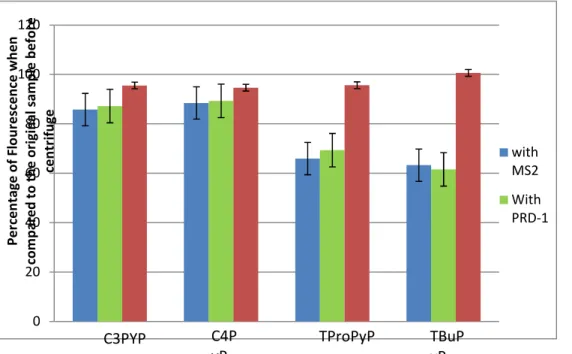
Table 4.5 MS2 Inactivation by fixed and flexible cationic porphyrins …………..73
Table 4.6 PRD-1 Inactivation by fixed and flexible cationic porphyrins …………73
Table 5.1 MS2 inactivation by porphyrins both in filtered light and in dark……...83
Table 5.2 Time needed to reach 3-log MS2 reductions with and without light filter………...84
Table 5.3 C4PyP inactivation of MS2 at 645 nm………...85
Table 5.4 Comparison of our study to previous literature on chlorine disinfection of MS2...………..89
ix
Table A3.1. The data collected in order to account for water evaporation
during the formation of porphyrin products……….110 Table A4.1 The paired t-test values for each porphyrin……….112 Table A4.2 The binding to MS2 and PRD-1 measured as percent
x
List of Figures
Figure1.1 A general diagram of the photodynamic process...………..5 Figure 1.2 An example of a fixed (right) and flexible (left) cationic meso
substituted porphyrins ……….……….10 Figure 2.1 The structure of a generic meso-substituted porphyrin……….20 Figure 2.2 General scheme for porphyrin synthesis, applied to C3PyP ………...22 Figure 2.3 1H NMR of ethyl- 4-bromo-butyrate from the spectral data base ……30 Figure 2.4 1H NMR of 4-bromo-1-butanol after the LiAlH4 reduction (CDCl3 )….30 Figure 2.5 1H NMR of 4-bromo-1-butanal after the PCC oxidation (CDCl3).…....31 Figure 2.6 1H NMR after the condensation step (CDCl3). ………..………32 Figure 2.7 1H NMR of the final product C3PyP (CD3OD)……….33 Figure 2.8 The name and structure of porphyrins used in inactivation
experiments with E. coli and MS2…………..……….35
Figure 3.1 The four porphyrins used in this study and their abbreviations………47 Figure 3.2 The degradation of C4PyP over time……….………..50 Figure 3.3 The absorbance of the porphyrin Soret over time………..51 Figure 3.4 The 1H NMR of C3PYP before and after 24 hrs irradiation…………..52 Figure 3.5 The 1H NMR of C4PYP before and after 24 hrs irradiation…………..53 Figure 4.1 The four porphyrins used in this study and their abbreviations………64 Figure 4.2 The porphyrin binding to bacteriophages MS2 and PRD-1
measured via analysis of the supernatant after ultracentrifugation ………...72 Figure 4.3 The porphyrin binding to bacteriophages measured via the
xi
Figure 5.1 C4PyP absorbance compared to filter light absorbance………...82 Figure 5.2 Control chromatogram of TProPyP (10µM), FFA (100µM) and IS (100µM) without light………..85 Figure 5.3 Chromatogram of TProPyP (10µM), FFA (100µM) and IS (100µM) with 5 minutes exposure to filtered light ……….86 Figure 5.4 Chromatogram of overlay of figures 5.2 and 5.3………86 Figure 5.5 Control chromatogram of FFA and IS with 5 minutes exposure to projector light………...87 Figure 5.6 Control chromatogram of FFA and IS without light………87 Figure 5.7 Control chromatogram of FFA and IS with 5 minutes exposure to projector light………...87 Figure 5.8 Chromatogram of overlay of graphs 5.5, 5.6, and 5.7………...88 Figure A2.11H NMR of TProPyP before(a) and after (b) 24
hours of exposure to light...……….104 Figure A2.2 1H NMR of TBuPyP before (a) and after (b) 24
hours of exposure to light...105 Figure A2.3 The mass spectra of C3PyP (a) and its photoproduct (b)...106 Figure A2.4 The mass spectra of C4PyP (a) and its photoproduct (b)...107 Figure A2.5 The mass spectra of TProPyP before (a) and
after (b) 24 hours of exposure to light...108 Figure A2.6 The mass spectra of TBuPyP before (a) and
after (b) 24 hours of exposure to light...109 Figure A4.1The increase in the absorbance of the Soret of
TMPyP over time...114 Figure A4.2 The absorbance of the Soret of TProPyP and
TBuPyP over time...114 Figure A5.1 A sample chromatogram of the porphyrin, photoproduct, furfuryl alcohol (FFA), and the internal standard...115 Figure A6.1 Graphs showing linear inactivation of E. coli and
xii
List of Abbreviations
C3PyP meso- tetrakis (3-[N-pyridiniumyl] propyl) porphyrin tetrabromide C4PyP meso- tetrakis (4-[N-pyridiniumyl] butyl) porphyrin tetrabromide C5PyP meso- tetrakis (5-[N- pyridiniumyl] pentyl) porphyrin tetrabromide C7PyP meso- tetrakis (7-[N- pyridiniumyl] heptyl) porphyrin tetrabromide C11PyP meso- tetrakis (11-[N- pyridiniumyl] undecyl) porphyrin tetrabromide CDC Centers for Disease Control and Prevention
CCl4 Carbon tetrachloride CFU Colony forming unit CHO Chinese Hamster Ovary DIW Deionized water
EC3000 E. coli strain C3000
FAmp Ampicillin resistant E coli strain FFA Furfuryl Alcohol
Hp Hematoporphyrin
HpD Hematoporphyrin derivative
HPLC High performance liquid chromatography 1
H NMR Proton nuclear magnetic resonance MDL Method detection limit
MS Mass spectrum
xiii
PCC Pyridinium chlorochromate PDI Photodynamic Inactivation PDT Photodynamic therapy PFU Plaque forming unit PS(s) Photosensitizer(s) ROS Reactive oxygen species SDS Sodium dodecyl sulfate SN Supernatant
STDEV Standard deviation
TBuPyP Meso-tetrakis (4-[N-butyl] pyridiniumyl) porphyrin tetrabromide TMPyP tetrakis (1-methy-4-pyridyl) porhyrine tetra- p- tosylate
TProPyP Meso-tetrakis (3-[N-propyl] pyridiuniumyl) porphyrin tetrabromide TSA Tryptic soy agar
TSB Tryptic soy broth
Chapter 1: Introduction
Review of Literature
After 100 years, disinfection remains a cornerstone of modern water treatment. Although current water treatments, such as chlorine, are relatively effective, there are still some inadequacies. In the United States alone, an estimated 4 million to 33 million cases of gastrointestinal illness resulting from contaminated water supplies occur annually 1,2. New challenges, such as biological contamination of water sources, an aging water distribution system, and increasing water reuse will require us to remain vigilant in protecting all aspects of our water supply. Many pathogens, such as various parasites, gram-negative bacteria, and non-enveloped viruses, have proven to be resistant to currently used disinfectants for water treatment. This along with any additional biological contamination from surface runoff, flooding, and/or the increase in water reuse, could lead to both temporary and perpetual inadequacies in drinking water treatment3.
2
currently used disinfectants to further improve water quality in developed countries.
This belief is strongly supported by the U.S. Centers for Disease Control and Prevention (CDC)4. One of the CDC’s major goals is that people be
prepared for emerging health threats. Their literature states the following: [one CDC goal is to] “Support and strengthen human and technological epidemiologic resources to prevent, investigate, mitigate, and control current, emerging, and new public health threats and to conduct research and development that lead to interventions for such threats.”
So, new methods of disinfection should be developed in order to protect people, not only from current waterborne pathogens, but also to protect us from new emerging heath threats due to drinking water.
Photodynamic inactivation (PDI) of pathogens is a unique approach to water treatment. PDI, in general, consists of a chromophore that absorbs energy from light, and ultimately uses that energy to inactivate pathogens via singlet oxygen. One method of PDI, called Photodynamic Therapy (PDT), is an
approved treatment for several cancers 5-9. While PDI and PDT both rely on the same photodynamic processes, in the present work, PDI is used to describe the application of those processes to disinfection where PDT refers to the cancer treatment.
3
Furthermore, the use of synthetic porphyrins, our PS of choice, was originally developed for PDT of cancers.
The oldest documented use of PSs was over 3000 years ago in India, where psoralens and light were used to treat vitiligo15. In 12th century AD,
Egyptians also used psoralens as a treatment for leucoderma8. Modern methods of PDI/PDT began to emerge in the 1900’s. In 1903 Herman Von Teppeiner used eosin and sunlight to treat skin tumors in mice. In 1907 he also showed that oxygen was required for the photodynamic killing of tumor cells. Similar observations were made with chlorophylls and erythrocyte hemolysis in 1907 and 19198. By 1911, experimentation on the photosensitization of mice with
hematoporphyrin (Hp), a compound naturally found in the body, was well
underway 6, 16. In 1913, Hp was found to cause photosensitization in man when Friedrich Meyer–Bertz injected himself with 200 mg of Hp and observed no adverse effects until he was exposed to light 6, 8, 16.
In 1924, it was found that natural porphyrins in tumors could give off fluorescence 6, 8, 16, while in 1942, Hp was found to accumulate in tumors. This ability of the natural porphyrins to accumulate in tumors and give off fluorescence was first developed to detect and quantify tumor growth. In 1960-61, Hp
derivatives (HpD) were synthesized and used for tumor detection 17. HpD would be used for tumor detection throughout the 1960’s.
4
To date photosensitizers activated by light are being used as a treatment to stop uncontrolled cell growth. PDT is an accepted treatment for macular
degeneration as well as several types of cancer 5-9. In PDT the photosensitizer is delivered to the target area and activated, often with lasers, to bring about a cytotoxic effect. PSs and light have been shown to kill all classes of pathogens: bacteria, viruses, parasites and fungi 23-26. Although PDI has been documented in various aqueous media, such as natural waters and blood plasma, its
application to drinking water is relatively new.
5
Figure1.1 A general diagram of the photodynamic process 8.
*Sn represents the various energy levels, and T1 represents the excited PS. This figure was obtained from Pushpan et al.8
The efficiencies of both PDI and PDT are dependent on the following PS properties 9:
I. Lipophilicity and ionization II. Molar extinction coefficient III. Quantum yield of triplet state
IV. Redox potential of the excited porphyrin V. Yield of singlet oxygen
I. Lipophilicity and ionization- The lipophilicity and ionization affect the PS’s
6
either the excited PS or the reactive oxygen species. Both are very short lived and must be in close proximity to the target to have a biocidal effect.
II. Molar extinction coefficient- The molar extinction coefficient is a measure of
the ability of the PS to absorb specific wavelengths of light. The extent of PS activation is proportional to the light energy absorbed by the PS.
III. Quantum yield of triplet state- The Quantum Yield of the triplet state is the
amount of excited PS produced relative to the photons absorbed from light. There are two ranges of light that seem most promising in PDI. The visible light range (400-650 nm) is the range of ambient light that can be detected by the human eye and electrically. This range allows for inexpensive light sources in which the PDI could take place in a natural environment supported by sunlight. The second range of long wavelength light (650-900 nm), although much more expensive to produce, can allow for deeper penetration into tissues (PDT) as well as through turbid waters (PDI). In terms of drinking water treatment, the
disinfection processes generally take place after coagulation and flocculation, which removes most, if not all, of the turbidity.
IV. Redox potential of the excited porphyrin- The redox potential of the excited
7
V. Yield of singlet oxygen- The yield of singlet oxygen pertains to the type II
reaction, in which the excited PS transfers its energy to molecular oxygen to produce the biocidal singlet oxygen that inactivates pathogens. Singlet oxygen is believed to be the active ROS in PDI.
Other important PS characteristics needed for the practical application of PDI include an economic synthesis, low native state toxicity, and rapid
elimination from the body 8. PSs used in water treatment will undoubtedly have to compete with the most inexpensive methods of disinfection, such as chlorine, which will require a reasonably simple and inexpensive synthesis. If used in drinking water treatment, some amount of the PS will be ingested, which will require the PS to be both nontoxic in the absence of light and rapidly eliminated from the body to reduce the potential adverse health effects due to PDI.
Since the development of Hp and HpDs for cancer treatment, several other PSs, naturally occurring and synthetic, have been selected to try to
increase the efficiency of PDT based on the above-mentioned characteristics of a good PS6. Each PS still has some drawbacks and there has not been one PS that is accepted as an agent for all PDT. In both PDT and PDI, increasing the PS association with target cells and/or pathogens is key; close association with the target pathogen is important given the short-lived ROS ultimately responsible for inactivation. Singlet oxygen, which is thought to be the biocidal species in most photodynamic processes, has a lifespan of 100-250 ns and an estimated
8
active agent will travel less distance than the diameter of the target malignant cells in PDT and the bacterial cells in PDI. For effective killing of both tumor cells and pathogens, the PS’s association with the target is likely equally or more important than its overall production of singlet oxygen.
Much of the latest developments in PDT have been geared toward the production of PSs that specifically target malignant cells so as to reduce the generalized killing of healthy cells. In general, PSs bound to conjugates for receptors on target cells have been used to increase the efficiency of PDT. This selectivity both increases the toxicity to malignant cells and reduces toxicity to healthy cells in PDT. For example, chlorin-bound microspheres have been found to increase PDT of human bladder carcinoma cells 27. Also anti-estrogen
conjugated porphyrins have been shown to increase the efficiency of PDT in MCF-7 breast cancer cells 28. Even PSs bound to internalizable ligands and proteins that are recognized by and actively transported into the cell nucleus have proven to improve PS association with and uptake by the cell 7. Subtle changes to the PS can also result in large increases in efficiency. Small increases in the aliphatic chain length bound to PSs were shown to increase lipophilicity of the compound and its uptake by cells 29.
The charge on the PS also plays a role in its interactions with the cell membrane 30. Recent studies have shown that cationic PSs, irradiated with light, are more efficient than anionic PSs in the inactivation of non-enveloped viruses and gram-negative bacteria14, 32-40, two types of pathogens that are more
non-9
enveloped counterparts. It is believed that the positive charge on cationic PSs help them to better associate with the predominantly negatively charged surfaces on non-enveloped viruses and gram-negative bacteria, thus bringing the PS in close proximity to the target pathogen. Furthermore, since the above and other parasitic pathogens, such as Cryptosporidium parvum and Giardia lamblia, that are most resistant to current methods of disinfection all have a net negative charge, cationic PSs are often the focus of PDI.
Using the latest developments in PDT as a model, it would seem that PSs could be developed to specifically inactivate pathogens in drinking water and other aqueous media. Small amendments to a PS’s structure and properties could make the PS more efficient for the PDI of aqueous media.
Porphyrins are one group of chromophores proven to be effective in the inactivation of viral, bacterial, and fungal and parasitic pathogens6, 8, 24, 32, 38-41 (See Appendix 1). Porphyrins are naturally occurring in the body and are believed to be less toxic than other PSs with chemical structures that are completely xenobiotic. A cardinal characteristic of porphyrins is their ability to accommodate various substituents bound to the macrocycle. Changing the substituents alters the reactivity of the porphyrin, and can allow for advanced disinfection control and specificity.
10
theory, could be used for treating various aqueous media such as drinking water and blood products 10-14.
Previously synthesized cationic porphyrins that have been proven to inactivate non-enveloped viruses and gram-negative bacteria in aqueous media have had fixed positive charges located on the periphery of the tetrapyrrole macrocycle 37-41. To date one study has included a porphyrin with a more flexible cation position; while in this study the flexible cation gave the most efficient PDI, the effect of the cation position was not directly observed39. In the present study, the positive charge is connected farther from the tetrapyrrole macrocycle through an aliphatic carbon chain to give the porphyrin added flexibility to associate with the negative regions on the surface of the target pathogens (See figure 1.2).
Figure 1.2 An example of a fixed (right) and flexible (left) cationic meso
substituted porphyrins.*
*The newly synthetisized porphyrins (left) vary in the chain length linking the cationic pyridinium. Porphyrins described in previous literature (right) have a more rigid cation position in which the pyridinium is directly attached to the macrocycle.
N
H N N
N H
N+
N+ N+
N+
CH3
H3C
H3C
CH3
N NH
N
HN N+
N+
11
A series of porphyrins were tested against model pathogens E. coli C3000 and FAmp, Salmonella LT2, and bacteriophages MS2 and PRD-1. MS2 and PRD-1 are examples of non-enveloped viruses, which have ssRNA and dsDNA respectively. MS2 has also been previously documented as a model for the hepatitis A virus 42, 44. The inactivation of non-infectious strains of E. coli is used to offer understanding of the porphyrins’ efficiency against bacteria commonly used as an indicator of fecal contamination45. Salmonella, which is the host for PRD-1, served as a second representative gram-negative bacteria.
In order for this class of porphyrins to become useful in a public health setting, this group of porphyrins must be characterized for use in water treatment. Specific questions must be answered as to better understand the porphyrins’ stability in storage and in use, native state toxicity, and other factors that influence PDI efficiency.
Porphyrin analogs to the above-described porphyrins that have the positive charge connected at the periphery of the porphyrins’ macrocycle were also synthesized in order to observe the effects of the positive charge location on various properties relevant to PDI. The singlet oxygen production and the final localization of porphyrin associated with the target cells were also measured
Specific Aims
12
mechanism of action, potential for toxicity, and stability in use is needed. A series of experiments was conducted to better define the porphyrins’ applicability to the disinfection of water and other aqueous media and answer the following questions.
(1) What is the optimum chain length for porphyrin inactivation of model
pathogens? The cationic porphyrins used in previous tests have carried fixed
positive charges on the periphery of the tetrapyrrole macro cycle37-41. Porphyrins with a more flexible cation linkage have been synthesized to adapt to various negative charge distributions on the surface of pathogens. These porphyrins have cations attached to the porphyrin through an aliphatic carbon chain. The inactivation of model pathogens by a range of meso substituted porphyrins with the cation positioned at various chain lengths was measured.
(2) What is the stability of the porphyrins under various conditions that
simulate likely real-life use? There is a need to better understand how long the
porphyrin maintains its integrity when irradiated as well as in the absence of light. In this study, the photostability of four porphyrins that differ only in the mobility of the cation was examined. The formation of degradation products was observed using 1H NMR, UV/Vis and mass spectrometry.
(3) What is the potential toxicity of the porphyrins and their degradation
products? The toxicity of the parent porphyrins and the photoproducts to model
13
4) How does the attachment of the cation to the porphyrin periphery affect
the binding to and subsequent inactivation of specific bacteria and
viruses? The efficiency of PDI of the above-mentioned porphyrins and their
fixed cation analogs was observed to better understand the effect of the flexible cation position. The amount of porphyrin bound to the target microbe was also measured and compared to each of the porphyrins’ inactivation of model pathogens.
(5) Will long-wavelength light excite the porphyrins to a level effective in
disinfection? Porphyrins have a maximum absorbance at 411-430 nm, but are
able to absorb longer wavelengths of light, up to 650 nm, to a lesser degree. If this longer wavelength light is still able to excite the porphyrins above the
14
References
1 J. M. Colford, S. Roy, M. J. Beach, A. Hightower, S. E. Shaw and T. Wade, A review of household drinking water intervention trials and an approach to the estimation of endemic waterborne gastroenteritis in the United States, Journal of
Water and Health, 2006, 4, 71-88.
2 M. Messner, S. Shaw, S. R. Regli Ken, V. Blank and J. Soller, An approach for developing a national estimate of waterborne disease due to drinking water and a national estimate model application, Journal of Water and Health, 2006, 4, 201-240.
3 Montgomery Watson Harza, ed. anonymous John Wiley and Sons Inc., Hoboken, New Jersey, 2nd edn., 2005.
4 CDC Morbidity and Mortality Weekly Report, Preventing Emerging Infectious Diseases: A Strategy for the 21st Century Overview of the Updated CDC Plan ,September 11, 1998 / 47(RR15);1-14.
5 K. Kurohane, A. Tominaga, K. Sato, J. R. North, Y. Namba and N. Oku, Photodynamic therapy targeted to tumor-induced angiogenic vessels, Cancer
Letters, 2001, 167, 49-56.
6 E. S. Nyman and P. H. Hynninen, Research advances in the use of tetrapyrrolic photosensitizers for photodynamic therapy, Journal of
Photochemistry and Photobiology B: Biology, 2004, 73, 1-28.
7 A. A. Rosenkrantz, D. A. Jans and A. S. Sobolev, Targeted intracellular
delivery of photosenstizers to enhance photodynamic efficiency. Immunology and
Cell Biology, 2000, 78, 452-464.
8 S. K. Pushpan, S. Venkatraman, V. G. Anand, J. Sankar, D. Parmeswaran, S. Ganesan and T. K. Chandrashekar, Porphyrins in Photodynamic Therapy - A Search for Ideal Photosensitizers, Current Medicinal Chemistry - Anti-Cancer
Agents, 2002, 2, 187-207.
9 T. Maisch, R. Szeimies, G. Jori and C. Abels, Antibacterial photodynamic therapy in dermatology. Photochemical & Photobiological Sciences, 2004, 3, 907-917.
15
11 J. Hirayama, H. Abe, N. Kamo, K. Ikebuchi and H. Ikeda, Comparison of the Effects of Different Antiviral Treatments on the Antioxidant Systems of Stroma-free Hemoglobin¶, Photochem. Photobiol., 2001, 74, 461-464
(DOI:10.1562/0031-8655(2001)074<0461:COTEOD>2.0.CO;2).
12 J. M. Obrien, D. K. Gaffney, T. P. Wang and F. Sieber, Merocyanine540-Sensitized Photoinactivation of Enveloped Viruses in Blood Products: Site and Mechanism of Phototoxicity, Blood, 1992, 80, 277-285.
13 A. N. Vzorov, D. W. Dixon, J. S. Trommel, L. G. Marzilli and R. W. Compans, Inactivation of Human Immunodeficiency Virus Type 1 by Porphyrins, Antimicrob.
Agents Chemother., 2002, 46, 3917-3925
(DOI:10.1128/AAC.46.12.3917-3925.2002).
14 L. L. Trannoy, J. W. Lagerberg, T. M. Dubbleman, H. J. Schuitmaker and A. Brand, Positively charged porphyrins: a new series of photosensitizers for sterilization of RBCs, Tranfusion, 2004, 44, 1186-1196.
15 T. B. Fitzpatrick and M. A. Pathak, Part IV: Basic Considerations of the Psoralens: Historical Aspects of Methoxsalen and Other Furocoumarins1, The
Journal of Investigative Dermatology, 1959, 32, 229-231.
16 R. Bonnett, Photodynamic therapy in historical perspective. Reviews in
Contemporary Pharmacotherapy., 1999, 10, 1-17.
17 R. L. Lipson, E. J. Blades and A. M. Olsen, The Use of a Derivative of Hematoporphyrin in Tumor Detection, Journal of the Ntional Cancer Institute, 1961, 26, 1-10.
18 J. Diamond, S. G. Granelli and A. F. McDonagh, Photodynamic therapy of malignant tumours, Lancet, 1972, ii, 1175-8.
19 T. Dougherty, C. J. Gomer and K. R. Weishaupt, Energetics and efficiency of Photoinactivation of Murine Tumor Cells Containing Hematoporphyrin, Cancer
Research, 1976, 36, 2330-2333.
20 J. F. Kelly and M. E. Snell, Hematoporphyrin derivative: a possible aid in the diagnosis and therapy of carcinoma of the bladder, Journal of Urology, 1976,
115, 150.
21 T. Dougherty, J. E. Kaufman, A. Goldfarb, K. R. Weishaupt, Boyle D.G. and A. Mitterlman, Photoradiation therapy for the treatment of malignant tumors. Cancer
Reasearch, 1978, 38, 2628-2635.
16
23 S. Ferro, F. Ricchelli, G. Mancini, G. Tognon and G. Jori, Inactivation of methicillin-resistant Staphylococcus aureus (MRSA) by liposome-delivered photosensitising agents, Journal of Photochemistry and Photobiology B: Biology, 2006, 83, 98-104.
24 Z. Alouini and M. Jemli, Destruction of Helminth Eggs by Photosensitized Porphyrin, Journal of Environmental Monitoring, 2001, 3, 548-551.
25 V. Carré, O. Gaud, I. Sylvain, O. Bourdon, M. Spiro, J. Biais, R. Granet, P. Krausz and M. Guilloton, Fungicidal properties of meso-arylglycosylporphyrins: influence of sugar substituents on photoinduced damage in the yeast
Saccharomyces cerevisiœ, Journal of Photochemistry and Photobiology B:
Biology, 1999, 48, 57-62.
26 K. Kassab, D. Dei, G. Roncucci, G. Jori and O. Coppelloti, Phthalocyanine-photosensitized inactivation of a pathogenic protozoan, Acanthamoeba
palestinensis, Photochemical & Photobiological Sciences, 2003, 2, 668-672.
27 R. Bachor, C. R. Shea, R. Gillies and T. Hasan, Photosensitized Destruction of Human Bladder Carcinoma Cells Treated with Chlorin e6-Conjugated
Microspheres, Proc. Natl. Acad. Sci. U. S. A., 1991, 88, 1580-1584. 28 A. F. Gacio, C. Fernandez-Marcos, N. Swamy, D. Dunn and R. Ray, Photodynamic cell-kill analysis of breast tumor cells with a tamoxifen-pyropheophorbide conjugate, J. Cell. Biochem., 2006, 99, 665-670.
29 I. Bronshtein, S. Aulova, A. Juzeniene, V. Iani, L. Ma, K. M. Smith, Z. Malik, J. Moan and B. Ehrenberg, In Vitro and In Vivo Photosensitization by
Protoporphyrins Possessing Different Lipophilicities and Vertical Localization in the Membrane, Photochemistry and Photobiology, 2006, 82, 1319-1325.
30 J. Bozja, J. Sherrill, S. Michielsen and I. Stojiljkovic, Porphyrin-Based, Light-Activated Antimicrobial Materials, Journal of Polymer Science: Part A: Polymer
Chemistry, 2003, 41, 2297-2303.
31 J. D. Hamlin, D. A. S. Phillips and A. Whiting, UV/Visible spectroscopic
studies of the effects of common salt and urea upon reactive dye solutions, Dyes
and Pigments, 1999, 41, 137-142.
17
33 M. Merchat, J. D. Spikes, G. Bertoloni and G. Jori, Studies on the mechanism of bacteria photosensitization by meso-substituted cationic porphyrins, Journal of
Photochemistry and Photobiology B: Biology, 1996, 35, 149-157.
34 A. Minnock, D. I. Vernon, J. Schofield, J. Griffiths, J. Howard Parish and S. B. Brown, Photoinactivation of bacteria. Use of a cationic water-soluble zinc
phthalocyanine to photoinactivate both Gram-negative and Gram-positive
bacteria, Journal of Photochemistry and Photobiology B: Biology, 1996, 32, 159-164 (DOI:DOI: 10.1016/1011-1344(95)07148-2).
35 T. Maisch, J. Baier, B. Franz, M. Maier, M. Landthaler, R. Szeimies and W. Bäumler, The role of singlet oxygen and oxygen concentration in photodynamic inactivation of bacteria, Proceedings of the National Academy of Sciences, 2007,
104, 7223-7228 (DOI:10.1073/pnas.0611328104).
36 M. B. Spesia, D. Lazzeri, L. Pascual, M. Rovera and E. N. Durantini, Photoinactivation of Escherichia coli using porphyrin derivatives with different number of cationic charges. FEMS Immunol. Med. Microbiol., 2005, 44, 289-295. 37 G. Valduga, B. Breda, G. M. Giacometti, G. Jori and E. Reddi,
Photosensitization of Wild and Mutant Strains of Escherichia coli by meso-Tetra (N-methyl-4-pyridyl)porphine. Biochem. Biophys. Res. Commun., 1999, 256, 84-88.
38 M. J. Casteel, K. Jayaraj, A. Gold, L. M. Ball and M. D. Sobsey,
Photoinactivation of Hepatitis A Virus by Synthetic Porphyrins¶, Photochemistry
and Photobiology, 2004, 80, 294-300.
39 D. A. Caminos, M. B. Spesia and E. N. Durantini, Photodynamic inactivation of Escherichia coli by novel meso-substituted porphyrins by
4-(3-N,N,N-trimethylammoniumpropoxy)phenyl and 4-(trifluoromethyl)phenyl groups.
Photochemical & Photobiological Sciences, 2006, 5, 56-65.
40 M. Magaraggia, F. Faccenda, A. Gandolfi and G. Jori, Treatment of microbiologically polluted aquaculture waters by a novel photochemical technique of potentially low environmental impact. Journal of Environmental
Monitoring, 2006, 8, 923-931.
41 E. Reddi, M. Ceccon, G. Valduga, G. Jori, J. C. Bommer, F. Elisei, L. Latterini and U. Mazzucato, Photophysical Properties and Antibacterial Activity of Meso-substituted Cationic Porphyrins¶, Photochemistry and Photobiology, 2002, 75, 462-470.
18
43 R. M. Hall and M. D. Sobsey, Inactivation of hepatitus A virus (HAV) and MS-2 by ozone and ozone-hydrogen peroxide in buffered water, Water Science and
Technology, 1993, 27, 371-378.
44 M. D. Sobsey, T. Fuji and P. A. Shields, Inactivation of hepatitus A virus and model viruses in water by free chlorine and monochloramine, Water Science and
Technology, 1988, 20, 385-391.
45 C. Chaidez, M. Moreno, W. Rubio, M. Angulo and B. Valdez, Comparison of the disinfection efficacy of chlorine-based products for inactivation of viral
indicators and pathogenic bacteria in produce wash water, International Journal
Chapter 2: Porphyrin Synthesis and Determination of Optimal Chain Length for Inactivation
Background
Photoactive disinfectants are of great interest as alternative measures against microbial contamination of aqueous media. To date, photosensitizers (PSs) and light have been shown to kill all classes of pathogens; bacteria,
viruses, parasites and fungi 1-4. While Photodynamic inactivation (PDI) has been used in various aqueous media, its application to drinking water is relatively new. Porphyrins prove to be an interesting PS for a number of reasons.
Perhaps the greatest reason that porphyrins demand closer investigation is the variety of substituents that can be placed on the porphine skeleton. The most familiar porphyrin, found in hemoglobin and myoglobin, is porphyrin IX, which has many substituents. Changing the substituents on the porphyrin can increase the physico-chemical interaction with specific molecules; thus, porphyrins can be tuned to promote PDI.
In general, the pathogens of concern in modern drinking water treatment all have surfaces with a net negative charge 5. Cationic substituents are believed to increase both the porphyrin association with the negatively charged surfaces and the photolytic activity against the pathogens of concern. Most of the
20
connecting the positive charge farther from the tetrapyrrole macrocycle through an aliphatic carbon chain, the porphyrin flexibility to associate with the negative regions on the surface of the target pathogens is increased (See figure 2.1).
Figure 2.1 The structure of a generic meso-substituted porphyrin.*
n=1 meso- tetrakis(1-methy-4-pyridyl) porhyrine tetra- p- tosylate (TMPyP) n=3 meso- tetrakis (3-[N-pyridiniumyl] propyl) porphyrin (C3PyP) n=4 meso- tetrakis (4-[N-pyridiniumyl] butyl) porphyrin (C4PyP) n=5 meso- tetrakis (5-[N- pyridiniumyl] pentyl) porphyrin (C5PyP) n=7 meso- tetrakis (7-[N- pyridiniumyl] heptyl) porphyrin (C7PyP) n=11 meso- tetrakis (11-[N- pyridiniumyl] undecyl) porphyrin (C11PyP) *Our synthetic porphyrins vary in the length of the aliphatic bridge which isolates the cationic center from the tetrapyrrolic macrocycle. The porphyrins used in this study (and their
abbreviations) based on the figure are as follows. The TMPyP porphyrin (from previous literature) has a tosylate counterion, whereas the others havebromine.
While there has been documentation of the potential of flexible cationic porphyrins14, few studies have observed the effect of the length of the aliphatic bridge between the porphyrin macrocycle and the cationic pyridinium. In pursuit of the first specific aim of this research, the above porphyrins were synthesized
N
NH N
HN R
R
R R
21
and tested against E. coli in order to determine an optimal chain length for porphyrin PDI.
Materials
Porphyrin Synthesis
Solvents methylene chloride, ether, and dimethyl formamide (DMF), and the reagent zinc acetate dihydrate were obtained from Fisher Scientific. Other solvents chloroform, hexane, ethyl acetate, and methanol were obtained from Mallinckrodt Chemicals. Reagents ethyl 4-bromobutyrate, ethyl
6-bromohexanoate, pyrrole (98%), pyridine (99%) HPLC grade, carbon
tetrachloride (99%) HPLC grade, lithium aluminum hydride (95%), trifluoroacetic acid (99%), and pyridinium chlorochromate (PCC) (98%) were obtained from Sigma Aldrich. The p-chloranil (PCA) was obtained from Kodak. Montmorillonite clay K10, florasil 100- 200 mesh, filter agent celite 521, alumina gel, standard grade, 50 mesh, 58 Ǻ and silica gel, merck grade 9385, 230-400 mesh, 60 Ǻ were also obtained from Sigma Aldrich. All solvents were evaporated using a Büchi Rotovapor R-114. ¹H NMR spectra were recorded on a Varian INOVA 500 spectrometer at 500 MHz and 25°C, using chloroform- d as the solvent. Mass spectra were obtained on a Finnigan LCQDECA quadrupole ion trap mass spectrometer with electrospray source operated in the positive mode. TMPyP was purchased directly from Aldrich Chemical Co., Milwaukee, WI.
Inactivation Experiments
22
obtained from Becton, Dickinson and Co. E. coli strain C3000 was obtained from ATCC 15597. A Phillips 40 W Hg fluorescent light (F40T12/DX), positioned 11.25 inches above the wells (measured by ruler), was used to irradiate samples at 0.48 mW/cm2 measured by light meter (Mannix DLM2000).
Methods
Porphyrin Synthesis
All porphyrins except TMPyP were synthesized in our laboratory according to Figure 2.2 which was adapted from literature methods15-20. The product
identity and purity was confirmed by 1H NMR between each step in the synthesis. The synthesis of the tetrakis(N-propyl) derivative, C3PyP, is described in detail below . Additional 1H NMR and MS spectra can be seen in Appendix 2.
Figure 2.2 General scheme for porphyrin synthesis, applied to C3PyP*
*All porphyrins were synthesized according to figure 2.2 using the appropriate ester except TMPyP, which is commercially available.
H
Step 4: Quaternization
N H N N N H
B r Br
B r Br N H N N N H
N + N +
N + N +
P y ridine
N H N N N H
B r B r
B r Br
Py rrol e
B r
O C C l 4, C
F3C O O H
or K 10 C l ay Step 3: Condensation
Br
O
O
Ether L iA lH4
Br
O H
Step1:Lithium Aluminum Hydride Reduction
Br
H
O
P C C
Br
O H
C H C l 2
23
1) Lithium Aluminum Hydride (LiAlH4) Reduction16
In a 500 mL flask, 15 g ethyl 4-bromobutyrate was combined with lithium aluminum hydride in a 1:1 molar ratio. The hydride was added to 100 mL of ether and refluxed at 40°C while the ethyl 4-bromob utyrate was diluted with 60 mL ether and added dropwise. The stirred solution was refluxed for 1 hour. The mixture was then cooled in ice and 10 mL saturated aqueous sodium chloride was added dropwise. Here the solution’s gray color may yield some white precipitate of LiOH. The LiOH was then removed by filtration under vacuum through a sintered glass funnel, and the clear solution was evaporated to yield a colorless alcohol stored at -20°C. The alcohol was then a nalyzed using 1H NMR before going to the second step of the process.
2) PCC Oxidation19
24 3) Condensation
The third step of this synthesis was a condensation of the above aldehyde and pyrrole in a 1:1 molar ratio17,18. 3 g of the aldehyde and the corresponding portion of dry pyrrole were mixed with 100 mL of carbon tetrachloride (CCl4) under argon for 15 min. 1 mL of trifluoroacetic acid was injected into the mixture as a catalyst. The argon was cut off and the mixture was allowed to stir for 24 hrs at room temperature. The CCl4 was removed by evaporation, and the dry residue was dissolved in 50 mL of methylene chloride and separated on a 1” x 6” alumina column eluted with chloroform. The eluate was then evaporated to 150 mL and refluxed with 300 mg p-chloranil for 40 min. The solution was separated on a 1” x 6” alumina column eluted with chloroform, evaporated to 10 mL, and further purified on a 1” x 7” silica column eluted with 1:1 hexane to chloroform. Finally, the solution containing the porphyrin freebase was evaporated and stored at -20°C.
A second method for condensation was also used in an effort to achieve a better yield15,20. In this method, 10 g of montmorillonite clay was activated at 120°C and below 0.5 Torr for 2 hrs in a 2 L round bo ttom flask. Next, the system was filled with argon and shielded from light. 950 mL of freshly distilled
methylene chloride and 10 mmol of the aldehyde dissolved in 50 mL of
methylene chloride were added to the flask. The mixture was stirred to uniform suspension, 10 mmol of pyrrole was introduced dropwise, and the mixture was allowed to stir for 24 hrs. 7.5 mmol of solid p- chloranil was added to the
25
under vacuum and washed with ethyl acetate. The filtrate was reduced and absorbed onto florisil. The absorbate was then purified by column
chromatography though two separate 1” x 6” silica columns using methylene chloride. The solution was then evaporated and stored at -20°C. The solid product was analyzed with 1H NMR and UV visible spectroscopy before the next step. The presence of the porphyrin can also be determined by the property of the porphyrin to yield a red fluorescence under long wave UV light.
4) Quaternization
The fourth and final step of this synthesis was a quaternization using the porphyrin produced from the above condensation procedure and pyridine. 50 mg of the porphyrin freebase was combined with 12 mL dry pyridine, and refluxed under argon gas for three hours. The solid pyridinium porphyrin was removed and washed with ether. Excess ether was removed and the solid was dried using a rotovapor, oil pump, and lyophilizer. The final product was confirmed via 1
H NMR.
26 Inactivation Tests
All steps of the inactivation tests except for microbe incubation were performed in a biological safety cabinet. All inactivation experiments were done in triplicate. Only the average, not the individual values of the replicates, are shown in the results section. The Thomas Equation was used to calculate the concentration of both the bacteria and viruses.
The above-mentioned porphyrins were used in experiments to measure their efficiency in killing E. coli C3000 according to the following protocol. The E.
coli stocks were made by first inoculating into 30 mL sterile TSB made to the
manufacturer’s specifications and then incubating at 37˚C for 18 hrs in a forced air incubator with a shaker. 100 µL of the E. coli culture was then re-inoculated in 30 mL of sterile tryptic soy broth and incubated for 4 hrs. This four-hour E. coli culture was diluted with glycerine in 3:1 ratio, E. coli to glycerin, and divided into 1mL aliquots and kept at -80˚ C until further use.
27
controls made with plates identical to that above, were shielded from all light for 30 min.
Duplicate serial dilutions were made from each well, experimental and control, at 10, 20, and 30-minute intervals. Once the final concentrations were achieved, 100 µL of the each dilution from each well was added to 2.5 mL of sterile molten top agar (0.7% agar) kept at 45˚C. The samples were then vortexed, poured onto a Petri dish containing 15 mL of TSA, made to the
manufacturer's specifications, and allowed to cool and solidify. The plates were then incubated for 20 hrs in a shelf incubator set at 37˚C. After this incubation, the E. coli colonies on each of the plates were counted. In most cases there were three consecutive countable 10-fold dilutions. All countable dilutions were used to obtain a value of colony forming units (CFU)/mL.
The porphyrins were also tested against the coliphage MS2. The MS2 stock was made by adding 10 µL of concentrated MS2 ( ~7 x 10¹¹/mL) and 300 µL of the undiluted four-hour E. coli culture to 5 mL top agar made of TSB with 0.3% agar. The top agar was gently mixed and poured onto a large Petri plate containing 25 mL of TSA. The plates were then incubated for 20 hours at 37˚C. 5mL of phosphate buffered saline (PBS; pH 7) was then added to each plate, and the loose top layer was scraped off and centrifuged at 4500 rpm for 20 minutes at 4˚C. The supernatant was then passed through a 0.45 µm filter, and 1 mL aliquots were made and stored at -80˚C until further use.
28
minute of light irradiation the test mixture was serially diluted. 100 µL of the diluted test mixture was added to 200 µL of the E. coli culture, described above, in 2.5 mL of top agar made of TSB with 0.7% agar kept in a water bath kept at 45˚C. This mixture was gently swirled and poured onto a Petri dish containing 15 mL of tryptic soy agar (TSA) made to the manufacturer's specifications and allowed to cool and solidify. These plates were then inverted and incubated for 20 hours at 37˚C. All countable plates were used to obtain a value of plaque forming units (PFU) per mL
Results
Synthesis
The results described below are for the synthesis of the C3PyP porphyrin. All of the porphyrins have similar 1H NMR spectra, however with each
29
Table 2.1 The percent yield for each reaction in the porphyrin synthesis.
LiAlH4 Reduction
The carbonyl group of the ethyl-4-bromobutyrate was reduced with LiAlH4 to give 4-bromobutanol. This process is done with 60-70% yield. This reduction was performed using different concentrations of LiAlH4 and ethyl
4-bromobutyrate and the more dilute solutions tended to give a better yield. 1H NMR spectra were used to confirm the new product and its purity. The most noticeable peaks in the 1H NMR spectrum consisted of two triplets at centered at 3.4 and 3.7 ppm and two quintets centered at 1.7 and 1.9 ppm. The triplets at 3.4 and 3.7 ppm are due to the CH2 groups next to the Br (C4) and the OH (C1) group, respectively. The two quintets correspond to the two interior CH2 groups on the alcohol (C2 & C3). Purity is noted from 1H NMR before measuring
amounts for the next step (See Figures 2.3 and 2.4). For example, if the product is 50% pure, then twice as much was used for the next step.
Reaction Starting Material Product % Yield
LiAlH4 Reduction 10.5g 7.4g 70.5%
PCC Oxidation 5.4g 3.1g 62.2%
Condensation (trifluoroacetic acid) 3.7g 22mg 0.6%
Condensation (clay) 2.85g 620mg 12%
30
Figure 2.3 1H NMR of ethyl- 4-bromo-butyrate from the spectral data base
Figure 2.4 1H NMR of 4-bromo-1-butanol after the LiAlH4 Reduction (CDCl3)
PCC Oxidation
31
at 9.8 ppm for the proton attached to the carbonyl group (C1), a triplet centered at 3.4 ppm for the hydrogens at C4, a quartet centered at 2.7 ppm for the protons on C2, and a quintet centered at 2.2 ppm for the protons on C3. The triplet at 9.8 ppm may appear as a single peak because of its very low coupling constant.
Figure 2.5 1H NMR of 4-bromo-1-butanal after the PCC oxidation (CDCl3)
Condensation
In this step the aldehyde was connected to the pyrrole at the meso position to form a porphyrin ring consisting of four pyrroles and four propyl chains. This procedure was done in very low yield (0.6%). The C1 that was once an aldehyde group now forms the methine bridge of the porphyrin ring. The 1
32
protons on C2 of the substituent chain. The presence of porphyrin was also noted by its dark purple color. In solution the porphyrin gives off red
fluorescence under long-wave UV light and it turns green in the presence of hydrochloric acid. The UV/Vis spectrum was also taken in chloroform to confirm the compound.
Figure 2.6 1H NMR after the Condensation step (CDCl3)
The clay condensation improved the percent yield by more than 20 fold with a 12% yield (Table 2.1). The clay absorbed and removed many of the oxidants and polymeric by-products. The pores in the clay acted as a template for the formation of the bulky porphyrin molecule20.
Quaternization of Pyridine
Here the bromine atom located at the end of each of the four substituent chains was replaced with pyridine where the nitrogen holds a positive charge. This reaction produces a water-soluble porphyrin with 1H NMR peaks as follows:
p p m ( f 1 )1 0 . 0 5 . 0 0 . 0
- 1 0 0 0 1 0 0 2 0 0 3 0 0 4 0 0 5 0 0 6 0 0 7 0 0
8
.0
6
8
.0
6
8
.0
0
1
.7
5
7
.6
7
N
H N N
N H
Br Br
33
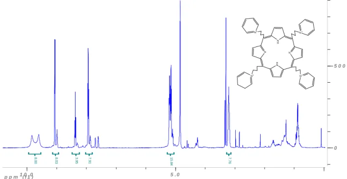
broad doublet centered at 9.2 ppm as pyrrole protons, a doublet at 9.1 ppm and triplets at 8.4 and 8.0 ppm that correspond to protons at the ortho, para, and meta positions on the pyridine respectively, a triplet at 5.2 ppm for protons on C1 and C3 of aliphatic substituent chain, and a quintet at 3.3 ppm for protons on C2 of the aliphatic chain. UV/Vis spectra were recorded in water and methanol. Figure 2.7 shows the 1H NMR of the final pyridinium porphyrin salt.
Figure 2.7 1H NMR of the final product C3PyP (CD3OD)
Inactivation Experiments
The most effective porphyrins against E. coli were found to be those where the cation is at the end of a propyl, butyl, or pentyl, substituent chain (Table 2.2). Several inactivation times were examined for both E. coli and MS2. The data shown is for 30 minutes irradiation for E. coli and 1 minute for MS2.
p p m ( f 1 )1 0 . 0 5 . 0
0 5 0 0
1
5
.8
4
8
.0
0
3
.9
5
7
.9
1
8
.0
3
7
.7
6
N
H N N
N H
N+ N+
N+
34
These times were chosen based on the observance of equally extensive inactivation of E. coli and MS2 (~6 logs) as well as time constraints of the
procedure. Since the porphyrins are more active in the light than in the dark, the MS2 dark samples are allowed 30 minutes rather than the 1 minute for irradiated samples to measure significant inactivation in the absence of light.
Table 2.2 Porphyrin inactivation of undiluted E. coli*
Porphyrin (1µM)
Log Reduction (Standard Deviation)
Light Dark
C3PyP 4.36(0.38) 0.38 (0.09)
C4PyP 4.30 (0.22) 0.09 (1.17)
C5PyP 4.94 (0.05) 0.15 (0.06)
C7PyP 3.45 (0.26) 0.48 (0.13)
C11PyP 0.30 (0.05) 0.08 (0.003)
*Data were obtained from 30 min irradiation with a 40 W Hg fluorescent light (0.48 mW/cm2) 11.25” (measured by ruler) from wells with porphyrin and E coli C3000. Experiments were done in triplicate with duplicate samples. There was no reduction in the concentration of E. coli samples exposed to light in the absence of porphyrin.
The E. coli inactivation was performed on two different E. coli mixtures. The original mixture (results shown in Table 2.3) was undiluted whereas in later experiments the mixture was diluted with 2:1 DIW to E. coli stock. From the original trials of the undiluted mixture, the C3PyP, C4PyP, and C5PyP exhibited efficient inactivation of E. coli while the C7PyP and C11PyP offered less
35
experiments that follow (See Figure 2.8). When the diluted E. coli mixture was used an increase in the inactivation was observed for the C3PyP and C4PyP, but the C5PYP decreased. See Table 2.3.
Figure 2.8 The name and structure of porphyrins used in inactivation experiments with E. coli and MS2
meso- tetrakis (3-[N-pyridiniumyl] propyl) porphyrin tetrabromide (C3PyP)
meso- tetrakis (4-[N-pyridiniumyl] butyl) porphyrin
tetrabromide (C4PyP)
meso- tetrakis (5-[N- pyridiniumyl] pentyl) porphyrin tetrabromide (C5PyP)
meso- tetrakis(1-methy-4-pyridyl) 21H, 23H- porhyrine tetra- p- tosylate (TMPyP) commercially available
N NH N HN N+ N+ N+ N+ N NH N HN N+ N+ N+ N+ N NH N HN N+ N+ N+ N+ N H N N N H N+ N+ N+ N+ CH3
H3C
H3C
36
Table 2.3 1µM porphyrin reduction of diluted E. coli *
Porphyrin Light log reduction ( Standard Deviation)
C3PyP 5.69 (1.27)
C4PyP 5.48 (0.37)
C5PyP 3.86 (0.68)
*The averages of three trials are shown. There was no observed dark inactivation in up to 30 min. E. coli solutions diluted 2:1, DIW: E. Coli was irradiated (40 W Hg fluorescent light,0.48 mW/cm2) with 1 µM porphyrin for 30 min.
The porphyrins had a much more rapid inactivation of MS2 than the E. coli. With the most efficient of the porphyrins, nearly 6 logs reduction of the E. coli was achieved in 30 minutes (Table 2.3) where it took just one minute with the MS2. Without the irradiation, each of the porphyrins had less than one log reduction of both E. coli and MS2. The MS2 reductions are shown in Table 2.4.
Table 2.4 MS2 reductions with and without light*
Porphyrin (1 µM)
Log reduction (Standard Deviation) Light (1min) Dark (30 min)
C3PyP 6.55 (0.71) 0.42 (0.17)
C4PyP 6.02 (0.19) 0.23 (0.08)
C5PyP 4.08 (1.14) 0.22 (0.14)
*MS2 was irradiated with a 40 W Hg fluorescent light,0.48 mW/cm2. Data shown is the average of three trials.
37
as at 1 and 30 minutes of contact time in the absence of light. Table 2.6 shows the results for the TMPyP experiments.
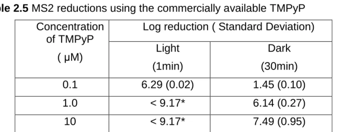
Table 2.5 MS2 reductions using the commercially available TMPyP
Concentration of TMPyP
( µM)
Log reduction ( Standard Deviation) Light
(1min)
Dark (30min)
0.1 6.29 (0.02) 1.45 (0.10)
1.0 < 9.17* 6.14 (0.27)
10 < 9.17* 7.49 (0.95)
*This data is below the limit of detection. *MS2 was irradiated with a 40 W Hg fluorescent light,0.48 mW/cm2. Data shown is the average of three trials.
Discussion
A total of five porphyrins were synthesized and tested against E. coli, and the three most efficient porphyrins, C3PyP, C4PyP, and C5PyP, were more extensively tested against bacteriophage MS2. Each of the porphyrins
synthesized has four identical substituents at the meso position of the porphine ring. This four-fold symmetry allows for a more straightforward procedure than would a synthesis of porphyrins with varying substituents. All data collected showed that the synthesis was successful in producing five distinct porphyrins. In each step of the synthesis, reactions seemed to have greater yield as the reaction mixtures were more dilute. In the condensation procedure, the use of the montmorillonite clay as a catalyst was much more effective than the method involving trifluoroacetic acid.
38
the porphyrins. The extent of inactivation and the ability to measure inactivation as a function of time was dependent on the rate of inactivation of each microbe, the concentration of microbes in solution, and the concentration of porphyrin used. A measurement of inactivation as a function of time was not feasible for MS2, which showed greater than six-logs reduction (99.9999%) with just one minute of light exposure. At five minutes the MS2 was reduced to below the limit of detection. Although the target concentration of porphyrin was 1 µM (~1 mg/L), which is comparable to drinking water limits on chlorine, inactivation was observed at 0.1 and 10 µM to ensure data within the limits of detection. The1 µM porphyrin concentration is also appealing because at this concentration there was a high level of inactivation of E. coli with light with no significant dark toxicity.
The bacteria and all host cells used must be in the log phase of growth. If non-viable bacteria compete for contact with the porphyrin, then the porphyrin will appear to be less efficient against the viable bacteria. While contaminated
waters in the environment would not contain bacteria in log phase growth, higher concentrations of bacteria are needed for a proof of concept for porphyrin use in PDI.
39
Turbidity, a measurement of cloudiness, was measured for both solutions of microbes. This will give some information as to the stages of water treatment at which the porphyrin will be most effective and allow for comparison of the porphyrin data to that of other disinfectants used in water treatment processes. The most common disinfectants for drinking water are generally used as the last step in water treatment. This occurs after most of the turbidity is removed from the water. It is assumed that the lower the turbidity, the higher the porphyrin’s efficiency of inactivating the test microbes.
40
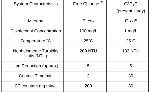
Table 2.6 Comparisons of C3PyP and free chlorine inactivation of E. coli
System Characteristics Free Chlorine 21 C3PyP (present study)
Microbe E. coli E. coli
Disinfectant Concentration 100 mg/L 1 mg/L
Temperature ˚C 25˚C 25˚C
Nephelometric Turbidity Units (NTU)
200 NTU 132 NTU
Log Reduction (approx) 5 5
Contact Time min 2 30
CT constant mg·min/L 200 30
Table 2.7 Comparisons of C3PyP and free chlorine inactivation of MS2.
System Characteristics Free Chlorine 21 C3PyP (present study)
Microbe MS2 MS2
Disinfectant Concentration 100 mg/L 1 mg/L
Temperature ˚C 25˚C 25˚C
Nephelometric Turbidity Units (NTU)
2 NTU 0.23 NTU
Log Reduction (approx) 4 6
Contact Time min 2 1
CT constant mg·min/L 200 1
41
concentration (mg/L) of the disinfectant and the time needed (min) for a specific level of inactivation. This value expresses a specific level of inactivation as a function of disinfectant concentration multiplied by contact time. Tables 2.6 and 2.7 show that the most efficient porphyrin offered more inactivation of the test microbes with a much smaller CT constant. These preliminary studies have shown that these porphyrin compounds could be a more effective disinfectant than free chlorine, especially in turbid waters.
With the comparison of TMPyP to our synthetic porphyrins in the inactivation of MS2, one can see that the TMPyP is much more lethal to MS2 than the synthetic porphyrins. This is true both in the presence and absence of light. The increase in the MS2 inactivation in the absence of light is due to an increase in natural toxicity of the porphyrin (toxicity in the absence of light). This shows that the TMPyP is not as inert in the absence of light as the other
porphyrins tested, and could potentially have more adverse effects if ingested. This could very well be due to the different counter ions in the commercially available TMPyP and our synthetic porphyrins. The synthetic porphyrins have a simple bromine counterion where as the TMPyP has a more complex, and potentially more toxic, tosylate counter ion. In order to directly measure the effect of cation position, a series of fixed-cation porphyrins with identical counterions were synthesized. These porphyrin analogs have the same
42
43
References
1 S. Ferro, F. Ricchelli, G. Mancini, G. Tognon and G. Jori, Inactivation of methicillin-resistant Staphylococcus aureus (MRSA) by liposome-delivered photosensitising agents, Journal of Photochemistry and Photobiology B: Biology, 2006, 83, 98-104.
2 Z. Alouini and M. Jemli, Destruction of Helminth Eggs by Photosensitized Porphyrin, Journal of Environmental Monitoring, 2001, 3, 548-551.
3 V. Carré, O. Gaud, I. Sylvain, O. Bourdon, M. Spiro, J. Biais, R. Granet, P. Krausz and M. Guilloton, Fungicidal properties of meso-arylglycosylporphyrins: influence of sugar substituents on photoinduced damage in the yeast
Saccharomyces cerevisiœ, Journal of Photochemistry and Photobiology B: Biology, 1999, 48, 57-62.
4 K. Kassab, D. Dei, G. Roncucci, G. Jori and O. Coppelloti, Phthalocyanine-photosensitized inactivation of a pathogenic protozoan, Acanthamoeba palestinensis, Photochemical & Photobiological Sciences, 2003, 2, 668-672. 5 Montgomery Watson Harza, in , ed. nonymous John Wiley and Sons Inc., Hoboken, New Jersey, 2nd edn., 2005,.
6 M. Merchat, G. Bertolini, P. Giacomini, A. Villaneuva and G. Jori,
Meso-substituted cationic porphyrins as efficient photosensitizers of gram-positive and gram-negative bacteria, Journal of Photochemistry and Photobiology B: Biology, 1996, 32, 153-157.
7 M. Merchat, J. D. Spikes, G. Bertoloni and G. Jori, Studies on the mechanism of bacteria photosensitization by meso-substituted cationic porphyrins, Journal of Photochemistry and Photobiology B: Biology, 1996, 35, 149-157.
8 A. Minnock, D. I. Vernon, J. Schofield, J. Griffiths, J. Howard Parish and S. B. Brown, Photoinactivation of bacteria. Use of a cationic water-soluble zinc
phthalocyanine to photoinactivate both Gram-negative and Gram-positive
bacteria, Journal of Photochemistry and Photobiology B: Biology, 1996, 32, 159-164 (DOI:DOI: 10.1016/1011-1344(95)07148-2).
9 T. Maisch, J. Baier, B. Franz, M. Maier, M. Landthaler, R. Szeimies and W. Bäumler, The role of singlet oxygen and oxygen concentration in photodynamic inactivation of bacteria, Proceedings of the National Academy of Sciences, 2007, 104, 7223-7228 (DOI:10.1073/pnas.0611328104).
44
11 L. L. Trannoy, J. W. Lagerberg, T. M. Dubbleman, H. J. Schuitmaker and A. Brand, Positively charged porphyrins: a new series of photosensitizers for sterilization of RBCs, Tranfusion, 2004, 44, 1186-1196.
12 G. Valduga, B. Breda, G. M. Giacometti, G. Jori and E. Reddi,
Photosensitization of Wild and Mutant Strains of Escherichia coli by meso-Tetra (N-methyl-4-pyridyl)porphine. Biochem. Biophys. Res. Commun., 1999, 256, 84-88.
13 M. J. Casteel, K. Jayaraj, A. Gold, L. M. Ball and M. D. Sobsey,
Photoinactivation of Hepatitis A Virus by Synthetic Porphyrins¶, Photochemistry and Photobiology, 2004, 80, 294-300.
14 D. A. Caminos, M. B. Spesia and E. N. Durantini, Photodynamic inactivation of Escherichia coli by novel meso-substituted porphyrins by
4-(3-N,N,N-trimethylammoniumpropoxy)phenyl and 4-(trifluoromethyl)phenyl groups. Photochemical & Photobiological Sciences, 2006, 5, 56-65.
15 M. ONAKA, T. SHINODA, Y. IZUMI and NOLEN Ernest, Porphyrin Synthesis In Clay Nanospaces, Chemistry Letters, 1993, , 117-120.
16 G. Kokotos and C. Noula, Selective One-Pot Conversion of Carboxylic Acids into Alcohols, J. Org. Chem., 1996, 61, 6994-6996 (DOI:10.1021/jo9520699). 17 R. G. Little, J. A. Anton, P. A. Loach and J. A. Ibers, Synthesis of some
substituted tetraarylporphyrins, Journal of Heterocyclic Chemistry, 1975, 12, 343-349.
18 J. S. Lindsey, I. C. Schreiman, H. C. Hsu, P. C. Kearney and A. M. Marguerettaz, Rothemund and Adler-Longo reactions revisited: synthesis of tetraphenylporphyrins under equilibrium conditions, J. Org. Chem., 1987, 52, 827-836 (DOI:10.1021/jo00381a022).
19 R. A. M. Gonsalves, J. M. T. B. Varejao and M. M. Pereira, Some new
aspects to the synthesis of meso-substituted porphyrins, Journal of Heterocyclic Chemistry, 1991, 28, 635.
20 O. Makoto, S. Tomotaka, I. Yusuke and E. Nole, Porphyrin synthesis in Clay nanospaces, Chemistry Letters, 1993, , 117-118.
21 C. Chaidez, M. Moreno, W. Rubio, M. Angulo and B. Valdez, Comparison of the disinfection efficacy of chlorine-based products for inactivation of viral
indicators and pathogenic bacteria in produce wash water, International Journal
Chapter 3: Porphyrin Photodegradation and Toxicity
Background
Photodynamic inactivation (PDI) of pathogens is an effective method of disinfection that has not been applied to water treatment. In PDI, a chromophore absorbs energy from light and ultimately transfers the energy to dioxygen to generate the singlet oxygen that inactivates pathogens. Additionally, PDI is an appealing method of disinfection because its mechanism of action is not likely to lead to resistance, and if the photosensitizer (PS) is ingested, its toxicity is reduced in the absence of light. The principles of PDI are evident in
photodynamic therapy (PDT), an approved treatment for several cancers 1-5, and they have more recently been applied to the disinfection of blood products6-10.
Cationic porphyrins are one class of chromophores that have proven to be effective in the inactivation of viral, bacterial, fungal and parasitic pathogens 2, 4, 11, 16-18, 20, 23
. In addition to being highly efficient PSs, the physicochemical properties of porphyrins may readily be tuned by altering the peripheral
46
negatively charged surface 24. To date, several studies have shown some connection between the cationic character of the PS and the association of that PS to gram-negative bacteria 11, 18, 20, 21.
The cationic porphyrins used in previous tests have rigidly attached positive charges on the periphery of the tetrapyrrole macrocycle. In the present study, it is hypothesized that attaching cationic groups via more flexible, aliphatic links will allow for increased cation mobility and better adaptation to the shape and negative charge distribution on the surface of target microorganisms. To evaluate this hypothesis we have synthesized porphyrin analogs to C3PyP and C4PyP, the most efficient porphyrins from the PDI experiments summarized in Chapter 2. These porphyrin analogs have the same molecular formula and differ only in the cation position and flexibility. The comparison of these porphyrins will allow for a direct observation of the effect of the cation position on various
properties that effect PDI.
47
spectrometry. The toxicity of the parent porphyrins and the photoproducts to model mammalian cells was measured in order to understand the potential of adverse effects of these porphyrins. The porphyrins incorporated into this study can be seen in Figure 3.1.
Figure 3.1 The four porphyrins used in this study and their abbreviations
Meso-tetrakis (3-[N-pyridiniumyl] propyl) porphyrin
tetrabromide (C3PyP)
Meso-tetrakis (4-[N-pyridiniumyl] propyl) porphyrin
tetrabromide (C4PyP)
Meso-tetrakis (3-[N-propyl] pyridiuniumyl) porphyrin
tetrabromide (TProPyP)
Meso-tetrakis (4-[N-butyl] pyridiniumyl) porphyrin
tetrabromide (TBuPyP)
N NH
N
HN N+
N+
N+ N+
N NH
N HN
N
N N
N
H3C CH3
CH3