http://dx.doi.org/10.11594/jtls.10.03.03
How to cite:
Research Article
Bactericidal Potentiality of Purified Terpenoid Extracts from the Selected Sea
Weeds and its Mode of Action
Sabira Siraj Sumayya 1, Abdulhadeef Shereefa Lubaina 2, Kumaraswamy Murugan 3*
1 Department of Botany, Sree Neelakanta Govt Sanskrit College, Pattambi, Kerala 695034, India 2 Department of Botany, Christian College, Kattakada, Kerala 695572, India
3 Center for Innovation in Science and Social Action (CISSA), Thiruvananthapuram, Kerala 695010, India
Article history: Submission April 2020 Revised July 2020 Accepted July 2020
ABSTRACT
Terpenoids are hydrocarbons involved in a variety of basic functions in plants such as growth, development and other physiological events. Terpenes and their associ-ated molecules safe guard the organisms from pest, pathogen and herbivores. Simi-larly, therapeutically terpenoids function as antimicrobial agents against bacteria, fungi and viruses. The mechanism of bactericidal activities may be via inhibiting the synthesis of essential molecules like proteins, nucleic acids, cell-wall compo-nents, cell membrane derailment, bacterial DNA replication or inhibition of meta-bolic pathways. The crude methanolic extracts of the seaweeds were subjected to silica gel column chromatographic purification and eluted with different combina-tions of ethyl acetate: petroleum ether solvent systems. The eluted fraccombina-tions were further subjected to thin layer chromatography and fractionated by GC-MS. The fractions obtained from Hypnea musciformis revealed the terpenoids such as eico-sane, heneicoeico-sane, 2-pentadecnone, hexadecanoic acid methyl ester, n- hexadeca-noic acid, hexadecahexadeca-noic acid ethyl ester, heptadecahexadeca-noic acid methyl ester, 11-octadecanoic acid metyl ester, whereas Kappapycus alvarezii displayed hexade-cane, eicosane, heptadehexade-cane, octadehexade-cane, heneicosane, tricosane, hexadecanoic acid, methyl ester and beta amyrin. Similarly, Gracillaria dura revealed hexadeca-noic acid methyl ester, n- hexadecahexadeca-noic acid, 11-octadecahexadeca-noic acid and phytol. Subsequently, the bactericidal activities of the purified terpenoid extracts from the sea weeds were carried. Initially, the extracts were tested for their in vitro antibac-terial activity against six bacantibac-terial strains such as three Gram-positive ( Staphylo-coccus aureus, Streptococcus mutans, Enterococcus faecalis) and three Gram-negative bacteria (Klebsiella pneumoniae, Escherichia coli, Pseudomonas aeru-ginosa) by disc diffusion method. The results revealed that the purified terpenoid extracts of G. dura exhibited significant bactericidal potentiality against S. mutans as compared to other strains. The zone of inhibition, MIC and MBC values narrate the efficacy of the purified terpenoid extract of the species. Remarkable leaching of metabolites like protein and DNA further substantiates the MIC and MBC results. Scanning electron microscopic observations such as clumping, irregularity of cells and ballooned walls reflect the possible membrane damage accounted in the cells by the terpenoid extracts. Further studies are planned to validate the above data by using molecular tools.
Keywords: Bactericidal activity, Disc diffusion method, Sea weeds, Terpenoids, Membrane damage
*Corresponding author: E-mail: harimurukan@gmail.com
Introduction
Terpenes are hydrocarbons consist of 5-carbon isoprenoid units as their building blocks. They are produced via non-mevalonate (precur-sor methylerythritol phosphate) or mevalonate
triterpenes [1]. Many terpenes display poor mi-crobicidal activities. Terpenoids are terpenes de-rivatives i.e., modified terpenes through addition or removal of functional groups [2]. Thus, the antimicrobial potentialities of terpenoids are reg-ulated by their functional groups. For example, the shifting or removal of a CH3 group and
addi-tion of O2 by enzymes result into derived
ter-penes. The OH group of the phenolic terpenoids and delocalized electrons are the microbicidal factors. Peppermint derived terpenes like linalool, menthol, carvacrol, thymol, citronella demon-strated synergistic activity against antibiotic re-sistant E. coli O157:H7, Salmonella
typhimuri-um, Staphylococcus aureus and Listeria
mono-cytogenes via disrupting Klebsiella pneumoniae
carbapenemase via oxidative stress [3]. There are challenges involved in experimenting with terpe-noids. Acquisition of terpenoids will demand higher cost when compared to synthetic chemi-cals. Natural phytochemicals have a molecular
mass ≤ 500 g/mol may have the calibre to act as
adjuvants for microbicides and exhibit synergistic roles [4]. Investigation of novel microbicidal agents via biotransformation is a recent trend practiced by molecular pharmacologists. Combi-nation therapy with a low molecular weighed biomolecules like terpene derivatives has showed optimal outputs. Terpenes derivatives have proved microbicidal activities against susceptible and resistant pathogens [5]. Wagner and Merzen-ich [6] documented that synergic research ap-proach was more effective in designing new gen-eration phytopharmaceuticals for effective anti-microbial activities. In this juncture, the present study was carried to analyse the bactericidal po-tentialities of purified terpenoid extracts from the selected seaweeds and their plausible mode of action.
Material and Methods
The marine seaweeds such as H. musciformis,
K. alvarezii and G dura were collected from the
Mandapam coast (latitude 9˚17’N, longitude79˚ 22’E), Gulf of Mannar. The algal thalli were
washed thoroughly to remove all the debris. Then, sliced into pieces, shade dried and pow-dered. Initially, each algal powder was subjected to Soxhlet hot continuous extraction with 250 ml of methanol as per the protocol narrated by
Oussalah
et al
. [3]. The supernatants weregen-tly decanted into a pre-weighed glass vials through Whatman No. 1 filter paper and concen-trated to dryness using a rotary evaporator. The dried extracts were made up to a dosage of 10 mg/ml (stock solution) to be used in subse-quent assays and stored at 4°C in tightly stop-pered glass tubes [3, 4].
The purification of methanolic extracts was carried out using silica gel Column Chromatog-raphy (CC) with different solvent combinations of petroleum ether: ethyl acetate. The fractions so obtained was collected and further analysed by thin layer chromatography and fractionated by GC-MS. The TLC was carried out by using the solvent combinations of toluene and ethyl acetate in 6:1 ratio [3,4].
GC-MS analysis
Adeyemi [7] protocol was adapted for GC-MS analysis. The column purified terpenoid ex-tract was injected into a HP-5 column (30 m X 0.25 mm i.d with 0.25 ìM film thickness), Ag-ilent technologies 6890 N JEOL GC Mate II GC-MS model. Experimental conditions of GC-GC-MS system were as follows. The flow rate of mobile phase (Helium: carrier gas) was set at 1 ml/min. In the gas chromatography part, temperature pro-gramme (oven temperature) was 40°C raised to
250°C at 5°C/min and injection volume was 1 μl.
Samples dissolved in chloroform were run fully at a range of 50-650 m/z and the results were compared by using NIST Spectral library.
Strains of bacteria
For the in vitro bactericidal tests, three Gram-positive (Staphylococcus aureus, S. mutans
En-terococcus faecalis) and three Gram-negative
bacteria (Klebsiella pneumoniae, Escherichia
coli, Pseudomonas aeruginosa) were purchased
from Microbial Type Culture Collection and Gene Bank (MTCC), Chandigarh. None of the strains were multidrug-resistant categories.
Bactericidal assay-Agar disc diffusion assay
The bactericidal activity of the purified terpe-noid extracts from the selected sea weeds was carried using the agar disc diffusion method of
Ster-ile Whatman No. 1 paper disc was used (6 mm in diameter). Varied concentrations of the purified terpenoid extracts were made with DMSO (100 mg/ml). Fresh bacterial cell suspensions were adjusted to 0.5 McFarland turbidity standards to
prepare 1 × 10⁸ bacterial/ml inoculum.
Bacterial suspension of each strain was in-oculated on Mueller-Hinton agar plates, and the
plates were then permitted to dry for 5 min. The sterile filter paper discs were soaked in 10 μL of each algal terpenoid extracts (concentrations of 0, 0.5, 1.0, 2.0, 3.0, and 5.0 mg/mL). The extract-soaked filter paper discs were then placed on the inoculated Mueller-Hinton agar plates. Gentami-cin (10 µg/disc) disc was used as the positive control, and 10% DMSO-soaked filter paper disc was used as the negative control. Plates were
in-cubated for 24 h at 32 ± 2°C. After incubation, the zones of inhibition were recorded as the di-ameter of the growth-free zones measured in mm using a Vernier caliper [9, 10]. The agar disc dif-fusion protocols were performed in triplicates.
Minimum inhibitory concentration (MIC) and Minimal bactericidal concentration (MBC)
The minimum inhibitory concentration (MIC) for terpenoid extract was evaluated according to method described by Bereksi et al. [9] and as per the Clinical and Laboratory Standards Institute (CLSI) protocol [11]. Sterile 96-well microplates were employed. Briefly, a 24-h culture of all bac-terial strains was prepared after incubation at 37°C. The Mueller-Hinton broth containing 1.5 × 10⁸ CFU/mL from each strain was prepared
equivalent to 0.5 McFarland standards and 100 mL of it was added to each well of the 96 well sterile microplate. Different dilutions were made from the terpenoid extract in each row by serial dilution; in well numbered from 0.0625 to 10.0 mg/mL. Positive control of culture medium con-taining bacteria was prepared without the extract and negative control of pure extract. Microplates were incubated for 24 h at 37°C and were exam-ined for turbidity Absorbance Microplate Readers at 570 nm. The lowest dilution of the extract with any turbidity was considered the MIC. All exper-iments were repeated thrice.
Minimum bactericidal concentration (MBC) was determined by the microplate dilution meth-od on the 96-well sterile plate by adding MTT reagent (3-(4, 5-dimethylthiazol-2-yl)-2,5
diphe-nyl tetrazolium bromide). The dose of the ex-tracts which could reduce the viable cell number was determined by MTT method. MTT colori-metric assay is based on the capacity of viable cell succinate dehydrogenase enzymes to reduce the water soluble yellow MTT into formazan, an insoluble colored product which is recorded by ELISA Microplate Reader. The reduction of MTT occurs only in metabolically active viable cells. Briefly, bacteria treated with extracts for 24 h at 36°C were incubated with 20 µl of MTT so-lution for 4 h at 36°C that was added to each well. (MTT reagent was prepared as 5 mg/mL in phosphate-buffered saline). Subsequently, the supernatant was slowly removed and 100 µL DMSO (dimethyl sulfoxide) was added to each well. DMSO is a solvent of formazan crystals and caused variable degrees of intensity of color spectrum from purple to white that is a measure of viable cells. The absorption of wells was measured at the wavelength of 570 nm by ELISA reader. In this method, the wells without any ab-sorbance due to formazan crystals were consid-ered to contain only dead cells. The last wells without formazan crystals were considered as MBCs.
Estimation of 260-nm absorbing material and protein release
Scanning electron microscopic analysis (SEM)
Kaya et al. [13] method was employed for the scanning electron microscopic study. The suscep-tible species to the terpenoid extracts such as E. coli and S. mutans were subjected to SEM analy-sis. Small agar pieces were cut out from the inhi-bition zone of the agar diffusion assay and were fixed in 3 % (v/v) glutaraldehyde buffered with 0.1 M sodium phosphate buffer (pH 7.2) for 1 h at room temperature and then washed thrice in sodium phosphate buffer. Further, the pieces were post-fixed in 1 % (w/v) osmium tetroxide for 1 h and then washed thrice in the buffer. Sub-sequently, the blocks were dehydrated in a grad-ed alcohol series. The last stages of dehydration were performed with propylene oxide. The sam-ples were dried and were mounted onto stubs us-ing double-sided carbon tape, and then were coated with a thin layer of gold by a Polaron SC 502 sputter coater. They were viewed in a Jeol JSM 6060 LV SEM.
Statistical analysis
The data were reported as mean ± SD values obtained from a minimum of three determina-tions. Data were analysed using one way Anova using GraphPad Prism5 software. A significant difference was considered at the level of P < 0.05
and 0.01.
Results and Discussions
The crude algal powder was extracted with 250 ml of methanol for 8h using hot continuous soxhlet method. The extract was filtered and con-centrated to dryness using rotary evaporator. The crude methanolic extract was subjected to purifi-cation by column chromatography (silica gel G of mesh size 230-400). Different combinations of petroleum ether: ethyl acetate (PE: EA) solvents were employed for the elution of terpenoids. Elu-tion was started with 100% PE and gradually in-creased the polarity. The eluted fractions were analysed for the presence of terpenoids and sub-jected to TLC using 6 ml toluene: 1ml EA sol-vent system as mobile phase. Further, the bands obtained from TLC were subjected to GC-MS analysis. In fraction eluted using 95:5 (PE: EA)
of H. musciformis showed a single prominent
band in TLC and in GC-MS revealed the compo-nents such as eicosane, heneicosane, 2-pentadecnone, hexadecanoic acid methyl ester, n-
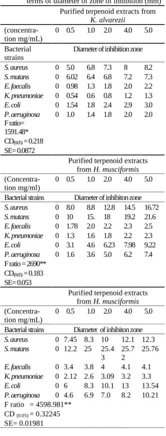
Table 1. Agar disc diffusion assay of purified terpe-noid extracts from the selected seaweeds in terms of diameter of zone of inhibition (mm)
Purified terpenoid extracts from K. alvarezii
(concentra-tion mg/mL) 0 0.5 1.0 2.0 4.0 5.0 Bacterial
strains Diameter of inhibition zone S. aureus 0 5.0 6.8 7.3 8 8.2 S. mutans 0 6.02 6.4 6.8 7.2 7.3 E. faecalis 0 0.98 1.3 1.8 2.0 2.2 K. pneumoniae 0 0.54 0.6 0.8 1.2 1.3 E. coli 0 1.54 1.8 2.4 2.9 3.0 P. aeruginosa
F ratio= 1591.48*
0 1.0 1.4 1.8 2.0 2.0
CD(0.05) = 0.218
SE= 0.0872
Purified terpenoid extracts from H. musciformis
(Concentra-tion mg/ml) 0 0.5 1.0 2.0 4.0 5.0 Bacterial strains Diameter of inhibiton zone S. aureus 0 8.0 8.8 12.8 14.5 16.72 S. mutans 0 10 15. 18 19.2 21.6 E. faecalis 0 1.78 2.0 2.2 2.3 2.5 K. pneumoniae 0 1.3 1.6 1.8 2.2 2.3 E. coli 0 3.1 4.6 6.23 7.98 9.22 P. aeruginosa 0 1.6 3.6 5.0 6.2 7.4 F ratio = 2690**
CD(0.05) = 0.183
SE= 0.053
Purified terpenoid extracts from H. musciformis
(Concentra-tion mg/mL)
0 0.5 1.0 2.0 4.0 5.0
Bacterial strains Diameter of inhibiton zone S. aureus 0 7.45 8.3 10 12.1 12.3 S. mutans 0 12.2 25 25.4
3
25.7 2
25.76
E. faecalis 0 3.4 3.8 4 4.1 4.1 K. pneumoniae 0 2.12 2.6 3.09 3.2 3.3 E. coli 0 6 8.3 10.1 13 13.54 P. aeruginosa 0 4.6 6.9 7.0 8.2 10.21 F ratio = 4598.981**
CD (0.05) = 0.32245
SE= 0.01981
hexadecanoic acid, hexadecanoic acid ethyl ester, heptadecanoic acid methyl ester, 11-octadecanoic acid metyl ester. 50:50 solvent combination of K.
GC-MS fractionation showed the presence of hexadecane, eicosane, heptadecane, octadecane, heneicosane, tricosane, hexadecanoic acid, me-thyl ester; beta amyrin. The 90:10 (PE: EA) puri-fied fraction of G. dura showed 2 bands on TLC with hexadecanoic acid methyl ester, n- hexadec-anoic acid, 11-octadechexadec-anoic acid and phytol mol-ecules in GC-MS.
Bactericidal assay – Agar disc diffusion assay
The bactericidal potentiality of different dos-es (0.0, 0.5, 1.0, 2.0, 4.0, 5.0 mg/mL) of the puri-fied terpenoid extracts from K. alvarezii, H.
mus-ciformis and G. dura against Gram-positive
(Staphylococcus aureus, S. mutans Enterococcus
faecalis) and Gram-negative bacteria (Klebsiella
pneumoniae, Escherichia coli, Pseudomonas
ae-ruginosa) were qualitatively and quantitatively
analysed in terms of diameter of inhibition zone (mm). As narrated in the Table1, the DIZ data for
S. mutans increased significantly (P < 0.01) with
the increasing dosage of G. dura terpenoid con-centrations as compared to K. alvarezii and H.
musciformis. The DIZ values for S. mutans
treat-ed with G. dura were ranged from 12.2 mm to 25.76 mm. DIZ values noticed for E. coli were 6 to 13.54 mm as compared to S. mutans. Mean-while, minimal response was displayed by E.
fae-calis, P. aeruginosa and K. pneumonia (resistant
strains). Antibacterial reports categorize the mi-crobicidal potentiality in to three classes based on zone of inhibition i.e., strong (DIZ > 20 mm), moderate (12 mm < DIZ < 20 mm) and weak ac-tivity (DIZ < 12 mm) [14, 15]. Based on this, G. dura displayed strong activity against S. mutans
and moderate to weak activity in other strains.
MIC and MBC of bacteria vs terpenoid extracts
The MIC values for purified terpenoid ex-tracts from G. dura, H. musciformis and K.
al-varezii, against S. mutans were 0.065, 0.5 and 1.5
mg/ml respectively. Similarly, the respective MBC values of the strain were 0.12, 1.25 and 3.0 mg/mL (Table 2). Generally, MIC values up to 0.5 mg/mL are considered strong, 0.6–1.5 mg/mL as moderate, and MIC above 1.5 mg/mL is with low antimicrobial power [16]. The results of the present study showed that the purified terpenoid extracts of the sea weeds had a strong bactericid-al activity against S. mutans, while low with E.
faecalis, P. aeruginosa and K.pneumoniae.
Simi-larly, the sea weeds showed variations in their MBC and MKC activities between the strains. This may be attributed due to the variations of terpenoid molecules and their content in the puri-fied extracts of the sea weeds. 11-octadecanoic acid and phytol were present only in the purified terpenoid extracts of G. dura. K. alvarezii re-vealed the unique molecules such as hexadecane, heptadecane, octadecane, tricosane and beta amyrin. H. musciformis showed the molecules such as 2-pentadecnone, hexadecanoic acid me-thyl ester, hexadecenoic acid eme-thyl ester and 11-octadecanoic acid methyl esters. Similar
varia-tions in the constituents such as sabinene, α
-pinene, β-pinene, limonene, β-caryophyllene, and caryophyllene were reported among the four ma-jor cultivars of black pepper [17]. It was previ-ously reported that the mode of action of essen-tial oils was through altering the bacterial cell membrane permeably and also capable to disin-tegrate the outer membrane of Gram-negative bacteria [18]. Further part of the study was re-stricted to the strains such as S. mutans and E. coliagainst the terpenoid extracts of G. dura on-ly.
Table 2. MIC and MBC of purified terpenoid extracts from the selected sea weeds (mg/mL)
Bacterial strains G. dura H. musciformis K. alvarezii
MIC MBC MIC MBC MIC MBC
S. aureus 1.5 3.0 2.5 4.0 3.5 7.0
S. mutans 0.065 0.12 0.5 1.25 1.5 3.0 E. faecalis 3.0 8.0 5.0 10.0 6.5 13.0 K. pneumoniae 6.0 12.0 7.5 15.0 8.5 18.0 P. aeruginosa 2.5 5.0 3.5 7.0 4.0 8.0
E. coli 1.25 2.5 2.0 4.0 3.0 5.0
Leakage of 260-nm absorbing material and pro-tein
The leakage of nucleic acid and proteins from the S. mutans and E. coli cells against purified terpenoid extracts from G. dura was displayed in the Table 3. The absorbance values for nucleic acid leakage of S. mutans increased remarkably at 24 and 48 h at 1 x MIC concentration (P < 0.01), whereas effectively from 8 to 48 h with 2 × MIC dose (P < 0.05) treatments. The OD260 nm
values of 2 × MIC were higher than that of 1 x MIC. However, the OD260 nm values for control
were marginal throughout the experimental peri-ods (P < 0.01). E. coli also showed a more or less similar trend but with less magnitude.
Similarly, the protein leakage values from
S. mutans and E. coli cells increased at par with duration and concentrations of the terpenoid ex-tract (1×MIC and 2×MIC (P < 0.01). The values of protein leakage at 2×MIC was maximal in S.
mutans than that of 1×MIC i.e., 125.3 µg/mL at
48 h
. Meanwhile, the similar value for E. coliwas 100.3 µg/mL (Table 4.) The values of pro-tein leak for control displayed the lowest values (P < 0.05). The 260 nm absorbing materials and proteins are used as biomarker for evaluating the irreversible disintegration to the cell membrane structure in the experimental groups when com-pared to control. Similar to the present data, the bactericidal potentiality of essential oil against membrane permeability of food-borne pathogens was recorded [19]. The vital biomolecules such as nucleic acids and proteins are compartmental-ized in the cell membrane and also in the
cyto-plasm and are the key molecules for the active phase of life in the bacterial cells. The obtained data revealed that the exposure of S. mutans and
E. coli to purified terpenoid extracts caused a
drastic loss of 260 nm absorbing materials and proteins, suggesting an irrecoverable damage ac-counted in the cell membranes. Further, the leak-age of nucleic acids and proteins may lead to functional disorders in the metabolic cycles of the cells and subsequently inhibits the bacterial growth.
Scanning electron microscopic observations
The morphological variation of S. mutans
and E. coli cells treated with purified terpenoid extracts of G. dura and untreated control cells was evaluated by SEM. S. mutans control cells displayed the smooth surface of shape and its rigidity to form normal cocci chains (Figure 1A). Meanwhile, abnormalities were noticed on the cells after exposure with the terpenoid extract which was reflected by the formation of cavities initiated to lyse, shrunk, and collapse of cells (Figure 1B, C, and D). The disruption of the cells has continuously occurred to form debris i.e., most of the cells were lysed, shrunk drastically, and completely collapsed. The normal and smooth cocci chains shape was no longer ob-served but only the crumpled cell residues. Treatment and these destructions in bacterial cells may be due to severe morphological disor-ganization and invaginations resulted in to clumping (secrete sticky mucus) as a response to stress condition.
Table 3. Release of 260 absorbing material in terms of OD260 from S. mutans and E. coli against purified
terpe-noid extracts from G. dura, Control, MIC 1×, and MIC 2×
0h 4h 8h 12h 16h 20h 24h 48h
Release of 260 absorbing material from S. mutans
Control 0.047 0.047 0.047 0.047 0.047 0.047 0.047 0.047 MIC X 0.089 0.143 0.241 0.276 0.298 0.323 0.376 0.489 MIC 2X
F ratio= 1904.27** CD(0.05)= 0.316 SE= 0.1076
0.095 0.187 0.294 0.365 0.504 0.599 0.631 0.754
Release of 260 absorbing material from E. coli
Control 0.04 0.04 0.04 0.04 0.04 0.04 0.04 0.04 MIC X 0.067 0.131 0.195 0.23 0.280 0.30 0.34 0.40 MIC 2X 0.072 0.143 0.21 0.279 0.38 0.46 0.49 0.52 F ratio= 785.47**
Table 4. Release of protein (µg/mL) from S. mutans and E. coli against purified terpenoid extracts from G. dura Control, MIC 1× and MIC 2×
0h 4 h 8 h 12 h 16 h 20 h 24 h 48 h Release of protein from S. mutans
Control 1.46 1.48 1.487 1.49 1.493 1.497 1.49 1.53
MIC X 31.4 43.2 47.5 49.2 54.3 57.5 66.9 70.23
MIC 2X 35 67.8 76.6 90 98.4 108 116.5 125.3
F ratio 590**
SE 0.1682
CD (0.05) 0.953
Release of protein from E. coli
Control 1.36 1.38 1.383 1.40 1.43 1.44 1.435 1.461
MIC X 26.5 30.2 36.4 39.2 43.3 46.5 54.2 60.3
MIC 2X 28.9 44.5 58.6 68.5 76.4 87.8 93 100.3
F ratio 1003**
SE 0.1743
CD (0.05) 0.4872
The control E. colicells were about 3.5 μm
long and showed smooth and intact surface (Fig. 2 A control). After exposure to terpenoid
ex-tracts, the cells were shortened to 1 μm with
re-markable compactness (Fig. 2 B, C & D). Blisters were seen followed by protrusion of numerous small bubbles. Further, alterations of cell mem-brane were seen with abnormal cell shapes. Sim-ilarly, sticky membranes, deep craters, lysed and completely burst cells. SEM micrographs showed asymmetric cytoplasmic turbidity and also the irregularity of distribution.
Similar changes in the bacterial cells treated with oregano and thyme essential oils and there by confirmed their antimicrobial power against E.
coli O157:H7 [19]. Burt [18] correlated the mode
of action of essential oils with cell membrane damages in bacterial cells. Further, the mode of action may be multiple such as the damage of proteins on the membrane, the binding molecules of the envelop, leakage of cell contents, cyto-plasmic coagulation and derailment of the proton motive force. The antibacterial activity of various leaf extracts of Merremia emarginata validates the present results displayed by the terpenoid ex-tracts [19]. All the above research outputs indi-cate that purified terpenoid extracts of G. dura
possess bactericidal activity as reflected by zone of inhibition, MIC and MBC values. The cell wall and cell membrane lysis further inhibit the bacterial growth. Leakage of nucleic acid and proteins further substantiates the SEM visuals. Further studies are warranted at clinical level to validate the obtained results. The current chal-lenges and perspectives of the usage of aqueous
Figure 1. SEM images showing the morphological variations in S. mutans A – control, B, C, and D treated cells with abnormalities
Figure 2. SEM images showing the morphological variations in E. coli. A – control, B, C, and D treated cells with damages, deformities & clumping
plant extracts were also validated against
mode of action of some plant extracts against food pathogens and the spoilage microbes, anti-microbial activity of plant extracts against antibi-otic susceptible and resistant bacterial strains causing wound infection, in vitro microbial pow-er of medicinal hpow-erbals against human pathogenic bacteria, anti-bacterial activity of Ricinus
com-munis against bacterial pathogens like
Escherich-ia coli and Klebsiella oxytoca and antimicrobial
activity of diverse extracts of Ocimum basilicum
supports the present mode of action of terpenoids from G. dura in terms of its antibacterial efficacy [22, 23, 24, 25, 26, 27].
Conclusion
Among the three sea weeds evaluated, the
Gracillaria dura presented potent bactericidal
activities as compared to others. The terpenoid fractions of the species showed strong antibacte-rial activity against S. mutans. The obtained vis-uals of SEM indicate the mode of the bacterial cell death of the evaluated S. mutans was due to loss of cellular membrane integrity or function. Leaching of 260 molecules and protein substanti-ates the SEM data. The present study brings an insight about the antibacterial activity of the red algae. Similarly, an understanding for the future researches of synergism, mode of action and bio-availability of various sea weed terpenoid com-ponents.
References
1. Christianson DW (2017) Structural and Chemical Biol-ogy of Terpenoid. Chemical Reviews 117(17):11570– 11648.doi: 10.1021 /acs.chemrev.7b00287.
2. Jiang Z, Kempinski C, Chappell J (2016) Extraction and Analysis of Terpenes/Terpenoids. Current Proto-cols Plant Biology 1(4):345–358.doi: 10.1002/cppb.20024.
3. Oussalah M, Caillet S, Saucier L, Lacroix M (2007) Inhibitory effects of selected plant essential oils on the growth of four pathogenic bacteria: E. coli O157:H7, Salmonella Typhimurium, Staphylococcus aureus and Listeria monocytogenes. Food Control 18(6): 414– 420.doi: 10.1016/j.foodcont.2005.11.009.
4. Barbieri R, Coppo E, Marchese A et al. (2016) Phyto-chemicals for human disease: An update on plant-derived compounds antibacterial activity. Microbiologi-cal Research 3 (7): 196 - 214. doi: 10.1016/j.micres.2016.12.003.
5. Mahizan NA, Yang SK, Moo CL, et al. (2019) Terpene
derivatives as a potential agent against antimicrobial re-sistance (AMR) Pathogens. Molecules 24(14): 2631 2641. doi :10.3390/molecules24142631.
6. Wagner H, Ulrich-Merzenich G (2009) Synergy re-search: Approaching a new generation of phytopharma-ceuticals. Phytomedicine 16(4): 97–110. doi: 10.1016/j.fitote.2010.11.016.
7. Adeyemi M (2017) Phytochemical analysis and GC-MS determination of Lagenaria breviflora R. fruit. Interna-tional Journal of Pharmacogonsy and Phytochemical
Research 9 (7):1045-1051. doi:
10.25258/phyto.v9i07.11178.
8. Bajpai VK, Sharma A, Baek KH (2013) Antibacterial mode of action of Cudrania tricuspidata fruit essential oil, affecting membrane permeability and surface
char-acteristics of food-borne pathogens. Food Control 32 :582–590 doi:10.3389/fmicb.2017.00552.
9. Bereksi MS, Hassaïne H, Bekhechi C, Abdelouahid DE (2018) Evaluation of antibacterial activity of some me-dicinal Plants extracts commonly used in Algerian tra-ditional medicine against some pathogenic bacteria. Pharmacogn Journal 10 (3):507-512. doi: 10.5530/pj.2018.3.83.
10. Humphries RM, Ambler J, Mitchell SL et al. (2018) CLSI methods development and standardization work-ing group best practices for evaluation of antimicrobial susceptibility tests. Journal of Clinical Microbiology 56 (4): e01934-17. doi:10.1128/JCM.01934-1.
11. Du W, Sun C, Liang Z et al. (2012) Antibacterial activi-ty of hypocrellin A against Staphylococcus aureus. World Journal of Microbiology and Biotechnology 28(3): 3151–3157. doi: 10.1007/s11274-012-1125-z. 12. Xu JG, Hu QP, Wang XD et al. (2010) Changes in the
main nutrients, phytochemicals, and antioxidant activity in yellow corngrain during maturation. J. Agriculture and Food Chemistry 58 (7): 5751–5756. doi: 10.1021/jf100364k.
13. Kaya I, Yigit N, Benli M (2008) Antimicrobial activity of various extracts of Ocimum basilicum L. and obser-vation of the inhibition effect on bacterial cells by use of scanning electron microscopy. African Journal of Traditional, Complementary and Alternative Medi-cines 5 (4): 363-369 doi: 10.4314/ajtcam.v5i4.31291. 14. Rota MC, Herrera A, Martinez RM et al. (2008)
Anti-microbial activity and chemical composition of Thymus vulgaris, Thymus zygis and Thymus hyemalis essential oils. Food Control 19 (4): 681–687. doi: 10.1016/j.foodcont.2007.07.00.
bac-teria. Food Control 21 (5): 1408–1414. doi: 10.1016/j.foodcont.2010.04.014.
16. Aligiannis N, Kalpoutzakis E, Mitaku S, Chinou IB (2001) Composition and antimicrobial activity of the essential oils of two Origanum species. Journal of Agri-culture and Food Chemistry 49 (2): 4168– 4170.doi:10.1021/jf001494m.
17. Menon AN, Padmakumari KP, Jayalekshmy A (2003) Essential oil composition of four major cultivars of black pepper (Piper nigrum L.). Journal of Essential Oil
and Research 15 (1): 155–157.
doi:10.1080/10412905.2003.9712099.
18. Burt S (2004) Essential oils: their antibacterial proper-ties and potential applications in foods- a review. Inter-national Journal of Food Microbiology 94(2):223–253. doi:10.1016/j.ijfoodmicro.2004.03.022.
19. Burt SA, Reinders RD (2003) Antibacterial activity of selected plant essential oils against Escherichia coli O157:H7. Letters in Applied Microbiology 36: 162-167.doi: 10.1046/j.1472-765X.2003.01285.x.
20. Elumalai EK, Ramachandran M, Thirumalai T, Vi-nothkumar P (2011) Antibacterial activity of various leaf extracts of Merremia emarginata. Asian Pacific Journal of Tropical Biomedicine 1 (5): 406–408. doi:10.1016/S2221-1691(11)60089-0.
21. Santos SAO, Martins C, Pereira C et al. (2019) Current challenges and perspectives for the use of aqueous plant extracts in the management of bacterial infections: the case-study of Salmonella enterica. International Journal of Molecular Science 20 (4): 940 – 947. doi:10.3390/ijms20040940.
22. Gonelimali FD, Lin J, Miao W et al. (2018) Antimicro-bial properties and mechanism of action of some plant extracts against food pathogens and spoilage microor-ganisms Frontiers in Microbiology 8 (2): 1639 -1643. doi: 10.3389/fmicb.2018.01639.
23. Atef NM, Shanab SM, Negm SI, Abbas YA (2019) Evaluation of antimicrobial activity of some plant extracts against antibiotic susceptible and resistant bacterial strains causing wound infection. Bulletin of the Natural Research Center 43 (5): 1-5.doi: 10.1186/s42269-019-0184-9.
24. Kalra SS Goldy Saxena (2011) Antimicrobial activity pattern of certain terpenoids. International Journal of Pharmaceutical and Biological Science 2 (1): 87-91. doi:10.1.1.294.1573.
25. Manandhar S, Luitel S, Dahal R (2019) In Vitro antimi-crobial activity of some medicinal plants against human pathogenic bacteria. Journal of Tropical Medicine 54(2):1-7.doi:org/10.1155/2019/1895340.
26. Hajrah N, Abdul WM, Sabir J et al. (2018) Anti-bacterial activity of Ricinus communis L. against bacte-rial pathogens Escherichia coli and Klebsiella oxytoca as evaluated by Transmission electron microscopy, Bio-technology and Biotechnological Equipment 32(3): 686-691.doi: 10.1080/13102818.2018.1451778. 27. Kaya I, Yiğit N, Benli M (2008) Antimicrobial activity