http://www.ijmedicine.com pISSN 2349-3925 | eISSN 2349-3933
Original Research Article
The total leucocyte count: its utility in dengue
Anagha A. Joshi, Gayathri B. R., Yashica Gowda R.*
INTRODUCTION
Dengue, an arboviral infection (DENVI-4) is transmitted by Aedes mosquito.1 The clinical manifestations include
headache, fever, retro orbital pain, myalgia, body pain, rash.2 Dengue has been included as an emergent public
health issue with 50 million infections annually, with 40% of population at risk in tropics and subtropics.3,4
In India, epidemics are frequent, straining the limited resources of public health system.5 Dengue is mostly
self-limiting, but mortality from complications can be about 20% if untreated.5 Dengue presents with a wide clinical
spectrum which includes undifferentiated fever, Classic
Dengue, Dengue Hemorrhagic Fever and Dengue Shock Syndrome.2
It may be confused with other febrile illnesses like Malaria, Typhoid, Leptospirosis, Chikungunya, Cox sackie, Infectious Mononucleosis, Rickettsia, Rubella, Influenza etc. So, clinical diagnosis in early phase is difficult.6-9 However, early rapid diagnosis is important
in-patient management as currently there is no specific treatment or vaccine available for dengue.8 In clinical
practice, diagnosis is based both on clinical features and lab tests.10 Serological tests, confirm dengue late in the
course of the disease.11 Simple hematological parameters
not only help in early diagnosis of dengue but also can predict onset of severe dengue and are useful in smaller
ABSTRACT
Background: Dengue infections are public health concerns in India, where they occur in epidemics and have a high mortality in the advanced stages. Clinical features are nonspecific, and diagnosis is supported by lab features. One of these lab tests include total leucocyte count-easily available, simple and cost effective which is useful in small rural set ups for early diagnosis and prognosis of dengue. The aim of this study is to analyse the total leucocyte count patterns in dengue and assess its utility as early marker of dengue and prognosticator of severe dengue.
Methods: A total of 132 serologically proven cases of dengue with total white cell counts (blood counts obtained by Automated hematology analyser) during November 2016 were analysed.
Results: In our study, most cases were noted in the younger age group with a male predominance. The range of total leucocyte count was 1.1x10⁹ /l to 14.3x10⁹/l. Most (55%) had normal counts, leucopenia was noted in 36%, had an
equal distribution in all ages and both sexes. 82% had mild leucopenia,52% of leucopenia cases were associated with severe thrombocytopenia. Almost half (47%) of leucopenia cases were NS1 positive while only 15% were antibody positive.59% of NS1 positive had leucopenia in contrast to 13% of antibody positive cases. While leucopenia was mostly (55%) associated with neutrophilia, lymphocytosis was seen in 74% of cases with a normal leucocyte count. Conclusions: Total leucocyte count is one of the simple, easily available, cost effective tests useful in small rural set ups for early diagnosis and prognosis in dengue and helps reduce the morbidity and mortality of dengue.
Keywords: Dengue fever, Leucopenia, Lymphocytosis, Thrombocytopenia
Department of Pathology, Kempegowda Institute of Medical Sciences and Research Centre, Bengaluru, Karnataka, India
Received: 26 August 2017 Accepted: 20 September 2017
*Correspondence: Dr. Yashica Gowda R.,
E-mail: yashica317@gmail.com
Copyright: © the author(s), publisher and licensee Medip Academy. This is an open-access article distributed under the terms of the Creative Commons Attribution Non-Commercial License, which permits unrestricted non-commercial use, distribution, and reproduction in any medium, provided the original work is properly cited.
rural setups with limited resources.12,13 The hematology
tests of importance are platelet counts, total leucocyte count and hematocrit.7,12 However changes in platelet
count and hematocrit occur in later stages of infection, after 3rd - 4th day.9,14 The earliest hematological
abnormality in dengue is a progressive decline in white cell counts.15-17
Leucopenia, defined as total leucocyte count <4x10⁹ /l is
a prominent and supposedly the second most common feature in dengue.9,18 It gives enough clue for diagnosis of
Dengue and helps in differentiation from other febrile illnesses thus aiding in reducing its morbidity and mortality.12,19,20 Some studies have observed that total
leucocyte counts/leucopenia could serve as a prognostic factor for dengue severity while others dispute it. 10,13-17,21,22
A few studies have observed that there is a progressive decline in white cell counts with sudden platelet drop which precedes plasma leakage and hence it could be the earliest prognosticator of severe dengue.23 Our study
focuses on the significance and patterns of one such simple, routine test the total white cell count in diagnosis of dengue. The aim of the study is to analyse the total white cell count patterns in dengue, assess its role in diagnosis and utility as early marker of dengue infection and prognosticator of severe dengue.
METHODS
This is a prospective study done on 132 patients with positive dengue serology in hematology department of KIMS hospital and research centre, Bengaluru over a one-month period in November 2016. All patients with serological confirmation of dengue (NS1 /IgM/IgG/all positivity) done by rapid card method (Standard diagnostics-Bioline Alera) with Total white cell counts along with hematocrit, differential leucocyte count, and platelet count were included in the study.
Exclusion criteria
Patients with concomitant infections like malaria, typhoid etc. along with Dengue and patients with normal or increased platelet count. The results of dengue tests were retrieved from microbiology registers. The hematological
data (obtained from Automated hematology Analyser-Sysmex 1800i) of these cases was tabulated for analysis. The differential leucocyte count was done on peripheral blood smears stained by Leishman's stain (done for confirmation of platelet counts as per hospital protocol).
RESULTS
A total of 132 dengue serology positive cases were analyzed. It showed an age range between 5 months to 65 years, the average being 32 years. The majority of patients were in the 12- 25 years group. There were 65% of cases above 14 years (86/132) and 35% of cases below 14 years (46/132) (Table 1).
Table 1: Age wise distribution of patients.
Age group Number of cases
(n=132)
(%)
Adults*(above 14 years) 86 65
Pediatric (below 14 years) 46 35
(*For simplicity age above 14 years has been categorized as adults in the study)
Males predominated over females with a male to female ratio of 1.2 :1 (Table 2).
Table 2: Gender wise distribution of patients.
Gender Number of cases (n=132) (%)
Males 73 55
Females 59 45
An analysis of white cell count in our study showed a range between 1.1x10⁹ /l to 14.3x10⁹/l, but there was one
peak at 33.4x10⁹/l, overall average was 17.8x10⁹/l (Table
3).
Table 3: Total white cell count distribution.
Total white cell count Number(n=132) (%)
< 4 x 10⁹ /l 47 36
4-11 x 109 73 55
> 11 x 109 /l 12 09
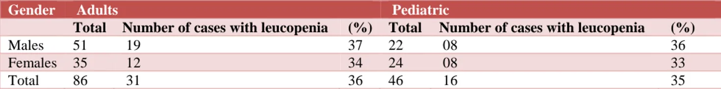
Age and gender distribution patterns in leucopenia is summarized in Table 4.
Table 4: Age and gender distribution in patients with leucopenia.
Gender Adults Pediatric
Total Number of cases with leucopenia (%) Total Number of cases with leucopenia (%)
Males 51 19 37 22 08 36
Females 35 12 34 24 08 33
Leucopenia was graded as mild, moderate, severe. Mild was total leucocyte count between 2x10⁹/l to 3.999x10⁹/l.
Moderate was counts between 1x10⁹/l to 1.999x10⁹/l and
severe was counts ≤0.999x10⁹/l (Table 5).
Table 5: Degree of leucopenia.
Grade (x 109 /l) Number (n=47) (%)
Mild (2 to 3.999 x 10⁹ /l) 39 82
Moderate (1-1.999 x 10⁹/l) 08 18
Severe (≤ 0.999 x 10⁹ /l) 0 0
It was noted that majority of cases showed mild leucopenia (82%). An analysis of leucopenia with thrombocytopenia was done. A total of 47 cases had leucopenia in association with thrombocytopenia. Thrombocytopenia was graded as mild with platelet
counts between 76-150x109/l, moderate with counts
between 50-75x109/l and severe with counts less than
50x109/l (Table 6).
Table 6: Leucopenia and degree of thrombocytopenia.
Degree of
thrombocytopenia (x 109 /l)
Number of cases with
leucopenia (n=47) (%)
76- 150 x 109 /l 11 23
50-75 x 109 /l 12 25
<50 x 109 /l 24 52
Almost over half the cases (52%) showed severe thrombocytopenia in association with leucopenia. 25% showed moderate thrombocytopenia in association with leucopenia.
We also analysed leucopenia with serology. The findings are tabulated in table 7 and 8. Serology pattern in leucopenia is shown in table 7.
Table 7: Serology patterns in leucopenia.
Serology pattern Number (n=47) (%)
Ns1 alone 22 47
Ns1 with IgG/IgM/both 18 38
IgG/IgM only 07 15
It was noted that almost half of the leucopenia (47%) cases were significantly associated with isolated NS 1 positivity in serology and 85% showed NS 1 with antibody association.
On the other hand, analysis of leucopenia within the serology spectrum is tabulated in table 8. Of a total of 37 cases of isolated NS 1 positivity 22 cases (59%) had significant leucopenia.
40% of NS 1 with antibody positivity cases showed leucopenia. Almost half (49%) of the cases with NS 1
positivity (isolated or in combination) showed leucopenia (40/81 cases with total NS1 positivity).
Table 8: Leucopenia and serology spectrum.
Serology pattern Total
cases
Number of cases with leucopenia
(n=47) (%)
Ns1 positive 37 22 59
Ns1 with
IgG/IgM or both 44 18 40
IgM/IgG/both 51 07 13
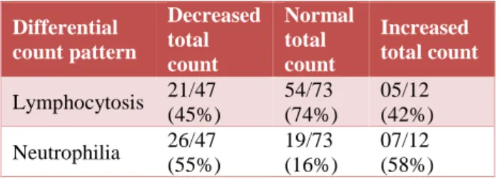
The differential count pattern was categorised based on predominant cell type as -'with lymphocytosis' and 'with neutrophilia'.
Our study showed that 45% (21 out of 47) cases of
leucopenia were associated with lymphocytosis.
Lymphocytosis was overall present in 61% (80 out of 132) of cases (Table 9).
Table 9: WBC count and differential count pattern.
Differential count pattern
Decreased total count
Normal total count
Increased total count
Lymphocytosis 21/47
(45%)
54/73 (74%)
05/12 (42%)
Neutrophilia 26/47
(55%)
19/73 (16%)
07/12 (58%)
It was noted that while significant number of cases of leucopenia were associated with neutrophilia (55%), only 16% of cases with normal leucocyte count had neutrophilia. Most of the normal total count patterns (74%) had lymphocytosis (Table 9).
DISCUSSION
Our study analyzing age was in concordance with others with most cases in the younger ages with slight male predominance probably due to occupational exposure and increased recreational activity in men.18,21
An analysis of white cell counts showed a range of 1.1x10⁹/l to 14.3x10⁹/l, in accordance with other
studies.2,7 Leucopenia (<4 x10⁹/l) 7 was noted in 36% in
accordance with other studies.7,11,12,17,21 A few studies
used a threshold of <5x10⁹/l for leucopenia.23,24 A few
studies had a lower and others, a higher proportion of cases with leucopenia.1,2,4,15,16,18,25 A normal leucocyte
count was observed in 55% of cases in our study in concordance with few studies but it was lower and higher in other studies.2,4,15,18,21,24
Leucocytosis was noted in 9% of cases in our study in concordance with few studies.25 It was seen in lower and
A demographic assessment of leucopenia revealed an equal distribution of cases across the ages and both sexes. Francisca et al and Thanachartwet et al noted increased risks of leucopenia in the ages ≥15 years. We had no data to compare these findings.
Our analysis of the degree of leucopenia showed most (82%) cases with mild leucopenia. Severe leucopenia was not noted in the study. We could not find data for comparison of these findings.
A few studies of the total and differential leucocyte count patterns have observed mild initial leucocytosis
accompanied by neutrophilia followed later by
leucopenia and lymphocytosis with atypical
lymphocytes.10,22
In our study lymphocytosis was noted in 61% in accordance with few studies, whereas it was seen in lower proportion of cases in others.18,21,25 Also we
observed leucopenia associated with lymphocytosis in 45% whereas it was noted in lower proportion of cases in others.3,12 Our study showed significant proportion (55%)
of cases with leucopenia associated with neutrophilia whereas 74% of cases with normal leucocyte count had lymphocytosis. Few studies claim that leucopenia with lymphocytosis is a major finding in dengue.12
Vibha et al have also observed that leucocyte count returns to normal by 9th-10th day post therapy and is an
important benchmark for clinical improvement.
Leucopenia is caused by bone marrow suppression by virus in acute phase and is due to decrease in polymorphs.7,17,18,20,25 Neutropenia is also attributed to
marked degeneration of mature neutrophils in febrile phase with shift to left.21 Stress accompanied with shock
may be the cause of mild initial leukocytosis.10
Our study revealed that 47 cases of leucopenia (36%) out
of 132 total cases were associated with
thrombocytopenia. 77% of the cases with leucopenia had platelet counts less than 75x109/l.
A few studies claim positive correlation between
leucopenia and thrombocytopenia which is not
statistically significant.16,17 In their study Juan Carlos et al
have observed that leucopenia is accompanied by a sudden drop in platelet count preceding plasma leakage. Our study suggests that leucopenia could be a marker of severe dengue as over half (52%) of the cases are associated with severe thrombocytopenia. It has been noted in some studies that thrombocytopenia is a risk factor for bleeding manifestations.7,18 However other
studies dispute the role of leucopenia as a
prognosticator.15,16,17 On the other hand, few studies
claim that leucopenia indicates good prognosis with
counts >5x10⁹/l being associated with severe
dengue.10,13,14 Vibha et al noted that leucopenia is
commoner in Dengue fever, Dengue hemorrhagic fever I/II but not in Dengue shock syndrome.
An analysis of serology patterns with respect to leucopenia showed that 85% of leucopenia cases were NS1 positive as against 15% of leucopenia cases which had antibody only (IgM/IgG) positivity.
A few studies showed mean total leucocyte count was lower in those with NS 1 positivity compared to (IgM/IgG) antibody positivity.27
The analysis of leucopenia cases associated with serology patterns showed that 22 of 37 NS1 positive cases (59%) had leucopenia whereas (13%) 7 of 51 antibody only (IgM/IgG) positive cases had leucopenia. This was in accordance with other studies.3,28
NS 1 antigen is known to be a marker for early diagnosis of the disease and is detected from first day, followed by IgM at 3-5 days and IgG from 1st week onwards.27,29,30
A few studies analysing thrombocytopenia with serology patterns also have observed a higher proportion of cases with thrombocytopenia in NS 1 positivity than with antibody positivity.29
Thus, our study in association with serology suggests that leucopenia is an early marker of dengue and association of leucopenia with thrombocytopenia suggests that it could be one of the prognosticator of severe dengue.17,21,22,25
Limitations of study-Our study is limited by
• Smaller study size,
• Use of uniform values for total leucocyte count
across all ages,
• Lack of sufficient data for comparision,
• Use of random, single samples for test.
Accurate, useful information could have been obtained with timed samples/serial testing of total leucocyte counts through recovery phase.
CONCLUSION
Early diagnosis is crucial to the management of dengue and helps in reduction of morbidity and mortality. Total leucocyte count is simple, easily available, cost effective test, very useful in small rural set ups with limited resources; not only for early diagnosis but also for prognosis in dengue.
Funding: No funding sources Conflict of interest: None declared
REFERENCES
1. Patel PM, Patel SK, Sabalpara MA, Shah CK, Shah
NR. Study of hematological and biochemical changes in dengue fever at tertiary care hospital at Ahmedabad. Inter J Medic Sci Pub Heal. 2016;5(9):1934-6.
2. Meena KC, Jelia S, Meena S, Arif M, Ajmera D, Jatav VS. A study of hematological profile in dengue fever at tertiary care center, Kota Rajasthan, India. Inter J Adv in Medic. 2016;3(3):621-4.
3. Dongre T, Karmarkar P. Hematological Parameters
and its utility in dengue–A prospective study. IOSR J Dent and Medic Sci. 2015;14(2):31-4.
4. Fazal F, Biradar S. Clinical and lab profile of Dengue fever. J Evidence based medicine and healthcare. 2015;2(9):1136-47.
5. Narayanan M, Aravind MA, Ambikapathy P, Prema
R and Jeyapaul MP. Dengue Fever-Clinical and
Laboratory Parameters Associated with
Complications. Dengue Bulletin. 2003;27:08-115
6. Malavige GN, Fernando S, Fernando DJ,
Seneviratne SL. Dengue viral infections.
Postgraduate medical J. 2004;80(948):588-601.
7. Khatri K, Rajani A, Khalla AR. Plasmacytoid
lymphocytes: a diagnostic clue to dengue infection. Int J Sci Res. 2016;5(3):1002-5.
8. Mehta RC, Goswami HM, Katara KR, Patel PS,
Parikh UV, Vegad MM, et al. Importance of complete blood count and peripheral smear examination in early diagnosis of dengue patients. J Infectious Diseases Letters. 2013;2(1):22-4.
9. Jayashree K, Manasa GC, Pallavi P, Manjunath GV.
Evaluation of platelets as predictive parameters in dengue fever. Ind J Hematol Blood Trans. 2011;27(3):127-30.
10. Pongpan S, Wisitwong A, Tawichasri C,
Patumanond J. Prognostic indicators for dengue
infection severity. Inter J Clinic Pedia.
2013;2(1):12-8.
11. Vulavala S, Reddy Y, Kamarthy P. Study of clinical
and laboratory profile of dengue fever patients. Eur J Pharma Medic Res. 2016;3(11):613-6.
12. Dhir G, Dhir T, Suri V, Dhir D, Khatri K.
Hematological and Serological test profile in dengue fever, dengue hemorrhagic fever and dengue shock syndrome in Bathinda region of Punjab. Scholars J Applied Medic Sci. 2015;3(8):2926-30.
13. Karyanti MR. Clinical manifestations and
hematological and serological findings in children with dengue infection. Paediatrica Indonesiana. 2011;51(3):157-62.
14. Ledika MA, Setiabudi D, Dhamayanti M.
Association between Clinical Profiles and Severe Dengue Infection in Children in Developing
Country. Americ J Epidemiol Infec Dis.
2015;3(3):45-9.
15. Mishra S, Ramanathan R, Agarwalla SK. Clinical
profile of dengue fever in children: a study from southern Odisha, India. Scientifica. 2016;2016.
16. Jayanthi HK, Tulasi SK. Correlation study between
platelet count, leukocyte count, nonhemorrhagic complications, and duration of hospital stay in dengue fever with thrombocytopenia. J Fam Medic Prim care. 2016;5(1):120.
17. Ch. Manoj Kumar, K. S. Keerthi Vyas, Y. Sai
Krishna. Clinical profile of dengue fever with severe
thrombocytopenia and its complications: a
retrospective study at a tertiary care hospital in South India. Inter J Res Medic Sci. 2017;5(5):1751-1755.
18. Patel K, Patel D, Das VK. Hematological
Parameters and Its Utility in Dengue fever: A prospective study. International Journal of Science and Research (IJSR). 2016;5(4):1077-9
19. Achalkar GV. Dengue-A clinicopathological study.
J Evolution of Medic Dent Sci. 2013;2(48):9380-5 20. Agrawal A, Pansuriya H, Dhruva G. Platelet count
and hematocrit as early indicators in acute dengue illness. Int J Res Med. 2013;2(2):63.
21. Gajera VV, Sahu S, Dhar R. Study of hematological
profile of dengue fever and clinical implications. Annuals of Applied Biosci. 2016;3(3):242-5.
22. Azin FR, Gonçalves RP, Pitombeira MH, Lima DM,
Castelo Branco I. Dengue: profile of hematological and biochemical dynamics. Revista brasileira de hematologia e hemoterapia. 2012;34(1):36-41. 23. Verdeal JC, Costa Filho R, Vanzillotta C, Macedo
GL, Bozza FA, Toscano L, et al. Guidelines for the management of patients with severe forms of dengue. Revista Brasileira de terapia intensiva. 2011;23(2):125-33.
24. Malathesha MK, Ashwini HN. Hematological
manifestations in dengue fever-an observational study. J Evol Medi Dent Sci. 2014;3(9):2245-50.
25. Pongpan S, Wisitwong A, Tawichasri C,
Patumanond J, Namwongprom S. Development of dengue infection severity score. ISRN pediatrics. 2013;2013.
26. Thanachartwet V, Oer-Areemitr N,
Chamnanchanunt S, Sahassananda D, Jittmittraphap A, Suwannakudt P, et al. Identification of clinical factors associated with severe dengue among Thai adults: a prospective study. BMC infectious diseases. 2015;15(1):420.
27. Jakribettu RP, Boloor R, Thaliath A, George SY, George T, Rai MP, et al. Correlation of clinicohaematological parameters in paediatric dengue: a retrospective study. Journal of Tropical Medicine. 2015;2015.
28. Kauser MM, Kalavathi GP, Radadiya M, Karthik
M, Afreen A, Kumaraswamy RC. A study of clinical and laboratory profile of Dengue fever in tertiary care hospital in central Karnataka, India. Global J Medic Res. 2014;14(5).
30. Cordeiro MT. Lab diagnosis for dengue. Revista do Instituto de Medicina Tropical de São Paulo. 2015;54(15):510-512.