Journal of Global Pharma Technology
Available Online at: www.jgpt.co.in
RESEARCH ARTICLE
Clinical Use of Ultrasound in Obstetrics
Saba Kamal Mahmood
1,Israa Adnan Falah Al-Qaseer
21. Baghdad Health Department Karkh Health Center Salhiya.
2. Ibn Al - Baladi Children's and Women's Hospital.
Abstract
During the past three years, ultrasound diagnostic procedures have been important and useful means in clinical practice of obstetrics and gynecology. Ultrasound images of the flow, whether color flow or spectral Doppler, are obtained from motion measurements. In ultrasound scanners, a series of pulses are transmitted to detect blood movement. Echoes of fixed tissues are the same from pulse to pulse. The echoes of the transition from scatter to slight differences appear at the time of signal return to the receiver. These differences can be measured as a direct time difference, or usually, in terms of the phase shift from which the Doppler frequency is obtained. It is then processed to produce either a color flow display or Doppler ultrasonography.
Introduction
In recent years, ultrasound imaging capabilities have increased significantly. Color-flow imaging is now common, and facilities such as "energy" or "energy" Doppler provide new ways for imaging flow [1]. With such diversity, it is tempting to use this technique for the most demanding applications and to try to measure the slight incremental changes in the circulation of the mother and fetus [2].
However, to avoid misinterpreting the results, it is necessary for a Doppler ultrasound user to be aware of the factors that affect the Doppler signal, whether it is a colored flow image or Doppler ultrasound imaging.
The efficient use of Doppler ultrasound techniques requires an understanding of three main elements: The capabilities and limitations of Doppler ultrasound;
The different parameters which contribute
to the flow display;
Blood flow in arteries and veins.
Doppler evaluation of the placental cycle plays an important role in detecting placental
weakness and complications from
preeclampsia and restricting intrauterine
growth and perinatal death [3]. Evaluation of fetal blood circulation is essential in a better understanding of the pathophysiology of a wide range of pathological pregnancies and their clinical management [4]. This paper provides a comprehensive account of ultrasound Doppler waves in obstetrics and will be of value to those involved in antenatal care and fetal medicine. As with the introduction of any new technology in routine clinical practice, [5] it is essential that those who evaluate Doppler for placental and fetal circulation are appropriately trained and that their results are subject to rigorous scrutiny [6].
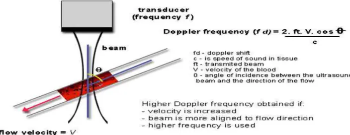
Figure 1: Doppler ultrasound. Doppler ultrasound measures the movement of the scatterers through the beam as a phase change in the received signal. The resulting Doppler frequency can be used to measure velocity if the beam/flow angle is known [2]
As can be seen from Figures 1 and 2, there has to be motion in the direction of the beam; if the flow is perpendicular to the beam, there is no relative motion from pulse to pulse [9]. The size of the Doppler signal is dependent on:
Blood velocity: as velocity increases, so does
the Doppler frequency;
Ultrasound frequency: higher ultrasound
frequencies give increased Doppler
frequency. As in B-mode, lower ultrasound frequencies have better penetration.
The choice of frequency is a compromise
between better sensitivity to flow or better penetration;
The angle of insonation: the Doppler
frequency increases as the Doppler ultrasound beam becomes more aligned to the flow direction (the angle between the beam and the direction of flow becomes smaller). This is of the utmost importance in the use of Doppler ultrasound [10]. The implications are illustrated schematically in Figure 3.
Figure 2: Effect of the Doppler angle in the sonogram. (A) higher-frequency Doppler signal is obtained if the beam is aligned more to the direction of flow. In the diagram, beam (A) is more aligned than (B) and produces higher-frequency Doppler signals. The beam/flow angle at (C) is almost 90° and there is a very poor Doppler signal. The flow at (D) is away from the beam and there is a negative signal [8]
All types of Doppler ultrasound equipment employ filters to cut out the high amplitude, low-frequency Doppler signals resulting from
altered by the user, for example, to exclude frequencies below 50, 100 or 200 Hz.
This filter frequency limits the minimum flow velocities that can be measured [11].
Clinical Recommendation for 3D and 4D Usages
3DUS is not yet widely used on a routine basis in perinatal medicine. It has been shown to be a problem-solving tool in selected circumstances and may well become a part of many obstetric ultrasound examinations in the future. However, the role of 3D ultrasound is still being evaluated in many areas. We will review here some examples of areas in which 3DUS and 4DUS might be helpful, as suggested by a recent report from AIUM.
Aliasing
Pulsed wave systems suffer from a fundamental limitation. When pulses are transmitted at a given sampling frequency (known as the pulse repetition frequency), the maximum Doppler frequency ƒd that can be measured unambiguously is half the pulse repetition frequency. If the blood velocity and beam/flow angle being measured combine to give a ƒd value greater than half of the pulse repetition frequency, ambiguity in the Doppler signal occurs. This ambiguity is known as aliasing [12]. A similar effect is seen in films where wagon wheels can appear to be going backwards due to the low frame rate of the film causing misinterpretation of the movement of the wheel spokes [13].
Figure 3: Aliasing of color doppler imaging and artefacts of color. Color image shows regions of aliased flow (yellow arrows) [8]
Ultrasound Flow Modes
Since color flow imaging provides a limited amount of information over a large region, and spectral Doppler provides more detailed information about a small region, the two modes are complementary and, in practice, are used as such [14]. Color flow imaging can be used to identify vessels requiring examination, to identify the presence and direction of flow, to highlight gross circulation anomalies, throughout the entire color flow image, and to provide beam/vessel angle correction for velocity measurements.
Pulsed wave Doppler is used to provide analysis of the flow at specific sites in the vessel under investigation. When using color flow imaging with pulsed wave Doppler, the color flow/ B mode image is frozen while the pulsed wave Doppler is activated .Recently,
some manufacturers have produced
concurrent color flow imaging and pulsed wave Doppler, sometimes referred to as triplex scanning. When these modes are used simultaneously, the performance of each is decreased. Because transducer elements are employed in three modes (B-mode, color flow and pulsed wave Doppler), the frame rate is decreased, the color flow box is reduced in size and the available pulse repetition frequency is reduced, leading to increased susceptibility to aliasing. Power Doppler is also referred to as energy Doppler, amplitude Doppler and Doppler angiography.
It complements the other two modes. Hybrid color flow modes incorporating power and velocity data are also available from some manufacturers. These can also have improved sensitivity to low flow. A brief summary of factors influencing the displays in each mode is given in the following sections. Most of these factors are set up approximately for a particular mode when the application (e.g. fetal scan) is chosen, although the operator will usually alter many of the controls during the scan to optimize the image.
Spectral Doppler
Examines flow at one site
Detailed analysis of distribution of flow
Good temporal resolution – can examine
flow waveform
Allows calculations of velocity and indices
Color Flow
Overall view of flow in a region
Limited flow information
Poor temporal resolution/flow dynamics
(frame rate can be low when scanning deep)
Color flow map (different color maps)
Direction information
Velocity information (high velocity & low
velocity)
Turbulent flows
Power/energy/amplitude Flow
Sensitive to low flows
No directional information in some modes
Very poor temporal resolution
Susceptible to noise
Spectral or Pulsed Wave Doppler
Pulsed wave Doppler ultrasound is used to provide a sonogram of the artery or vein under investigation. The sonogram provides a measure of the changing velocity throughout the cardiac cycle and the distribution of velocities in the sample volume (or gate) (Figure 5). If an accurate angle correction is made, then absolute velocities can be measured. The best
resolution of the sonogram occurs when the B-mode image and color image are frozen, allowing all the time to be employed for spectral Doppler. If concurrent imaging is used (real-time duplex or triplex imaging), the temporal resolution of the sonogram is compromised [16].
Blood Flow Measurements Velocity Measurement
Theoretically, once the beam/flow angle is known, velocities can be calculated from the Doppler spectrum as shown in the Doppler equation. However, errors in the measured velocity may still occur.
(1) Errors can arise in the formation of the Doppler spectrum due to:
Use of multiple elements in array
transducers;
Non-uniform insonation of the vessel
lumen;
Insonation of more than one vessel;
Use of filters removing low-velocity
components.
(2) Errors can arise in the measurement of the ultrasound beam/flow velocity angle.
Use of high angles (Ɵ> 60°) may give rise to
error because of the comparatively large changes in the cosine of the angle which occur with small changes of angle (Figure 6).
The velocity vector may not be in the
direction of the vessel axis.
(3) Errors can arise in the calculation packages provided by the manufacturers for analysis of the Doppler spectrum (for instance, of intensity weighted mean velocity).
While efforts can be made to minimize
The effort applied to produce accurate velocity measurements should be balanced against the importance of absolute velocity measurements for an investigation.
Changes in velocity and velocity waveform
shape are often of more clinical relevance when making a diagnosis. In this and other
cases, absolute values of velocity
measurement may not be required.
Calculation of Absolute Flow
Total flow measurement using color or duplex
Doppler ultrasound is fraught with
difficulties, even under ideal conditions 5. Errors that may arise include:
Those due to inaccurate measurement of
vessel cross-sectional area, for example the cross-sectional area of arteries which pulsate during the cardiac cycle;
Those originating in the derivation of
velocity (see above).
These errors become particularly large
when flow calculations are made in small vessels; errors in measurement of diameter are magnified when the diameter is used to derive cross-sectional area. As with velocity measurements, it is prudent to be aware of possible errors and to conduct repeatability tests.
Non-Dimensional
Non-dimensional analysis of the flow waveform shape and spectrum has proved to be a useful technique in the investigation of many vascular beds. It has the advantage that derived indices are independent of the beam/flow angle. Changes in flow waveform shape have been used to investigate both
proximal disease (e.g. in the adult peripheral arterial circulation) and distal changes (in the fetal circulation and uterine arteries). While the breadth of possible uses shows the technique to be versatile, it also serves as a reminder of the range of factors which cause changes to the local Doppler spectrum.
If waveform analysis is to be used to observe changes in one component of the proximal or distal vasculature, consideration must be given to what effects other components may have on the waveform.
Indices of Measurement
Indices have been used to describe the shape of flow waveforms. Techniques range from simple indices of systolic to diastolic flow to feature extraction methods such as principal component analysis. All are designed to describe the waveform in a quantitative way, usually as a guide to some kind of classification. In general, they are a compromise between simplicity and the amount of information obtained.
Conclusion
These indices are all based on the maximum Doppler shift waveform and their calculation is described. The PI takes slightly longer to calculate than the RI or S/D ratio because of the need to measure the mean height of the waveform. It does, however, give a broader range of values, for instance in describing a range of waveform shapes when there is no end-diastolic flow. In addition to these indices, the flow waveform may be described or categorized by the presence or absence of a particular feature, for example the absence of end-diastolic flow and the presence of a post-systolic notch.
References
1.
2. Abdul-Khaliq H, PE Lange, M Vogel
(2000) Feasibility of brain volumetric analysis and reconstruction of images by
transfontanel three-dimensional
ultrasound. J. Neuroimaging, 10: 147.
3. Bonilla-Musoles F, LE Machado, NG
Osborne, F Raga, F Bonilla, MJ Puig, et al (2002) Morphological assessment of the umbilical cord with three-dimensional ultrasonography. Ultrasound Rev. Obstet. Gynecol., 2: 17.
4. Abuhamad A (2006) Doppler Ultrasound
in Obstetrics. Ultrasound Clinics.
5. Krupinski EA (2016) Medical imaging. In:
Handbook of Visual Display Technology.
6. Mone F, McAuliffe FM, Ong S (2014) The
clinical application of Doppler ultrasound in obstetrics. Obstet Gynaecol.
7. Chan FY, A Taylor, B Soong, B Martin, J
Clark, P Timothy, et al (2002) Randomized comparison of the quality of realtime fetal ultrasound images transmitted by ISDN and by IP videoconferencing. J. Telemed Telecare, 2: 91.
8. The Use of Fetal Doppler in Obstetrics. J.
9. Timmerman D, Testa AC, Bourne T, Ameye L, Jurkovic D, Van Holsbeke C, et al (2008) Simple ultrasound-based rules for the diagnosis of ovarian cancer. Ultrasound Obstet Gynecol.
10. Vollgraff Heidweiller-Schreurs CA, De
Boer MA, Heymans MW, Schoonmade LJ, Bossuyt PMM, Mol BWJ, et al (2018) Prognostic accuracy of cerebroplacental ratio and middle cerebral artery Doppler for adverse perinatal outcome: systematic review and meta-analysis. Ultrasound in Obstetrics and Gynecology.
11.Baschat AA, Gembruch U, Harman CR
(2001) The sequence of changes in Doppler and biophysical parameters as severe fetal growth restriction worsens. Ultrasound Obstet Gynecol.
12.Cruz-Martinez R, Savchev S, Cruz-Lemini
M, Mendez A, Gratacos E, Figueras F (2015) Clinical utility of third-trimester uterine artery Doppler in the prediction of brain hemodynamic deterioration and adverse perinatal outcome in
small-for-gestational-age fetuses. Ultrasound Obstet Gynecol.
13.Kak AC, Slaney M (2011) 5. Aliasing
Artifacts and Noise in CT Images. In: Principles of Computerized Tomographic Imaging.
14.Maciejewski MW, Qui HZ, Rujan I, Mobli
M, Hoch JC (2009) Nonuniform sampling and spectral aliasing. J. Magn. Reson.
15.Dmochowski J, Benesty J, Affès S (2009)
On spatial aliasing in microphone arrays. IEEE Trans Signal Process.
16.Prieto-Ramos J, Parkin TDH, French AT
(2016) Evaluation of a novel
echocardiographic view for the assessment of the pulmonary artery in dogs. J. Vet. Cardiol.
17.Csutak R, L Unterassinger, C
Rohrmeister, M Weninger, KA Vergesslich Three-dimensional volume measurement of the lateral ventricles in preterm and term infants: evaluation of a standardised computer-assisted method in vivo.
![Figure 3: Aliasing of color doppler imaging and artefacts of color. Color image shows regions of aliased flow (yellow arrows) [8]](https://thumb-us.123doks.com/thumbv2/123dok_us/8104369.2148378/3.892.83.813.448.714/figure-aliasing-doppler-imaging-artefacts-color-regions-aliased.webp)
