Research Article
Role of predictors and rapid diagnosis of fungal peritonitis
in CAPD patients
Upma Narain
1*, Arvind Gupta
2INTRODUCTION
Fungal peritonitis (FP) is a rare but serious complication of peritoneal dialysis (PD).1 It is associated with temporary and frequently permanent cessation of PD. The lethality, although variable, remains very high2 because the fungi form a biofilm on the surface of the silastic catheters that reduces the penetration of antifungal agents.3 Fungus penetrates the peritoneal cavity through intraluminal or periluminal pathways and cross the intestinal mucosa, or enters through the hematogenic pathway due to a distant fungal infection.4 The reported incidence of FP accounted for 3%-30% with high morbidity and mortality ranging between 20%-30%5. International societies of PD guidelines for peritonitis
2010 recommend immediate catheter removal once fungus is identified by microscopy or culture.6 Risk factors for developing fungal peritonitis have not been clearly determined. Numerous situations have been listed which play an important role in the appearance of the mycotic infection. Therefore the focus of this study was to analyze the demographic features, incidence, predictors, causative fungi and outcome of PD associated fungal peritonitis.
METHODS
It is a retrospective study involving patients undergoing CAPD at our centre who developed peritonitis over a period of 16 years, from Jan 2000 to August 2015. Out of
ABSTRACT
Background: Fungal peritonitis represents one of the most serious complications in patients on CAPD therapy. In the present study we analyze PD patients who developed fungal peritonitis.
Methods: In between January 2000 to August 2015, we retrospectively identified fungal PD peritonitis episodes, examined the demographic features, incidence, predictors, causative fungi and outcome.
Results: We analyzed 65 episodes of PD associated fungal peritonitis which includes 89.3% Candida species, 1.5% yeast and 9.2% dimorphic fungi. Association between predictors and pathogens was found significant at 0.05 levels. Catheter removal (47.1%) and loss of life (20.6%) was significantly more frequent in patients with diabetes, glomerulonephritis required maximum hospitalization while cure rate (55.6%) was more significant in patients with hypertension. Data were significant at p value of 0.05.
Conclusions: Our results suggest that prompt identification of predictors may force us for early intervention to reduce mortality and technique failure in PD associated fungal peritonitis. Early availability of direct microscopy of dialysate pellet became useful for the rapid, aggressive and judicious management of the fungal peritonitis and was the reason of high cure (27.7%) and low mortality (15.7%) in our case series.
Keywords: Fungal peritonitis, End stage renal disease, CAPD, Predictors, Candida albicans, Non albicans
1Microbiologist & Immunologist, Tejas Micro diagnostics, Allahabad, Uttar Pradesh, India 2Division of Nephrology, Moti Lal Nehru Medical College, Allahabad, Uttar Pradesh, India
Received: 22 December 2015
Accepted: 15 January 2016
*Correspondence:
Dr. Upma Narain,
E-mail: upmanarain@gmail.com
Copyright: © the author(s), publisher and licensee Medip Academy. This is an open-access article distributed under the terms of the Creative Commons Attribution Non-Commercial License, which permits unrestricted non-commercial use, distribution, and reproduction in any medium, provided the original work is properly cited.
402 patients, 65 end stage renal disease (ESRD) patients developed fungal peritonitis. Aerobic, Anaerobic, mycobacterium and polymicrobial peritonitis were excluded from the analysis due to their different outcomes.
As per the peritoneal dialysis related infection recommendations published by ISPD in 20106, the patient’s exchange bags, containing effluent dialysate were received in the microbiology laboratory for macro level examination, microscopic examination and culturing simultaneously. From these bags, 100 ml of fluid was withdrawn with a sterile needle and syringe under aseptic conditions. The fluid was centrifuged in sterile tubes at a rate of 3000 g for 15 minutes and supernatant was discarded, leaving 0.5 ml. In the centrifuged deposit, 10 ml of sterile distilled water was added together and the mixture was shaken vigorously on vortex for 30 sec. This mixture then was divided into 4 parts of 1 ml, 3 ml, 3 ml and 3 ml each. 1 ml was further divided for staining characteristic like gram stain, Z.N. stain, and lacto phenol cotton blue film, while 3 ml in FA bottle for isolation of aerobes and fungi, 3 ml in FN bottle and remaining 3 ml in MP bottle for the isolation of anaerobes and mycobacterium respectively. These three inoculated bottles were further incubated in Bact Alert 3D system following standard protocols. The isolated fungi were re-examined microscopically to ensure the staining and morphologic characteristic. Each positive specimen was inoculated on Sabouraud dextrose agar (M286) and Sabouraud cycloheximide chloramphenicol agar (M664).
Cultures were routinely incubated at 250 c and 370 c and examined daily for up to 4 weeks. The identification of individual fungi was based on standard methods such as microscopy, morphology, colonial characterization, pigment production, rate of growth while yeast identification was done by Vitek-2 (Biomeurix, France). Hence the noteworthy observation of our case series was, microscopy became available within 3 to 5 hrs. and identification along with antibiogram of Candida species (89.3%) and yeast(10.8%) was made available within 48 hrs. remaining 9.2% dimorphic fungi was identified within 4-6 days after the clinical diagnosis was made.
The following definitions were used in the article. Previous bacterial peritonitis episodes have been suggested that fungal peritonitis appears most of all after episodes of bacterial peritonitis. Prior antibiotic use was defined as the use of an antibiotic for a suspected infectious disease in the 30 days, 3 months and 6 months prior to development of FP. Prolonged time in the dialysis program means when time is elapsed from insertion of the peritoneal catheter. Prolonged time with the peritoneal catheter inserted means maintaining the catheter after detecting the fungal infection. Use of immunosuppressive agents referred to steroid or immunosuppressive use for at least 2 weeks prior to the diagnosis of FP. Hospitalization is considered to be a risk
factor when an infection of nosocomial origin occurs in the 30 days, 3 months, and 6 months prior to development of FP. Co-existence of an extra peritoneal fungal infection explains when patient is suffering of extra peritoneal fungal infection that causes a fungal peritonitis through a haematogenic pathway.
Statistical analysis was performed using chi square test and contingency coefficient. Data were expressed as mean + standard deviation. Statistical significance was defined at a p value of 0.05.
RESULTS
During the period from Jan 2000 to June 2015, 402 ESRD patients were initially on CAPD. The total no. of episodes of fungal peritonitis during the entire period was 65. The average rate of fungus peritonitis for the entire period was 2.6 episodes/CAPD year. Their base line and demographic data are summarized below.
Baseline characteristic of the patients:
Sex (M/F) 89.2%/10.8% Age (Year) 58.29 + 8.845
Duration of dialysis (Months) 18.26 + 8.080 Body height 1.56 + 0.040
Body weight 63.68 + 6.806 Primary causes of ESRD Glomerulonephritis 32.3% Diabetes 52.3%
Hypertension 13.9% Other/Unknown 1.5%
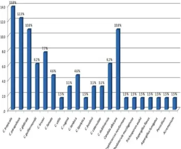
In our case series fungal peritonitis was presented with abdominal pain, sub-acute intestinal obstruction, nausea, vomiting, often fever and cloudy dialysate. The 65 episodes of FP were caused by various fungi (Figure 1).
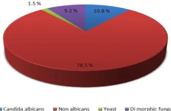
Candida species remained the most common pathogen, accounting for 89.3% of the fungal peritonitis among which 78.5% were non albicans and 10.8% were C. albicans. Among non albicans C. tropicalis 13.8% was the most frequent fungi isolated. Other than Candida
species, yeast was isolated in 1.5% cases while 9.2% was dimorphic fungi (Figure 2).
Figure 2: Percentage of candida albicans, nonalbicans, yeast and dimorphic fungi.
Association between predictors and pathogen reveals 30.8% de novo episodes and remaining 60.2% episodes of FP predisposed by different predictors. Among de novo non albicans were 23.1. The strongest predictor to predispose the fungal infection was previous bacterial peritonitis episode (16.9%). It was first time when analysis was done between predictors and pathogen and was found significant at 0.05 levels (Table 1).
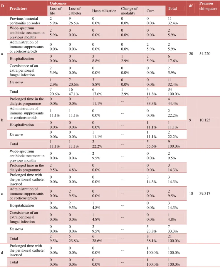
Statistical analysis of primary cause of ESRD, predictors and outcome showed that catheter removal (47.1%) and loss of life (20.6%) was significantly more frequent in patients with diabetes while in glomerulonephritis, patient required maximum hospitalization for resolution of peritonitis. Cure rate (55.6%) was more significant in patients with hypertension. It was also noticed that de novo infection was more frequent with glomerulonephritis. Statistical analysis has been calculated separately in all the primary causes of ESRD with outcome and predictors and was found significant in case of diabetes and glomerulonephritis at p value of 0.05 (Table 2).
Table 1:Association between predictors and pathogen.
Predictors
Fungi
Total df
Pearson chi-square
Contingency coefficient
Candida albicans
Non
albicans Yeast
Dimorphic fungi Previous bacterial
peritonitis episodes 0 0.0%
7 10.8%
1 1.5%
3 4.6%
11 16.9%
21 51.990* 0.667 Wide-spectrum
antibiotic treatment in previous months
0 0.0%
3 4.6%
0 0.0%
1 1.5%
4 6.2%
Prolonged time in the dialysis programme
0 0.0%
7 10.8%
0 0.0%
0 0.0%
7 10.8%
Prolonged time with the peritoneal catheter inserted
4 6.2%
0 0.0%
0 0.0%
0 0.0%
4 6.2%
Administration of immune
suppressants or corticosteroids
0 0.0%
6 9.2%
0 0.0%
0 0.0%
6 9.2%
Hospitalisation 0 0.0%
10 15.4%
0 0.0%
0 0.0%
10 15.4% Coexistence of an
extra peritoneal fungal infection
0 0.0%
3 4.6%
0 0.0%
0 0.0%
3 4.6%
De novo 3
4.6%
15 23.1%
0 0.0%
2 3.1%
20 30.8%
Total 7
10.8%
51 78.5%
1 1.5%
6 9.2%
65 100.0%
Table 2: Correlation among primary cause of ESRD, predictors and outcome.
D
Predictors
Outcomes
Total df
Pearson chi-square Loss of
life
Loss of
catheter Hospitalization
Change of
modality Cure
a Previous bacterial peritonitis episodes 2 5.9% 9 26.5% 0 0.0% 0 0.0% 0 0.0% 11 32.4%
20 54.220 Wide-spectrum
antibiotic treatment in previous months 2 5.9% 0 0.0% 0 0.0% 0 0.0% 0 0.0% 2 5.9% Administration of immune suppressants or corticosteroids 0 0.0% 0 0.0% 0 0.0% 0 0.0% 2 5.9% 2 5.9% Hospitalization 0
0.0% 0 0.0% 3 8.8% 1 2.9% 2 5.9% 6 17.6% Coexistence of an
extra peritoneal fungal infection 2 5.9% 0 0.0% 0 0.0% 0 0.0% 0 0.0% 2 5.9%
De novo 1
2.9% 7 20.6% 3 8.8% 0 0.0% 0 0.0% 11 32.4%
Total 7
20.6% 16 47.1% 6 17.6% 1 2.9% 4 11.8% 34 100.0% b
Prolonged time in the dialysis programme 0 0.0% 0 0.0% 1
11.1% --
3 33.3%
4 44.4%
9 10.125 Administration of immune suppressants or corticosteroids 1 11.1% 1 11.1% 0
0.0% --
0 0.0%
2 22.2% Hospitalization 0
0.0% 0 0.0%
0
0.0% --
1 11.1%
1 11.1%
De novo 0
0.0% 0 0.0%
1
11.1% --
1 11.1%
2 22.2%
Total 1
11.1% 1 11.1%
2
22.2% --
5 55.6% 9 100.0% c Wide-spectrum antibiotic treatment in previous months 0 0.0% 0 0.0% 2
9.5% --
0 0.0%
2 9.5%
18 39.317 Prolonged time in the
dialysis programme 2 9.5% 1 4.8% 0
0.0% --
0 0.0%
3 14.3% Prolonged time with
the peritoneal catheter inserted 0 0.0% 0 0.0% 0
0.0% --
3 14.3% 3 14.3% Administration of immune suppressants or corticosteroids 0 0.0% 2 9.5% 0
0.0% --
0 0.0%
2 9.5% Hospitalization 0
0.0% 2 9.5%
1
4.8% --
0 0.0%
3 14.3% Coexistence of an
extra peritoneal fungal infection 0 0.0% 0 0.0% 1
4.8% --
0 0.0%
1 4.8%
De novo 0
0.0% 0 0.0%
2
9.5% --
5 23.8%
7 33.3%
Total 2
9.5% 5 23.8%
6
28.6% --
8 38.1%
21 100.0%
d
Prolonged time with the peritoneal catheter inserted 0 0.0% 0 0.0% 0
0.0% --
1 100.0%
1 100.0%
Total 0
0.0% 0 0.0%
0
0.0% --
1 100.0%
1 100.0%
DISCUSSION
Fungi are widely found in human environment being part of the normal flora of the skin and mucosa but in certain conditions, they can become pathogenic.7 The rupture of the coetaneous barrier as a result of the presence of peritoneal catheter and decrease cellular immunity due to uremia also predispose to FP.1 Moreover continuous exposure of peritoneal cells to non-physiological peritoneal dialysis solution may result in an impairment of the local peritoneal host defense mechanism increasing susceptibility to fungal infection.7 Fungi enter the peritoneal cavity through touch contamination at the time of PD exchange of invasion of skin by the offending organisms from the exit site through the tunnel of the peritoneum.8
Fungal peritonitis frequently resulted in catheter removal (33.8%), hospitalization (21.5%), change in modality (1.5%), loss of life (15.4%) and cure 27.7%.
Patients with fungal peritonitis present a higher rate of bacterial peritonitis episodes than patients without FP9-13, sometimes more than two times higher,11-13 owing to the fact that the peritoneal inflammation can increase susceptibility to a fungal invasion. In our case series 16.9% episodes followed previous bacterial peritonitis.
Four (6.2%) patients had received broad spectrum antibiotics with in the preceding months due to different clinical conditions like UTI, upper respiratory infection and bacterial vaginosis.
Other predictors are prolonged time in the peritoneal dialysis programme and the time elapsed from insertion of the peritoneal catheter.10,11,14 According to our case series 10.8% and 6.2% FP episodes occurred due to prolonged time in dialysis program and prolonged time with the insertion of the peritoneal catheter respectively.
Maintaining the catheter after detecting the fungal infection is related to a worse prognosis, and on some occasions has been the main factor causing failure of the technique and mortality.11,15-19 In the present case series out of 65 patients 18 patients had the catheter left in situ whereas 22 patients had the catheter removed immediately after the fungal diagnosis was made and in 14 patients catheter was removed within 3 to 5 days during hospitalization. Cheng et al reviewed 58 patients with fungal peritonitis reported in the literature who continued CAPD without catheter removal.20 However, Wang et al reported out of 67 patients 11 patients had the catheter left in situ however; they believed this approach may only be applicable in very mild cases of FP.
For the timing of catheter removal, immediate after pathogen identification was recommended both in the 20106 and 200519 ISPD guidelines, compared with the 200022 ISPD guidelines that suggested catheter removal if
clinical improvement did not occur after 4-7 days of therapy.
Hospitalization is considered to be a predictor when an infection of nosocomial origin occurs in our case series 15.4% FP occurred after hospitalization.12,23
In six (9.2) of the fungal episodes patients received an immunosuppressive agents within two weeks prior to FP. Coexistence of an extra peritoneal fungal infection causes peritoneal infection through a hematogenic pathway.12,23 In our case series 4.6% FP episodes were found in females all three were partially treated with vaginal candidiasis.
CONCLUSION
In conclusion careful and continuous training of patients, especially in hand washing technique and in doing the connection, are critical for preventing exogenous factors causing peritonitis with rare environmental fungi and with those colonizing the skin because dimorphic fungi did not require antibiotic pressure.
We analyzed the association between the predictors and pathogens and conclude that prompt identification of these predictors provides opportunities for early aggressive intervention for fungal peritonitis and helps to reduce mortality and technique failure. In addition, FP is highly suspected with previous bacterial peritonitis and attention should be paid to those with diabetes and glomerulonephritis owing to high risk of mortality. Direct microscopy of the dialysate pellet should be available within few hours following the diagnosis of the infectious episode, which may become extremely useful for the prompt, aggressive and judicious management of the fungal episodes and probably that was the reason of high cure (27.7%) and low mortality (15.7%) in our case series.
Funding: No funding sources Conflict of interest: None declared Ethical approval: Not required
REFERENCES
1. Rowinska JM. Update on fungal peritonitis and its treatment. Perit Dial Int. 2009;29(2):S161-5. 2. Prasad N, Gupta A. Fungal peritonitis in peritoneal
dialysis patients. Perit Dial Int. 2005;25:207-22. 3. Bibashi E, Memmos D, Kokolina E, Tsakiris D,
Sofianou D, Papadimitriou M. Fungal peritonitis complicating peritoneal dialysis during an 11-Year Period: Report of 46 Cases. Clin Infect Dis. 2003;36:927-31.
4. Agudo RG, Martos PG. Clinical and microbiological aspects of fungal peritonitis in peritoneal dialysis. Nefrologia. 2009;29(6):506-17. 5. Kumar KV, Mallikarjuna HM, Gokulnath, Jayanthi
peritoneal dialysis: The impact of antifungal prophylaxis on patient and technique outcomes. Indian J Nephrol. 2014;24(5):297-301.
6. Li PK, Szeto CC, Piraino B, Bernardini J, Figueiredo AE, Gupta A, et al. Peritoneal dialysis-related infections recommendations: 2010 Update. Perit Dial Int. 2010;30:393-423.
7. Michel C, Courdavault L, Khayat RA, Viron B, Roux P, Mignon F. Fungal peritonitis in patients on peritoneal dialysis. Am J Nephrol. 1994;14:113-20. 8. Miles R, Hawley CM, McDonald SP, Brown FG,
Rosman JB, Wiggins KJ, et al. Predictors and outcomes of fungal peritonitis in peritoneal dialysis patients. Kidney Int. 2009;76:622-8.
9. Piraino B. Peritonitis as complication of peritoneal dialysis. J Am Soc Nephrol. 1998;9:1956-64. 10. Bren A. Fungal peritonitis in patients on continuous
ambulatory peritoneal dialysis. Eur J Clin Microbiol Infect Dis. 1998;17:839-43.
11. Troidle L, Gorban-Brennan N, Kliger A, Finkelstein FO. Continous peritoneal dialysis-associated peritonitis: a review and current concepts. Semin Dial. 2003;6:428-37.
12. Molina P, Puchades MJ, Aparicio M, Ramón RG, Miguel A. Experiencia en peritonitis fúngica en una unidad de diálisis durante diez años. Nefrología. 2005;25:393-8.
13. Kocak Z, Bulut C, Kinikli S, Yilmaz GR, Irmak H, Demdroz AP. Fungal peritonitis in patients undergoing continuous ambulatory peritoneal dialysis: a report of three cases. Turk Med J. 2007;1:30-3.
14. Liu SW, Chern CH, Yen DH, Huang CI, How CK. Abdominal wall and intraperitoneal abscesses complicating Aspergillus peritonitis in peritoneal dialysis. Am J Med Sci. 2009;337:56.
15. The Turkish multicenter peritoneal dialysis study group (TULIP). The rate, risk factors, and outcome of fungal peritonitis in CAPD patients: experience in Turkey. Perit Dial Int. 2000;20:338-40.
16. Chen CM, Ho MW, Yu WL, Wang JH. Fungal peritonitis in peritoneal dialysis patients: effect of
fluconazole treatment and use of the twin-bag disconnect system. J Microbiol Immunol Infect. 2004;37:115-20.
17. Felgueiras J, Peso GD, Bajo A, Hevia C, Romero S, Celadilla O, et al. Risk of technique failure and death in fungal peritonitis is determined mainly by duration on peritoneal dialysis: single-center experience of 24 years. Adv Perit Dial. 2006;22:77-81.
18. Predari SC, De Paulis AN, Verón D, Zucchini A, Santoianni JE. Fungal peritonitis in patients on peritoneal dialysis: twenty five years of experience in a teaching hospital in Argentina. Rev Argent Microbiol. 2007;39:213-7.
19. Piraino B, Bailie GR, Bernardini J, Boeschoten E, Gupta A, Holmes C, et al. ISPD guidelines/recommendations. Peritoneal dialysis related infections: 2005 Update. Perit Dial Int. 2005;25:107-31.
20. Indhumathi E, Chandrasekaran V, Jagadeswaran D, Varadarajan M, Abraham G, Soundararajan P. The risk factors and outcome of fungal peritonitis in continuous ambulatory peritoneal dialysis patients. Indian J Med Microbiol. 2009;27:59-61.
21. Cheng IKP, Fang GX, Chan TM, Chan PCK, Chan MK. Fungal peritonitis complicating peritoneal dialysis: report of 27 cases and review of treatment. Q J Med. 1989;265:407-16.
22. Keane WF, Bailie GR, Boeschoten E, Gokal R, Golper TA, Holmes CJ, et al. Adult peritoneal dialysis-related peritonitis treatment recommendations: 2000 Update. Perit Dial Int. 2000;20:396-411.
23. Fourtounas C, Marangos M, Kalliakmani P, Savidaki E, Goumenos DS, Vlachojannis JG. Treatment of peritoneal dialysis related fungal peritonitis with caspofungin plus amphotericin B combination therapy. Nephrol Dial Transplant. 2006;21:236-7.