R E S E A R C H
Open Access
Role of the ion channel, transient receptor
potential cation channel subfamily V
member 1 (TRPV1), in allergic asthma
Katie Baker
1, Kristof Raemdonck
1,2,3, Bilel Dekkak
1, Robert J. Snelgrove
4, John Ford
5, Fisnik Shala
1,
Maria G. Belvisi
1,6and Mark A. Birrell
1,6*Abstract
Background:Asthma prevalence has increased world-wide especially in children; thus there is a need to develop new therapies that are safe and effective especially for patients with severe/refractory asthma. CD4+T cells are thought to play a central role in disease pathogenesis and associated symptoms. Recently, TRPV1 has been demonstrated to regulate the activation and inflammatory properties of CD4+cells. The aim of these experiments was to demonstrate the importance of CD4+T cells and the role of TRPV1 in an asthma model using a clinically ready TRPV1 inhibitor (XEN-D0501) and genetically modified (GM) animals.
Methods:Mice (wild type, CD4−/−or TRPV1−/−) and rats were sensitised with antigen (HDM or OVA) and subsequently topically challenged with the same antigen. Key features associated with an allergic asthma type phenotype were measured: lung function (airway hyperreactivity [AHR] and late asthmatic response [LAR]), allergic status (IgE levels) and airway inflammation.
Results:CD4+T cells play a central role in both disease model systems with all the asthma-like features attenuated. Targeting TRPV1 using either GM mice or a pharmacological inhibitor tended to decrease IgE levels, airway inflammation and lung function changes.
Conclusion:Our data suggests the involvement of TRPV1 in allergic asthma and thus we feel this target merits further investigation.
Keywords:Lung, Asthma, Ion channel, Inflammation
Background
Asthma is an inflammatory airway disease characterised by variable expiratory flow limitation and is associated with respiratory symptoms, such as wheeze, shortness of breath, chest tightness and cough [1]. Asthma preva-lence has increased worldwide. It is estimated that it affects approximately 300 million people and it is believed that over 100 million more people will be af-fected by 2025 [2]. Therefore, there is a need to develop new therapies that are safe and effective, especially for
patients with severe/refractory asthma. CD4+ T cells are thought to play a central role in the disease pathogenesis and associated symptoms [3, 4]. Recently, it has been shown that the ion channel, transient receptor potential cation channel subfamily V member1 (TRPV1) is present on T cells [5] and it regulates the activation and inflamma-tory properties of CD4+cells [6, 7].
TRPV1 is a non-selective cation channel and is a member of a large family of TRP ion channels. It can be activated by a diverse range of endogenous and exogen-ous chemical ligands, low pH and high temperatures [8]. In addition to the recent data demonstrating a role in CD4+T cell function, previous reports have indicated an association with asthma. For example, TRPV1 poly-morphism has been associated with childhood asthma [9] and a loss of function. The TRPV1 genetic variant * Correspondence:m.birrell@imperial.ac.uk
1Respiratory Pharmacology, Airway Disease Section, National Heart and Lung Institute, Faculty of Medicine, Imperial College London, Exhibition Road, London SW7 2AZ, UK
6Asthma UK Centre in Allergic Mechanisms of Asthma, Imperial College London, London, UK
Full list of author information is available at the end of the article
has been shown to be associated with a lower risk of childhood asthma or the presence of wheezing [10]. In another study, TRPV1 gene expression and Th1/Th2 cy-tokines were found to be increased compared to controls and associated with childhood onset asthma [11]. Fur-thermore, inflammatory stimuli have been reported to increase the expression of TRPV1 [12] and other studies have suggested an increase in TRPV1 expression in asth-matic patients [13] and also in animal models of asthma [14]. Based on this body of data, we, and others, have hypothesised that TRPV1 plays a role in allergic asthma [15–17]. The aim of this study was to determine the role of TRPV1 in two distinct models of allergic asthma by adopting two different approaches; incorporating the use of genetically modified TRPV1−/−animals and utilising a selective, potent, clinically ready, small molecule TRPV1 inhibitor [18] to test the hypothesis.
Methods Animals
Male and female C57BL/6 mice (16–20 g) were originally obtained from Harlan UK Limited (Bicester, UK). Male CD4−/− mice were obtained via the Swiss Immunological Mouse Repository. TRPV1−/− mice were purchased from Jackson Labs.TRPA1−/−mice were obtained from a gener-ous donation from Prof. David Julius (University of California) via Prof Peter Zygmunt (Lund University). All of the genetically modified (GM) lines were on a C57BL/6 background and colonies of sufficient size, including the wild type (WT) controls, were established in house. Age matched male mice were used for the studies.Male Brown Norway rats (175-225gm) were purchased from Charles River, Germany, and housed for at least 5 days before be-ginning treatments with food and water supplied ad libi-tum.All protocols were approved by a local ethical review process (Animal Welfare and Ethical Review Body) and strictly adhered to the Animals (Scientific Procedures) Act 1986 UK Home Office guidelines. The in vivo work was performed under a project licence (PPL70/7212) by staff holding personal licences that were trained in the relevant techniques and according to the ARRIVE guidelines [19].
Compounds and materials
XEN-D0501 was a gift from Dr J. Ford at ArioPharma Ltd (Unit 3, Iconix Park, Pampisford, Cambs, CB22 3EG). He also provided the pharmacokinetic data to guide dose se-lection (along with internally generated pharmacodynamic data [18]). Reagents were purchased from Sigma-Aldrich (Poole, UK) unless otherwise described.
Confirmation of phenotype/genotype of the GM lines While establishing the colony, the phenotype of the CD4−/−mice was confirmed by assessing cell types in the lung. Wild type (WT) and CD4−/− male mice (18–22 gm)
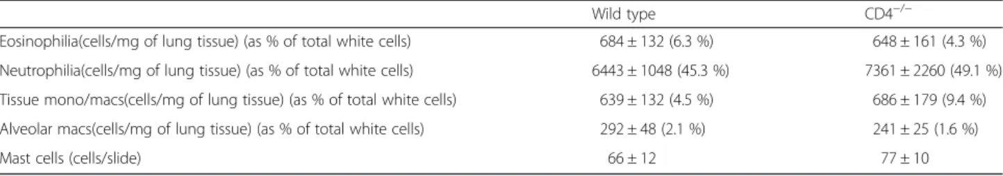
were culled with an overdose of pentobarbitone (200 mg/kg, i.p.). The blood was removed from the lung vessels by perfusing with normal saline prior to harvesting. The tissue was then cleaned, chopped and the cells collected via an enzymatic digestion based on a method described previously [20]. The numbers of CD4+ cells, CD8+ T cells, CD19+ cells (B cells), eosinophils, neutrophils and alveolar macrophages were determined by flow cytometry. Lung mast cell populations were determined by Toluidine blue histological analysis (see below). The genetic status of the TRP knockout lines was confirmed using a standard genotyping procedure.
Flow cytometry
Single-cell suspensions were stained for surface markers in PBS containing 0.1 % sodium azide and 1 % BSA for 30 min at 4 °C and fixed with 2 % paraformaldehyde. Data was acquired on a BD FACS Fortessa machine (BD Biosystems, UK). Forward scatter and side scatter gates were used to exclude debris and dead cells were ex-cluded using a fixable near IR dead cell stain kit for 633 or 635 nm excitation. Cell types were characterised by their forward and side scatter profiles and by their phenotypes (Table 1).
Mast cell enumeration
Mast cells were identified using a standard Toluidine blue histological stain. Mice were culled via overdose with Sodium Pentobarbitone and the systemic circula-tion perfused with saline. The trachea was then can-nulated and the lungs perfused with formalin before being placed in formalin for 24 h. Following this, they were transferred into 70 % ethanol until paraffin wax embedding and slicing could take place. 4 μm sec-tions were cut from the processed lung samples. The sections were stained using a standard toluidine blue staining protocol [21]. Briefly, the lung sections were dewaxed using Histochoice clearing agent ® (Sigma, UK) and rehydrated in a series of ethanol dilutions (100 %, 90 %, 70 %). The slices were then washed in deionised water and stained in 0.1 % Toluidine Blue (Sigma, UK) for 5mins. Sections were then washed in distilled water before the slices were dehydrated using a series of ethanol dilutions (70 %, 90 %, 100 %). The slices were left to dry at room temperature and mounted onto glass slides. The stained sections were analysed under light microscopy at ×40 magnification, the observer blinded to the specimen identities. The numbers of mast cells per slide (3 slides per lung sample) were counted.
mouse i.p.) on day 0 and 14. On days 24, 25 and 26 mice were challenged daily either with vehicle (saline, intranasally) or 1.25μg/kg HDM (in 50μl dose volume, intranasally) under light anaesthesia (inhaled isoflurane) as described previously [22]. 72 h after the final HDM challenge airway reactivity (AR) to inhaled 5-HT was assessed using whole body plethysmography (WBP; Penh). Previous work by our group, and others, have highlighted the important role airway sensory nerves play in respiratory disease [23], this is the reason lung function measurements are performed in conscious an-imals. One hour after AR assessment (to allow recovery from the bronchospasm) the mice were culled with an over dose of pentobarbitone (200 mg/kg, i.p.). Tissue was collected for genotyping. Heparinised blood sam-ples were collected via cardiac puncture for plasma IgE levels. The lungs were lavaged via a tracheal can-nula (3 times with 0.3 ml of RPMI, pooled) and total white cell number and differential percentage in the BAL fluid assessed (as described previously, [24]). The remaining lavage samples were kept at −80 °C. Total IgE levels were measured using BD OptEIATM set
for mouse immunoglobulin E (BD Biosciences,
Oxford, UK) in accordance with the manufacturer’s instructions.
OVA-driven allergic asthma rat model
Male Brown-Norway rats (200–250 g) were sensitised on day 0, 14 and 21 with vehicle (saline with Alum 50:50, 1 ml/rat, i.p.) or OVA (100 μg/rat) administered with Alum (20 mg/rat aluminium hydroxide and 20 mg/rat magnesium hydroxide). All rats were challenged with OVA (1 % w/v, aerosolised for 30 min) on day 28 as previ-ously described [23, 25]. Rats received vehicle (0.5 % MC + 0.2 % tween 80 in saline, 10 ml/kg, i.p., 2 sites) or TRPV1 inhibitor (XEN D0501, 10 mg/kg) 1 h before and 30 min after challenge. One hour after the antigen chal-lenge the rats were placed in plethysmography chambers and Penh levels monitored over night. Following this the animals were given an overdose of pentobarbitone (200 mg/kg, i.p.) and a heparinised plasma sample col-lected via cardiac puncture. Bronchoalveolar lavage (BAL) was carried out (2 × 3 ml RPMI, 30 s each) and total/dif-ferential leukocyte numbers assessed.
OVA specific IgE levels were measured using ELISA. Briefly, 96 well plates were coated with OVA (20 ug/ml) and then blocked. Samples were added and left over night at room temperature. After washing Biotin anti-IgE was added for an hour. After washing anti-IgE was detected using horseradish peroxidase conjugated to streptavidin and visualised with tetramethylbenzidine substrate. Table 1Characterisation of immune cells by flow cytometry
Cell Type Surface Marker Phenotype Monoclonal Antibody Conjugate Catalogue Number Dilution
B Cells CD19+ CD19-FITC (BD Biosciences) 557398 1/100
CD3− CD3-PECy7 (eBioscience) 25–0031 1/200
Natural Killer (NK) Cells NKp46+ NKp46-PE (eBioscience) 12–3351 1/200
CD3− CD3-PECy7 (eBioscience) 25–0031 1/200
CD4+T Cells CD4+ CD4-PerCP (BD Biosciences) 553052 1/200
CD3+ CD3-PECy7 (eBioscience) 25–0031 1/200
CD8+T Cells CD8+ CD8-APC (BD Biosciences) 553035 1/200
CD3+ CD3-PECy7 (eBioscience) 25–0031 1/200
Neutrophils Ly-6Ghigh Ly6G-FITC (BD Biosciences) 551460 1/100
CD11bhigh CD11b-PerCP (eBioscience) 45–0112 1/400
CD11clow CD11c-APC (BD Biosciences) 550261 1/200
F4/80low F4/80-PE (eBioscience) 12–4801 1/50
Alveolar Macrophages CD11blow-int. CD11b-PerCP (eBioscience) 45–0112 1/400
CD11chigh CD11c-APC (BD Biosciences) 550261 1/200
F4/80high F4/80-PE (eBioscience) 12–4801 1/50
Inflammatory monocytes/ macrophages CD11bhigh CD11b-PerCP (eBioscience) 45–0112 1/400
CD11clow CD11c-APC (BD Biosciences) 550261 1/200
F4/80high F4/80-PE (eBioscience) 12–4801 1/50
Eosinophils CD11bhigh CD11b-PerCP (eBioscience) 45–0112 1/400
CD11clow CD11c-APC (BD Biosciences) 550261 1/200
The level of airway inflammation was assessed on his-tologically prepared lung samples from the rat and mouse models using PAS (for mucus load) staining and inflammatory scoring. The scoring was performed by a trained technician blinded to the treatment groups. Full methods have been described previously [22].
Data analysis and statistics
Data was expressed as mean ± S.E.M of n observations. A p value < 0.05 was taken as statistically significant and the actual statistical test employed indicated in the figure legends.
Results
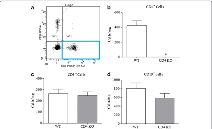
Confirmation of phenotype/genotype of the GM lines To confirm the phenotype of the CD4+ T cell KO mice and to investigate whether this impacted on other key allergic effector cells in the lung, we performed FACS analysis. Figure 1a shows an example FACS plot of lung cells from a naïve wild type mouse, the highlighted box shows the presence of CD4+ cells. The CD4+ T cell KO mice were devoid of CD4+cells (Fig. 1b). This did not im-pact on the levels of other adaptive immune cell types such as CD8+ T cells and B cells (Fig. 1c-d) and tissue, mast cells, eosinophil, neutrophil and alveolar macrophage
numbers (Table 2). Genotyping data confirmed the status of the TRPV1 and TRPA1−/− lines (data not shown) and the typical functional phenotype of these lines has been demonstrated in previous publications [26].
Role of CD4+cells in the House Dust Mite driven allergic asthma mouse model
Initially, to elucidate the role of CD4+ T cells in our murine model of allergic airway disease, WT and CD4−/− mice were sensitised and challenged with HDM. HDM challenge significantly increased serum total/specific IgE (Figure2a and b), BAL eosinophilia (Fig. 2c) and airway reactivity to inhaled 5-HT (Fig. 2d) relative to vehicle challenged controls. CD4−/−mice did not display such an augmentation in these parameters, being indistinguish-able from vehicle controls (Fig. 2); highlighting the crit-ical function of this cell in the development of the allergic asthma phenotype.
Role of TRPV1 in the House Dust Mite driven allergic asthma mouse model
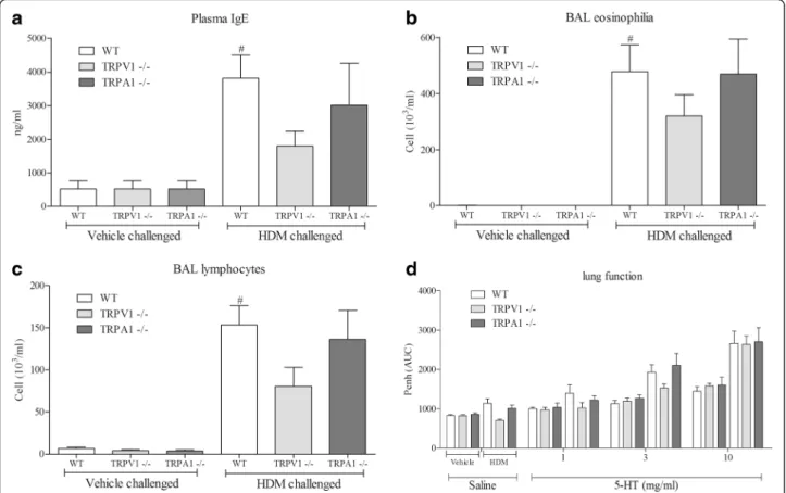
Subsequently, we addressed the role of key CD4+ T cell regulator, TRPV1, in our murine model of allergic airway disease. As anticipated, HDM challenge significantly in-creased serum IgE (Fig. 3a), BAL eosinophil (Fig. 3b),
Fig. 1Characterising the CD4+−/−mice.aRepresentative scatter plot of CD4+and CD8+cells in the lungs of naive mice and the mean data for
total CD4+T cell numbers in wild type and CD4+KO mice (n= 4, expressed at cells/mg of lung tissue) (b).c&d) show the mean numbers of
lymphocytes (Fig. 3c) and airway reactivity to inhaled 5-HT (Fig. 3d) in wild type mice. TRPV1,but not TRPA1, KOs developed an attenuated allergic asthma phenotype although these effects failed to reach statistical signifi-cance (Fig. 3). There was no difference in cellular in-flammation or airway reactivity in non-challenged animals (WT vs TRPV1 or TRPA1 KO lines).
Role of TRPV1 channels in the allergic asthma rat model Given the promising amelioration in pathological pheno-type in the TRPV1−/− mice, we investigated the thera-peutic potential of manipulating this pathway in a distinct pre-clinical animal model with a disparate aller-gen. To determine the role of TRPV1 channels in the rat
model of allergic asthma we compare responses in ve-hicle treated rats with those dosed with a selective TRPV1 antagonist, XEN-D0501. The compound was dosed after the animals were sensitised to focus the in-vestigation on the allergic response rather than the de-velopment of sensitisation. Antigen (OVA) sensitisation caused a statistically significant increase in plasma OVA specific IgE (Fig. 4a), BALF total cell numbers (Fig. 4b), eosinophils (Fig. 4c), neutrophils (Fig. 4d) and lympho-cytes (Fig. 4e) relative to vehicle control. Whilst OVA-specific IgE levels were not impacted on by the TRPV1 inhibitor (Fig. 4a), blockade of the TRPV1 channel resulted in a general reduction in the airway cellular inflammation profile (Fig. 4), although as seen in the Table 2Characterisation of naive CD4−/−mouse line
Wild type CD4−/−
Eosinophilia(cells/mg of lung tissue) (as % of total white cells) 684 ± 132 (6.3 %) 648 ± 161 (4.3 %)
Neutrophilia(cells/mg of lung tissue) (as % of total white cells) 6443 ± 1048 (45.3 %) 7361 ± 2260 (49.1 %)
Tissue mono/macs(cells/mg of lung tissue) (as % of total white cells) 639 ± 132 (4.5 %) 686 ± 179 (9.4 %)
Alveolar macs(cells/mg of lung tissue) (as % of total white cells) 292 ± 48 (2.1 %) 241 ± 25 (1.6 %)
Mast cells (cells/slide) 66 ± 12 77 ± 10
Fig. 2Role of CD4+T cells in the House Dust Mite driven allergic asthma mouse model.a & b) Total and HDM specific IgE levels in the plasma,
Fig. 3Role of TRPV1/TRPA1 in the mouse model. IgE levels in the plasma as measured by ELISA (a), eosinophils (b) and lymphocytes (c) in the BAL as determined by microscopy using standard morphological criteria in the House Dust Mite driven allergic asthma mouse model in TRPV1 or TRPA1 KO mice compared to wild-type. The percentages of the total white cells for each treatment group are: 0, 0, 0, 65, 61 & 78; 9, 6, 6, 21, 15&23, respectively, for eosinophils and lymphocytes. Lung function to inhaled 5-HT is shown in panel (d). Data (n= 8) expressed as mean +/− s.e.m. # =p< 0.05 using studentsT-test (Mann-Whitney)
mouse model this failed to reach significance. These changes were associated with a reduction in the LAR signal (Fig. 5).
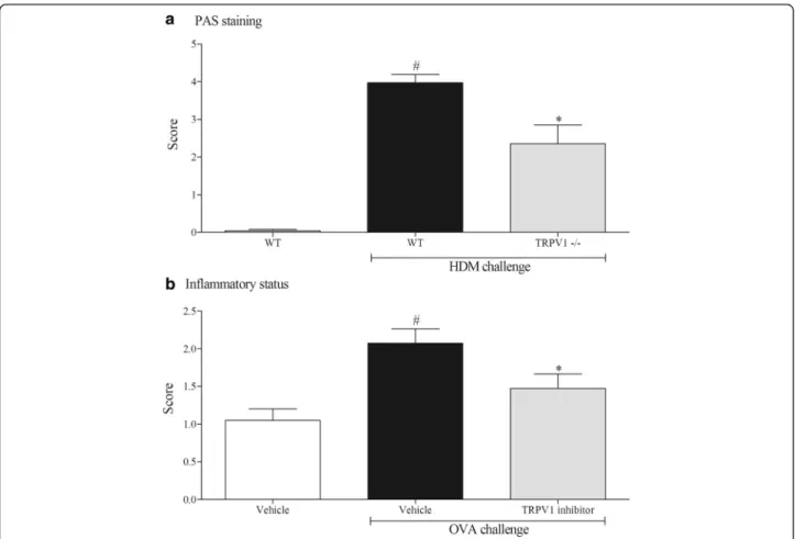
Histological assessment of the lung tissue from the model systems are shown in Fig. 6. The levels of mucus in the lung tissue were increased in the murine-HDM model (panel A) but not in the rat-OVA model (data not shown). The mice missing functional TRPV1 had re-duced levels of mucus compared to the HDM challenged wild type controls (Panel A). In both model systems we measured an increase in the inflammatory score. This was reduced by the TRPV1 inhibitor (Panel B).
Discussion
CD4+T cells are thought to play a prominent role in the development of allergic asthma, thus targeting them is considered to be a viable means to combat the disease. Re-cently it has been shown that human and mouse CD4+T cells express the ion channel TRPV1 [7]. Furthermore, modulating TRPV1 can influence activation status, sur-vival and, importantly, the release of mediators thought to be important in the allergic phenotype [5, 6, 27]. Thus we hypothesised that inhibiting the TRPV1 channel would re-duce the activity of CD4+ T cells thereby modulating the allergic phenotype. To test this hypothesis we used models of allergic asthma in which T cells are known to play a prominent role. We have previously shown that T cells play a dominant role in the OVA-driven Brown Norway rat model which recapitulates an asthma-like phenotype with regard the lung inflammatory response and the LAR (a clinical endpoint that is often used to
trial new asthma therapeutics) [20]. To parallel this model system utilising an alternative antigen and a dif-ferent species, we recently developed a HDM-driven model in the mouse [22]. This model has a number of advantages over the rat model including: the ability to utilise a ‘disease-relevant antigen’, the fact that Alum is not required for the sensitising phase and because it ex-hibits one of the cardinal characteristics of the clinical asthma phenotype, AHR.
Before profiling the role of TRPV1 in these systems, we confirmed a role for CD4+ cells in the HDM-driven model by utilising genetically GM mice that do not pos-sess CD4+T cells. Using this model we compared the al-lergic phenotype in wild type mice and in mice missing functional TRPV1 (and TRPA1) channels. The data sug-gests that TRPV1, but not TRPA1, plays a discrete role in several of the functional endpoints assessed in this model system including a reduction in plasma IgE levels, airway cellular inflammation and AHR. In order to con-firm a role for TRPV1 in the asthma phenotype we employed an alternative model system/species config-ured in the Brown Norway rat and in this case utilised a clinically-ready pharmacological inhibitor, XEN-D0501. This enabled us to avoid any possible developmental is-sues associated with using GM mice and study the effect on the challenge phase, rather than both the sensitisa-tion and challenge phase of the model (which is what is studied in developmental KO mice). Interestingly, while the impact on cellular inflammation is reminiscent of the data in the mouse, the levels of IgE were not altered. This may suggest that TRPV1 plays a role in both the
sensitisation and challenge phases; this would parallel the concept of published data demonstrating a role for TRPV1 on T cell function [6, 7]. The reduction in the LAR signal was associated with reduced inflammatory response. In summary, the data suggests that blockade of TRPV1 attenuates the allergic asthma phenotype which is consistent with some previous studies [28–30]. Conversely, others have published that TRPV1 has no role, or can protect against the development of allergic inflammation in the airways [31, 32]. It is not clear why there are these differences; potentially it could be due the different allergens used to provoke the phenotype, the species, whether validation utilised developmental KOs or small molecule inhibitors (and which small mol-ecule inhibitors) and the end-points recorded. However, we have taken into account the differences in experi-mental settings and have performed one of the most comprehensive studies to date and have obtained a broadly similar picture regardless of the parameters of the model which all points to a discrete role for TRPV1 in the development of the asthma phenotype.
We have suggested a role for TRPV1 in modulation of CD4+T cell function, but cannot rule out a role on other cell types. Indeed, TRPV1 has been reported to be expressed on many cell types in the airway including those thought to play a key role in allergic asthma such as: mast cells, macrophages, epithelial cells, smooth muscle cells, leukocytes and dendritic cells [33–37]. Indeed Rehman et al. have suggested TRPV1 inhibition could be beneficial in attenuating airway epithelial injury and thus reduces asthma features [27]. Furthermore, the TRPV1 receptor is expressed on the peripheral terminals of airway specific, vagal afferent nerves [38] and it has been suggested that the LAR [23] and AHR [30, 39] are reflex events. In addition, activation of this ion channel on sensory afferents has been linked with the release of tachykinins/neuropep-tideswhich could be involved in certain functional aspects of the asthma phenotype such as inflammation and bronchoconstriction [40]. It is not clear how the TRPV1 channel is being activated in the model systems. TRPV1 is known to be activated by various pro-inflammatory media-tors such as lipoxygenase metabolites which are reported
to be increased in the asthmatic airway [8]. Furthermore, the airway of asthmatic patients is thought to be at a lower pH (from 5.2 to 7.1) and the pH is normalised with cor-ticosteroid, treatment [41]. Thus this lower pH could be an endogenous activator of TRPV1 in the diseased airway.
Conclusion
In conclusion, our data supports the view that targeting TRPV1 could be of clinical benefit in patients with allergic asthma, via a possible role in the modulation of CD4+ T cell function. This data, along with other recent publica-tions, informs our thinking on the role of TRP channels in the airways expanding their remit from the control of sen-sory nerve function to a modulator of key cells involved in adaptive immunity.
Abbreviations
AHRairway hyperresponsiveness,LARlate asthmatic response,TRPV1 transient receptor potential cation channel subfamily V member1,TRPA1 transient receptor potential cation channel, subfamily A, member 1,GM genetically modified,HDMhouse dust mite
Sources of support
Studies were supported by the Medical Research Council; (MRC, UK) (MR/K020293/1). KB was supported by an MRC studentship.
Authors’contributions
Conception and design;MAB, MGB; data generation, analysis and interpretation; MAB, MGB, KB, KR, RS, BD; writing the paper; MGB, MAB, provided intellectual input and advice that was taken into consideration in the drafting of the manuscript; JF. All authors reviewed the manuscript and approved the final draft.
Competing interests
JF is employed by and MGB, MAB are consultants for Ario Pharma. Conflict of interest: Dr J. Ford works at ArioPharma Ltd who supplied the TRPV1 inhibitor, all other authors declare that they have no competing interests.
Author details
1Respiratory Pharmacology, Airway Disease Section, National Heart and Lung Institute, Faculty of Medicine, Imperial College London, Exhibition Road, London SW7 2AZ, UK.2Department of Anatomy, Faculty of Medicine, University of Porto, Alameda Prof. HernâniMonteiro, 4200-319 Porto, Portugal. 3Center for Health Technology and Services Research (CINTESIS), Faculty of Medicine, University of Porto, Rua Dr. Plácido da Costa, 4200-450 Porto, Portugal.4Leukocyte Biology, Imperial College, London, UK.5ArioPharma Limited, Iconix Park, London Road, Pampisford CB22 3EG, UK.6Asthma UK Centre in Allergic Mechanisms of Asthma, Imperial College London, London, UK.
Received: 28 August 2015 Accepted: 26 May 2016
References
1. Bateman ED, Hurd SS, Barnes PJ, Bousquet J, Drazen JM, FitzGerald M, Gibson P, Ohta K, O’Byrne P, Pedersen SE, Pizzichini E, Sullivan SD, Wenzel SE, Zar HJ. Global strategy for asthma management and prevention: GINA executive summary. Eur Respir J. 2008;31:143–78.
2. Masoli M, Fabian D, Holt S, Beasley R. The global burden of asthma: Executive summary of the GINA Dissemination Committee Report. Allergy Eur J Allergy Clin Immunol. 2004;59(5):469–78.
3. Robinson DS, Hamid Q, Ying S, Tsicopoulos A, Barkans J, Bentley AM, Corrigan C, Durham SR, Kay AB. Predominant TH2-like bronchoalveolar T-lymphocyte population in atopic asthma. N Engl J Med. 1992;326:298–304. 4. Ngoc PL, Gold DR, Tzianabos AO, Weiss ST, Celedón JC. Cytokines, allergy,
and asthma. Curr Opin Allergy Clin Immunol. 2005;5:161–6.
5. Bertin S, de Jong PR, Jefferies W a, Raz E. Novel immune function for the TRPV1 channel in T lymphocytes. Channels. 2015;8:479–80.
6. Majhi RK, Sahoo SS, Yadav M, Pratheek BM, Chattopadhyay S, Goswami C. Functional expression of TRPV channels in T cells and their implications in immune regulation. FEBS J. 2015;282:2661–81.
7. Bertin S, Aoki-Nonaka Y, de Jong PR, Nohara LL, Xu H, Stanwood SR, Srikanth S, Lee J, To K, Abramson L, Yu T, Han T, Touma R, Li X, González-Navajas JM, Herdman S, Corr M, Fu G, Dong H, Gwack Y, Franco A, Jefferies Wa, Raz E. The ion channel TRPV1 regulates the activation and proinflammatory properties of CD4(+) T cells. Nat Immunol. 2014;15:1055–63.
8. Grace MS, Baxter M, Dubuis E, Birrell M a, Belvisi MG. Transient receptor potential (TRP) channels in the airway: Role in airway disease. Br J Pharmacol. 2014;171:2593–607.
9. Wang Q, Bai X, Xu D, Xu D, Li H, Fang J, Zhu H, Fu W, Cai X, Wang J, Jin Z, Wang Q, Xu C, Chang J. TRPV1 UTR-3 polymorphism and susceptibility of childhood asthma of the Han Nationality in Beijing. Wei Sheng Yan Jiu. 2009;38:516–21. 10. Cantero-Recasens G, Gonzalez JR, Fandos C, Duran-Tauleria E, Smit L a M,
Kauffmann F, Antó JM, Valverde Ma. Loss of function of transient receptor potential vanilloid 1 (TRPV1) genetic variant is associated with lower risk of active childhood asthma. J Biol Chem. 2010;285:27532–5.
11. Ren Y-F, Li H, Xing X-H, Guan H-S, Zhang B-A, Chen C-L, Zhang J-H. Preliminary study on pathogenesis of bronchial asthma in children. Pediatr Res. 2015;77:506–10.
12. Sadofsky LR, Ramachandran R, Crow C, Cowen M, Compton SJ, Morice AH. Inflammatory stimuli up-regulate transient receptor potential vanilloid-1 expression in human bronchial fibroblasts. Exp Lung Res. 2012;38:75–81.
13. McGarvey LP, Butler CA, Stokesberry S, Polley L, McQuaid S, Abdullah H, Ashraf S, McGahon MK, Curtis TM, Arron J, Choy D, Warke TJ, Bradding P, Ennis M, Zholos A, Costello RW, Heaney LG. Increased expression of bronchial epithelial transient receptor potential vanilloid 1 channels in patients with severe asthma. J Allergy Clin Immunol. 2014;133:704–12. 14. Watanabe N, Horie S, Spina D, Michael GJ, Page CP, Priestley JV.
Immunohistochemical localization of transient receptor potential vanilloid subtype 1 in the trachea of ovalbumin-sensitized Guinea pigs. Int Arch Allergy Immunol. 2008;146:28–32.
15. Jia Y, McLeod RL, Hey JA. TRPV1 receptor: a target for the treatment of pain, cough, airway disease and urinary incontinence. Drug News Perspect. 2005; 18:165–71.
16. Brito R, Sheth S, Mukherjea D, Rybak LP, Ramkumar V. TRPV1: A Potential Drug Target for Treating Various Diseases. Cells. 2014;3:517–45. 17. Aoki H, Mogi C, Okajima F. Ionotropic and metabotropic proton-sensing
receptors involved in airway inflammation in allergic asthma. Mediators Inflamm. 2014;2014:712962.
18. Wortley MA, Birrell MA, Maher SA, Round P, Ford J, Belvisi MG: Profiling Of XEN-D0501, A Novel TRPV1 Antagonist, In Pre-Clinical Models Of Cough. Am J Respir Crit Care Med. 2014;18:9A4979.
19. Kilkenny C, Browne W, Cuthill I. The ARRIVE guidelines. ReqartoCom. 2010;8:1–2. 20. Underwood SL, Haddad E-B, Birrell MA, McCluskie K, Pecoraro M, Dabrowski
D, Webber SE, Foster ML, Belvisi MG. Functional characterization and biomarker identification in the Brown Norway model of allergic airway inflammation. Br J Pharmacol. 2002;137:263–75.
21. Yagil Z, Hadad Erlich T, Ofir-Birin Y, Tshori S, Kay G, Yekhtin Z, Fisher DE, Cheng C, Wong WSF, Hartmann K, Razin E, Nechushtan H. Transcription factor E3, a major regulator of mast cell-mediated allergic response. J Allergy Clin Immunol. 2012;129:1357–66.
22. Raemdonck K, Baker K, Dale N, Dubuis E, Shala F, Belvisi MG, Birrell MA. CD4+ and CD8+T cells play a central role in a HDM driven model of allergic asthma.
Respir Res. 2016;17:45.
23. Raemdonck K, de Alba J, Birrell M a, Grace M, Maher S a, Irvin CG, Fozard JR, O’Byrne PM, Belvisi MG. A role for sensory nerves in the late asthmatic response. Thorax. 2012;67:19–25.
24. Birrell MA, De Alba J, Catley MC, Hardaker E, Wong S, Collins M, Clarke DL, Farrow SN, Willson TM, Collins JL, Belvisi MG. Liver X receptor agonists increase airway reactivity in a model of asthma via increasing airway smooth muscle growth. J Immunol. 2008;181:4265–71.
26. Birrell MA, Belvisi MG, Grace M, Sadofsky L, Faruqi S, Hele DJ, Maher SA, Freund-Michel V, Morice AH. TRPA1 Agonists Evoke Coughing in Guinea Pig and Human Volunteers. Am J Respir Crit Care Med. 2009;180:1042–7. 27. Basu S, Srivastava P. Immunological role of neuronal receptor vanilloid
receptor 1 expressed on dendritic cells. Proc Natl Acad Sci U S A. 2005;102: 5120–5.
28. Mabalirajan U, Rehman R, Ahmad T, Kumar S, Singh S, Leishangthem GD, Aich J, Kumar M, Khanna K, Singh VP, Dinda AK, Biswal S, Agrawal A, Ghosh B. Linoleic acid metabolite drives severe asthma by causing airway epithelial injury. Sci Rep. 2013;3:1349.
29. Rehman R, Bhat YA, Panda L, Mabalirajan U. TRPV1 inhibition attenuates IL-13 mediated asthma features in mice by reducing airway epithelial injury. Int Immunopharmacol. 2013;15:597–605.
30. Delescluse I, Mace H, Adcock JJ. Inhibition of airway hyper-responsiveness by TRPV1 antagonists (SB-705498 and PF-04065463) in the unanaesthetized, ovalbumin-sensitized guinea pig. Br J Pharmacol. 2012;166:1822–32. 31. Mori T, Saito K, Ohki Y, Arakawa H, Tominaga M, Tokuyama K. Lack of
transient receptor potential vanilloid-1 enhances Th2-biased immune response of the airways in mice receiving intranasal, but not intraperitoneal, sensitization. Int Arch Allergy Immunol. 2011;156:305–12.
32. Caceres AI, Brackmann M, Elia MD, Bessac BF, del Camino D, D’Amours M, Witek JS, Fanger CM, Chong JA, Hayward NJ, Homer RJ, Cohn L, Huang X, Moran MM, Jordt S-EE. A sensory neuronal ion channel essential for airway inflammation and hyperreactivity in asthma. Proc Natl Acad Sci. 2009;106: 9099–104.
33. Wang JP, Tseng CS, Sun SP, Chen YS, Tsai CR, Hsu MF. Capsaicin stimulates the non-store-operated Ca2+ entry but inhibits the store-operated Ca2+ entry in neutrophils. Toxicol Appl Pharmacol. 2005;209:134–44. 34. Chen C-W, Lee ST, Wu WT, Fu W-M, Ho F-M, Lin WW. Signal transduction
for inhibition of inducible nitric oxide synthase and cyclooxygenase-2 induction by capsaicin and related analogs in macrophages. Br J Pharmacol. 2003;140:1077–87.
35. Bíró T, Maurer M, Modarres S, Lewin NE, Brodie C, Acs G, Acs P, Paus R, Blumberg PM. Characterization of functional vanilloid receptors expressed by mast cells. Blood. 1998;91:1332–40.
36. Zhao L, Zhang X, Kuang H, Wu J, Guo Y, Ma L. Effect of TRPV1 channel on the proliferation and apoptosis in asthmatic rat airway smooth muscle cells. Exp Lung Res. 2013;39:283–94.
37. Jia X, Zhang H, Cao X, Yin Y, Zhang B. Activation of TRPV1 mediates thymic stromal lymphopoietin release via the Ca2+/NFAT pathway in airway epithelial cells. FEBS Lett. 2014;588:3047–54.
38. Gunthorpe MJ, Benham CD, Randall A, Davis JB. The diversity in the vanilloid (TRPV) receptor family of ion channels. Trends Pharmacol Sci. 2002; 23:183–91.
39. Tränkner D, Hahne N, Sugino K, Hoon M a, Zuker C. Population of sensory neurons essential for asthmatic hyperreactivity of inflamed airways. Proc Natl Acad Sci U S A. 2014;111:11515–20.
40. Nasra J, Belvisi MG. Modulation of sensory nerve function and the cough reflex: Understanding disease pathogenesis. Pharmacol Ther. 2009;124:354–75. 41. Hunt JF, Fang K, Malik R, Snyder A, Malhotra N, Platts-Mills T aE, Gaston B.
Endogenous airway acidification: Implications for asthma pathophysiology. Am J Respir Crit Care Med. 2000;161:694–9.
• We accept pre-submission inquiries
• Our selector tool helps you to find the most relevant journal
• We provide round the clock customer support
• Convenient online submission
• Thorough peer review
• Inclusion in PubMed and all major indexing services • Maximum visibility for your research
Submit your manuscript at www.biomedcentral.com/submit