http://www.ijpediatrics.com pISSN 2349-3283 | eISSN 2349-3291
Research Article
Study of significance of thrombocytosis in lower
respiratory tract infections in children
Sreenivasa B
1, Kumar GV
2*, Manjunatha B
1INTRODUCTION
Lower Respiratory Tract Infections (LRTI) continues to threaten the health of children worldwide and are exacerbated by global environmental problems. In the developing world where nutrition remains poor and access to healthcare is scarce, LRTIs are the most common cause of illness and death in children. Outcome of LRTI are far better in developed countries, but the
overall morbidity of LRTI is still high. However, while the incidence of lower respiratory tract infections in developed countries may be as low as 3-4%, its incidence in developing countries range between 20% and 30%. In India, in the states and districts with high infant and child mortality rates, LRTI is one of the major causes of death. Upto 13% of in-patient deaths in pediatric wards are due to LRTI.1
ABSTRACT
Background: Lower Respiratory Tract Infections (LRTI) continues to threaten the health of children worldwide. Up to 13% of inpatient deaths in pediatric wards are due to LRTI. Platelets play a major role in antimicrobial host defence, the induction of inflammation and tissue repair. Inflammatory thrombocytosis is related to increased levels of several cytokines such as thrombopoietin, interleukin-6, interleukin-1alpha, interleukin-8 and tumour necrosis factor alpha.
Methods: A prospective study was done on thrombocytosis in children aged from 2 months to 5 years admitted with lower respiratory tract infections meeting the criteria of ARI control programme.
Results: Among 220 cases, 63.2% of the cases were males and 36.8% of the cases were females, with male to female ratio being 1.7: 1. Among total 220 admitted cases of LRTI, 50% of the cases were severe pneumonia, 26.4% were very severe pneumonia and 23.6% were pneumonia. Among 220 cases, thrombocytosis was found in 77 children. Among 4 children with severe thrombocytosis (9.01-10.00 L/mm3), 3 had very severe pneumonia and 1 had severe pneumonia. It indicates that platelet count increases as the severity of pneumonia increases. All 4 cases of severe thrombocytosis (9.01-10.00 Lakhs/mm3) and 1 case of extreme thrombocytosis (>10.00 Lakhs/mm3) had TLC >15000/mm3.
Conclusions: Children admitted with LRTI having thrombocytosis had longer duration of hospital stay, which indirectly indicate more severity of pneumonia. Children with thrombocytosis have more severe clinical condition and longer hospitalization. Platelet count may be used as a useful marker associated with severity of lower respiratory tract infection and its complications.
Keywords: Lower respiratory tract infection, Thrombocytosis, Pneumonia, Plural effusion
1Department of Paediatrics, Basaveshwara medical College, Chitradurga-577501, Karnataka, India
2Department of Paediatrics, Sri Siddhartha Medical College, Tumkur-572107, Karnataka, India
Received: 03 April 2015
Accepted: 22 April 2015
*Correspondence:
Dr. Kumar GV,
E-mail: kumargowripura@yahoo.co.in
Copyright: © the author(s), publisher and licensee Medip Academy. This is an open-access article distributed under the terms of the Creative Commons Attribution Non-Commercial License, which permits unrestricted non-commercial use, distribution, and reproduction in any medium, provided the original work is properly cited.
Platelets play a major role in antimicrobial host defense, the induction of inflammation and tissue repair.2 Activation of platelets by agonists enhances platelet interactions with complement proteins and humoral immune components, as well as leukocytes and
endothelial cells. They are capable of binding,
aggregating and internalizing microorganisms, which enhances the clearance of pathogens from bloodstream and also participate in antibody dependent cell cytotoxicity functions to kill protozoal pathogens by releasing array of potent antimicrobial peptides. Inflammatory thrombocytosis is related to increased levels of several cytokines such as thrombopoietin,
interleukin-6, interleukin-1alpha, interleukin-8 and
tumour necrosis factor alpha.3 Severe community
acquired pneumonia is associated in the literature with
significant increment of plasma levels of the
inflammatory cytokines TNF-α, IL-1b, IL-6, IL-8. The TNF-α, IL-1b and IL-6 were also elevated in the bronchoalveolar lavage fluid of patients with community acquired pneumonia.4,5
Normal platelet counts range between 150000/mm3 and 450000/mm3. This definition is for healthy neonates, infants, children and adolescents.6 Thrombocytosis or elevation in the peripheral blood platelet count to values >500000/mm3 is common in infancy and childhood,
occurring in 3 to 13% of children.2 Reactive
thrombocytosis has an estimated incidence of 6-15% among hospitalized children. Bacterial or viral infections (acute or chronic) are most common cause for reactive thrombocytosis (37-78%) at any age during childhood. Within this group, infections of the respiratory tract account for 60-80% of reactive thrombocytosis, followed by infections of the gastrointestinal and urinary tract.7,8
METHODS
A prospective study was done on thrombocytosis in children admitted with lower respiratory tract infections aged between 2 months and 5 years at Basavehwara medical college hospital, Chitradurga between February
2014 to February 2015. A total of220 children meeting
the inclusion criteria were included for the present study. All children from 2 months to 5 years admitted with lower respiratory tract infections meeting the criteria in ARI control programme (WHO),1 were included in the study. Children <2 months and >5 years and children with associated anaemia (Hb <11g/dl), neuroinfections, connective tissue disorders, or congenital heart diseases. Young infants (<2 months), frequently have non-specific signs, such as poor feeding, fever or low body temperature and any pneumonia in young infant is considered to be severe and were excluded from the study. The clinical condition at admission, course in the hospital, complications and outcome of the children with thrombocytosis and children without thrombocytosis were analysed. Children with platelet count from 4.51-4.99 Lakhs/mm3 were also included in this group, as according to definition, thrombocytosis in children is
>5.00 Lakhs/mm3.1,6 Thrombocytosis was further divided as mild (5.00-7.00 Lakhs/mm3), moderate (7.01-9.00
Lakhs/mm3), severe (9.01-10.00 Lakhs/mm3) and
extreme (>10.00 Lakhs/mm3) thrombocytosis.6 A
predesigned proforma was used to collect information regarding age, sex, socio demographic profile, presenting complaints like duration of fever, cough, hurried breathing, chest indrawing, and decreased feeding, lethargy, convulsions. Relevant past and family history were also taken. Birth history which included term/preterm, mode of delivery, birth weight and any history of NICU admission were documented. Complete general physical examination, systemic examination with special orientation towards respiratory system like Respiratory Rate, SpO2, capillary refilling time, chest
indrawing, stridor, grunting, crepitations, rhonchi were noted. Routine and relevant investigations such as Hb%, total leukocyte count, ESR, platelet count and chest X-ray
were done. Complications like parapneumonic
effusion/empyema, pneumatoceles, septic shock and respiratory failure were noted. Numbers of deaths were also noted.
RESULTS
Among total 220 cases, 70% (154) of the cases were in the age group of 2 months to 12 months, and rest 30% (66) of the cases were in the age group of 13 months to 60 months (5 years). Among 220 cases, 63.2% (139) of the cases were males and 36.8% (81) of the cases were females, with male to female ratio being 1.7:1. Among total 220 admitted cases of LRTI, 50% (110) of the cases were severe pneumonia, 26.4% (58) were very severe pneumonia and 23.6% (52) were pneumonia. Out of 154 cases in the age group of 2 months to 12 months, majority of the cases were admitted with severe pneumonia (80 children) followed by very severe pneumonia (43 children), whereas in the age group of 13 months to 60 months majority were admitted with severe pneumonia (30 children) and pneumonia (21 children). This indicates that severity of pneumonia is more in infancy. Among 220 cases, thrombocytosis was found in 77 (35%) children. Out of total 77 cases of LRTI with thrombocytosis, 68.8% of the children were in the age group of 2 months to 12 months and remaining 31.2% were in the age group of 13 months to 60 months. 143 children were without thrombocytosis (Table 1).
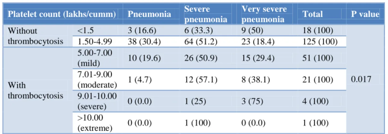
Among 4 children with severe thrombocytosis (9.01-10.00 Lakhs/mm3), 3 had very severe pneumonia and 1 had severe pneumonia. It indicates that platelet count increases as the severity of pneumonia increases (Table 2). All 4 cases of severe thrombocytosis (9.01-10.00
Lakhs/mm3) and 1 case of extreme thrombocytosis
children with chest X-ray feature suggestive of bronchopulmonary infiltrates and lobar pneumonia,
majority had platelet count within normal the range (Table 4).
Table 1:Relationship between platelet count and severity of pneumonia.
Platelet count Pneumonia Severe
pneumonia
Very severe
pneumonia Total P value
Without thrombocytosis 41 (78.8) 70 (48.9) 32 (55.2) 143 (65)
0.017
With thrombocytosis 11 (21.2) 40 (51.1) 26 (44.8) 77 (35)
Total 52 (100) 110 (100) 58 (100) 220 (100)
Figures in the parentheses indicate percentage. Significant association between platelet count and severity of pneumonia was seen (P value 0.017)
Table 2:Relationship between thrombocytosis and severity of pneumonia.
Platelet count (lakhs/cumm) Pneumonia Severe pneumonia
Very severe
pneumonia Total P value
Without thrombocytosis
<1.5 3 (16.6) 6 (33.3) 9 (50) 18 (100)
0.017
1.50-4.99 38 (30.4) 64 (51.2) 23 (18.4) 125 (100)
With
thrombocytosis
5.00-7.00
(mild) 10 (19.6) 26 (50.9) 15 (29.4) 51 (100)
7.01-9.00
(moderate) 1 (4.7) 12 (57.1) 8 (38.1) 21 (100)
9.01-10.00
(severe) 0 (0.0) 1 (25) 3 (75) 4 (100)
>10.00
(extreme) 0 (0.0) 1 (100) 0 (0.0) 1 (100)
Figures in the parentheses indicate percentage
Table 3:Relationship between platelet count and total leukocyte count.
Platelet count (lakhs/mm3) Total leukocyte count(/mm 3)
Total P value
<5000 5000 to 15000 >15000
Without thrombocytosis
<1.5 2 (11.1) 11(61.1) 5 (27.8) 18 (100)
0.001
1.50-4.99 3 (2.4) 105 (84) 17 (13.6) 125 (100)
With
thrombocytosis
5.00-7.00
(mild) 1 (1.9) 34 (66.7) 16 (31.4) 51 (100)
7.01-9.00
(moderate) 0 (0.0) 8 (38) 13 (62) 21 (100)
9.01-10.00
(severe) 0 (0.0) 0 (0.0) 4 (100) 4 (100)
>10.00
(extreme) 0 (0.0) 0 (0.0) 1 (100) 1 (100)
Figures in the parentheses indicate percentage
Table 4:Relationship between platelet count and chest X-ray findings.
Platelet count Chest X-ray P value
Normal BPI LP PE
Without thrombocytosis 18 (75) 113 (64.2) 11 (78.6) 1(16.7)
0.004
With thrombocytosis 6 (25) 63 (34.8) 3 (21.4) 5 (83.3)
Total 24 (100) 176 (100) 14 (100) 6 (100)
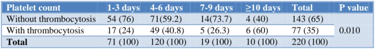
Mean duration of stay was 4.52 days. Significant association of thrombocytosis with duration of hospital stay was noted (P value = 0.010). Children admitted with LRTI having thrombocytosis had longer duration of
hospital stay, which indicate more severity of pneumonia (Table 5).Mortality was more common in children with thrombocytosis, but this association of thrombocytosis with mortality was not statistically significant (Table 6).
Table 5:Relationship between platelet count and duration of hospital stay.
Platelet count 1-3 days 4-6 days 7-9 days ≥10 days Total P value
Without thrombocytosis 54 (76) 71(59.2) 14(73.7) 4 (40) 143 (65)
0.010
With thrombocytosis 17 (24) 49 (40.8) 5 (26.3) 6 (60) 77 (35)
Total 71 (100) 120 (100) 19 (100) 10 (100) 220 (100)
Figures in the parentheses indicate percentage
Table 6:Relationship between platelet count and outcome of the study.
Platelet count Outcome Total P value
Relieved Death
Without thrombocytosis 141 (98.6) 2 (1.4) 143 (100)
0.354
With thrombocytosis 72 (93.5) 5 (6.5) 77 (100)
Total 213 (96.8) 7 (3.2) 220 (100)
Figures in the parentheses indicate percentage
DISCUSSION
Infections of the respiratory tract are perhaps the most common human ailment. Its incidence in developing countries ranges between 20 and 30 percent. Pneumonia is one of the leading causes of mortality among under five children in most developing countries, accounting for almost 18 percent of under five deaths. Platelets have long been recognized for their importance in maintaining hemostasis and for their contribution to wound healing.
However, platelets have historically been
underappreciated for their contributions to antimicrobial host defense.
Thrombocytosis was noted in 35% of the study population, among which 68.8% of the children were in the age group of 2 months to 12 months and remaining 31.2% were in the age group of 13 months to 60 months. In study conducted by Vlacha et al. in 2006, thrombocytosis was observed in 34.5% of the children aged <12 months.9 Study conducted by Dodig S et al. in 2005, thrombocytosis was noted in slightly younger children (3.0 ± 1.8 years) than in children with normal platelet count (3.8 ± 2.4 years).10
In our study, significant association was noted between platelet count and severity of pneumonia (P value 0.017). As the severity of pneumonia increases, platelet count
also increases. Among 4 cases with severe
thrombocytosis (9.01-10.00 Lakhs/mm3), 3 had very
severe pneumonia and 1 had severe pneumonia. In present study, 8.2% had low platelet count, 56.8% had normal platelet count and 35% had thrombocytosis.
Among children with thrombocytosis, 66.2% had mild thrombocytosis, 27.3% had moderate thrombocytosis, 5.7% had severe thrombocytosis and 1.3% had extreme thrombocytosis. This is comparable to studies done by Sutor et al. (1999), Yohannan et al. (1994), Heng and Tan (1998), where mild thrombocytosis was seen in 72-86%, moderate thrombocytosis in 6-8%, severe thrombocytosis in 3%, and extreme thrombocytosis in 0.5%.7,8,11
All 4 cases of severe thrombocytosis and 1 case of extreme thrombocytosis, had total leukocyte count >15000/mm3. This shows that, with increase in total leukocyte count, platelet count also increases (P value <0.001). Study done by Mirsaeidi M et al. (2009), on community acquired pneumonia showed that platelet count was strongly associated (P value 0.009) with 30 day mortality, whereas no association was observed for leukocyte count (P value 0.5114) and concluded that an abnormal platelet count is a better predictor of outcome than an abnormal leukocyte count.12
patients with pleural effusion had thrombocytosis (P-value 0.03).9 Study by Prina E et al. in 2012 showed a significant association of pleural effusion/empyema with thrombocytosis (P value <0.001).14
Significant association of thrombocytosis with duration of hospital stay was noted (P value 0.010). Similar finding was noted in a study done by Vlacha V et al. in 2006, where the association of thrombocytosis with length of hospital stay was significant (P value 0.03).9 Another study in 2012 by Prina E et al. also shows similar results with P value of 0.004.14 In contrast to above findings, study in 1990 by Wolach B et al. showed no associations of thrombocytosis with increased duration of hospital stay.13
CONCLUSION
Thrombocytosis is a common finding among children with lower respiratory tract infection, especially in infancy. Children with thrombocytosis have more severe clinical condition and longer hospitalization. Platelet count increases as the severity of pneumonia increases. Importantly, thrombocytosis occurs almost exclusively in children with pleural effusion. Platelet count may be used as a useful marker associated with severity of lower respiratory tract infection and its complications.
Funding: No funding sources Conflict of interest: None declared
Ethical approval: The study was approved by the institutional ethics committee
REFERENCES
1. Park K. Park’s textbook of preventive and social medicine. In: Park K, eds. A Book. 22nd ed. Jabalpur (India): Banasidas Bhanot; 2013.
2. Klinger MH, Jelkmann W. Role of blood platelets in
infection and inflammation. J Interferon Cytokine Res. 2002;22(9):913-22.
3. Yeaman MR. The role of platelets in antimicrobial host defence. Clin Infect Dis. 1997;25:951-70.
4. Greene C, Lowe G, Taggart C, Gallagher P,
McElvaney N, O’Neill S. Tumor necrosis factor- alpha converting enzyme: its role in community
acquired pneumonia. J Infect Dis.
2002;186(12):1790-6.
5. Ishiguro A, Ishikita T, Shimbo T, Matsubara K,
Baba K, Hayashi, et al. Elevation of serum
thrombopoietin precedes thrombocytosis in
Kawasaki disease. Thromb Hemost. 1998;79:1096-100.
6. Dua Vikas, Sachdev A, Yadav SP. Platelet counts in
healthy neonates, infants, children and adolescents. In: Dua Vikas, Sachdev A, Yadav SP, eds. Practical Pediatric Hematology (IAP). 2nd ed. Delhi, India: Jaypee; 2012: 90-97.
7. Dame C, Sutor AH. Primary and secondary
thrombocytosis in childhood. Br J Haematol. 2005;129:165-77.
8. Heng JT, Tan AM. Thrombocytosis in childhood.
Singapore Med J. 1998;39:485-7.
9. Vlacha V, Feketea G. Thrombocytosis in pediatric patients is associated with severe lower respiratory tract infections. Arch Med Res. 2006;37:755-9. 10. Dodig S, Raos M, Kovac K, Nogalo B, Benko B,
Glojnaric I, et al. Thrombopoietin and interleukin-6
in children with pneumonia-associated
thrombocytosis. Arch Med Res. 2005;36:124-8.
11. Yohannan MD, Higgy KE, Al-Mashhadani SA,
Santhosh-Kumar. Thrombocytosis. Etiologic
analysis of 663 patients. Clin Pediatr. 1994;33:340-3.
12. Mirsaeidi M, Peyrani P, Aliberti S, Filardo G, Bordon J, Blasi F, et al. Thrombocytopenia and thrombocytosis at time of hospitalization predict
mortality in patients with CAP. Chest.
2010;137(2):416-20.
13. Wolach B, Morag H, Drucker, Sadan N.
Thrombocytosis after pneumonia with empyema and other bacterial infections in children. Pediatr Infect Dis J. 1990;9:718-21.
14. Prina E, Ferrer M, Ranzani OT, Polverino E,
Cilloniz C, Moreno E, et al. Thrombocytosis is a marker of poor outcome in community-acquired pneumonia. Chest. 2013;143:767-75.
DOI: 10.5455/2349-3291.ijcp20150508
Cite this article as: Sreenivasa B, Kumar GV,
Manjunatha B.Study of significance of thrombocytosis