MICRO-CT VOLUMETRIC ANALYSIS OF VOIDS AT THE MARGIN AFTER MANUFACTURER RECOMMENDED CEMENTATION PROCESS
Brandon Craig Peters
A thesis submitted to the faculty at the University of North Carolina at Chapel Hill in partial fulfillment of the requirements for the degree of Master of Science in the Adams School of
Dentistry (Prosthodontics)
Chapel Hill 2020
Approved by:
Taiseer Sulaiman
Ryan Cook
ABSTRACT
Brandon Craig Peters: Micro-CT volumetric analysis of voids at the margin after manufacturer recommended cementation process.
(Under the Direction of Taiseer Sulaiman)
Purpose: The purpose is to use Micro-Computed Tomography (Micro-CT) to evaluate the
volume of voids left at the margin after cement cleanup.
Materials and Methods: Two methods of cement cleanup were studied using two
cements making 4 groups of 5 crown-die samples. The buccal margins were examined with
Micro-CT to measure the volume and width of voids remaining at the margin. The outcomes
were compared by the Wilcoxon Ranked Sum test at P = .05.
Results: There were statistically significant differences for all outcomes between the
types of cement (P < .05). There were no statistically significant differences for all outcomes
between the methods of cement cleanup (P >.05).
Conclusion: The choice of cement may be more important to the clinician than the
method of cement cleanup when considering voids left at the margin. Micro-CT is an excellent
ACKNOWLEDGEMENTS
First, I would like to thank God for blessing me with the gifts he has bestowed upon me.
Thank you to my parents who nurtured me, pushed me. Without their unrelenting belief in me
when everyone else would have given up, I wouldn’t be here today. Most of all, thank you to my
wife who, through the first years of our marriage, supported me through the hardships of
residency.
Thank you to Dr. Ryan Cook for his leadership, mentorship, motivation, and unending
enthusiasm as my Graduate Prosthodontics Program Director and as my committee member.
You made me the best clinician I could be. I would like to express my gratitude to Dr. Taiseer
Sulaiman, thank you for your patience, guidance, and words of encouragement when things were
tough. Thank you to Dr. Terry Donovan, who, over many years of education, has shaped my
outlook on what quality research is and what I expect of myself as a researcher.
Also, I’d like to extend my thanks to Brandon Rodgers, without your laboratory support
this project would not have been possible. Thank you for your hard work and endless support.
To Dr. Ceib Phillips, thank you for your patience with me and help with my statistical
analysis.
To the Graduate Prosthodontics staff, thank you for your help and support. I greatly
Dan Bailey, Dr. Matthew Hopfensperger, and Dr. Kevin Lim, thank you for your time, patience
and knowledge. You have made an impact in my career and I will always be grateful for it.
Thank you to my amazing co-residents for always getting me through the day. Especially
Gabrielle Jackson and Renata Camino Navarro, I am grateful to have shared this journey with
you and cannot imagine accomplishing it without your support and most importantly friendship,
TABLE OF CONTENTS
LIST OF TABLES ... VIII LIST OF FIGURES ... IX
CHAPTER 1: REVIEW OF THE LITERATURE ... 1
1. Introduction ... 1
2. Literature Review ... 5
2.1. History of Dental Cements ... 5
2.2. Classification of Dental Cements ... 6
2.3. Solubility ... 10
2.4. Cements in This Study ... 10
2.4.1. RelyX Unicem 2 (Automix) ... 10
2.4.2. RelyX Luting Plus (Automix) ... 11
3. History of Dental Ceramics ... 11
3.1. Ceramics Used in This Study ... 12
4. Marginal Fit ... 13
4.1. Measurement of Margin Adaptation ... 13
4.2. Measurement of Cement Porosity ... 14
4.3. Microleakage ... 14
5. History of Imaging in Dental Research ... 15
5.1. Cone Beam Computer Tomography ... 15
CHAPTER 2: MANUSCRIPT ... 17
1. Introduction ... 17
2. Materials and Methods ... 18
2.1. Die Preparation ... 19
2.2. Digital Impression ... 19
2.3. Crown Design and Manufacture ... 20
2.4. Specimen Grouping ... 21
2.5. Cementation Procedure ... 22
2.6. Cement Cleanup ... 22
2.6.1. Light to Tack-Cure Method Unicem 2 (Group LU) ... 23
2.6.2. Light to Tack-Cure Method Luting Plus (Group LL) ... 23
2.6.3. Time to Partial Set method Unicem 2 (Group TU) ... 24
2.6.4. Time to Partial Set Method Luting Plus (Group TL) ... 24
2.7. Micro-CT Scan ... 24
2.8. Segmentation and Measurement ... 24
2.9. Statistical Analysis ... 26
3. Results ... 27
3.1. Average Values by Group ... 27
3.2. Comparison of Cleanup Method ... 27
3.3. Comparison of Cements ... 27
4. Discussion ... 28
LIST OF TABLES
Table 1: Typical properties of dental cements used for luting applications. ... 7
Table 2: The median values for each outcome and group. ... 27
Table 3: The median values for each outcome by method of cement cleanup. ... 27
LIST OF FIGURES
Figure 1: Picture of digital design ... 5
Figure 2: Picture of die and cemented crown ... 21
Figure 3: Flow chart of specimen distribution ... 21
CHAPTER 1: REVIEW OF THE LITERATURE
1. Introduction
Full-coverage restorations are a vital part of every dental practice. There are many steps
involved in executing a full coverage restoration which will be successful long-term. Accuracy in
the fabrication of the restoration, geometry of the preparation, crown morphology, margin
position, and material choice are among the many important variables which contribute to the
longevity of the restoration.1 Equally as important as the qualities of the restoration is an
adequate luting agent. The methods employed by researchers to elucidate these truths have been
varied and inventive and have evolved as our technology evolved.2 It is in the spirit of innovation
that we intend to investigate new methods of evaluation of our materials and methods in
dentistry by employing Micro Computed Tomography to make volumetric analyses of voids at
the margins of full coverage restorations in an attempt to validate our current clinically accepted
methods of cementation.
An incredible amount of research has been conducted on the longevity of full coverage
restorations. An area of great interest, in particular has been evaluation of the quality of margin
adaptation. The systematic review by Contrepois et al details the myriad of methods used in the
past to determine the amount of margin discrepancy, margin gap, cement film thickness, or the
overall misfit of a crown. The most widely used method of examination of the marginal area in
the dental literature is direct microscopic examination, which could entail in vitro or in vivo
without altering the specimen. This is problematic, however, because it limits the criteria which
can be measured. Another popular method consists of cross-sectioning cemented specimens and
examining the marginal area under a microscope. This has the advantage of being able to
measure in more dimensions, but this is a destructive process and it limits the number of sites
which can be measured. In an effort to be non-destructive, another method has been devised by
which a light-bodied silicone replica of the gap between the crown and the tooth is created. This
replica is then sectioned and the area that relates to the margin is observed with microscopy.
Again, this has its own limitations such as inaccuracies inherent to the silicone impression
material and its lowest accuracy of reproduction.2 In recent years, advancements in technology
have allowed us to overcome some of the limitations of the methods used in the past. A new
non-destructive imaging modality has begun to be used in dental research. Micro Computer
Tomography (Micro-CT) is a high-resolution version of medical CT. In Micro-CT an x-ray
source rotates around an object taking multiple x-ray projections. These multiple projections are
received by an x-ray detector and the projections are reconstructed with software into a 3-D
image made up of small slices. Micro-CT allows for visualization and measurement of an
object’s structures in three dimensions without destruction of the sample.3 The spatial resolution
of Micro-CT can reach below 1μm per voxel, depending on the scanner.4 Having a wide range of
applications in dental research, it has been used to analyze root canal systems,5 to study
maxillary and mandibular bone microstructure,6 and to evaluate the results of guided bone
regeneration procedures.7,8 Implant osseointegration has also been studied with Micro-CT.9 In
recent years, many studies have been published evaluating marginal fit or internal fit of full
coverage all ceramic restorations.3,10–13 It has its limitations in respect to resolution when
nondestructive tool to investigate the relationship of dental materials and tooth structure in three
dimensions.2,3,10–16
While the fit of a restoration is of vital importance, equally important is the
implementation of an adequate luting agent. The basic function of a dental luting agent has
historically been to fill the space between the prepared tooth and the restorative material being
placed. Dental cements, as we know them today, have been used as luting agents to retain
indirect dental restorations since 1878 when Pierce invented Zinc Phosphate dental cement. A
luting agent is expected to fill the gap between the tooth surface and restoration in order to retain
the restoration. A seal between the oral environment and the internal aspects of the
preparation/restorative interface is desired to prevent penetration of bacteria and oral fluids.
These concerns are integral to the long-term success of a restoration.17
For over 100 years, dental researchers have been evaluating the properties of luting
agents and attempting to improve them in hopes of achieving perfection. A perfect dental cement
would have a very low film thickness, a long working time, a short setting time, high
compressive strength, an elastic modulus similar to natural tooth structure, low pulp irritation,
very low solubility, very low microleakage, high retention, and easy removal of excess.18 Many
past efforts to evaluate the qualities of dental cement have focused on the ability of a luting agent
to make and maintain a seal. The metric used to express the effectiveness of the seal is
microleakage. Microleakage is defined as the clinically undetectable passage of bacteria, fluids,
molecules, or ions between a cavity wall and the restorative material applied to it.19 Kydd and
Alani, among others, have published reviews of microleakage of various types of restorations
and luting agents. Microleakage has long been correlated with the loss of the integrity of the
secondary caries, post-operative sensitivity, pulpal inflammation, staining, and plaque
accumulation.19–21 Another common area of focus has been voids in the cement. Cementation
procedure, polymerization shrinkage and the hand-mixing of luting cements can lead to
formation of voids in the cement layer which exponentially affect the mechanical properties of
the restoration. If these voids are located in the marginal area and are exposed to the oral
environment, penetration of fluids and bacteria could possibly lead to breakdown and washout of
the cement leading to potential loss of structural integrity, microleakage, and development of
secondary caries.22
Manufacturers of dental cements have made advancements in material sciences and
methods of application that are intended to diminish the shortcomings associated with dental
cements. New classes of cement have been invented including glass ionomers, resin modified
glass ionomers, and adhesive resin cements, all of which exhibit improvements over zinc
phosphate in at least two areas; the bond to tooth structure and decreased solubility.18 One aspect
of dental cements on which manufacturers have done much research and development has been
the decrease of technique sensitivity which can contribute greatly to the success of a dental
cement. In their efforts to make their cements more user friendly, ease of cement cleanup has
become one of the main areas of focus. In today’s clinics, the prevailing cement cleanup strategy
is to clean up the cement in its partially set state because it is solid enough to come away from
the restoration in large, mostly complete pieces, leaving behind a clean restoration, but not so set
that it is too hard to be removed and is adhered to the restoration. This method is appealing but
there are inherent questions to be answered. What is the quality of the cement at the margin
produced by this method?
for cement cleanup by conducting Micro-CT volumetric analysis of any voids remaining at the
buccal margins of full coverage restorations after employing manufacturer recommended
cementation procedures. Micro-CT was used to evaluate the specimens in three dimensions in
order to gain highly accurate measurements in a non-destructive way. Many studies have been
conducted to study the marginal discrepancies and internal fit of crowns, and many have been
conducted on microleakage and cement dissolution, leaving voids at the margins. However, no
studies have been conducted investigating voids left at the margin during the cementation
cleanup process using Micro-CT.10,11,13 This experiment could elucidate the validity of the
manufacturer recommended procedures of cementation in producing margins free of voids
exposed to the environment and it could also prove the usefulness of Micro-CT analysis as a tool
for future research into marginal integrity of indirect restorations.
2. Literature Review
2.1. History of Dental Cements
Dental luting agents as we know them today have been used to retain fixed restorations
since Pierce invented a cement by mixing zinc oxide powder with phosphoric acid. The
components were mixed to the consistency of a paste which was then placed on the intaglio
surface of the cast restoration and cemented to the prepared tooth. The paste would set in 6-8
minutes to a hard mass and the excess would then be removed.23 It has long been known that full
coverage restorations, no matter the fabrication method, cannot be made to fit perfectly.24
Therefore, the main function of a dental luting agent, or dental cement, has been to fill the space
and seal the gap between the restoration and the prepared tooth structure that must inherently
Since cement was first introduced as a solution for affixing indirect restorations, constant
attempts to make improvements on existing formulas and materials have been undertaken. In
1902, Fleck documented improvements to Pierce’s zinc phosphate cement which made it more
popular in the dental community.25
The early practitioners of restorative dentistry recognized that a significant problem
associated with luting cements was their high rate of solubility. It was suggested that the way to
minimize the effects of the problems associated with dissolution of cement was to decrease the
space available, and therefore the amount of cement by fabricating extremely closely fitting
crowns. Lane, worried about the dissolution rates of dental luting agents, stated that even the best
fitting crowns have a cement line which would eventually dissolve and lead to the failure of the
restoration. His slogan for the cementation of crowns was that one should “reduce the bulk of
cement to the smallest possible minimum.” Dissolution of dental luting agents has been
recognized as an issue for more than 100 years.26 In that time, many researchers have been
making improvements and innovating new dental materials in an effort to produce the ideal
luting agent. Rosenstiel described the ideal luting agent as being biocompatible, anticariogenic,
resistant to microleakage, having sufficient strength to resist functional forces over the lifetime
of the restoration, having low water solubility and no water sorption, adherent to the natural
tooth structure and to the restorative material, radiopaque, esthetic, easy to manipulate, low in
cost, and having low viscosity at mixing. It has been reiterated many times in the literature, also,
that no material exists that satisfies all the criteria for an ideal luting agent.18
2.2. Classification of Dental Cements
upon Zinc Phosphate cement. It has been the gold standard by which all cements have been
compared since. However, innovations and inventions of new materials has brought about
countless different cements in many new classes of cements. Each new cement touting new and
improved properties. The most significant improvement might be the development of a bond to
tooth structure. Other improvements in strength, solubility, and pulpal response have been made.
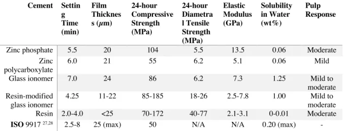
A review of the major cement classes can be seen in Table 1.
Cement Settin g Time (min) Film Thicknes s (µm) 24-hour Compressive Strength (MPa) 24-hour Diametra l Tensile Strength (MPa) Elastic Modulus (GPa) Solubility in Water (wt%) Pulp Response
Zinc phosphate 5.5 20 104 5.5 13.5 0.06 Moderate
Zinc polycarboxylate
6.0 21 55 6.2 5.1 0.06 Mild
Glass ionomer 7.0 24 86 6.2 7.3 1.25 Mild to
moderate Resin-modified
glass ionomer
4.25 11-22 85-185 18-26 2.5-7.8 1.00 Mild to
moderate
Resin 2.0-4.0 <25 70-172 40-77 2.1-3.1 0-0.01 Moderate
ISO 9917 27,28 2.5-8 25 (max) 50 N/A N/A 0.20 (max) -
Table 1: Typical properties of dental cements used for luting applications.29
Zinc phosphate cement is the oldest dental luting cement, first appearing in the literature
in 1879 by Pierce.23 The modern-day chemistry of the cement is very similar to the improved
formula made by Fleck in 1902.25It has a long record of clinical success and it is the standard by
which contemporary cements are compared. Zinc phosphate cement consists of a powder and
liquid which are mixed just before use. The powder contains more than 75% of zinc oxide and up
to 13% of magnesium oxide, which is added for its white color. The liquid contains a varying
mixture of phosphoric acid, water, and a small amount of aluminum phosphate. The formula
oxide, which reacts with the aluminum phosphate and forms zinc aluminophosphate gel on the
remaining undissolved zinc oxide particles. Zinc Phosphate had to me mixed on a glass slab,
kept cool but above the dewpoint so that no condensation on the glass would dilute the cement.
The consistency had to be mixed carefully so as not to be too viscous or too thin. After
placement of the restoration and after setting of the cement, excess cement could easily be
cleaned off as the cement does not bond to either tooth structure or restorative materials. The
retention was based purely on mechanical means.29 The main drawbacks with zinc phosphate are
its high acidity until it is set, which can lead to pulpal irritation; and high solubility.18 In spite of
high solubility and no bonding, however, zinc phosphate has proven to have an excellent track
record.30
Zinc polycarboxylate cement is a powder and liquid cement which is mixed immediately
before cementation. It sets via an acid-base reaction. The liquid is an aqueous solution of
polyacrylic acid and the powder is principally zinc oxide. It was the first dental cement with the
potential to form a chemical bond to tooth structure. This quality was an improvement over zinc
phosphate. The disadvantages of zinc polycarboxylate are that the viscosity could easily be
increased when mixing, causing incomplete seating of the restoration and it is also relatively
soluble in an acidic environment.29,31
Glass Ionomer Cement is a class of cements which are based on the reaction of glass
powder and polyacrylic acid. Wilson and Kent developed the first glass ionomer cement in 1969.
The liquid is composed of polyacrylic acid, and polyprotic carboxylic acid and the powder is a
fluoroaluminosilicate glass. It sets by an acid-base reaction. It is also able to bond to tooth
structure by chelation of the carboxyl groups of the polyacrylic acids with the calcium in the
after the cement has set, it will release fluoride ions. It is proposed that this can combat the
demineralization caused by biofilm and reduce the occurrence of secondary decay.29,31
The success of attaching unfilled resin to etched enamel gave rise to the concept of using
resins to bond indirect fixed prostheses to prepared abutment teeth. They consist of the adhesive
monomers HEMA, 4-META, carboxylic acids, and an organo- phosphate, such as MDP.
Countless resin cements are on the market. These cements are virtually insoluble in oral fluids,
but the brands vary widely in physical properties because of the variety and proportions of resins
and fillers in the formulas. Polymerization of the resin cement occurs by auto-cure (chemical
cure), light-cure, or dual-cure (both light- and auto-cure) mechanisms. The majority of resin
cements today are of the dual-cure variety.29
Resin modified glass ionomer cements replace port of the liquid component of
conventional glass ionomer cement with water-soluble methacrylate-based monomers. The
monomers can be polymerized by a chemical or light activation or both, and the glass ionomer
cement acid-base reaction will occur along with polymerization. This class of cement attempts to
combine the benefits of chemical bonding to the tooth surface with the insolubility of a resin
cement.29
Self-adhesive cements are the newest category of cement on the market. They are
formulated with specific adhesive monomers which are acidic enough to etch the tooth surface
sufficiently while curing that they eliminate the adhesive bonding agent. This greatly simplifies
the cementation process making it much less technique sensitive. They are a dual-cure cement
and are indicated for use on all full coverage restorations, inlays, and onlays regardless of
2.3. Solubility
Cements in the oral environment must continually endure exposure to acids produced by
foods, drinks or by microorganisms. Nearly all cements exhibit some degree of solubility in the
oral environment, except perhaps some resin cements. Of the current cements being used in
clinics, glass ionomer and resin modified glass ionomer cements exhibit the most solubility while
resin cements exhibit virtually no solubility.29 It is of note that the presence of porosity and voids
in the cement increase the rate of solubility in other classes of cements.32
2.4. Cements in This Study
2.4.1. RelyX Unicem 2 (Automix)
RelyX Unicem 2 is a self-adhesive resin-based dental cement (3M, St. Paul, Minnesota,
USA). The manufacturer advocates no pre-treatment of tooth surfaces. This is due to the
presence of self-etching phosphoric acid methacrylates which react with hydroxyapatite from
tooth structure, therefore no separate bonding agent is require. The setting reaction is started by
light and/or by a chemical reaction of the initiator system (dual-cure). The setting reaction is a
radical polymerization during which the single monomer molecules are chemically cross-linked
to form a three-dimensional polymer network.33 The cement has been recommended for luting all
metal-based and ceramic crowns, as well as partial coverage ceramic and indirect composite
restorations, with the exception of veneers.34 Good marginal adaptation of all-ceramic crowns
cemented with Rely-X Unicem 2 has been documented.20 The American Dental Association
states in Specification No. 8 that the film thickness should not exceed 25μm for Type 1
cements.35 RelyX Unicem 2 meets that specification with a film thickness that has been
2.4.2. RelyX Luting Plus (Automix)
RelyX Luting Plus Cement is a self-curing, radiopaque, fluoride-releasing, resin-modified
glass ionomer luting cement (3M, St. Paul, Minnesota, USA). It is indicated for the permanent
cementation of metal-based and strengthened-core ceramic restorations, posts, and orthodontic
appliances.
Two setting reactions occur, an acid-base reaction between the fluoroaluminosilicate
glass and the methacrylate functionalized polycarboxylic acid (this is the true glass ionomer
setting reaction) and a free radical polymerization. According to 3M, RelyX Luting Plus
Automix has an added photoinitiator. Therefore, the free radical polymerization reaction can be
initiated with a 5 second light cure, or it will takes place without light activation, therefore it is
dual-curing.37
Resin-modified glass-ionomer cements contain conventional glass-ionomer cement
components, i.e. glass and aqueous solutions of polyacids, as well as additional monomeric
ingredients, usually 2-hydroxyethyl methacrylate (HEMA). These monomers contribute to the
setting reaction of the cements. RelyX Luting Plus meets the ADA Specification No. 8 with a
film thickness that has been documented to be 19µm experimentally.36
3. History of Dental Ceramics
Ceramics were first introduced to dentistry in 1744 by Alexis Duchateau, a pharmacist
who, with the help of Parisian dentist Nicholas Dubois de Chemant, invented a porcelain formula
which was used to create the first porcelain denture. In 1808, Guiseppangelo Fonzi improved on
Chemant’s idea and fabricated the first porcelain denture teeth by firing the teeth to platinum
introductions of ceramic were not widely successful as these early formulations of porcelain
were very fragile. In 1962, Weinstein-Katz-Weinstein developed a new ceramic formula which
contained leucite. This allowed porcelain to be fired to cast metal copings by allowing the
thermal coefficients of expansion of the materials to be compatible.40 While a great improvement
in esthetics, larger demands for nonmetallic materials have steadily increased through the years.
While many materials have been produced with varying levels of success, in today’s clinic
zirconia, due to improvements in its formulations has had a surge in popularity among clinicians
for this material. Also, lithium disilicate, introduced in 1998, has overcome many of the
limitations of past generations, fracture strength potentially being its main weakness, but it
remains a popular choice for clinicians.
3.1. Ceramics Used in This Study
IPS e.max CAD – Ivoclar Vivodent - Lithium disilicate glass ceramic.
Classified as special silicate glass, this material is available in a pressable version and a
partially crystallized block for CAD/CAM design (IPS e.max CAD). It is a translucent material
which boasts a fairly high flexural strength (> 360 MPa) which is why this system continues to
be one of the most popular in terms of use.41 The CAD/CAM blocks are available in four
translucency levels (MO,LT,MT,HT) all in a crystalline intermediate stage. For this study, e.max
4. Marginal Fit
Marginal fit is an essential factor for clinical success of indirect restorations. Poor
marginal adaptation can result in dissolution of cement; increase plaque accumulation,
periodontal inflammation, and secondary caries.21 The study by Holmes, et al. (1989) measuring
the marginal fit of restorations states that marginal fit should be considered as the angular
combination of the vertical and horizontal error43. Christensen (1966) found that clinically
detectable subgingival margins in a range of 34-119 microns and supragingival margins were in
the range of 2-51 microns. No consensus yet exists regarding clinically acceptable marginal fit.
However, McLean (1971), among others, has suggested that 120 microns should be the limit for
clinically acceptable marginal discrepancies.44
4.1. Measurement of Margin Adaptation
Different measurement methods have been used among various studies across the
literature. The first and most widely used method is direct microscopic examination of the
marginal area. This method has two important disadvantages. First, identification of reference
points from which to measure can be difficult. Second, projection errors can occur which can
skew the measurements. Another popular method involves cross-sectioning cemented specimens,
then examining them with microscopy. The main issues with this method are that a limited
number of sections could be cut on any one specimen and it is a destructive process. The most
recent technique used is x-ray microtomography. This is a nondestructive technique which
delivers 2-dimensional and 3-dimensional imaging of the restoration and the die. It can provide
high resolution sections of the marginal area, which allows for many measurement sites and for
4.2. Measurement of Cement Porosity
Until the use of Micro-CT in dental research, this author is unaware of any research
conducted on the volumetric measurement of porosities within the cement layer which employs a
method that approximates the clinical situation of a die-preparation configuration. Namoto
(2004) used CT to analyze voids in the cylindrical cement specimens, proving that
Micro-CT could be used to effectively image dental cement with a high degree of resolution.15 Malkoç
(2015) demonstrated that Micro-CT is a nondestructive method of analysis that allows high
resolution of the dental cement, where porosities can be found between the prepared dentin and
the ceramic coping. They state that micro-CT was very useful for developing a standard method
to examine the number of porosities and where they were located under ceramic restorations in
vitro.14
4.3. Microleakage
Microleakage has been studied extensively since the first attempts to examine the
amalgam marginal contraction using microscopy were made by Tomes. The failure of the
adhesion or seal of the luting agent to the tooth structure has been a concern to dentists since the
advent of the luting agent.45 Microleakage is defined as the clinically undetectable passage of
bacteria, fluids, molecules, or ions between a cavity wall and the restorative material applied to
it.46 Countless different methods have been derived in an attempt to measure microleakage. In
1912 Harper submerged amalgam filled steel dies in water and air-pressure was applied to see if
bubbles escaped around the margins. Restored teeth have been subjected to bacterial cultures and
examined for the presence of bacteria past the margin or for secondary decay-like lesions.45 The
analysis. Direct microscopic observations have been made in vivo and in vitro. The importance
of microleakage is that voids in the cement layer can lead to an increase in the rate of cement
degradation and bond degradation.46
5. History of Imaging in Dental Research
Depending on the research being conducted, intra-oral radiographs including periapical,
bitewing and occlusal projections, as well as extra-oral radiographs including panoramic and
cone beam CT are commonly used. Their resolution, however, is limited and therefore other in
vitro modalities have been utilized in dental research. Scanning electron microscopes have very
high resolutions but can only visualize a surface in two dimensions. Computer tomography has
been in use as a medical imaging tool for many years. Later, the introduction of cone beam
computer tomography gave the dental practitioner the ability to image his patient in three
dimensions. However, technological advances in the area of computer tomography have allowed
researchers to conduct a three dimensional analysis of a specimen in vitro with Micro-CT at
resolutions that were impossible before.47
5.1. Cone Beam Computer Tomography
Computer Tomography has been in use in the medical field since the mid 1970’s. There
have been many advancements over the years, but the principal has remained relatively
unchanged. It creates a three dimensional image of the subject by taking many x-ray images,
which are imaged in thin axial slices, and the computer combines the images to form a digital,
three dimensional representation of the subject.48 Dentists have used a version of this technology
with visibility of details of only 500 microns, i.e. 0.5 mm.49
5.2. Micro Computer Tomography
Micro-CT, which utilizes differences in X-ray attenuation properties of materials to
reconstruct 3D structure, is similar to medical CT except it employs a much smaller field of view
with a high-resolution detector. The 3D images can have a voxel size down to 1µm or smaller.
Micro-CT is used to study diverse materials including bone, teeth, medical implants, textiles,
concrete and precious stones. Micro-CT reveals in great detail the internal structure of these
materials, such as the trabecular architecture within bone or grain within wood, allowing
quantitative analysis of properties such as density and volume.4 In dental research, it is useful as
a nondestructive method of analysis that allows for measurements in different sections and
distances along the specimen, providing reliable three-dimensional reconstructions. The main
drawbacks of Micro-CT are low resolution when materials have similar x-ray absorption
CHAPTER 2: MANUSCRIPT
1. Introduction
The basic function of dental cement has historically been to fill the space between the
prepared tooth and the restorative material being placed. The cement is expected to create a seal
between the oral environment and the internal aspects of the preparation/restorative interface to
prevent penetration of bacteria and degradation of the cement. An intimate fit and seal between
the tooth and restoration are integral to the long-term success of a restoration.31 A perfect cement
would have a very thin film thickness, the ability to adhere durably to both tooth structure and
restorative material, and also be able to stand up to both the chemical and physical abuse of the
oral cavity. However, all these things would be undermined if there were any voids created at the
margin during the cement cleanup process. Porosities incorporated into the materials may lead to
inhibition zones with unpolymerized materials, which may result in higher water solubility and
microleakage, which in turn, can lead to secondary caries, loss of retention, or periodontal
issues.14,20,22 As much as a manufacturer can improve their product, proper implementation of the
product is important. A luting agent which is highly technique sensitive is more likely to be less
effective. Clinicians need a clear process for cementation which, through extensive research, has
proven to be consistently effective.15
The purpose of this in vitro study is to use Micro Computed Tomography (Micro-CT) to
evaluate, in three dimensions, the volume of any voids left at the margin when the
manufactured by 3M were chosen for this study; RelyX Unicem 2, which is a dual-cure
self-adhesive resin cement, and RelyX Luting Plus, which is an auto-cure resin-modified glass
ionomer (3M, St. Paul, Minnesota, USA). Currently, there are no clear guidelines in the literature
for cement cleanup protocol, therefore, for this study we chose to follow 3M’s recommended
instructions for their dental cements. They recommend the same two optional methods for
cement cleanup for each cement to be studied. The first technique involves light-curing the
excess cement until tack-cure is reached and then removing the excess with a sharp sickle scaler.
The second technique involves waiting sufficient time for the excess cement to reach a partial
cure, and then removing the excess with a sharp sickle scaler.
It is understood that, during the cement cleanup process, the excess cement must “break
away” from the margin. What is not known is the quality of the cement at the margin
immediately afterward. In the past, research has been conducted using a variety of techniques to
evaluate the quality of a crown margin such as stereo microscopy, electron scanning microscopy,
and computed tomography, to name a few.2 However, there is very little research to date using
Micro-CT to evaluate voids at the margin in three dimensions and there is no consensus on what
volume of void is clinically significant in regards to the longevity of indirect restorations.12
Null Hypothesis: There will be no clinically significant difference in the average volume
per void between the methods tested for cement cleanup.
2. Materials and Methods
Two different cement cleanup techniques were evaluated for their ability to produce a
margin lacking voids which were open to the oral cavity. These two methods were used with two
these techniques and materials in three dimensions, focusing on the volumetric measurement of
any voids at the buccal margin exposed to the environment. Twenty specimens were divided into
four groups of five specimens. For each group, five prepared left mandibular first molar Ivorine
(Columbia Dentoform® Teaching Solutions, Lancaster, Pennsylvania, USA) dies were acquired
and individually designed lithium disilicate crowns were cemented to each die using the one of
two manufacturer recommended cementation techniques and using one of two different dental
cements. Those specimens were then scanned using a Micro-CT machine and volumetric
analysis of voids in the cement at the margin was performed.
2.1. Die Preparation
Twenty left mandibular first molar Ivorine typodont prepared dies were acquired from the
manufacturer. Each die was prepared by Ivorine according to Ivoclar’s specifications for Ivoclar
IPS e.max CAD lithium disilicate crowns (Ivoclar Vivadent, Schaan, Germany) with a
circumferential reduction of 1.5 mm, occlusal reduction of 1.5 mm, chamfer finish line of 1.0
mm and convergence angle of the axial walls of 6˚ per wall. The dies were milled to Ivoclar’s
specifications using CNC milling machines.
2.2. Digital Impression
A digital impression was made of each die using a 3Shape D810 digital laboratory
desktop scanner (3Shape, Copenhagen, Denmark) which has an accuracy of less than 7µm.50
Before scanning, the scanner was calibrated according to the manufacturer’s instructions and the
dies were lightly sprayed with CEREC® Optispray powder spray (Dentsply Sirona, Charlotte,
scanning, the scanning powder was removed from all the dies using flour pumice, water, and a
prophy cup to prepare them for cementation. The digital impression of each die and design of
each crown was conducted by an experienced prosthodontic resident.
2.3. Crown Design and Manufacture
An individually designed crown was made for each die using the 3Shape TRIOS Design
Studio software version 2.2.1 (3Shape, Copenhagen, Denmark). The design parameters were set
to 20µm space at the margin and an internal cement space of 60µm in accordance with the
instructions of the CAD/CAM milling machine. Each crown was designed so as not to violate
minimal material thicknesses of the Ivoclar IPS e.max CAD lithium disilicate material set by the
manufacturer. Axial and occlusal thicknesses were at least 1.5mm and thickness at the depth of
the margin was at least 1.0mm. The crowns were designed with proper occlusal anatomy.42 After
the design, each crown was milled in the same 5-axis Wieland Zenotec Select Hybrid milling
machine (Wieland Dental + Technik GmbH & Co. KG, Pforzheim, Germany) from individual
partially crystalized Ivoclar IPS e.max CAD lithium disilicate blocks. After milling, the crowns
were test fitted to their respective dies and examined under a microscope at 20x to ensure proper
seating. The margins were polished by an experienced operator to be clinically non-detectable,
which is standard laboratory procedure for finishing milled lithium disilicate margins. The
crowns were then crystalized using the manufacturer recommended crystallization cycle
parameters.42 Each crown and die were randomly assigned to their experimental group and
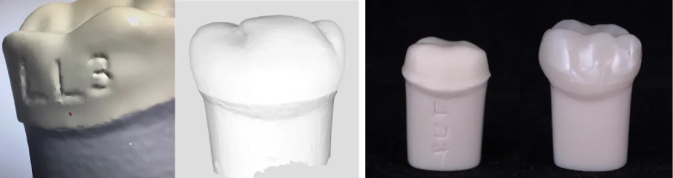
uniquely marked so that they would not be incorrectly paired before cementation. A design is
Figure 1: Picture of digital design Figure 2: Picture of die and cemented crown
2.4. Specimen Grouping
Each specimen consists of one lithium disilicate crown paired with its respective die. The
20 specimens were divided into 2 groups of 10 corresponding to the cement cleanup method
being evaluated. Each group was then divided into 2 subgroups of 5 specimens, each subgroup
corresponding to the cement being used. Group LL refers to light to tack-cure with RelyX Luting
Plus cement. Group LU refers to light to tack-cure with RelyX Unicem 2 cement. Group TL
refers to waiting an allotted time for partial set with RelyX Luting Plus cement. Group TU refers
to waiting an allotted time for partial set with RelyX Unicem 2 cement. (Figure 3)
Figure 3: Flow chart of specimen distribution
Specimens: N20
Light to Tack Cure
N10 Time to Partial Set N10
Group LU Light Cure – Unicem 2
N5
Group LL Light Cure – Luting Plus
N5
Group TU Time Cure – Unicem 2
N5
Group TL
2.5. Cementation Procedure
Cementation was executed by an experienced prosthodontics resident using 3M’s
recommended cementation methods for cementation of lithium disilicate restorations. Before
cementation the crowns were cleaned and prepared for cementation according to 3M’s protocol
for each cement to be used. The crowns to be cemented with the self-adhesive resin cement
(RelyX Unicem 2) were etched with 5% hydrofluoric acid gel for 20 seconds and then rinsed
with water and air dried. The crowns to be cemented with the resin modified glass ionomer
(RelyX Luting Plus) were rinsed with water and dried. No other preparation is indicated for the
restoration.
For all groups, enough volume of cement was applied to the internal surface of each
crown with a microbrush to ensure excess cement past the margin and then the crown was seated
on its Ivorine die with finger pressure for 3-4 seconds. The crown and die were then placed on
the bottom compression platen of an Instron 4411 Universal Testing Machine (Instron, Norwood
Massachusetts, USA). A custom acrylic resin positioning jig was used on the bottom platen in
order to position each die in the same, fixed position. A custom seating plate made of a layer of
light-body silicone over a layer of medium body silicone was affixed against the top compression
platen. This was used to apply a consistent, even seating pressure of 100N/mm2 across the
occlusal surface of each crown during cementation. Goracci et al described his pressure as the
mean finger pressure for crown cementation.51 The seating pressure was maintained for the
duration of the cement cleanup process.
2.6. Cement Cleanup
methods. 3M recommends two cement cleanup methods for both cements being used. The “Light
to Tack-Cure” method indicates using a light curing device to reach tack-cure, removal of the
excess cement in large chunks with a sharp sickle scaler, and then light curing to full cure. The
3M EliparTM DeepCure LED curing light was used in this experiment. The irradiance of the
curing light was measured at 1250 mW/cm2 with a radiospectrometer (MARC resin calibrator,
BlueLight Analytics, Halifax, Canada). The “Time to Partial Set” method indicates waiting a
specified amount of time for the cement to be sufficiently set for the excess cement to be
removed in large chunks with a sharp sickle scaler, and then allowing sufficient time to reach full
cure. The amount of time suggested for each method was different for the two cements. The
crowns were allowed to reach full cure under pressure of the Instron Machine.
2.6.1. Light to Tack-Cure Method Unicem 2 (Group LU)
For Group LU the buccal surface of the specimen was light cured for 2 seconds to reach
tack-cure. The excess cement was then removed with a sharp sickle scaler and then the specimen
was light cured for 20 seconds per surface to reach full cure.52
2.6.2. Light to Tack-Cure Method Luting Plus (Group LL)
For Group LL the buccal surface of the specimen was light cured for 5 seconds to reach
tack-cure. The excess cement was then removed with a sharp sickle scaler and then the specimen
2.6.3. Time to Partial Set method Unicem 2 (Group TU)
For Group TU the cement was allowed to partially set by waiting 3 minutes after the
crown was seated. The excess cement was then removed with a sharp sickle scaler and then the
specimen was light cured for 20 seconds per surface to reach full cure.52
2.6.4. Time to Partial Set Method Luting Plus (Group TL)
For Group TL the cement was allowed to partially set by waiting 2 minutes. The excess
cement was then removed with a sharp sickle scaler and then the specimen was allowed to cure
for a total of 5 minutes after placement to reach full cure.53
2.7. Micro-CT Scan
All 20 specimens were scanned for marginal void analysis using a microcomputed
tomography scanner (Scanco micro-CT 40 scanner; Scanco Medical AG, Zurich, Switzerland) at
the Biomedical Research Imaging Center (BRIC) at the University of North Carolina. Digital
Imaging and Communications in Medicine (DICOM) files were generated using a 70-kilovolt
peak (kVp); the voxel size for the slice width was 8μm.
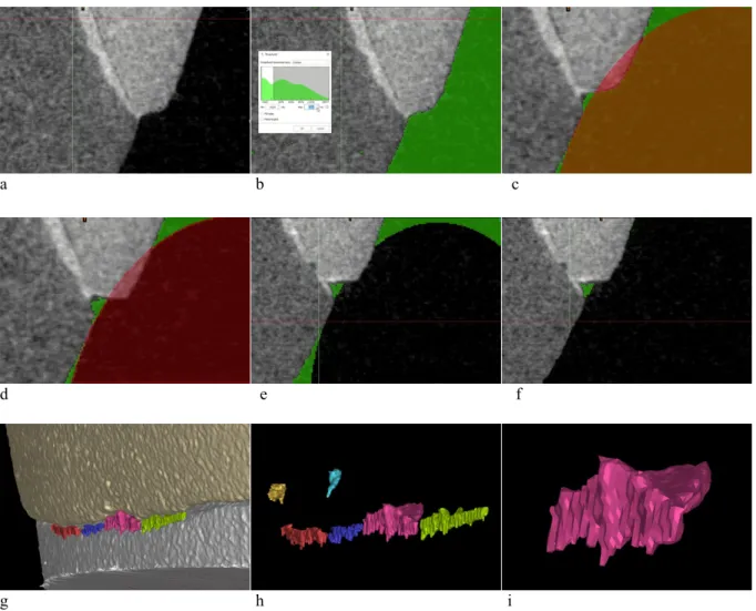
2.8. Segmentation and Measurement
Each sample was analyzed using the Materialise MIMICS (Materialise Interactive
Medical Image Control System) version 22.0 software (Materialise Medical, Leuven, Belgium).
MIMICS is an image processing software which can be used to create 3D surface models, 3D
design, 3D measurements and analysis of images from DICOM data. For each sample, the buccal
chosen for analysis. A trained operator used the software to section out from the sample any void
at the margin. A void was defined as space between the crown and the die which was not filled
with cement that extended inward past a virtual straight line drawn from outermost edge of the
die margin, in line with the emergence profile of the die, to the restoration. The first step in
separating out the voids is to apply a threshold range that represents air to create a mask. The
operator manually demarcated separation of the void from the air outside the crown. The
software automatically extrapolates from one demarcation to the next so that the operator could
make delineations approximately every 30-40µm instead of every 8µm slice. Once the voids
were separated from the mask of the air the software converted the masks of the voids into
Stereolythography (.STL) three dimensional objects. The MIMICS software was then used to
measure the volume of each .STL object which represents a void. The width of each void was
a b c
d e f
g h i
Figure 4: Insight into segmentation and measurements
a) 2D slice of sample at margin; b) Intensity threshold adjustment; c-e) Selective differentiation of voids from environment; f) Mask of environment discarded leaving only masks of voids; g) 3D model of die, crown, and voids, h) 3D models of separate voids; i) Final 3D model of segmented void.
2.9. Statistical Analysis
Analysis of the data was conducted by the Wilcoxon Ranked Sum due to the lack of a
normal distribution and the heterogeneity of the dataset. Outcome of interest was the volume per
void, however, the number of voids and the width per void were also analyzed using the
Wilcoxon Ranked Sum. Each outcome was compared separately by type of cement and by
3. Results
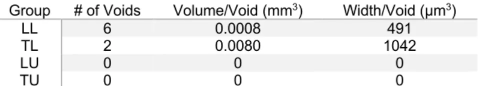
3.1. Average Values by Group
For an overview, the average values for each of the four individual groups are provided in
Table 2. The median value of “0” for the average number of voids, average volume/void, and
average width/void for the LU and TU groups may be a product of a low sample size.
Group # of Voids Volume/Void (mm3) Width/Void (µm3)
LL 6 0.0008 491
TL 2 0.0080 1042
LU 0 0 0
TU 0 0 0
Table 2: The median values for each outcome and group.
3.2. Comparison of Cleanup Method
The average values, the lower quartile, and the upper quartile for the method of cement
cleanup irrespective of cement type are represented in Table 3. There was no statistically
significant between the two cement cleanup methods for all outcomes.
Method # of Voids
Median (Q1, Q3)
Volume/Void (mm3)
Median (Q1, Q3)
Width/Void (µm3)
Median (Q1, Q3)
Light to Tack-Cure 2 (0,6) 0.0004 (0, 0.0008) 279 (0, 492)
Time to Partial Set 1 (0,4) 0.0008 (0, 0.008) 235 (0, 1042)
P value 0.67 0.91 0.97
Table 3: The median values for each outcome by method of cement cleanup.
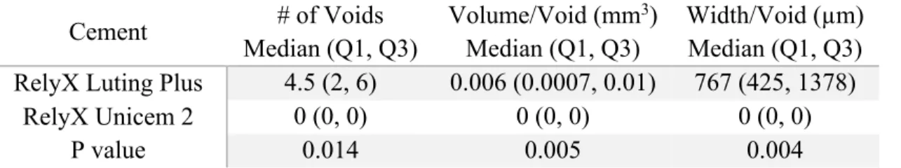
3.3. Comparison of Cements
The average values, the lower quartile, and the upper quartile for the cement used
irrespective of method of cement cleanup are provided in Table 4. There was a statistically
median was larger for RelyX Luting Plus than for RelyX Unicem 2.
Cement # of Voids
Median (Q1, Q3)
Volume/Void (mm3) Median (Q1, Q3)
Width/Void (µm) Median (Q1, Q3) RelyX Luting Plus 4.5 (2, 6) 0.006 (0.0007, 0.01) 767 (425, 1378)
RelyX Unicem 2 0 (0, 0) 0 (0, 0) 0 (0, 0)
P value 0.014 0.005 0.004
Table 4: The median values for each outcome by type of cement.
4. Discussion
The purpose of this study was to use Micro-CT to compare two cement cleanup
procedures which were conducted with a resin modified glass ionomer and a self-adhesive resin
cement, making volumetric measurements of voids at the margins of full coverage restorations.
Our results show that there was no statistically significant difference in the volume per void
between the two cleanup methods, irrespective of cement type. Therefore, we must accept our
Null Hypothesis.
However, during the course of our research we analyzed other outcomes, such as number
of voids and width per void. It is interesting to note that no statistically significant difference
existed for all outcomes (number of voids, volume per void, and width per void) when
comparing the two cement cleanup methods. Another interesting finding, although not the
original focus of this study, is that a statistically significant difference in the number of voids, the
volume per void and the width per void were found between the two cements, irrespective of the
cleanup method used, the RMGI having the higher value for all outcomes.
The results of this study constitute a contribution to the advancement in research
methodology, considering that, to date only one study has measured the volume of voids at the
network full-coverage restorations, using digital scans and CAD/CAM technology, and cemented
with RelyX Unicem. They found an average volume of marginal porosity between 0.363mm3
and 0.517mm3.12 In their study, though, the entire margin was scanned and the excess cement
was wiped from the margins with a foam pellet before it was set instead of following the
manufacturer’s recommended method of cement cleanup, making our studies difficult to
compare. However, if our data were extrapolated to estimate the total void for the entire margin,
we find an average void of 0.051 mm3 which is an order of magnitude lower in average
volume/sample. The use of Micro-CT to evaluate voids at the margin allowed the quantification
of several parameters in three dimensions in a non-destructive manner. With a resolution of
8µm/voxel and sufficient differences in x-ray attenuations of the crown, die, and cement layer,
we were able to clearly identify and measure the number, volume, and width of each void present
along the entire margin. This study focused on the buccal margin due to its ease of access within
our cementation rig and to the fact that the cleanup method employed experimentally does not
translate to the clinical method of cleaning cement interproximally. Nevertheless, we may come
to some conclusions. It is important to remember the clinically relevant fact that open voids in
the marginal areas can support the penetration of fluids and bacteria in the cement space leading
to cement dissolution, secondary caries, periodontal inflammations up to loss of restoration or
tooth.54 If we consider the clinical implications of our findings, they suggest that the type of
cement has a greater influence on the outcome of void formation at the margin than does the
cement cleanup method. Our results would suggest that the clinician may choose either method
of cement cleanup investigated in this study and would potentially choose the adhesive resin
cement over the resin modified glass ionomer. Also, the successful implementation of a novel
of researching hitherto uninvestigated aspects of dental practices and materials.
The experiments in this study, and those that plan to use Micro-CT, must be limited to the
in vitro design. Standardized laboratory conditions were employed to minimize bias as much as
possible. Factors such as blood, saliva, gingiva, and adjacent teeth in the clinical setting are
certainly a limitation of the study and would certainly impact the cementation process in vivo. It
is recognized that the sample sizes in this study are inadequate to lend substantial statistical
power to the results.
The first suggestion of ways to expand on this experiment would be to increase the
sample size and variety of dental cements investigated. This would greatly improve the power of
the results obtained. Also, while milled lithium disilicate was adequate for this study, it may be
worthwhile to attempt to replicate this study using polymer infiltrated ceramic network crowns
because their x-ray attenuation levels were shown to be superior when differentiating the resin
die, the cement layer, and the crown from each other.12 Lastly, it may be beneficial to incorporate
an additional experimental method of cement cleanup which is widely performed clinically. This
method involves placing the crown and immediately wiping the unset cement away from the
margin before it has begun to set. It would be interesting to compare accepted manufacturer’s
REFERENCES
1. Manhart J, Chen H, Hamm G, Hickel R. Buonocore Memorial Lecture. Review of the clinical
survival of direct and indirect restorations in posterior teeth of the permanent dentition. Oper Dent. 2004 Oct;29(5):481–508.
2. Contrepois M, Soenen A, Bartala M, Laviole O. Marginal adaptation of ceramic crowns: a
systematic review. J Prosthet Dent. 2013 Dec;110(6):447–454.e10.
3. Pelekanos S, Koumanou M, Koutayas S-O, Zinelis S, Eliades G. Micro-CT evaluation of the
marginal fit of different In-Ceram alumina copings. Eur J Esthet Dent. 2009;4(3):278–92.
4. Scanco Micro-CT general information [Internet]. Scanco.ch. [cited 2020 Apr 15]. Available from:
http://www.scanco.ch/en/support/faq-general.html
5. Amano M, Agematsu H, Abe S, Usami A, Matsunaga S, Suto K, et al. Three-dimensional analysis
of pulp chambers in maxillary second deciduous molars. J Dent. 2006 Aug;34(7):503–8.
6. Fanuscu MI, Chang T-L. Three-dimensional morphometric analysis of human cadaver bone:
microstructural data from maxilla and mandible. Clin Oral Implants Res. 2004 Apr;15(2):213–8.
7. Nevins ML, Camelo M, Rebaudi A, Lynch SE, Nevins M. Three-dimensional micro-computed
tomographic evaluation of periodontal regeneration: a human report of intrabony defects treated with Bio-Oss collagen. Int J Periodontics Restorative Dent. 2005 Aug;25(4):365–73.
8. Lee B-S, Lee C-C, Wang Y-P, Chen H-J, Lai C-H, Hsieh W-L, et al. Controlled-release of
tetracycline and lovastatin by poly(D,L-lactide-co-glycolide acid)-chitosan nanoparticles enhances periodontal regeneration in dogs. Int J Nanomedicine. 2016 Jan 18;11:285–97.
9. Bissinger O, Probst FA, Wolff K-D, Jeschke A, Weitz J, Deppe H, et al. Comparative 3D micro-CT
and 2D histomorphometry analysis of dental implant osseointegration in the maxilla of minipigs. J Clin Periodontol. 2017 Apr;44(4):418–27.
10. Neves FD, Prado CJ, Prudente MS, Carneiro TAPN, Zancopé K, Davi LR, et al. Micro-computed tomography evaluation of marginal fit of lithium disilicate crowns fabricated by using chairside CAD/CAM systems or the heat-pressing technique. J Prosthet Dent. 2014 Nov;112(5):1134–40.
11. Duqum IS, Brenes C, Mendonca G, Carneiro TAPN, Cooper LF. Marginal Fit Evaluation of CAD/CAM All Ceramic Crowns Obtained by Two Digital Workflows: An In Vitro Study Using Micro-CT Technology. J Prosthodont. 2019 Dec;28(9):1037–43.
12. Dauti R, Cvikl B, Lilaj B, Heimel P, Moritz A, Schedle A. Micro-CT evaluation of marginal and internal fit of cemented polymer infiltrated ceramic network material crowns manufactured after conventional and digital impressions. J Prosthodont Res. 2019 Jan;63(1):40–6.
14. Malkoç MA, Sevimay M, Tatar İ, Çelik HH. Micro-CT Detection and Characterization of Porosity in Luting Cements. J Prosthodont. 2015 Oct;24(7):553–61.
15. Nomoto R, Komoriyama M, McCabe JF, Hirano S. Effect of mixing method on the porosity of encapsulated glass ionomer cement. Dent Mater. 2004 Dec;20(10):972–8.
16. Stock SR. Microcomputed tomography: methodology and applications. CRC Press; 2018.
17. Mesu FP. Degradation of luting cements measured in vitro. J Dent Res. 1982 May;61(5):665–72.
18. Rosenstiel SF, Land MF, Crispin BJ. Dental luting agents: A review of the current literature. J Prosthet Dent. 1998 Sep;80(3):280–301.
19. Kydd WL, Nicholls JI, Harrington G, Freeman M. Marginal leakage of cast gold crowns luted with zinc phosphate cement: an in vivo study. J Prosthet Dent. 1996 Jan;75(1):9–13.
20. Ibarra G, Johnson GH, Geurtsen W, Vargas MA. Microleakage of porcelain veneer restorations bonded to enamel and dentin with a new self-adhesive resin-based dental cement. Dent Mater. 2007 Feb;23(2):218–25.
21. Alani AH, Toh CG. Detection of microleakage around dental restorations: a review. Oper Dent. 1997 Aug;22(4):173–85.
22. Felton DA, Kanoy BE, Bayne SC, Wirthman GP. Effect of in vivo crown margin discrepancies on periodontal health. J Prosthet Dent. 1991 Mar;65(3):357–64.
23. Pierce CN. Filling materials of oxide of zinc and glacial phosphoric acid. Dent Cosmos. 1897;21:696.
24. Rosner D. Function, placement, and reproduction of bevels for gold castings. J Prosthet Dent. 1963 Nov;13(6):1160–6.
25. Fleck H. The chemistry of oxyphosphates. Dent Items Interest. 1902;906–35.
26. Lane JG. Crowns. Dent Dig. 1910 Feb;16(2):71–82.
27. ISO. ISO 9917-1: dentistry-water-based cements—part 1: powder/liquid acid–base cements. International Organization for Standardization, Geneva, Switzerland. 2007;
28. ISO. ISO 9917-2: dentistry-water-based cements—part 2: resin-modified cements. International Organization for Standardization, Geneva, Switzerland. 2010;
29. Anusavice KJ. Phillips’ science of dental materials. 12th ed. Elsevier/saunders; 2012.
30. Donovan TE, Cho GC. Contemporary evaluation of dental cements. Compend Contin Educ Dent. 1999 Mar;20(3):197–9, 202.
31. Ramaraju DV S, Krishna Alla R, Ramaraju Alluri V, Makv R. A review of conventional and contemporary luting agents used in dentistry. AJMSE. 2014 Aug 12;2(3):28–35.
2012 Oct;28(10):e187-98.
33. RelyX Unicem 2 Technical Data Sheet [Internet]. [cited 2020 Apr 14]. Available from:
https://multimedia.3m.com/mws/media/669183O/3m-relyx-unicem-2-automix-self-adhesive-resin-cement-technical-data-sheet.pdf
34. Behr M. Marginal adaptation in dentin of a self-adhesive universal resin cement compared with well-tried systems. Dental Materials. 2004 Feb 1;20(2):191–7.
35. Revised american national standards institute/american dental association specification no. 8 for zinc phosphate cement. The Journal of the American Dental Association. 1978 Jan;96(1):121–3.
36. Sulaiman TA, Abdulmajeed AA, Altitinchi A, Ahmed SN, Donovan TE. Physical Properties, Film Thickness, and Bond Strengths of Resin-Modified Glass Ionomer Cements According to Their Delivery Method. J Prosthodont. 2019 Jan;28(1):85–90.
37. RelyX Luting Plus Technical Product Profile [Internet]. [cited 2020 Apr 14]. Available from: https://multimedia.3m.com/mws/media/250363O/3m-relyx-luting-cement-technical-product-profile.pdf
38. Kelly JR, Benetti P. Ceramic materials in dentistry: historical evolution and current practice. Aust Dent J. 2011 Jun;56 Suppl 1:84–96.
39. Rosenblum MA, Schulman A. A review of all-ceramic restorations. J Am Dent Assoc. 1997 Mar;128(3):297–307.
40. Weinstein M, Katz S, Weinstein AB. Permanent Manufacturing Corporation, assignee. Fused Porcelain-to-Metal Teeth. US Patent No 3,052,982. 1962 Sep 11;
41. Willard A, Gabriel Chu T-M. The science and application of IPS e.Max dental ceramic. Kaohsiung J Med Sci. 2018 Apr;34(4):238–42.
42. Ivoclar e.max CAD solutions chairside [Internet]. IvoclarVivadent. [cited 2020 Apr 14]. Available from:
https://www.ivoclarvivadent.us/mam/celum/celum_assets/9514750640158_IPS_e-max_CAD_Monolithic_Solutions_Chairside_pdf_4774.pdf?2
43. Holmes JR, Bayne SC, Holland GA, Sulik WD. Considerations in measurement of marginal fit. J Prosthet Dent. 1989 Oct;62(4):405–8.
44. McLean JW, von Fraunhofer JA. The estimation of cement film thickness by an in vivo technique. Br Dent J. 1971 Aug 3;131(3):107–11.
45. Taylor MJ, Lynch E. Microleakage. J Dent. 1992 Feb;20(1):3–10.
46. Kidd EAM. Microleakage : a review. J Dent. 1976 Sep;4(5):199–206.
47. Boeddinghaus R, Whyte A. Current concepts in maxillofacial imaging. Eur J Radiol. 2008 Jun;66(3):396–418.
49. Brüllmann D, Schulze RKW. Spatial resolution in CBCT machines for dental/maxillofacial applications-what do we know today? Dentomaxillofac Radiol. 2015;44(1):20140204.
50. Factsheet 3Shape D800/810. [cited 2020 Apr 14]; Available from:
http://s3-eu-west-1.amazonaws.com/core3d-website/content/pdfs/core3dcentres-3shape-d800_810-product-sheet.pdf
51. Goracci C, Cury AH, Cantoro A, Papacchini F, Tay FR, Ferrari M. Microtensile bond strength and interfacial properties of self-etching and self-adhesive resin cements used to lute composite onlays under different seating forces. J Adhes Dent. 2006 Oct;8(5):327–35.
52. RelyX Unicem 2 Automix Technique Guide [Internet]. [cited 2020 Apr 15]. Available from: https://multimedia.3m.com/mws/media/684611O/3m-relyx-unicem-2-automix-self-adhesive-resin-cement-technique-guide.pdf
53. RelyX Luting Plus Technique Guide [Internet]. [cited 2020 Apr 15]. Available from:
https://multimedia.3m.com/mws/media/735193O/3m-relyx-luting-plus-automix-resin-modified-glass-ionomer-cement-technique-guide.pdf