Role of Endoplasmic Reticulum Stress in Cystic
Fibrosis–Related Airway Inflammatory Responses
Carla M. P. Ribeiro1 and Richard C. Boucher1
1
Cystic Fibrosis/Pulmonary Research and Treatment Center and the Department of Medicine, University of North Carolina at Chapel Hill, Chapel Hill, North Carolina
Chronic airway infection and inflammation are hallmarks of cystic fibrosis (CF) pulmonary disease. The altered airway environment resulting from infection and inflammation can affect the innate defense of the airway epithelia. Luminal bacterial and inflammatory stimuli trigger an adaptation in human airway epithelia, character-ized by a hyperinflammatory response to inflammatory mediators, which is mediated by an expansion of the endoplasmic reticulum (ER) and its Ca21stores. Recent studies demonstrated that a form of
ER stress, the unfolded protein response (UPR), is activated in airway epithelia by bacterial infection–induced airway inflammation. UPR-dependent signaling is responsible for the ER Ca21store
expansion-mediated amplification of airway inflammatory responses. These studies highlight the functional importance of the UPR in airway inflammation and suggest that targeting the UPR may be a thera-peutic strategy for airway diseases typified by chronic inflammation. This article reviews the contribution of airway epithelia to airway inflammatory responses, discusses how expansion of the ER Ca21
stores in inflamed airway epithelia contributes to airway inflamma-tion, describes the functional role of the UPR in these processes, and discusses how UPR activation might be relevant for CF airways inflammatory disease.
Keywords:airway inflammation; airway epithelia; cystic fibrosis; endo-plasmic reticulum calcium stores; unfolded protein response
Chronic airway infection and inflammation are hallmarks of cystic fibrosis (CF) pulmonary disease. The altered airway en-vironment resulting from infection and inflammation can affect the airways epithelial innate defense. In accord with this notion, we have described an adaptive response in human bronchial epithelia (HBE) triggered by luminal bacterial and inflamma-tory stimuli. This HBE adaptation reflected a hyperinflamma-tory response to inflammahyperinflamma-tory mediators (e.g., increased uridine triphosphate [UTP] or bradykinin [BK]-induced IL-8 secretion, which was mediated by an expansion of the endoplasmic re-ticulum [ER] and its Ca21stores) (1, 2). Our previous studies suggested that a form of ER stress, the unfolded protein re-sponse (UPR), is activated in airway epithelia by bacterial infection–induced airway inflammation and mediates airway inflammatory responses such as secretion of inflammatory mediators.
In this article, we review the contribution of airway epithelia to airway inflammatory processes, discuss how expansion of the ER Ca21 stores in inflamed airway epithelia contributes to airway inflammation, and describe the functional importance of the UPR in these processes. In addition, we discuss recent findings suggesting that activation of the UPR is functionally
important for the pathophysiology of CF airways inflammatory disease. Some of the results of these studies have been pre-viously reported in the form of abstracts (3, 4).
THE CONTRIBUTION OF AIRWAY EPITHELIA TO AIRWAY INFLAMMATION IN CHRONICALLY INFECTED AND INFLAMED AIRWAYS
CF is a multiorgan disease caused by mutations in the cystic fibrosis transmembrane conductance regulator (CFTR). In airway epithelia of patients with CF, the absence of CFTR-mediated Cl2secretion coupled with increased Na1absorption (5) results in dehydration of the periciliary layer (6), decreased mucus clearance, and accumulation of thickened mucus in airway lumens. As a consequence of these alterations, CF air-ways are plagued by persistent infection (7–10) and inflamma-tion (9, 11–13). CF airways disease is associated with increased cytokine concentrations of IL-1, IL-6, IL-8, and tumor necrosis factor (TNF)-a, whose syntheses are dependent on activation of the transcription factor nuclear factor (NF)-kB (14–16), and neutrophil infiltration (17). The CF airway epithelium plays a central role in the airway inflammatory response, as suggested by studies showing persistent activation of NF-kB (18), elevated production of IL-6 and IL-8, and decreased secretion of antiinflammatory factors by CF airway epithelia (18 –21). These observations suggested that alterations in CF airway epithelial cytokine production contribute to the inflammatory response of CF airways.
THE ROLE OF INTRACELLULAR Ca21MOBILIZATION
IN AIRWAY EPITHELIAL SECRETION OF INFLAMMATORY MEDIATORS
The role of intracellular Ca21(Ca21
i) signaling in inflammatory
responses is well established (22). Inflammatory and infectious stimuli can activate phospholipase C (23, 24) and increase Ca21
i
through two pathways: (1) inositol 1,4,5-trisphosphate–induced Ca21 release from the ER, which is the major Ca21 storing, buffering, and signaling compartment within cells, and (2) Ca21 entry mediated by plasma membrane channels (25–27). Ca21
i
mobilization resulting from heterotrimeric G protein–coupled receptor activation by inflammatory mediators or infectious agents is functionally relevant to inflammatory responses in several systems, including airway epithelia (22).
Activation of G protein–coupled receptors results in Ca21
i
mobilization associated with activation of NF-kB and secretion of inflammatory mediators (28–31). In addition, infectious agents, or their products, activate NF-kB in a Ca21
i
mobiliza-tion–dependent way (32–34). The proinflammatory mediator BK triggers Ca21
imobilization (35) and induces IL-8 secretion
in non-CF and CF human airway epithelia (2, 36). Likewise, activation of airway epithelial purinoceptors by UTP results in increased IL-8 secretion (2). Furthermore, the CF pathogens
Pseudomonas aeruginosa andStaphylococcus aureus promote IL-8 secretion by a Ca21
imobilization–dependent mechanism
in airway epithelial cells (37). The delay between Ca21
i
(Received in original form January 28, 2010; accepted in final form August 17, 2010)
Supported by grants RIBEIR00Z0, RIBEIR00G0, and RIBEIR07G0 from the Cystic Fibrosis Foundation (C.M.P.R.) and grant HL34322 from the National Institutes of Health (R.C.B.).
Correspondence and requests for reprints should be addressed to Carla M. P. Ribeiro, Ph.D., CB #7248, 7013 Thurston-Bowles Building, Chapel Hill, NC 27599-7248. E-mail: [email protected]
Proc Am Thorac Soc Vol 7. pp 387–394, 2010 DOI: 10.1513/pats.201001-017AW
mobilization and the onset of cytokine secretion reflects the kinetics of activation of Ca21
i–dependent transcription (38) and
protein translation. After rises in Ca21
i, NF-kB is freed from
IkB inhibition and translocates to the nucleus, where it can reside for tens of minutes, resulting in persistent transcriptional activation (38), despite the relaxation of Ca21
i levels toward
baseline levels, as occurs in human airway epithelia (1, 2). Ca21
isignals induced by airway epithelial activation by
in-flammatory or bacterial factors can affect airway inin-flammatory responses by affecting mucin secretion. For example, purino-ceptor activation with ATP, released in part by inflammatory cells (39), triggers mucin secretion in airway goblet cells (40– 42).
THE RELATIONSHIP AMONG AIRWAY INFECTION AND
INFLAMMATION, ER/Ca21STORE EXPANSION, AND
HYPERINFLAMMATORY AIRWAY EPITHELIAL RESPONSES
Although the two phases of Ca21
imobilization (i.e., ER Ca21
release and Ca21 influx) induced by inflammatory and in-fectious stimuli can act in concert to modulate NF-kB, the quantity of ER-releasable Ca21is a major factor controlling the magnitude of Ca21
i signals and, thus, NF-kB activation.
For instance, presenillin mutations in Alzheimer’s disease have been associated with increases in ER size, Ca21 stores, and cytokine production (43, 44). The role of ER-sequestered Ca21in inflammatory responses is further supported by studies illustrating a link between infection and ER expansion. For example, viral infection promotes ER proliferation associated with the massive production of viral proteins (45–47), and bacterial infection increases ER size (48).
These data led to the hypothesis that airway epithelia exposed to infectious and inflammatory stimuli exhibited increased ER mass and ER Ca21 storage, with resulting larger ER-derived Ca21
isignals triggered by external stimuli mediating increased
Ca21
i–dependent transcriptional activity of inflammatory
medi-ators. Indeed, it has been shown that apical purinoceptor (P2Y2)
or BK receptor activation induced greater Ca21
isignals in
short-term (6–11 d old) primary cultures of CF versus normal HBE (1), which resulted from an increased density and Ca21 storage capacity of the apical ER (1). The ER expansion was similar in native airway epithelia from patients with CF and patients with primary ciliary diskynesia who had chronic airway infection and inflammation (1), was independent of ER retention of mutated DF508 CFTR (1), revertedin vitrowith time in the absence of infection and inflammation (1), and was induced in normal HBE by luminal exposure to supernatant from mucopurulent material (SMM) from human CF airways infected withP. aeruginosaand
Staphyloccocus aureus(SMM contains infectious and inflamma-tory factors derived from bacteria, inflammainflamma-tory cells and inflamed epithelia [1, 2]).
Short-term primary cultures of DF508 CF HBE exhibited hyperinflammation (e.g., an amplified mucosal BK-dependent IL-8 secretion), which was lost in long-term (30–40 d-old) cultures, suggesting that this phenotype can revert to normal and be dissociated from theDF508 CFTR mutation (2). A CF-like hyperinflammatory response could be induced in long-term cultures of normal HBE by luminal exposure to SMM (2). Thus, the increased BK-induced IL-8 secretion in short-term primary cultures of CF HBE was associated with larger ER Ca21stores and increased BK-triggered, ER-derived Ca21
isignals, and the
loss of the hyperinflammatory phenotype in long-term CF HBE correlated with the reversal of ER Ca21 stores and ER-dependent Ca21
isignals to normal levels (1, 2). Together, these
published observations led to the conclusion that the
up-regulation of the ER Ca21stores in inflamed airway epithelia reflected an epithelial adaptation to chronic airway infection and inflammation (1, 2).
RELEVANCE OF AIRWAY INFLAMMATION–INDUCED ACTIVATION OF THE UPR IN AIRWAY EPITHELIA
Previous studies have demonstrated that luminal infection increases total intracellular protein synthesis in HBE, reflecting the increased epithelial synthesis of inflammatory mediators (2). The up-regulation of protein synthesis in infected and inflamed airway epithelia affects ER function because the increased load of new, unfolded inflammatory mediators and defensive factors on the protein folding machinery alters ER homeostasis and triggers an ER stress response, the UPR (49–55). In eukaryotic cells, three different mechanisms have evolved to deal with the accumulation of unfolded proteins in the ER: (1) transcriptional induction of genes whose functions are associated with increas-ing the ER volume and capacity for protein foldincreas-ing, (2) trans-lational attenuation to decrease the ER protein folding load, and (3) degradation of misfolded proteins. Thus, the UPR plays a key role in the folding, processing, export, and degradation of proteins originating from the ER, and it can be regarded as an adaptive, cell survival mechanism. However, if one or all of these mechanisms cannot relieve cell stress resulting from accu-mulation of unfolded proteins in the ER, the cell will undergo apoptosis or necrosis (50, 51, 53).
Eukaryotic cells exhibit three major UPR-sensing pathways, namely inositol-requiring enzyme 1 (IRE1), activating tran-scription factor 6 (ATF6), and PKR-like ER kinase/pancreatic eIF2akinase (PERK), whose activation during ER stress result in downstream activation of different signaling pathways. An additional review of UPR signaling is provided in Ron and Walter (56). Changes in ER homeostasis driven by increases in cellular protein synthetic rates, such as those experienced during airway epithelial inflammation, are transduced in the ER by sensors that detect the higher levels of unfolded, nascent proteins. These ER stress sensors activate signaling pathways responsible for the expression of several genes associated with the UPR. In mammalian cells, the requirement for increased folding of proteins is detected by two transmembrane ER stress sensors: IRE1 (57, 58), which exists in two isoforms, aandb, and ATF6 (50, 53, 59). Recent studies have shown that IRE1 signaling plays a key role in gut and airway inflammatory responses (60, 61). UPR activation leads to IRE1 dimerization and activation of its C-terminal endoRNase activity (51, 53). The IRE1 endoRNase activity splices the leucine zipper tran-scription factor X-box binding protein-1 (XBP-1) mRNA via removal of a 26-nucleotide intron, resulting in a frameshift of the XBP-1 mRNA transcript (62, 63). The spliced XBP-1 mRNA is translated into a transcription factor that up-regulates genes encoding ER chaperones (52, 62–64). Albeit mediated by a different mechanism of activation as compared with that from IRE1, activation of ATF6 also leads to the up-regulation of ER chaperones that facilitate the folding requirements of newly synthesized proteins (56).
activation of the UPR, and ER expansion (65, 71). The high rate of Ig secretion by plasma cells requires ER expansion (70), a process dependent on XBP-1 mRNA splicing (65). These find-ings agree with earlier data suggesting a role for the UPR in the coordination of the synthesis of phospholipids and new mem-branes and the up-regulation of a wide spectrum of genes of the secretory pathway (72, 73). Previous studies have demonstrated that the spliced XBP-1 promotes phospholipid biosynthesis, which is necessary for ER expansion (66–68). After UPR ac-tivation, new membranes are produced, and the resulting increased ER volume serves to dilute unfolded proteins and prepare the compartment to receive newly synthesized folding components, thereby restoring ER homeostasis (73). In addition to its role in ER expansion, the spliced XBP-1 triggers increases in protein synthesis, ribosomal number, lysosomal content, Golgi compartment, mitochondrial mass and function, and cell size (66). The function of spliced XBP-1 in coordinating structural and functional features characteristic of secretory cells is relevant for inflammatory responses of airway epithelia. Besides IRE1 and ATF6, PERK serves as an additional sensor of ER stress (50, 53, 59). PERK has a lumenal domain similar to that of IRE1 and a cytoplasmic domain similar to that of other eIF2a kinases. UPR activation stimulates PERK’s kinase activity, resulting in PERK-mediated phosphorylation of eIF2aand attenuation of general protein synthesis (59). How-ever, phosphorylation of eIF2ais also associated with selective translation and expression of the activating transcription factor 4 (ATF4) (74). Of importance for airway inflammatory re-sponses, ATF4 can induce the transcription of genes associated with the improvement of cellular metabolism and survival (e.g., amino acid import, glutathione biosynthesis, and resistance to oxidative stress) (75).
The following sections focus on IRE1-dependent XBP-1 signaling and PERK-mediated activation of the ATF4 pathway because recent data have uncovered key functional roles of these branches of the UPR in airway inflammatory responses in models relevant to CF airways disease.
AIRWAY INFLAMMATION INDUCES XBP-1 mRNA SPLICING IN CF HUMAN AIRWAYS AND IN INFLAMED
MURINE AIRWAYSIN VIVO
The role of XBP-1 mRNA splicing in coordinating structural and functional features characteristic of secretory cells, including those in the intestinal tract and the airways, can be summarized as follows.
A XBP-1 conditional knockout mouse was mated to a
Villin-Cretransgenic mouse to delete XBP-1 in the intestinal epithe-lial cells (60). The resulting mouse lacking XBP-1 specifically in intestinal epithelia exhibited spontaneous enteritis, an approx-imately 95% decrease in the number of Paneth cells, and a decrease in the granule protein lysozyme. In addition, the in-testinal epithelia XBP-12/2mouse exhibited a decreased goblet cell staining by periodic acid Schiff and a decreased number of goblet cells per villus (60). Electron micrographs demonstrated that deletion of XBP-1 in intestinal epithelia resulted in a contracted ER and rudimentary electron-dense granules in Paneth cells and, similarly, a contracted ER and smaller cyto-plasmic mucin granules in goblet cells (60). These studies demonstrated the importance of XBP-1 in secretory processes associated with intestinal inflammation, such as secretion of defense proteins (e.g., lysozyme) and mucins.
Earlier studies have suggested that the XBP-1 pathway is functionally relevant in CF airways inflammatory disease by demonstrating that exposure of primary HBE cultures to SMM promotes XBP-1 mRNA splicing (2). In addition, chronically
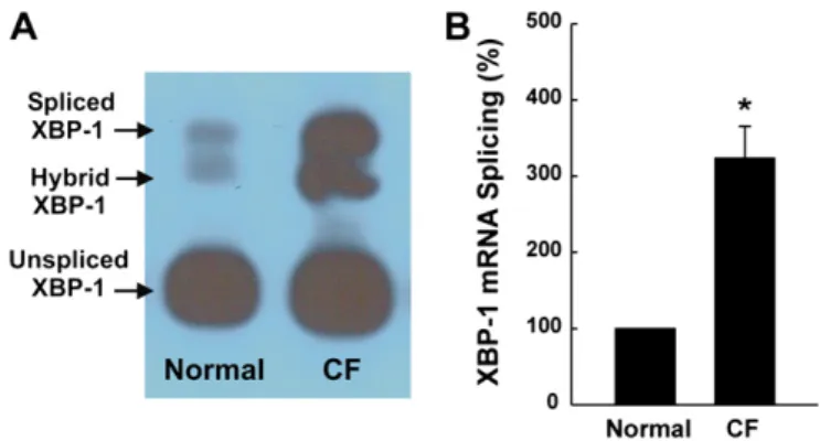
infected and inflamed native CF airway epithelia exhibit an increased protein expression of the UPR markers calreticulin and protein disulfide isomerase, which are gene targets of XBP-1 (2). A follow-up study provided evidence that mRNA splicing of XBP-1 occurs in airway inflammationin vivo, based on the following observations. (1) Native infected/inflamed CF human bronchial airway epithelia exhibit increased levels of spliced XBP-1 as compared with noninfected/inflamed normal human bronchial epithelia (Figure 1). (2) ER stress-activated indicator (ERAI) mice (76) exhibit evidence of ER stress-dependent XBP-1 mRNA splicing after intrapulmonary bacterial challenge (61). ERAI mice are a transgenic line of mice where UPR activation-dependent XBP-1 mRNA splicing couples to expres-sion of Venus (a variant of GFP) fluorescence (76). The ER stress indicator was constructed by fusing XBP-1 and Venus. Upon UPR activation, the spliced indicator mRNA is translated into an XBP-1–Venus fusion protein, whose expression can be detected by its fluorescence. Hence, there is no Venus expres-sion in the absence of XBP-1 mRNA splicing, but, in the presence of XBP-1 splicing, Venus expression occurs, and its fluorescence is an index of spliced XBP-1. ERAI mice have been successfully used to monitor physiological and patholog-ical ER stressin vivo(76).
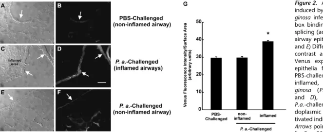
To test whether airway infection promoted XBP-1 mRNA splicing in vivo, ERAI mouse airways were exposed to 40 ml PBS or PBS containing 1 3 106 colony-forming units of
P. aeruginosastrain PAK (77). Although this is a model of acute pneumonia (77), it also induces airway inflammation 24 hours after infection. XBP-1 mRNA splicing, as reflected by increased Venus fluorescence, was increased 24 hours afterP. aeruginosa
infection in lining epithelia from inflamed airways (Figures 2C and 2D). In contrast, XBP-1 splicing/Venus fluorescence was low in noninflamed airway sites in PBS- and P. aeruginosa– challenged airways (Figures 2A, 2B, 2E, and 2F). Quantification of Venus fluorescence intensity, expressed per surface airway epithelial area, strengthened the notion that XBP-1 mRNA splicing was increased in airway epithelia from inflamed airways (Figure 2G). These findings demonstrated that luminal
P. aeruginosainfection–induced airway inflammation activates the UPR and promotes XBP-1 mRNA splicing in airway epitheliain vivo.
ACTIVATION OF XBP-1 BY AIRWAY INFLAMMATION
TRIGGERS THE ER Ca21STORE EXPANSION IN
AIRWAY EPITHELIA
Data linking inflammation with airway epithelial ER Ca21store expansionin vivowere recently provided by findings from wild-type mouse airways exposed toP. aeruginosastrain PAK (77). Histological analyses of P. aeruginosa–challenged lungs at 24 hours revealed areas that were clearly inflamed and areas that showed no evidence of inflammation (61). As visualized by calreticulin (ER Ca21 store marker) staining, ER Ca21 store expansion was only observed in epithelia from airways that were inflamed 24 hours after inoculation with P. aeruginosa
(61). These data illustrated that epithelial ER expansion and UPR activation coupled to XBP-1 mRNA splicing are a feature of inflamed airways.
We have recently investigated the role of XBP-1 in human airway epithelial inflammatory processes mediated by ER Ca21 store expansion by overexpressing the spliced XBP-1 or a dom-inant negative XBP-1 (DN-XBP-1) in human bronchial airway epithelia (61). Overexpression of spliced XBP-1 promoted mor-phological and functional expansion of the ER and its Ca21stores in the absence of extracellular stimulus (61). In contrast, the ER Ca21 store expansion resulting from exposure to mucosal in-fectious and inflammatory factors from CF airways was blunted in epithelia expressing the DN-XBP-1 (61). These data demonstrate that activation of XBP-1 is a key mechanism responsible for the ER/Ca21store expansion in inflamed human airway epithelia. In addition, these studies indicate that activation of XBP-1–dependent ER/Ca21store expansion is a mechanism responsible for the am-plified inflammatory mediator secretion we have previously re-ported in inflamed human bronchial epithelia (2, 61).
ACTIVATION OF XBP-1 BY AIRWAY INFLAMMATION MEDIATES INCREASED AIRWAY EPITHELIAL CYTOKINE SECRETORY RESPONSES
We have considered that activation of XBP-1 mRNA splicing during airway inflammation may mediate ER Ca21 store expansion–dependent hyperinflammation. Because a hyperin-flammatory response was, in part, typified by an increased mucosal BK-dependent IL-8 secretion in airway epithelia (2), we investigated IL-8 secretory responses induced by mucosal BK in cultures of 16HBE14o2–expressing control, spliced XBP-1, or DN-XBP-1 vectors. In control vector–expressing cultures,
mucosal BK induced a modest increase in IL-8 secretion in comparison with vehicle-treated cultures (Figure 3). In contrast, cultures expressing the spliced XBP-1 exhibited a higher IL-8 secretory response, as compared with control vector expressing cultures in the absence of BK, and potentiation of BK-induced IL-8 secretion (Figure 3). Finally, expression of DN-XBP-1 inhibited mucosal BK-induced IL-8 secretion (Figure 3). These data illustrate that constitutive activation of spliced XBP-1 induces a hyperinflammatory response (e.g., increased IL-8 secretion in airway epithelia).
CFTR MUTATIONS ARE NOT ASSOCIATED WITH ACTIVATION OF XBP-1–DEPENDENT SIGNALING
Freshly isolated CF airway epithelia exhibit increased levels of XBP-1 mRNA splicing (Figure 1). However, our previous work (2) and the findings from other investigators suggest that CFTR mutations are not linked to the higher levels of spliced XBP-1 found in inflamed CF airway epithelia. Although CF human bronchial epithelia homozygous for theDF508 CFTR mutation exhibit an increased IL-8 secretion in short-term primary
Figure 2. Airway inflammation induced byPseudomonas
aeru-ginosa infection promotes
X-box binding protein-1 mRNA splicing (activation) in murine airway epitheliain vivo. (A,C, andE) Differential interference contrast and (B, D, and F) Venus expression in airway epithelia from noninflamed, PBS-challenged (A and B), inflamed, Pseudomonas
aeru-ginosa (P.a.)-challenged (C
and D), and noninflamed, P.a.-challenged (Eand F) en-doplasmic reticulum stress-ac-tivated indicator mice airways.
Arrowspoint to airway
epithe-lia. Bar, 20mm. (G) Compiled data for Venus fluorescence intensity, expressed per surface epithelial area, from all groups. *P,0.05, P.a.-challenged and inflamed versus PBS-challenged, noninflamed. Reproduced with permission from Ref. 61.
cultures, their higher IL-8 secretion is lost in long-term cultures (2). Moreover, exposure of long-term CF cultures to SMM reproduced the increased IL-8 secretion found in short-term CF cultures, and this response was coupled with the ability of SMM to induce XBP-1 mRNA splicing (2).
Follow-up studies have also suggested a dissociation of CFTR mutations from XBP-1 mRNA splicing. For instance, no differences in XBP-1 mRNA splicing were found in passage 1 normal versus CF airway epithelial cultures (78). The endog-enous levels of CFTR expression in primary cultures of airway epithelia are low, as observed in airway epithelia in vivo. Dissociation of theDF508 CFTR mutation from IRE1/XBP-1 signaling and airway epithelial inflammatory responses has been further documented in a study showing no significant differ-ences in basal and P. aeruginosa (or flagellin)-induced IL-8 secretion, intracellular Ca21mobilization, and IRE1 activity in CF15 cells overexpressing wild-type or DF508 CFTR (79). In contrast, high-level expression of recombinantDF508 CFTR in Calu-3 cells led to increased XBP-1 mRNA splicing (80). We speculate that the absence versus presence of activation of the IRE1/XBP-1 pathway in the latter two studies may have re-sulted from cell-specific differences or differences in DF508 CFTR expression levels induced in cell lines (i.e., whereas lower levels of expression of mutated CFTR do not trigger ER stress, high levels of expression ofDF508 CFTR induce XBP-1 mRNA splicing). The relevance of in vitro–promoted exaggerated expression ofDF508 CFTR-triggered activation of XBP-1 sig-naling in airway epithelial inflammatory responses remains to be established. Nevertheless, the findings from primary cultures of normal and CF epithelia expressing endogenous CFTR muta-tions suggest that airway epithelial inflammation, rather than mutated CFTR, is responsible for activation of ER stress mediated by XBP-1 mRNA splicing.
SIGNIFICANCE OF INFECTION-TRIGGERED AIRWAY EPITHELIAL UPR ACTIVATION FOR CF AIRWAY
INFLAMMATIONIN VIVO
A model linking airway epithelial inflammation-induced ER stress-dependent XBP-1 mRNA splicing–and the consequent
XBP-1–dependent increased airway epithelial secretion of in-flammatory mediators–is presented in Figure 4. Under normal conditions, these XBP-1–mediated functions are beneficial for the host by amplifying the inflammatory response to clear the airways of infection. However, we speculate that XBP-1– dependent functions can mediate adaptive and maladaptive responses during chronic airway inflammation. For example, expansion of ER Ca21stores can provide a compensatory and protective function for infected airways via an increased Ca21
i–
dependent mucociliary clearance (1). The higher Ca21
isignals
resulting from ER Ca21 store expansion may be particularly beneficial to patients with CF, who depend on Ca21–activated Cl2 channels (CaCC) to compensate for the lack of CFTR-dependent Cl2secretion (81). Because of the increased expres-sion of ER Ca21 stores at the apical domain in CF airway epithelia (1), luminal infectious and inflammatory stimuli gen-erate greater Ca21
isignals in close proximity to CaCC, allowing
the airways to transiently restore defective mucus clearance. However, UPR-induced ER Ca21 store expansion also amplifies the airway epithelial inflammatory response, which may adversely affect the homeostasis of CF airways. After intraluminal infection-triggered ER Ca21 store expansion, the airway epithelium is primed to secrete higher levels of in-flammatory factors (e.g., IL-8) in response to the larger Ca21
i
signals triggered by Ca21
i–mobilizing stimuli such as BK or
ATP/UTP (1). The amplified cytokine response resulting from persistent bacterial infection in CF may be maladaptive because inflammatory cells cannot rid the lumen of bacteria that are protected in the thickened mucus environment characteristic of CF airways (82). Thus, rather than eradicating the bacterial infection, the chronic, but ineffective, UPR-mediated hyper-inflammatory response of CF airways may have harmful conse-quences by promoting paradoxical airway wall destruction due to proteolytic enzyme release.
It is plausible to speculate that the resulting activation of the PERK/eIF2a/ATF4 UPR pathway should also be functionally relevant for the pathophysiology of CF airways inflammatory disease. The increased synthesis of inflammatory mediators in CF airways likely causes profound consequences for the meta-bolic and oxidative status of inflamed airway epithelia. Protein
secretion can constitute an irreversible loss of amino acids into the extracellular environment and produce net loss of equiva-lents from the cell. The foldase PDI catalyzes oxidative protein folding in the ER, resulting in the transfer of electrons from the nascent polypeptide chain to the oxidoreductase ERO1 (75). ERO1 is then oxidized by molecular oxygen, which functions as the terminal electron acceptor. Therefore, the greater the secretory burden, the greater the loss of amino acids and reducing equivalents. Previous studies have revealed that the PERK/ATF4 UPR pathway plays a key role in protecting cells against amino acid deprivation and oxidative stress (75, 83). In cells devoid of PERK, activation of the UPR results in accu-mulation of reactive oxygen species, and ATF4 and PERK knockout cells require amino acid and cysteine supplementa-tion, most likely to replenish amino acids lost during secretion and increase glutathione levels, respectively (75). The impor-tance of ATF4 in regulation of amino acid metabolism and oxidative stress responses was not only revealed by these studies (PERK2/2cells cannot activate eIF2a-dependent translational up-regulation of ATF4, and ATF42/2cells lack ATF4 protein) but was also underscored in microarrays with ATF42/2 cells, illustrating that ATF4 induces the transcription of genes in-volved in amino acid import, glutathione biosynthesis, and resistance to oxidative stress (75).
ATF4-induced amino acid transport likely plays a protective role in inflamed CF airway epithelia because SMM-exposed HBE exhibit an augmented metabolic response (e.g., increased protein synthesis [2] and lactate production) (C.M.P. Ribeiro, unpublished observation). Moreover, the antioxidant functions of ATF4 may also be relevant for infected or inflamed CF airways because oxidative stress is a hallmark of CF airways disease (84, 85). Glutathione levels in the airway surface liquid from patients with CF are reduced (86), which may result, at least in part, from the lack of CFTR-mediated glutathione transport (reviewed in [86]). However, infected/inflamed CF airway epithelia may also contribute for the oxidative stress of CF airways through increased generation of reactive oxygen species, independently of mutated CFTR. Indeed, primary cul-tures of human bronchial airway epithelia exposed to SMM ex-hibit a higher expression of oxidative stress genes, including protective genes that are targets of ATF4 (87). A model depic-ting the functional role of ATF4 signaling in airway epithelial inflammatory responses is shown in Figure 4.
FUTURE DIRECTIONS
Because the knowledge generated by the studies suggesting a functional role for XBP-1 in airway inflammatory responses was limited to primarilyin vitro studies of selected cell types,
in vivo models that allow the study of UPR in mammalian airways and, specifically, secretory cell types (e.g., mucin-secret-ing cells) are necessary. Glimcher and colleagues have generated a LoxP-flanked XBP-1 mouse, which allows for conditional knockdown of XBP-1, as demonstrated by the specific knockout of XBP-1 in intestinal epithelia (60). The generation of the airway epithelia XBP-12/2mouse, using the LoxP-flanked XBP-1 mouse, will advance the understanding of cellular pathways relevant to chronic airway inflammation.
Previous studies have linked XBP-1 with a genetic risk for human inflammatory bowel disease by showing that several single-nucleotide polymorphisms within the XBP-1 gene locus conferred risk for Crohn’s disease and ulcerative colitis (60). Similar studies are necessary to address whether XBP-1 is linked with genetic risks for human airway inflammatory diseases.
An unresolved area involves the relative roles of IRE1aand IRE1b in UPR-mediated signaling in airway inflammatory
re-sponses. A central question is whether IRE1b has a similar activation scheme to IRE1a. Structural studies with the core region of the ER luminal domain of IRE1a suggest that di-merization of this domain, resulting from IRE1a activation during ER stress, creates a groove reminiscent of the peptide-binding domains of histocompatibility complexes (88). Additional studies have shown that oligomerization is central to IRE1a function and is an intrinsic attribute of its cytosolic domains (89). Studies that address whether IRE1bexhibits a similar sequence of activation during airway inflammation and whether it has a distinct functional role versus IRE1ain the airways will expand the understanding of airway secretory mechanisms.
An additional role for the UPR in CF airway inflammatory responses has been suggested by initial studies investigating the PERK/eIF2a/ATF4 pathway, indicating that it plays a key role in innate immune responses of human airway epithelia by in-ducing ATF4-dependent expression of genes involved in pro-tection against amino acid loss and oxidative stress (87) and NF-kB–mediated transcription of inflammatory mediators (3, 4). Future studies addressing the role of the PERK/eIF2a/ATF4 pathway in airway inflammatory responses may result in novel therapies for CF airway inflammation associated with oxidative stress. A previous study has shown that the eIF2aUPR pathway can be modulated by salubrinal, a selective inhibitor of eIF2a dephosphorylation that does not affect ATF6- or IRE1-dependent signaling and promotes cell protection (90). This raises the possibility that a therapeutic strategy for conferring protection against airway epithelial inflammation may be based on small molecules, like salubrinal, to up-regulate the ATF4 pathway.
The functional consequences of UPR activation for inflam-matory responses relevant to CF airways disease deserve further investigation. Studies with mice exhibiting airway epi-thelial deletion of UPR pathways will provide insight into the relevance of ER stress responses in the pathogenesis of CF. Future studies are required to establish how the UPR is triggered in CF; whether activation of the IRE1-dependent XBP-1, ATF6, or PERK signaling pathways plays a favorable or a negative role in CF pathophysiology; and whether manipula-tion of these pathways can be therapeutically beneficial for patients with CF.
Author Disclosure:C.M.P.R. received grant support from Cempra Pharmaceuti-cals ($50,001–$100,000). R.C.B. was a consultant for Gilead Sciences ($1,001– $5,000), Parion Sciences, Inspire Pharmaceuticals ($10,001–$50,000), Pulmatrix ($5,001–$10,000). He is on the Board of Parion Sciences ($10,001–$50,000) and owns patents through Inspire Pharmaceuticals and Parion Sciences. He owns stocks or options of Parion Sciences and Inspire Pharmaceuticals (more than $100,001).
Acknowledgments: The authors thank Lisa Brown for editorial assistance during the preparation of this manuscript.
References
1. Ribeiro CMP, Paradiso AM, Carew MA, Shears SB, Boucher RC. Cystic fibrosis airway epithelial Ca21
isignaling: the mechanism for the larger agonist-mediated Ca21i signals in human cystic fibrosis airway epithelia.J Biol Chem2005;280:10202–10209.
2. Ribeiro CMP, Paradiso AM, Schwab U, Perez-Vilar J, Jones L, O’Neal W, Boucher RC. Chronic airway infection/inflammation induces a Ca21
i-dependent hyperinflammatory response in human cystic fibrosis airway epithelia.J Biol Chem2005;280:17798–17806. 3. Braun M, Ribeiro CP. Airway epithelial infection/inflammation
acti-vates an unfolded protein response coupled to ATF4 signaling: relevance for airway inflammation and oxidative stress [abstract]. Pediatr Pulmonol2007;Suppl. 30:260–261.
5. Boucher RC, Cotton CU, Gatzy JT, Knowles MR, Yankaskas JR. Evidence for reduced Cl2and increased Na1permeability in cystic
fibrosis human primary cell cultures.J Physiol1988;405:77–103. 6. Matsui H, Grubb BR, Tarran R, Randell SH, Gatzy JT, Davis CW,
Boucher RC. Evidence for periciliary liquid layer depletion, not abnormal ion composition, in the pathogenesis of cystic fibrosis airways disease.Cell1998;95:1005–1015.
7. Konstan MW, Hilliard KA, Norvell TM, Berger M. Bronchoalveolar lavage findings in cystic fibrosis patients with stable, clinical mild lung disease suggest ongoing infection and inflammation.Am J Respir Crit Care Med1994;150:448–454.
8. Khan TZ, Wagener JS, Bost T, Martinez J, Accurso FJ, Riches DWH. Early pulmonary inflammation in infants with cystic fibrosis.Am J Respir Crit Care Med1995;151:1075–1082.
9. Muhlebach MS, Stewart PW, Leigh MW, Noah TL. Quantitation of inflammatory responses to bacteria in young cystic fibrosis and control patients.Am J Respir Crit Care Med1999;160:186–191.
10. Ziady AG, Davis PB. Infection versus inflammation. In: Bush A, Alton EWFW, Davies JC, Griesenbach U, Jaffe A (eds): Cystic fibrosis in the 21st century. Prog Respir Res. Basel, Karger; 2006, vol 34, pp. 122–130.
11. Muhlebach MS, Noah TL. Endotoxin activity and inflammatory markers in the airways of young patients with cystic fibrosis.Am J Respir Crit Care Med2002;165:911–915.
12. Koller DY, Nething I, Otto J, Urbanek R, Eichler I. Cytokine concentra-tions in sputum from patients with cystic fibrosis and their relation to eosinophil activity.Am J Respir Crit Care Med1997;155:1050–1054. 13. Taggart C, Coakley RJ, Greally P, Canny G, O’Neill SJ, McElvaney NG.
Increased elastase release by CF neutrophils is mediated by tumor necrosis factor-aand interleukin-8.Am J Physiol2000;278:L33–L41. 14. Baeuerle PA, Baltimore D. NF-kB: ten years after.Cell1996;87:13–20. 15. Karin M. How NF-kappaB is activated: the role of the IkB kinase (IKK)
complex.Oncogene1999;18:6867–6874.
16. Jobin C, Sartor RB. The IkB/NF-kB system: a key determinant of mucosal inflammation and protection.Am J Physiol2000;278:C451–C462. 17. Berger M. Inflammatory mediators in cystic fibrosis lung disease.Allergy
Asthma Proc2002;23:19–25.
18. Tabary O, Escotte S, Couetil JP, Hubert D, Dusser D, Puchelle E, Jacquot J. High susceptibility for cystic fibrosis human airway gland cells to produce IL-8 through the IkB kinaseapathway in response to extracellular NaCl content.J Immunol2000;164:3377–3384. 19. Bonfield TL, Konstan MW, Berger M. Altered respiratory epithelial cell
cytokine production in cystic fibrosis.J Allergy Clin Immunol1999; 104:72–78.
20. Tabary O, Zahm JM, Hinnrasky J, Couetil JP, Cornillet P, Guenounou M, Gaillard D, Puchelle E, Jacquot J. Selective up-regulation of chemokine IL-8 expression in cystic fibrosis bronchial gland cells in vivoandin vitro. Am J Pathol1998;153:921–930.
21. Kammouni W, Figarella C, Marchand S, Merten M. Altered cytokine production by cystic fibrosis tracheal gland serous cells.Infect Immun 1997;65:5176–5183.
22. Ribeiro CM. The role of intracellular calcium signals in inflammatory responses of polarised cystic fibrosis human airway epithelia.Drugs R D2006;7:17–31.
23. Chun J, Prince A. Activation of Ca2+-dependent signaling by TLR2.J Immunol2006;177:1330–1337.
24. Rozengurt E. Mitogenic signaling pathways induced by G protein-coupled receptors.J Cell Physiol2007;213:589–602.
25. Putney JW Jr. Capacitative calcium entry. Austin, TX: R.G. Landes Company; 1997.
26. Putney JW Jr, Ribeiro CM. Signaling pathways between the plasma membrane and endoplasmic reticulum calcium stores.Cell Mol Life Sci2000;57:1272–1286.
27. Meldolesi J, Pozzan T. The endoplasmic reticulum Ca21store: a view
from the lumen.Trends Biochem Sci1998;23:10–14. 28. Hu Q, Deshpande S, Irani K, Ziegelstein RC. [Ca21]
i oscillation frequency regulates agonist-stimulated NF-kB transcriptional activity. J Biol Chem1999;274:33995–33998.
29. Quinlan KL, Naik SM, Cannon G, Armstrong CA, Bunnett NW, Ansel JC, Caughman SW. Substance P activates coincident AT- and NF-kB-dependent adhesion molecule gene expression in microvascular endothelial cells through intracellular calcium mobilization.J Immu-nol1999;163:5656–5665.
30. Han B, Logsdon CD. CCK stimulates mob-1 expression and NF-kB activation via protein kinase C and intracellular Ca21.Am J Physiol
2000;278:C344–C351.
31. Ouellet M, Barbeau B, Tremblay MJ. p56(lck), ZAP-70, SLP-76, and calcium-regulated effectors are involved in NF-kB activation by bisperoxovanadium phosphotyrosyl phosphatase inhibitors in human T cells.J Biol Chem1999;274:35029–35036.
32. Gewirtz AT, Rao AS, Simon PO Jr, Merlin D, Carnes D, Madara JL, Neish AS. Salmonella typhimurium induces epithelial IL-8 expression via Ca21- mediated activation of the NF-kB pathway.J Clin Invest
2000;105:79–92.
33. Jefferson KK, Smith MF Jr, Bobak DA. Roles of intracellular calcium and NF-kB in the Clostridium difficile toxin A-induced up-regulation and secretion of IL-8 from human monocytes.J Immunol1999;163: 5183–5191.
34. Hsuan SL, Kannan MS, Jeyaseelan S, Prakash YS, Malazdrewich C, Abrahamsen MS, Sieck GC, Maheswaran SK. Pasteurella haemoly-tica leukotoxin and endotoxin induced cytokine gene expression in bovine alveolar macrophages requires NF-kB activation and calcium elevation.Microb Pathog1999;26:263–273.
35. Paradiso AM, Cheng EHC, Boucher RC. Effects of bradykinin on intracellular calcium regulation in human ciliated airway epithelium. Am J Physiol1991;261:L63–L69.
36. Rodgers HC, Pang L, Holland E, Corbett L, Range S, Knox AJ. Bradykinin increases IL-8 generation in airway epithelial cells via COX-2-derived prostanoids.Am J Physiol2002;283:L612–L618. 37. Ratner AJ, Bryan R, Weber A, Nguyen S, Barnes D, Pitt A, Gelber S,
Cheung A, Prince A. Cystic fibrosis pathogens activate Ca21
-dependent mitogen-activated protein kinase signaling pathways in airway epithelial cells.J Biol Chem2001;276:19267–19275.
38. Lewis RS. Calcium oscillations in T-cells: mechanisms and consequences for gene expression.Biochem Soc Trans2003;31:925–929.
39. Esther CR Jr, Alexis NE, Clas ML, Lazarowski ER, Donaldson SH, Ribeiro CM, Moore CG, Davis SD, Boucher RC. Extracellular purines are biomarkers of neutrophilic airway inflammation. Eur Respir J2008;31:949–956.
40. Abdullah LH, Davis SW, Burch L, Yamauchi M, Randell SH, Nette-sheim P, Davis CW. P2Upurinoceptor regulation of mucin secretion in SPOC11 cells, a goblet cell line from the airways.Biochem J1996; 316:943–951.
41. Rossi AH, Salmon WC, Chua M, Davis CW. Calcium signaling in human airway goblet cells following purinergic activation. Am J Physiol Lung Cell Mol Physiol2007;292:L92–L98.
42. Rossi AH, Sears PR, Davis CW. Ca(21) dependency of ‘Ca(21 )-independent’ exocytosis in SPOC1 airway goblet cells. J Physiol 2004;559:555–565.
43. Guo Q, Sopher BL, Furukawa K, Pham DG, Robinson N, Martin GM, Mattson MP. Alzheimer’s presenilin mutation sensitizes neural cells to apoptosis induced by trophic factor withdrawal and amyloid b-peptide: involvement of calcium and oxyradicals.J Neurosci1997; 17:4212–4222.
44. Patterson PH. Cytokines in Alzheimer’s disease and multiple sclerosis. Curr Opin Neurobiol1995;5:642–646.
45. Ho LJ, Wang JJ, Shaio MF, Kao CL, Chang DM, Han SW, Lai JH. Infection of human dendritic cells by dengue virus causes cell maturation and cytokine production.J Immunol2001;166:1499–1506. 46. Su HL, Liao CL, Lin YL. Japanese encephalitis virus infection initiates endoplasmic reticulum stress and an unfolded protein response. J Virol2002;76:4162–4171.
47. Charlier N, Leyssen P, Paeshuyse J, Drosten C, Schmitz H, Van Lommel A, de Clercq E, Neyts J. Infection of SCID mice with MontanaMyotis leukoencephalitisvirus as a model for flavivirus encephalitis.J Gen Virol2002;83:1887–1896.
48. Eremeeva ME, Silverman DJ. Rickettsia rickettsii infection of the EA.hy 926 endothelial cell line: morphological response to infection and evidence for oxidative injury.Microbiology1998;144:2037–2048. 49. Kaufman RJ. Stress signaling from the lumen of the endoplasmic reticulum: coordination of gene transcriptional and translational controls.Genes Dev1999;13:1211–1233.
50. Mori K. Tripartite management of unfolded proteins in the endoplasmic reticulum.Cell2000;101:451–454.
51. Patil C, Walter P. Intracellular signaling from the endoplasmic reticulum to the nucleus: the unfolded protein response in yeast and mammals. Curr Opin Cell Biol2001;13:349–355.
52. Lee AS. The glucose-regulated proteins: stress induction and clinical applications.Trends Biochem Sci2001;26:504–510.
54. Rutkowski DT, Kaufman RJ. A trip to the ER: coping with stress. Trends Cell Biol2004;14:20–28.
55. Zhang K, Kaufman RJ. Signaling the unfolded protein response from the endoplasmic reticulum.J Biol Chem2004;279:25935–25938. 56. Ron D, Walter P. Signal integration in the endoplasmic reticulum
unfolded protein response.Nat Rev Mol Cell Biol2007;8:519–529. 57. Tirasophon W, Welihinda AA, Kaufman RJ. A stress response pathway
from the endoplasmic reticulum to the nucleus requires a novel bifunctional protein kinase/endoribonuclease (IRE1p) in mammalian cells.Genes Dev1998;12:1812–1824.
58. Wang XZ, Harding HP, Zhang Y, Jolicoeur EM, Kuroda M, Ron D. Cloning of mammalian Ire1 reveals diversity in the ER stress responses.EMBO J1998;17:5708–5717.
59. Harding HP, Zhang Y, Ron D. Protein translation and folding are coupled by an endoplasmic-reticulum-resident kinase. Nature1999; 397:271–274 (erratum:Nature1999;398:90).
60. Kaser A, Lee AH, Franke A, Glickman JN, Zeissig S, Tilg H, Nieuwenhuis EE, Higgins DE, Schreiber S, Glimcher LH, et al. XBP1 links ER stress to intestinal inflammation and confers genetic risk for human inflammatory bowel disease.Cell2008;134:743–756. 61. Martino MEB, Olsen JC, Fulcher NB, Wolfgang MC, O’Neal WK,
Ribeiro CMP. Airway epithelial inflammation-induced endoplasmic reticulum Ca21store expansion is mediated by X-box binding
pro-tein-1.J Biol Chem2009;284:14904–14913.
62. Yoshida H, Matsui T, Yamamoto A, Okada T, Mori K. XBP1 mRNA is induced by ATF6 and spliced by IRE1 in response to ER stress to produce a highly active transcription factor.Cell2001;107:881–891. 63. Calfon M, Zeng H, Urano F, Till JH, Hubbard SR, Harding HP, Clark
SG, Ron D. IRE1 couples endoplasmic reticulum load to secretory capacity by processing theXBP-1mRNA.Nature2002;415:92–96. 64. Urano F, Bertolotti A, Ron D. IRE1 and efferent signaling from the
endoplasmic reticulum.J Cell Sci2000;113:3697–3702.
65. Reimold AM, Iwakoshi NN, Manis J, Vallabhajosyula P, Szomolanyi-Tsuda E, Gravallese EM, Friend D, Grusby MJ, Alt F, Glimcher LH. Plasma cell differentiation requires the transcription factor XBP-1. Nature2001;412:300–307.
66. Shaffer AL, Shapiro-Shelef M, Iwakoshi NN, Lee AH, Qian SB, Zhao H, Yu X, Yang L, Tan BK, Rosenwald A,et al.XBP1, downstream of Blimp-1, expands the secretory apparatus and other organelles, and increases protein synthesis in plasma cell differentiation.Immunity 2004;21:81–93.
67. Sriburi R, Jackowski S, Mori K, Brewer JW. XBP1: a link between the unfolded protein response, lipid biosynthesis, and biogenesis of the endoplasmic reticulum.J Cell Biol2004;167:35–41.
68. Ron D, Hampton RY. Membrane biogenesis and the unfolded protein response.J Cell Biol2004;167:23–25.
69. Lee AH, Chu GC, Iwakoshi NN, Glimcher LH. XBP-1 is required for biogenesis of cellular secretory machinery of exocrine glands.EMBO J2005;24:4368–4380.
70. Wiest DL, Burkhardt JK, Hester S, Hortsch M, Meyer DI, Argon Y. Membrane biogenesis during B cell differentiation: most endoplasmic reticulum proteins are expressed coordinately.J Cell Biol1990;110: 1501–1511.
71. Iwakoshi NN, Lee AH, Vallabhajosyula P, Otipoby KL, Rajewsky K, Glimcher LH. Plasma cell differentiation and the unfolded protein response intersect at the transcription factor XPB-1. Nat Immunol 2003;4:321–329.
72. Cox JS, Chapman RE, Walter P. The unfolded protein response coordinates the production of endoplasmic reticulum protein and endoplasmic reticulum membrane. Mol Biol Cell 1997;8:1805– 1814.
73. Travers KJ, Patil CK, Wodicka L, Lockhart DJ, Weissman JS, Walter P. Functional and genomic analyses reveal an essential coordination
between the unfolded protein response and ER-associated degrada-tion.Cell2000;101:249–258.
74. Harding HP, Calfon M, Urano F, Novoa I, Ron D. Transcriptional and translational control in the mammalian unfolded protein response. Annu Rev Cell Dev Biol2002;18:599.
75. Harding HP, Zhang Y, Zeng H, Novoa I, Lu PD, Calfon M, Sadri N, Yun C, Popko B, Paules R, et al.An integrated stress response regulates amino acid metabolism and resistance to oxidative stress. Mol Cell2003;11:619–633.
76. Iwawaki T, Akai R, Kohno K, Miura M. A transgenic mouse model for monitoring endoplasmic reticulum stress.Nat Med2004;10:98–102. 77. Smith RS, Wolfgang MC, Lory S. An adenylate cyclase-controlled
signaling network regulates Pseudomonas aeruginosa virulence in a mouse model of acute pneumonia.Infect Immun2004;72:1677–1684. 78. Nanua S, Sajjan U, Keshavjee S, Hershenson MB. Absence of typical unfolded protein response in primary cultured cystic fibrosis airway epithelial cells.Biochem Biophys Res Commun2006;343:135–143. 79. Hybiske K, Fu Z, Schwarzer C, Tseng J, Do J, Huang N, Machen TE.
Effects of cystic fibrosis transmembrane conductance regulator and DeltaF508CFTR on inflammatory response, ER stress, and Ca21of
airway epithelia. Am J Physiol Lung Cell Mol Physiol 2007;293: L1250–L1260.
80. Bartoszewski R, Rab A, Jurkuvenaite A, Mazur M, Wakefield J, Collawn JF, Bebok Z. Activation of the unfolded protein response bydF508 CFTR.Am J Respir Cell Mol Biol2008;39:448–457. 81. Ribeiro CMP, Paradiso AM, Lazarowski E, Boucher RC. P2Y2
re-ceptors and Ca21-dependent Cl2 secretion in normal and cystic
fibrosis human airway epithelia. In: Salathe M, editor. Cilia, mucus and mucociliary interactions. New York: Marcel Dekker, Inc.; 2001. pp. 303–314.
82. Worlitzsch D, Tarran R, Ulrich M, Schwab U, Cekici A, Meyer KC, Birrer P, Bellon G, Berger J, Weiss T,et al.Effects of reduced mucus oxygen concentration in airway Pseudomonas infections of cystic fibrosis patients.J Clin Invest2002;109:317–325.
83. Siu F, Bain PJ, LeBlanc-Chaffin R, Chen H, Kilberg MS. ATF4 is a mediator of the nutrient-sensing response pathway that activates the human asparagine synthetase gene. J Biol Chem 2002;277:24120– 24127.
84. Bowler RP, Crapo JD. Oxidative stress in airways: is there a role for extracellular superoxide dismutase?Am J Respir Crit Care Med2002; 166:538–543.
85. Winklhofer-Roob BM. Oxygen free radicals and antioxidants in cystic fibrosis: the concept of an oxidant-antioxidant imbalance. Acta Paediatr Suppl1994;395:49–57.
86. Hudson VM. Rethinking cystic fibrosis pathology: the critical role of abnormal reduced glutathione (GSH) transport caused by CFTR mutation.Free Radic Biol Med2001;30:1440–1461.
87. Ribeiro CMP, Hurd H, Wu Y, Martino MEB, Jones L, Brighton B, Boucher RC, O’Neal WK. Azithromycin treatment alters gene expression in inflammatory, lipid metabolism, and cell cycle pathways in well-differentiated human airway epithelia. PLoS ONE 2009;4: e5806.
88. Credle JJ, Finer-Moore JS, Papa FR, Stroud RM, Walter P. On the mechanism of sensing unfolded protein in the endoplasmic reticulum. Proc Natl Acad Sci USA2005;102:18773–18784.
89. Korennykh AV, Egea PF, Korostelev AA, Finer-Moore J, Zhang C, Shokat KM, Stroud RM, Walter P. The unfolded protein response signals through high-order assembly of IRE1.Nature2009;457:687– 693.