Traumatic stress and accelerated DNA methylation age: A meta-analysis
Erika J. Wolf
a,b,⁎, Hannah Maniates
a, Nicole Nugent
c,d, Adam X. Maihofer
e, Don Armstrong
f,
Andrew Ratanatharathorn
g, Allison E. Ashley-Koch
h, Melanie Garrett
i, Nathan A. Kimbrel
i,j,k,
Adriana Lori
l, VA Mid-Atlantic MIRECC Workgroup
j, Allison E. Aiello
m, Dewleen G. Baker
e,n,o,
Jean C. Beckham
i,j,k, Marco P. Boks
p, Sandro Galea
q, Elbert Geuze
p,r, Michael A. Hauser
h,
Ronald C. Kessler
s, Karestan C. Koenen
t,u, Mark W. Miller
a,b, Kerry J. Ressler
v,
Victoria Risbrough
e,n,o, Bart P.F. Rutten
w, Murray B. Stein
e,n,x, Robert J. Ursano
y,
Eric Vermetten
p,r,z, Christiaan H. Vinkers
p, Monica Uddin
f,A, Alicia K. Smith
l,B,
Caroline M. Nievergelt
e,n,o, Mark W. Logue
a,b,CaNational Center for PTSD at VA Boston Healthcare System, United States bDepartment of Psychiatry, Boston University School of Medicine, United States cBradley Hasbro Children’s Research Center, Rhode Island Hospital, United States
dDepartments of Psychiatry and Human Behavior and Pediatrics, Brown Medical School, United States eUniversity of California San Diego, Department of Psychiatry, United States
fUniversity of Illinois Urbana-Champaign, Carl R. Woese Institute for Genomic Biology, United States gColumbia University, Department of Epidemiology, United States
hDuke Molecular Physiology Institute, Duke University School of Medicine, United States iDepartment of Psychiatry & Behavioral Sciences, Duke University Medical Center, United States jVA Mid-Atlantic, Mental Illness Research, Education, and Clinical Center, United States kDurham VA Medical Center, United States
lDepartment of Psychiatry and Behavioral Sciences, Emory University, United States
mDepartment of Epidemiology, University of North Carolina at Chapel Hill Gillings School of Global Public Health, United States nVeterans Affairs San Diego Healthcare System, United States
oVeterans Affairs Center of Excellence for Stress and Mental Health, United States
pUniversity Medical Center Utrecht, Brain Center Rudolf Magnus, Department of Psychiatry, Utrecht, The Netherlands qBoston University School of Public Health, United States
rMinistry of Defence, Military Mental Healthcare, Utrecht, The Netherlands sHarvard Medical School, Department of Health Care Policy, United States
tHarvard T.H. Chan School of Public Health, Department of Epidemiology, United States
uMassachusetts General Hospital, Psychiatric and Neurodevelopmental Genetics Unit, Center for Human Genetic Research, and Department of Psychiatry, United States vDepartment of Psychiatry, Harvard Medical School and McLean Hospital, Belmont, MA, United States
wSchool for Mental Health and Neuroscience and the European Graduate School of Neuroscience (EURON), Department of Psychiatry and Neuropsychology, Maastricht University Medical Centre, Maastricht, The Netherlands
xUniversity of California San Diego, Department of Family Medicine and Public Health, United States
yCenter for the Study of Traumatic Stress, Department of Psychiatry, Uniformed Services University of the Health Sciences, United States zArq Psychotrauma Expert Group, The Netherlands
AUniversity of Illinois Urbana-Champaign, Department of Psychology, United States BDepartment of Gynecology and Obstetrics, Emory University, United States CBiomedical Genetics, Boston University School of Medicine, United States
A R T I C L E I N F O
Keywords: DNA methylation Traumatic stress PTSD
Accelerated aging Meta-analysis Epigenetic clock
A B S T R A C T
Background: Recent studies examining the association between posttraumatic stress disorder (PTSD) and
ac-celerated aging, as defined by DNA methylation-based estimates of cellular age that exceed chronological age,
have yielded mixed results.
Methods:We conducted a meta-analysis of trauma exposure and PTSD diagnosis and symptom severity in as-sociation with accelerated DNA methylation age using data from 9 cohorts contributing to the Psychiatric
Genomics Consortium PTSD Epigenetics Workgroup (combinedN= 2186). Associations between demographic
⁎Corresponding author at: National Center for PTSD (116-2), VA Boston Healthcare System, 150 South Huntington Ave., Boston, MA, 02130, United States. E-mailaddress:[email protected](E.J.Wolf).
https://doi.org/10.1016/j.psyneuen.2017.12.007
Received 8 August 2017; Received in revised form 7 November 2017; Accepted 12 December 2017
and cellular variables and accelerated DNA methylation age were also examined, as was the moderating
in-fluence of demographic variables.
Results:Meta-analysis of regression coefficients from contributing cohorts revealed that childhood trauma ex-posure (when measured with the Childhood Trauma Questionnaire) and lifetime PTSD severity evidenced
sig-nificant, albeit small, meta-analytic associations with accelerated DNA methylation age (ps = 0.028 and 0.016,
respectively). Sex, CD4T cell proportions, and natural killer cell proportions were also significantly associated
with accelerated DNA methylation age (allps < 0.02). PTSD diagnosis and lifetime trauma exposure were not
associated with advanced DNA methylation age. There was no evidence of moderation of the trauma or PTSD variables by demographic factors.
Conclusions:Results suggest that traumatic stress is associated with advanced epigenetic age and raise the possibility that cells integral to immune system maintenance and responsivity play a role in this. This study highlights the need for additional research into the biological mechanisms linking traumatic stress to accelerated DNA methylation age and the importance of furthering our understanding of the neurobiological and health consequences of PTSD.
1. Introduction
Traumatic stress (e.g., psychiatric symptoms related to traumatic experiences) may precipitate a host of negative outcomes inclusive of
psychological and medical conditions (Schnurr et al., 2000;Afari et al.,
2014). Theory (Miller and Sadeh, 2014;Lohr et al., 2015;Williamson
et al., 2015) and empirical research (Roberts et al., 2017,Li et al., 2017; Wolf et al., 2016, 2017) suggest that traumatic stress may also advance the pace of cellular aging such that it exceeds that of chronological aging and this may potentially lead to, or be a marker for, negative
health outcomes (Horvath, 2013).
There are highly reliable age-related changes in DNA methylation
(DNAm) throughout the epigenome (Christensen et al., 2009). Recent
research has capitalized on these associations and on the substantial information available from state-of-the-art DNAm arrays, capturing methylation levels at hundreds of thousands of CpG (Cytosine-phos-phate-Guanine) loci using just a single beadchip and a small amount of DNA, to develop methylation-based estimates of chronological age.
Specifically,Hannum et al. (2013)developed a DNAm agealgorithm
derived from whole blood that was based on 71 probes (89 in the“all
data”model) and it correlated with chronological age atr= 0.96.
In-dependently, Horvath (2013) identified 353 DNAm loci that when
combined into a weighted summary score also evidenced very strong
correlations (r= 0.96) with chronological age across multiple tissues.
Support for the utility and validity of DNAm age is evident in research demonstrating that DNAm age estimates that are higher than expected
given chronological age (i.e., “accelerated DNAm age”) are associated
with age-related disorders and mortality (Chen et al., 2016;
Christiansen et al., 2016; Horvath et al., 2015; Levine et al., 2015; Marioni et al., 2016;Marioni et al., 2015). Collectively, this suggests that accelerated DNAm age may be a biomarker for a generalized pa-thological cellular aging process, with a variety of environmental
con-ditions and diseases associated with this basic epigenetic“clock.”
Emerging research raises the possibility that traumatic stress may be associated with advanced DNAm age, though results to date have been
mixed. Specifically,Wolf et al. (2016, 2017)reported that symptoms of
posttraumatic stress disorder (PTSD) were associated with DNAm age acceleration (relative to chronological age) per the Hannum, but not the
Horvath (Wolf et al., 2016), algorithm in two samples of predominately
male U.S. military veterans (sample sizes ranged from 281 to 339). In
contrast, in a Dutch sample of 96 male military veterans,Boks et al. (2015)
reported that PTSD was negatively associated with Horvath DNAm age estimates over time, while combat trauma was positively associated with Horvath DNAm age. In that study, the relationship between DNAm age
and chronological age was not factored in to the definition of age
accel-eration. In a largely female civilian sample (n= 392),Zannas et al. (2015)
found no evidence of an association between childhood trauma exposure or PTSD and accelerated Horvath DNAm age relative to chronological age. However, the authors did report an association between personal life stressors and advanced Horvath DNAm age, particularly among older
participants. The variability in the approach to measuring DNAm age across these studies (e.g., Horvath versus Hannum metrics; inconsistent
use of operational definitions that model the relationship between DNAm
age and chronological age) and the variability in results across studies to
date make it difficult to discern a clear pattern of association between
traumatic stress and accelerated aging in DNAm.
Given this, the aim of this study was to bring the strengths of the Psychiatric Genomics Consortium (PGC) PTSD Epigenetics Workgroup (Ratanatharathorn et al., 2017) to bear on the evaluation of the asso-ciation between traumatic stress and accelerated DNAm age using data
from nine cohorts encompassing 2186 subjects. The PGC (Sullivan
et al., 2017), which began in 2007 and added the PTSD Working Group in 2012, represents the largest consortium in biological psychiatry; this
study used a subset of PGC-affiliated datasets with relevant data.
1.1. Study aims
The specific aims of the study were to: (1) evaluate DNAm age and
DNAm age acceleration1in association with key demographic (i.e., sex,
ancestry, age) and cellular variables (i.e., white blood cell proportions); (2) examine associations between trauma exposure and PTSD with ac-celerated DNAm age; and (3) examine demographic variables that might moderate the association between traumatic stress and accelerated DNAm age. Given that PTSD-related accelerated DNAm age has only
been observed with the Hannum et al. DNAm age algorithm (Wolf et al.,
2016, 2017), we hypothesized that PTSD diagnosis and severity would be associated with accelerated Hannum et al. DNAm age, but we in-vestigated Horvath DNAm age in parallel. These goals were accom-plished by deploying a standardized script and instructions to individual investigators with relevant data who participate in the PGC PTSD
Epi-genetics Workgroup and thenmeta-analyzing results across cohorts.
2. Method
2.1. Participants
Table 1lists the individual cohorts included in the meta-analysis and key demographic and methodological details of the studies. There were seven military samples. These included: (1) the National Center
for PTSD cohort (NCPTSD;Logue et al., 2013), a sample of 465 white,
non-Hispanic trauma-exposed male and female veterans from mixed
war eras and a subset of their trauma-exposed spouses2; (2)
1While the primary variable of interest in this study captured the extent to which DNAm age deviated from chronological age along a spectrum ranging from under-pre-dicted age to preunder-pre-dicted age, for the sake of simplicity, we refer here to the over-predicted/accelerated end of this spectrum.
Table 1 Study Characteristics of each Cohort Included in the Meta-Analysis. Study Total N (% male) M Age ( SD ; Range) Ancestry Sample Description Source of PCs for ancestry covariates % PTSD Cases % Trauma Exposure %C h Trauma Exposure LT Trauma Measure Ch Trauma Measure PTSD DX Measure
PTSD Severity Measure
number of different types of traumatic experiences was based on the
need to use a consistent definition of trauma across the various cohorts
(which utilized different trauma inventories) to aid in interpretation of
results. It was also based on evidence that the number of different types
of traumatic experiences poses the strongest risk for the development of PTSD and other psychopathology, compared to other metrics of trauma
exposure (e.g.,Hedtke et al., 2008). Current (past month) and lifetime
(worst period of symptoms) DSM-IV PTSD diagnostic determinations
were based on the established scoring instructions for each measure employed by each study, and PTSD severity was based on total self-report or interviewer-rated severity (e.g., total score on the PTSD Checklist (Weathers et al., 1993) or total frequency + intensity ratings on the
Clinician Administered PTSD Scale (Blake et al., 1995)). As not all studies
had data pertaining to each variable of interest (Table 1), the sample size
for each meta-analysis varied as detailed below.
2.3. Procedure
The last author, in consultation with thefirst author, developed an R
script to deploy to data analysts at each study. The analysts followed the aforementioned guidance for scoring each variable, executed the
script, and sent summarized results to thefirst and last author for
meta-analysis. Results were carefully screened for potential errors (e.g., missing or mis-coded variables, inconsistent sample sizes, etc.) prior to meta-analysis.
DNA was extracted from peripheral blood samples according to the
procedures identified in the original publications for each cohort
(Table 1). Genotyping was completed on a variety of genome-wide arrays and for this study, is relevant only for the development of
principal components (PCs) for use in ancestry evaluation (Table 1). All
studies employed the Illumina Infinium Human Methylation BeadChip,
with details on methylation procedures for each study available7in the
original publications (Table 1) and the DNAm processing pipeline
de-scribed inRatanatharathorn et al. (2017). Each study site obtained local
IRB approval and all participants provided written informed consent. The IRB of the VA Boston Healthcare System approved the meta-ana-lytic procedures of the summarized data.
2.4. Statistical procedures
2.4.1. Ancestry
PCs were calculated to reflect ancestry within each cohort from
either the genotyped data (in most cases) or the DNAm data (Table 1).
Thefirst three PCs were controlled for in all analyses in GWAS-based
ancestry inferences, whereas PCs 2–4 were controlled for in the
DNAm-based ancestry analyses as described inRatanatharathorn et al. (2017).
Both sets of PCs reflect ancestry. While SNP-based PCs are more
common, as detailed by Ratanatharathorn et al., when DNAm probes located within 1 base pair of a SNP are used to index ancestry (i.e.,
essentially proxies for the SNP), the resulting PCs are significantly
correlated with genome-wide SNP-based PCs. Ratanatharathorn et al. showed that in the PGC PTSD Epigenetics workgroup data that is also
featured in this study, PCs 2–4 derived from probes within 1 base pair
of SNPs aligned with SNP-based PCs 1–3. These ancestry PCs from
DNAm data are distinct from quantification of cell mixtures, batch
ef-fects, and white blood cell counts (see below).
2.4.2. DNAm age, white blood cell counts
Two metrics of DNAm age were computed. Horvath DNAm age
estimates were computed per the instructions on Dr. Horvath’s website
3Examination of this cohort byWolf et al. (2016)employed a different definition of PTSD than that used here.
4Examination of this cohort byBoks et al. (2015)was based on a larger sample, did not include evaluation of the Hannum DNAm age index, and was based on a different op-erational definition of the association between DNAm age and chronological age than that employed here.
5The number of controls differs as a function of which datasets had current and/or lifetime PTSD diagnostic data. For analyses involving lifetime PTSD, we only included cohorts that specifically assessed history of the disorder (worst period of symptoms) as it is unknown if current controls had a history of PTSD and we had no index of the severity of past symptoms unless the study explicitly assessed lifetime symptoms.
6One study (DNHS) combined items across the CTQ with another measure (Table 1) and therefore was not included in CTQ-specific analyses. Army STARRS used a near-identical version of the CTQ and was included in CTQ-specific analyses.
7Details concerning the use of the Infinium beadchip in the DNHS cohort are not yet available in the published literature, thus the references listed inTable 1for this cohort are included to provide a more general overview of the study methodology. An overview of the DNAm processing pipeline employed for all cohorts included in this meta-analysis are provided in the methods section of this paper and inRatanatharathorn et al. (2017).
TranslationalResearchCenterforTBIandStressDisorders(TRACTS)
cohort,asampleof289primarilywhite,non-Hispanicmaleandfemale
veteranswhodeployedtoIraqand/orAfghanistan(McGlincheyetal.,
2017;Sadehetal.,2016;Wolfetal.,2016)3;(3)theMarineResiliency
Study(MRS;Bakeretal.,2012;Nievergeltetal.,2015),asampleof126
malemixed-ancestryMarineswhoweredeployedtoAfghanistanand
whowereassessedpre,andat3-and6-monthspost-deployment(PTSD
dataandDNAmdataforthisstudyreflectthetimepointwiththemost
severesymptomspost-deployment);(4)theArmyStudytoAssessRisk
andResilienceinServiceMembers(ArmySTARRS;Ursanoetal.,2014;
Steinet al., 2016), asample of 102maleArmy servicemembers of
primarilyEuropeanancestrywhowereassessedpre-and
post-deploy-menttoAfghanistan(dataforthisstudywerefromthe3-month
post-deployment assessment);(5& 6)theMid-Atlantic MentalIllness
Re-search Education and Clinical Center PTSD Study (Mid-Atlantic
MIRECC; Ashley-Koch etal.2015), tworelatedcohorts ofmaleand
femaleveteranswhodeployedtoIraqand/orAfghanistan(onecohort,
MIRECC-a,waswhite,non-Hispanic[n=176]andthesecondcohort,
MIRECC-b, was black, non-Hispanic [n=369]); and (7) the Dutch
Prospective Research in Stress-related Military Operations(PRISMO)
study(Boksetal.,2015),asampleof62maleDutchsoldierswhowere
assessedpre-and6-monthspost-deploymenttoAfghanistan(datafor
this studycome fromthepost-deploymentPTSDassessment).4 There
weretwo non-militarycohorts:(1)theDetroit NeighborhoodHealth
Study(DNHS;Ruggieroetal.,2003;Uddinetal.,2010),asampleof
179primarilyblackmaleandfemaleurbancommunitymembers;and
(2)theGradyTraumaProject(GTP;Gillespieetal.,2009;Binderetal.,
2008),acohort of418primarily blackmaleandfemale urban
com-munitymembers. Thetotalsample sizeacross allcohortswas 2186.
This included855 lifetimePTSDcasesand516lifetimecontrolsand
876 current PTSDcasesand1228 current controls.5 Allparticipants
were adults. Additional detailson these cohorts andthe PGCPTSD
approach toDNAm analysesareavailableinRatanatharathorn etal.
(2017).
2.2. Measures
Givenvariabilityinthemeasuresusedtoassesstraumaexposureand
PTSDsymptoms(Table1),weprovidedguidanceandinstructiononthe
scoring ofthesevariablesinordertoharmonizethephenotypesof
in-terest.Totaltraumaexposurereflectedacountofthenumberofdifferent
types of lifetime traumatic experiences (though studies differedwith
respecttothespecifictypeandnumberofeventsthatwereincludedin
eachmeasure).Measuresofchildhoodtraumareflectedacountofthe
number ofdifferenttypes oftraumaticevents (e.g.,witnessingfamily
violence,sexualabuse,physicalabuse,orneglectoccurringpriortoage
18).ThreestudiesemployedtheChildhoodTraumaQuestionnaire(CTQ;
Bernsteinetal.,2003) oranearidenticalversionofthemeasure.6In
ordertoharmonizetheapproachtoscoringthismeasureandkeepthe
scaling consistentwith theothermeasures of childhood trauma, this
scalewasscoredtoreflecta0–3countofanyexposuretosexualabuse,
childhood trauma, total lifetime trauma, current and lifetime PTSD diagnosis, and current and lifetime PTSD severity. In each of these six regressions (per each DNAm age metric), we covaried for WBC
pro-portions, sex, and the three ancestry PCs reflecting either population
stratification (in mixed ancestry samples) or population substructure
(in homogenous ancestry samples). This allowed us to meta-analyze associations between these key demographic factors and advanced DNAm age. Interaction terms between age and each trauma exposure and PTSD variable and between sex and each trauma exposure and PTSD variable were computed and the interaction term added to each model predicting DNAm age residuals. This step was omitted for samples with no variability in sex and/or age. Regressions were
pro-tectedF-tests given that all covariates and predictors (and interaction
terms) were included in the same analysis; the difference in the
exo-genous variables across regressions was based on which trauma or PTSD variable was included, as determined by the available data for
each cohort. All study-specific results were summarized and sent to
thefirst author for meta-analysis.
Meta-analysis was conducted in R using the rma function from the
metafor package (Viechtbauer, 2010). A random effects model was used
to meta-analyze across cohorts. All regression parameter coefficients
re-ported are unstandardized. To evaluate potential heterogeneity of effects,
we also conducted a meta-analysis of interaction terms for the moderating
influence of sex and chronological age on the association between each
traumatic stress variable and DNAm age residuals. Finally, to explore potential methodological sources of variation on meta-analytic results, meta-analyzed across studies that used the same assessment tool (i.e., the most common measure employed to assess each phenotype of interest across studies). For childhood trauma, we meta-analyzed across the
subset of studies using the CTQ (n= 487 across 3 studies), as differential
effects for the CTQ compared to other childhood trauma measures have
previously been reported (Polanczyk et al., 2009). Separately, we also
meta-analyzed results across studies that employed select childhood items
from the Traumatic Life Events Questionnaire (TLEQ;n= 1295 across 4
studies). For lifetime trauma exposure, we focused on studies that
em-ployed the TLEQ (n= 1296 across 4 studies) and for current PTSD
di-agnosis and severity, we focused on studies that employed the
gold-standard Clinician Administered PTSD Scale (CAPS; diagnosis:n= 1216
across 4 studies; severity:n= 1210 across 4 studies). As the majority of
the studies (3 out of 5 for PTSD diagnosis; 3 out of 4 for PTSD severity) with any lifetime PTSD diagnostic and/or severity data used the CAPS, we
did not prioritize a CAPS-specific lifetime PTSD analysis in the results, but
instead footnote these results for completeness.
Given that parallel tests were conducted for the Horvath and
Hannum algorithms, we corrected thep-value threshold in a manner
that also took into account the correlation between the two metrics
by adjusting for 1.8 tests (adjusted p-value threshold = 0.028),9
though we note that prior studies examining both metrics have not
adjusted for multiple sets of tests (e.g.,Chen et al., 2016;Quach et al.,
2017;Marioni et al., 2015). For significant effects of interest (i.e.,
those relating to trauma and PTSD), we report the I2 statistic, an
index of the proportion of the variance across studies that is due to true heterogeneity across populations (i.e., not an absolute index of
the heterogeneity of effect size;Borenstein et al., 2017). We include
this statistic for completeness, but note that caution is warranted in interpreting it because meta-analyses with a small number of con-tributing cohorts tend to yield inexact and systematically biased
va-lues (Von Hippel, 2015).
8Information on missing probe values is not output by the Horvath website and thus we were only able to obtain this information for the Hannum et al. algorithm.
9Specifically, using a permutation testing procedure for use in raw data (seeMiller et al., 2016), we determined the effective number of tests given the correlation across the Hannum and Horvath age residuals as evaluated in the TRACTS data cohort (raw data from that cohort were available to thefirst and last author and this procedure can only be performed on raw, not summarized, data). This revealed that the adjustedp-value re-presented 1.8 tests based on ther= 0.49 DNAm age residual association in that dataset. (https://labs.genetics.ucla.edu/horvath/dnamage/) on the raw
(non-normalized) probe data as theHorvath program includes a
normal-ization stepatpartof itsprocedures. Thealgorithmis basedon353
probes. TheHorvath websiteoutputsDNAmageestimatesaswellas
proportional white bloodcell (WBC)counts estimated from the
me-thylationdataaccordingtotheHousemanmethod(Housemanetal.,
2012;JaffeandIrizarry,2014.)TheseWBCestimates(CD8Tcells,CD4
Tcells,Bcells,naturalkiller[NK]cells,andmonocytes)wereretained
as covariates in the analyses and their potential associations with
DNAm age residuals were evaluated accordingly. Cleaning and
im-putationofthemethylationdataateachsitefollowedtheprotocolfor
the PGC-PTSD EWAS group. This protocol is described in detail by
Ratanatharathornetal.(2017),andwillonlybedescribedbrieflyhere.
Individual methylation values failing to meet a detection p-value
thresholdof0.001weresettomissing.Subjectsandprobeswithmore
than10%missingdatawereexcluded. Sampleswithintensityofless
than50%oftheexperiment-widemeanorwithintensity<2000
ar-bitraryunitswereexcluded. Probesthatcrosshybridizebetween
au-tosomes and sex chromosomes (Chen et al., 2013) were excluded.
Normalization was performed using beta mixture quantile dilation
methodasimplementedinthewateRmelon(TouleimatandTost,2012;
Pidsleyetal.,2013)Bioconductorpackage.Missingdatawereimputed
usingtheimputepackagewithaknearestneighbormethod(http://
www.bioconductor.org/packages/release/bioc/html/impute.html). An
empirical Bayes batch-correction method (ComBat; Johnson et al.,
2007),asimplementedintheBioconductorsvapackage(Leeketal.,
2013),wasusedtoremovechipeffectsandsystematicvariationdueto
chipandsamplepositiononthechip.Hannumetal.’s“alldata”
algo-rithm(inclusiveof89probes)wasusedtocalculateDNAmageonthe
batch-corrected andnormalizedprobe values.This algorithmreflects
thelinearweightedcombinationofmethylationlevelsattheselectloci.
Acrossstudies,themaximumpercentageofsubjectswithmissingdata
on a given probe in the Hannum et al. algorithmwas 3.29%, with
0.11%asthemaximumnumberofimputedprobesacrosssubjectsand
probes inanystudy.8Oneprobe (cg25428494)in theHannum
algo-rithmwasexcludedfromallstudiesbecauseitwasidentifiedasa
cross-reactiveprobethatbindswithelementsonthesexchromosomes(see
above).
2.4.3. Dataanalyses
We first evaluated the meta-correlations between Horvath and
HannumDNAm ageestimateswithchronologicalageandthe
corre-lationsbetweenHorvathandHannumageestimatesusingthemetacor
packageinR.EachstudycalculatedDNAmageresidualsbyregressing
each DNAm age estimate on chronological age and saving the
un-standardized residuals from this equation. As described elsewhere
(Wolf et al., 2016), when DNAm age is over-estimated relative to
chronologicalage,thisyieldspositiveDNAmageresidualsandcanbe
conceptualizedasacceleratedDNAmage.Likewise,whenDNAmage
is under-estimated relative tochronological age,thisis reflected in
negativeage residualsandis thoughttodenotedecelerated cellular
aging. These residualized variables were the primary dependent
variablesfor useintheanalyses.We examinedthe averageage
re-sidual across studies and the correlationbetween the Horvath and
Hannumageresiduals.Analystsforeachcohortthenexecutedaseries
ofmultipleregressionanalyses,withtheparametercoefficientsfrom
each cohortsubsequently meta-analyzed.Specifically,the script
Table 2 Associations between DNAm Age, Chronological Age, and DNAm Age Residuals and Descriptive Statistics for DNAm Age Residuals. DNAm Age with Chron. Age Horvath with Hannum DNAm Age M DNAm Age Residuals Horvath with Hannum DNAm Age Residuals Hannum Horvath Hannum Horvath rrr M S D Range MS D Range r NCPTSD 0.893 0.894 0.878 < 0.001 4.84 − 14.37 to 14.72 < 0.001 4.11 − 15.91 to 25.85 0.392 TRACTS 0.873 0.881 0.883 < 0.001 4.24 − 11.84 to 19.23 < 0.001 3.82 − 13.00 to 16.99 0.493 MRS 0.646 0.703 0.722 < 0.001 3.04 − 7.97 to 8.67 < 0.001 3.42 − 9.75 to 8.67 0.493 Army STARRS 0.727 0.756 0.756 < 0.001 3.52 − 9.12 to 9.67 < 0.001 4.12 − 13.71 to 11.23 0.460 MIRECC-a 0.895 0.885 0.932 < 0.001 3.92 − 9.11 to 12.22 < 0.001 3.95 − 11.40 to 8.57 0.671 MIRECC-b 0.897 0.864 0.928 < 0.001 4.07 − 11.34 to 11.32 < 0.001 4.55 − 12.11 to 13.39 0.688 PRISMO 0.945 0.929 0.932 < 0.001 3.23 − 7.89 to 7.89 < 0.001 3.98 − 9.30 to 11.15 0.448 DNHS 0.820 0.871 0.908 < 0.001 6.81 − 18.85 to 37.04 < 0.001 5.65 − 17.96 to 24.70 0.690 Grady Trauma Project (GTP) 0.916 0.915 0.930 < 0.001 5.32 − 19.41 to 20.33 < 0.001 5.03 − 13.68 to 20.81 0.565 Meta stats 0.87 (0.82 – 0.90) 0.87 (0.83 – 0.90) 0.89 (0.85 – 0.92) < 0.001 4.33 − 12.21 to 15.68 < 0.001 4.33 − 12.98 to 15.71 .56 (0.47 – 0.63) Note : Study name abbreviations are found in Table 1 . Mean Hannum and Horvath DNAm age residuals are < 0.001 because, on average, estimated DNAm age was very close to chronological age. Chron = chronologi cal; DNAm = DNA methylation.
10When these two analyses were restricted to the three studies that employed the CAPS for lifetime PTSD assessment, results were largely unchanged: metaβfor lifetime PTSD dx = 0.58,p= 0.066; metaβfor lifetime PTSD severity = 0.01,p= 0.019.
3. Results
3.1. AssociationsbetweenDNAmageandchronologicalageacrossHannum andHorvathmodels
Themeta-correlationbetweenHannumDNAmageandchronological
agewasr= 0.87 (SD=0.09).Themeta-correlationbetweenHorvath
DNAmageandchronologicalagewasr= 0.87 (SD=0.07;Table2).In
general,thestrengthofthiscorrelationwasassociatedwiththevariance
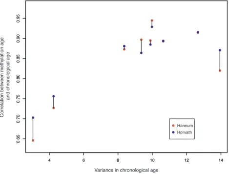
inchronologicalageineachsample.ThiseffectisshowninFig.1, which
plotstheassociationbetweenthevarianceinageineachsampleandthe
correlationbetweenDNAmageandchronologicalageforbothDNAm
age algorithms (linked via barbell for each study). TheHannum and
Horvath age estimatescorrelatedwith eachother atr = 0.89in the
meta-analysis. Across studies,the meanHannum age residualwas <
0.001years(SD=4.33years,range:−12.21to15.68years),andthe
meanHorvathageresidualwas<0.001years(SD=4.33years,range:
−12.98to15.71years).Uponmeta-analysis,theHannumandHorvath
age residualsweremoderatelycorrelatedwitheach other(r =0.56).
Detailsonthesesta-tisticsareprovidedinTable2.
3.2. AssociationsbetweenDNAmageresidualsanddemographicand cellularvariables
Tables Table 3and S1 show the patterns of association between
DNAm age residuals and sex, ancestry, and WBC proportions,
meta-analyzedfromthecovariateportionoftheregressionequations.Ofnote,
atthemeta-analyticlevel(Table3),thereweresignificantasso-ciations
between sex and both DNAm age residuals, such that women had
deceleratedagingrelativetomen.CD4Tcellproportionswereinversely
associated with both DNAm age residuals, and NK cell counts were
positivelyassociatedwithHannumDNAmageresiduals.Forestplotsfor
thesesignificantmeta-analyticeffectsareshowninFig.2.
3.3. Associationsbetweentrauma,PTSD,andDNAmageresiduals
TablesS2andTable3 showtheindividualandmeta-analyticresults,
respectively, for the regressions of each DNAm age residual on the
trauma and PTSD variables. There wereno significant meta-analytic
effectsofchildhood(orlifetime)traumaexposureoneitherDNAmage
residual (smallest p = 0.12) when evaluating all metrics of trauma
exposureacrossallstudies.Toevaluatepotentialmethodologicalsources
of variability (see above), we then examined associations between
childhood traumaas assessedbytheCTQ (3 studies) andseparately,
itemsontheTLEQ(4studies).ThisanalysisrevealedthatCTQ-defined
childhood trauma ex-posure was positively associated with Hannum
DNAmageresiduals,withoverlapping95%confidenceintervalsforthe
magnitudeoftheeffectacrosscohorts(metaβ=0.46,p=0.028which
wasexactlyequaltotheadjustedthreshold,I2 = 0, see Fig.3,Tables
Table3,S3)whiletherewasnosignificanteffectforstudiesutilizingthe
TLEQ(TablesTable3, S3). No significanteffectsemergedwhenlimiting
the evaluation of lifetime trauma history to theTLEQ (Tables Table
3, S3).
Meta-analysesrevealedanearsignificantassociationbetween
life-timePTSDdiagnosisandHannumDNAmageresiduals(metaβ=0.53,
p=0.06,totaln=1371)andasignificantassociationbetweenlifetime
PTSD severity and Hannum DNAm age residuals (meta β=0.01,
p=0.016,I2 =0,totaln=1317,seeTablesTable3,S3,Fig.3),with
overlapping95%confidenceintervalsacrossthecohorts.10Therewere
nosignificantassociationsbetweencurrentPTSDdiagnosisorseverity
andDNAmageresiduals(smallestp=0.13;TablesTable3,S3).The
DNAm age residuals just missed the unadjusted threshold for statistical
significance (p= 0.06); there was no significant association for current
PTSD diagnosis when limiting the analysis to studies using the CAPS
(TablesTable 3, S3).
All significant trauma and PTSD effects noted above were with the
Hannum DNAm age residuals. There were no significant associations
between any of the trauma or PTSD variables and Horvath DNAm age
residuals (Tables Table 3, S2, S3). There were also no significant
meta-analytic interaction effects to suggest a moderating influence of sex or
chronological age on the associations between trauma and PTSD with
either set of DNAm age residuals (smallestp= 0.10; Table S4).
4. Discussion
This was the largest and most demographically heterogeneous
evaluation of the associations between trauma exposure, PTSD, and accelerated aging in DNAm, spanning nine studies and over 2000 par-ticipants. Results of meta-analyses suggested that both childhood trauma exposure (when assessed with the CTQ) and lifetime PTSD se-verity (assessed with the CAPS for the majority of studies with lifetime PTSD severity data), were associated with accelerated epigenetic aging. Associations between lifetime PTSD diagnosis and current PTSD se-verity (when assessed with the CAPS) and DNAm age residuals just
failed to meet the unadjusted threshold for statistical significance,
though the direction of the effect was consistent with those for lifetime
PTSD severity and CTQ-measured childhood trauma. Though there was
variability in the p-values associated with each cohort, collectively,
results suggest an overlapping pattern of association between traumatic stress and acceleration of the pace of the epigenetic clock, albeit the relationship was modest in magnitude. Each additional exposure to a
Fig. 1.shows the relationship between variance in each study in chronological age and the magnitude of the correlation between chronological age and Horvath and Hannum DNAm age estimates (linked via barbell for each study).
Table 3
Meta-analytic associations between Demographic, Cellular, and Traumatic Stress Variables in Association with DNAm Age Residuals.
N Hannum Horvath
beta se p beta se p
Sex 2185 −1.1455 0.389282 0.003255 −0.93492 0.334843 0.005236
PC1 2152 0.23162 2.367165 0.922054 1.0379 2.391098 0.664239
PC2 2152 0.47236 2.046801 0.817486 −0.24999 2.016886 0.901355
PC3 2152 1.666092 2.499842 0.505105 2.673311 2.79451 0.338754
CD8T 2158 −0.00293 0.050745 0.954011 2.946052 2.345547 0.209109
CD4T 2143 −12.1967 3.610022 0.000729 −4.94381 2.107359 0.018978
Bcell 2127 0.031993 0.043265 0.459626 −5.40386 4.355332 0.214699
NK 2142 8.444542 3.10706 0.006571 −0.08119 0.083031 0.328179
Mono 2170 0.139378 0.109426 0.20276 0.034167 0.107404 0.750396
Childhood Trauma 2009 0.060947 0.096259 0.526634 −0.03471 0.110594 0.753619
Childhood Trauma (CTQ) 487 0.459596 0.209814 0.028488 −0.11822 0.237175 0.618178
Childhood Trauma (TLEQ) 1295 −0.1468 0.173309 0.396967 −0.16058 0.132627 0.225986
Lifetime Trauma 2024 0.017713 0.036284 0.625429 −0.05254 0.033963 0.121898
Lifetime Trauma (TLEQ) 1296 −0.02009 0.040609 0.620864 −0.05992 0.043818 0.171457
Current PTSD Dx 2104 0.12899 0.189075 0.495103 −0.11764 0.193403 0.542997
Current PTSD Sev 2081 0.004478 0.002969 0.131459 0.000469 0.003043 0.877467
Current PTSD Dx (CAPS) 1216 0.341131 0.262793 0.194254 0.010832 0.251552 0.965654
Current PTSD Sev (CAPS) 1210 0.008573 0.004587 0.061615 0.004787 0.004356 0.271811
Lifetime PTSD Dx 1371 0.529147 0.276291 0.05547 0.122311 0.410977 0.766
Lifetime PTSD Sev 1251 0.011099 0.004608 0.01601 0.005694 0.006226 0.3604
new type of childhood trauma on the CTQ was associated with nearly a
half-year of age acceleration; the difference with respect to age
accel-eration between someone with a score of 0 on the CAPS versus a highly symptomatic patient with a score of 100 would be expected to be about
1.1 years. Small effects in DNAm studies are commonplace and it is
critical to next examine the functional effects of such differences, which
may be considerable with respect to gene expression, and to model the
potential cumulative effects of advanced DNAm over time (Breton et al.,
2017).
As many as one-third of patients with PTSD exhibit a chronic form
of the disease that persists for years (Kessler, 2000) and the chronicity
and severity of such symptoms would be expected to magnify the ne-gative health correlates of PTSD. Indeed, one study that modeled PTSD
burden as defined by both symptom severity and duration (e.g., chronicity) found that the burden of the disease evidenced stronger negative associations with cortical thickness than did PTSD severity
alone (Lindemer et al., 2013). Based on this, it is possible that effect size
estimates in this study underestimate the association between traumatic stress and cellular age because they do not account for the burden of
traumatic stress across time. Future research may benefit from
quanti-fying the burden of PTSD as a function of both disorder severity and chronicity. Consistent with this interpretation, we found that
childhood, but not lifetime, trauma exposure was associated with ac-celerated aging and this could suggest: (a) that there are critical
win-dows during childhood in which the effects of trauma exposure are
particularly damaging; and/or (b) that childhood trauma could be a marker for a more prolonged period of psychiatric distress that con-tributes to overall greater burden. Longitudinal studies are necessary to evaluate these possibilities.
Associations between traumatic stress and advanced epigenetic age were observed for the Hannum et al. algorithm but not for the Horvath metric. To our knowledge, prior positive associations between PTSD and epigenetic age have only been shown for the Hannum et al. metric (Wolf et al., 2016, 2017), while positive effects for general life stressors, violence, and trauma exposure have previously been reported for the
Horvath index (Zannas et al., 2015;Boks et al., 2015;Jovanovic et al.,
2017). It is remarkable that the two algorithms show differential
pat-terns of association given that they are highly correlated with each other and with chronological age. Despite these strong correlations, the components of each age algorithm that do not index chronological age, as captured by the age residuals, appear to be fairly distinct from each other: the two age residuals shared approximately 25% of the variance
and evidenced differential patterns of association with the WBCs in this
study. The two algorithms have just 6 loci and 11 genes in common
(Wolf et al., 2016) and together with the results of this study, this
implies that each algorithm may be sensitive to different pathogenic
environmental and biological processes, though more research is needed to test this. The strong meta-analytic correlations between both DNAm age estimates and chronological age in the context of the re-lationship between variance in age in each sample and the strength of
the DNAm age/chronological age correlation (Fig. 1) are also
in-formative. This supports the accuracy of the DNAm age estimates and raises the possibility that in instances in which the correlation between DNAm age and chronological age is weaker than expected, that this may be due to limited age variance in the sample, and may not
ne-cessarily reflect a failure in the algorithm.
Results of this study underscore the importance of identifying the biological mechanisms that link traumatic stress to age acceleration. Zannas et al. (2015)examined the responsivity of the loci included in the Horvath algorithm to the glucocorticoid receptor agonist dex-amethasone and found that approximately 31% of the DNAm loci were responsive to dexamethasone while over 80% of genes located near the DNAm loci evidenced dexamethasone-related changes in gene tran-scription. Consistent with this, about a quarter of the DNAm loci were shown to be localized to glucocorticoid response elements. As well, Davis et al. (2017)found advanced Horvath DNAm age to be correlated with diurnal cortisol levels. This pattern of results is consistent with a
central role of stress hormones in PTSD (Yehuda, 2009).
Dex-amethasone-regulated genes also showed enrichment in gene networks associated with age-related diseases (i.e., coronary artery disease,
ar-teriosclerosis, leukemia, etc.;Zannas et al., 2015). This raises the
pos-sibility that accelerated cellular aging in DNAm may link traumatic stress to increased risk for pre-mature disease onset.
Complementary research on the loci included in the Hannum al-gorithm has yet to be conducted and comparing the responsivity of these loci to those in the Horvath algorithm is critical for understanding
differential patterns of association across the two metrics. As well,
evaluation of the sensitivity of the loci to other dynamic biological
processes implicated in PTSD, such as pro-inflammatory cytokines
(Passos et al., 2015), and catecholamines (Highland et al., 2015), would help to elucidate the biological mechanisms involved in traumatic-stress related accelerated aging. This type of evaluation is also im-portant for determining whether epigenetic age acceleration is a me-chanism for, or simply a biomarker of, early health decline. Behavioral pathways to epigenetic aging are also critical to evaluate further, given evidence that insomnia (which is also a symptom of PTSD) and obesity
(which is highly comorbid with PTSD;Pagoto et al., 2012) are
asso-ciated with accelerated DNAm age (Carroll et al., 2017; Nevalainen
et al., 2017).
We also observed meta-analytic effects suggesting that women, on
average, had decelerated aging relative to men (using both indices).
This has been reported previously in blood and brain tissue (Horvath
et al., 2016;Hannum et al., 2013) but the interpretation of the effect is
unclear. Thisfinding could reflect an underlying differential rate of
epigenetic aging in men versus women, and/or differential
suscept-ibility to factors that cause epigenetic age to deviate from chronological age across the sexes. These explanations are consistent with the
common finding that women tend to outlive men (Beltrán-Sánchez
et al., 2015), perhaps because their pace of cellular aging is attenuated.
On the other hand, thefinding could indicate that the age algorithms
are simply more accurate in one sex over the other. There is preliminary
evidence to suggest that differential age acceleration across the sexes
does not alter the strength of the association between epigenetic age and important clinical correlates, including traumatic stress in this
study, and time to death in prior work (Chen et al., 2016). This suggests
that regardless of the reason for the differential age acceleration across
sexes, the predictive power of accelerated epigenetic age is not di-minished in one sex compared to the other.
on published summary statistics. Results suggested that childhood trauma (when assessed with a detailed instrument) and lifetime PTSD severity were both associated with advanced DNAm age, though the
magnitude of the effect was small and additional research is needed to
determine how the chronicity of psychiatric symptoms might contribute to accelerated aging. As genome-wide and epigenome-wide testing move from the research to the clinical domains, assessment of
epige-netic age may become a practical approach for tracking an individual’s
cellular age, and testing the utility of interventions designed to slow the pace of cellular aging. In the future, we may develop traumatic-stress
specific epigenetic age profiles that contribute to our understanding of
the neurobiological consequences of trauma and PTSD.
Disclosures
In the past 3 years, Dr. Kessler received support for his
epidemio-logical studies from SanofiAventis; was a consultant for Johnson &
Johnson Wellness and Prevention, Sage Pharmaceuticals, Shire, Takeda; and served on an advisory board for the Johnson & Johnson Services Inc. Lake Nona Life Project. Kessler is a co-owner of DataStat,
Inc., a market researchfirm that carries out healthcare research.
Conflict of interest
All other authors report nofinancial conflicts of interest relevant to
this work.
Acknowledgements
The Psychiatric Genomics Consortium (PGC) PTSD Epigenetics Workgroup is supported by the U.S. Army Medical Research and Materiel Command and the National Institute of Mental Health (NIMH; R01MH108826).
This work was supported in part by: Merit Review Award Number I01 CX-001276-01 to EJW from the United States (U.S.) Department of
Veterans Affairs Clinical Sciences R&D (CSR&D) Service, National
Institute On Aging of the National Institutes of Health under Award Number R03AG051877-02S1 to EJW, Presidential Early Career Award for Scientists and Engineers to EJW as administered by U.S. Department
of Veterans Affairs Office of Research and Development (PECASE
2013A).
Nicole Nugent’s effort on this project was supported by NIMH
R01MH105379 & R01MH108641.
Funding for the TRACTS study was supported in part by NIMH grant
R21MH102834 “Neuroimaging Genetics of PTSD” to MWM and the
Translational Research Center for TBI and Stress Disorders (TRACTS), a VA Rehabilitation Research and Development Traumatic Brain Injury Center of Excellence (B9254-C). This research is the result of work
supported with resources and the use of facilities at the
Pharmacogenomics Analysis Laboratory, Research and Development Service, Central Arkansas Veterans Healthcare System, Little Rock, Arkansas.
Funding for the NCPTSD cohort was supported by U.S. Department of VA CSRD Merit Review award5I01CX000431-02 and by U.S. Department of VA Biomedical Laboratory Research & Development Program award 1I01BX002150-01, both to MWM. This project was also supported by NIMH RO1MH079806 to MWM.
Funding for the Mid-Atlantic MIRECC cohorts was provided in part by grant #I01 BX002577 from the Biomedical and Laboratory Research and Development (BLR&D) Service of the Department of Veterans
Affairs Office of Research and Development (VA ORD), grant
#IK2CX000525 and #11S-RCS-009 from the CSR&D Service of VA ORD, and the VA Mid-Atlantic Mental Illness Research, Education, and Clinical Center (MIRECC).
Funding for MRS was provided by the Marine Corps, Navy Bureau of Medicine and Surgery (BUMED), VA Health Research and Development
WealsoobservedanegativerelationshipbetweenestimatesofCD4T
cellsandbothDNAmageresidualsandapositiveassociationbetween
NKcellsandHannumageresiduals.Similarpatternsofassociationwere
reportedinmeta-analysesof13,000individualsbyChenetal.(2016)
andinover4600individualsbyMarionietal.(2015).Thesefindings
raisethepossibilitythatcellsthatareintegraltoimmunesystem
maintenanceandresponsivityplayaroleinalteringthepaceofthe
epigeneticclocksothatitlosessynchronicitywiththepaceof
chron-ologicalaging.Alternatively,Chenetal.andHorvathetal.(2016)
combinedinformationfromageresidualsandestimatedWBCcounts
andfoundthatthismetricwasabetterpredictoroftimetodeath(Chen
etal.,2016)andmetabolicandinflammatorymarkers(Horvathetal.,
2016)comparedtotheDNAmageresidualsalone.Thecombinedindex
wasreferredtoas“extrinsicepigeneticageacceleration”and
con-ceptualizedasamarkerofthebiologicalageoftheimmunesystemin
blood(Chenetal.,2016),however,itwasnotclearwhichcomponents
ofthiscombinedmetricwereresponsiblefortheincreasedpredictive
strengthoftimetilldeath.StudiesoftheresponsivityofDNAmage
probestoinflammatoryandanti-inflammatoryagentscouldshedlight
onhowinflammationisassociatedwiththeDNAmageclock.
4.1. Studylimitations
Theseresults shouldbeconsideredinlight ofanumberof
limita-tions.First,thiswasacross-sectionalstudyand,assuch,wecannotinfer
causalassociationsbetweentraumaticstressandacceleratedDNAmage.
Second,giventhatthecontributingstudieshaddiverseavailabledata,
thereweredifferentsamplescomprisingeachmeta-analysis.Thisalso
meantthatwewereunabletodiscerntherelativeeffectsofchildhood
traumaexposureversuslifetimePTSDseverityasonlyonestudy(Grady
TraumaProject)hadboththeCTQchildhoodtraumaexposurevariable
andlifetimePTSDseverity.Forthesamereasons,wewereunableto
includechildhoodtraumaandadulttraumaexposureinthesamemodel
ortoevaluatetheinfluenceofchronicinterpersonalviolenceacrossthe
lifetime.Thus,asthislineofworkisdevelopedandnewdatabecome
available, it will be important to disentangle the relative effects of
trauma exposure versus PTSD on accelerated aging. As this
meta-analysis was based on a relativelynascent field withfewer than10
contributing studies,statisticalpowerwas alsoapotentiallimitation.
However, power for meta-analyses based on summary data across
individualstudieshasbeenshowntobesimilartopowerforanalysesin
whichdatafromdistinctstudiesarepooledtoformonelarge sample
(OlkinandSampson,1998;MathewandNordstrom,1999;LinandZeng, 2010a,b)andthisappliestoge-neticassociationstudiesaswell(Linand Zeng,2010a,b;Sungetal.,2014).Ourpooledsamplesizeshouldhave
provided adequate power in this context, though inclusion of more
cohortsintothemeta-analysiswouldstrengthenresults.Aswell,while
weattemptedtolookforsourcesofdemographicvariationintheresults,
thereareundoubtedlyothervariablesthatwedidnothaveaccesstothat
might moderate the primary associations (or otherwise serve as
importantcovariates).Thisconcernisoffsetbythestrengthofthe
meta-analytic approach which yields improved statistical power and
generalizability ofresults acrosspopulationsrelativetoanindividual
cohort.
4.2. Conclusions
Evaluationofepigeneticcellularagingisinitsinfancy,withthevast
majority ofresearch todatefocusedon theutilityof theDNAmage
calculatorstopredictmortalityandhealthoutcome.Thisstudywasthe
largest toevaluate traumatic stress-related advanced epigenetic age,
andtoourknowledge,wasthelargestexaminationofanypsychiatric
symptomdomaininassociationwithDNAmagetodate.Bydeployinga
uniform data analytic script and harmonizing critical variables, our
analytic approachwas able toeliminatesome sourcesof
Affairs Mid-Atlantic Mental Illness Research, Education, and Clinical Center Workgroup, Beckham, J.C., Hauser, M.A., 2015. Genome-wide association study of posttraumatic stress disorder in a cohort of Iraq-Afghanistan era veterans. J. Affect. Disord. 184, 225–234.
Baker, D.G., Nash, W.P., Litz, B.T., Geyer, M.A., Risbrough, V.B., Nievergelt, C.M., O’Connor, D.T., Larson, G.E., Schork, N.J., Vasterling, J.J., Hammer, P.S., Webb-Murphy, J.A., Team, M.R.S., 2012. Predictors of risk and resilience for posttraumatic stress disorder among ground combat Marines: methods of the Marine Resiliency Study. Prev. Chronic Dis. 9, E97.
Barfield, R.T., Almli, L.M., Kilaru, V., Smith, A.K., Mercer, K.B., Duncan, R., Klengel, T., Mehta, D., Binder, E.B., Epstein, M.P., Ressler, K.J., Conneely, K.N., 2014. Accounting for population stratification in DNA methylation studies. Genet. Epidemiol. 38, 231–241.
Beltrán-Sánchez, H., Finch, C.E., Crimmins, E.M., 2015. Twentieth century surge of excess adult male mortality. Proc. Natl. Acad. Sci. U. S. A. 112, 8993–8998.
Bernstein, D.P., Stein, J.A., Newcomb, M.D., Walker, E., Pogge, D., Ahluvalia, T., Stokes, J., Handelsman, L., Medrano, M., Desmond, D., Zule, W., 2003. Development and validation of a brief screening version of the Childhood Trauma Questionnaire. Child Abuse Negl. 27, 169–190.
Binder, E.B., Bradley, R.G., Liu, W., Epstein, M.P., Deveau, T.C., Mercer, K.B., Tang, Y., Gillespie, C.F., Heim, C.M., Nemeroff, C.B., Schwartz, A.C., Cubells, J.F., Ressler, K.J., 2008. Association of FKBP5 polymorphisms and childhood abuse with risk of post-traumatic stress disorder symptoms in adults. JAMA 299, 1291–1305.
Blake, D.D., Weathers, F.W., Nagy, L.M., Kaloupek, D.G., Gusman, F.D., Charney, D.S., Keane, T.M., 1995. The development of a Clinician-Administered PTSD Scale. J. Trauma Stress 8, 75–90.
Boks, M.P., van Mierlo, H.C., Rutten, B.P., Radstake, T.R., De Witte, L., Geuze, E., Horvath, S., Schalkwyk, L.C., Vinkers, C.H., Broen, J.C., Vermetten, E., 2015. Longitudinal changes of telomere length and epigenetic age related to traumatic stress and post-traumatic stress disorder. Psychoneuroendocrinology 51, 506–512. Borenstein, M., Higgins, J.P., Hedges, L.V., Rothstein, H.R., 2017. Basics of meta-analysis:
I2is not an absolute measure of heterogeneity. Res. Synth. Methods 8, 5–18.
Bremner, J.D., Bolus, R., Mayer, E.A., 2007. Psychometrics properties of the Early Trauma Inventory-Self Report. J. Nervous Ment. Dis. 195, 211–218.
Breslau, N., Kessler, R.C., Chilcoat, H.D., Schultz, L.R., Davis, G.C., Andreski, P., 1998. Trauma and posttraumatic stress disorder in the community: the 1996 Detroit Area Survey of Trauma. Arch. Gen. Psychiatry 55, 626–632.
Breton, C.V., Marsit, C.J., Faustman, E., Nadeau, K., Goodrich, J.M., Dolinoy, D.C., Herbstman, J., Holland, N., LaSalle, J.M., Schmidt, R., Yousefi, P., Perera, F., Joubert, B.R., Taylor, M., Yang, I.V., Chen, R., Hew, K.M., Freeland, D.M., Miller, R., Murphy, S.K., 2017. Small-magnitude effect sizes in epigenetic end points are important in children’s environmental health studies: the children’s environmental health and disease prevention research center’s epigenetics working group. Environ. Health Perspect. 125, 511–526.
Carroll, J.E., Irwin, M.R., Levine, M., Seeman, T.E., Absher, D., Assimes, T., Horvath, S., 2017. Epigenetic aging and immune senescence in women with insomnia symptoms:
findings from the Women’s Health Initiative Study. Biol. Psychiatry 81, 136–144.
Chen, Y.A., Lemire, M., Choufani, S., Butcher, D.S., Grafodatskaya, D., Zanke, B.W., Gallinger, S., Hudson, T.J., Weksberg, R., 2013. Discovery of cross-reactive probes and polymorphic CpGs in the Illumina Infinium Human Methylation 450 microarray. Epigenetics 8, 203–209.
Chen, B.H., Marioni, R.E., Colicino, E., Peters, M.J., Ward-Caviness, C.K., Tsai, P.C., Roetker, N.S., Just, A.C., Demerath, E.W., Guan, W., Bressler, J., Fornage, M., Studenski, S., Vandiver, A.R., Moore, A.Z., Tanaka, T., Kiel, D.P., Liang, L., Vokonas, P., Schwartz, J., Lunetta, L.L., Murabito, J.M., Bandinelli, S., Hernandez, D.G., Melzer, D., Nalls, M., Pilling, L.C., Price, T.R., Singleton, A.B., Gieger, C., Holle, R., Kretschmer, A., Kronenberg, F., Kunze, S., Linseisen, J., Meisinger, C., Rathmann, W., Waldenberger, M., Visscher, P.M., Shah, S., Wray, N.R., McRae, A.F., Franco, O.H., Hofman, A., Uitterlinden, A.G., Absher, D., Assimes, T., Assimes, T., Levine, M.E., Lu, A.T., Feinberg, A.P., Levy, D., Baccarelli, A., van Meurs, J., Bell, J.T., Peters, A., Deary, I.J., Pankow, J.S., Ferrucci, L., Horvath, S., 2016. DNA methylation-based measures of biological age: meta-analysis predicting time to death. Aging (Albany, NY) 8, 1844–1865.
Christensen, B.C., Houseman, E.A., Marsit, C.J., Zheng, S., Wrensch, M.R., Wiemels, J.L., Nelson, H.H., Karagas, M.R., Padbury, J.F., Bueno, R., Sugarbaker, D.J., Yeh, R.F., Wiencke, J.K., Kelsey, K.T., 2009. Aging and environmental exposures alter tissue-specific DNA methylation dependent upon CpG island context. PLoS Genet. 5, e1000602.
Christiansen, L., Lenart, A., Tan, Q., Vaupel, J.W., Aviv, A., McGue, M., Christensen, K., 2016. DNA methylation age is associated with mortality in a longitudinal Danish twin study. Aging Cell 15, 149–154.
Davidson, J.R., Book, S.W., Colket, J.T., Tupler, L.A., Roth, S., David, D., Hertzberg, M., Mellman, T., Beckham, J.C., Smith, R.D., Davison, R.M., Katz, R., Feldman, M.E., 1997. Assessment of a new self-rating scale for post-traumatic stress disorder. Psychol. Med. 27, 153–160.
Davis, E.G., Humphreys, K.L., McEwen, L.M., Sacchet, M.D., Camacho, M.C., MacIsaac, J.L., Lin, D.T.S., Kobor, M.S., Gotlib, I.H., 2017. Accelerated DNA methylation in adolescent girls: associations with elevated diurnal cortisol and reduced hippocampal volume. Transl. Psychiatry 7, e1223.
First, M.B., Spitzer, R.L., Gibbon, M., Williams, J.B., 1994. Structured Clinical Interview for Axis I DSM-IV Disorders. Biometrics Research, New York.
Gillespie, C.F., Bradley, B., Mercer, K., Smith, A.K., Conneely, K., Gapen, M., Weiss, T., Schwartz, A.C., Cubells, J.F., Ressler, K.J., 2009. Trauma exposure and stress-related disorders in inner city primary care patients. Gen. Hosp. Psychiatry 31, 505–514. Gray, M.J., Litz, B.T., Hsu, J.L., Lombardo, T.W., 2004. Psychometric properties of the life
events checklist. Assessment 11, 330–341.
Hannum, G., Guinney, J., Zhao, L., Zhang, L., Hughes, G., Sadda, S., Klotzle, B., Bibikova, M., Fan, J.B., Gao, Y., Deconde, R., Chen, M., Kajapakse, I., Friend, S., Ideker, T., Zhang, K., 2013. Genome-wide methylation profiles reveal quantitative views of human aging rates. Mol. Cell. 49, 359–367.
Hedtke, K.A., Ruggiero, K.J., Fitzgerald, M.M., Zinzow, H.M., Sauders, B.E., Resnick, H.S., Kilpatrick, D.G., 2008. A longitudinal investigation of interpersonal violence in re-lation to mental health and substance use. J. Consult. Clin. Psychol. 76, 633–647. Highland, K.B., Costanzo, M., Jovanovic, T., Norrholm, S.D., Ndiongue, R., Reinhardt, B.,
Rothbaum, B., Roy, M.J., 2015. Biomarkers of post-deployment resilience among military service members. Neurobiol Stress 2, 62–66.
Horvath, S., Pirazzini, C., Bacalini, M.G., Gentilini, D., Di Blasio, A.M., Delledonne, M., Mari, D., Arosio, B., Monti, D., Passarino, G., De Rango, F., D’Aguila, P., Giuliani, C., Marasco, E., Collino, S., Descombes, P., Garagnani, P., Franceschi, C., 2015. Decreased epigenetic age of PBMCs from Italian semi-supercentenarians and their offspring. Aging (Albany, NY) 7, 1159–1170.
Horvath, S., Gurven, M., Levine, M.E., Trumble, B.C., Kaplan, H., Allayee, H., Ritz, B.R., Chen, B., Lu, A.T., Rickabaugh, T.M., Jamieson, B.D., Sun, D., Li, S., Chen, W., Quintana-Murci, L., Fagny, M., Kobor, M.S., Tsao, P.S., Reiner, A.P., Edlefsen, K.L., Absher, D., Assimes, T.L., 2016. An epigenetic clock analysis of race/ethnicity, sex, and coronary heart disease. Genome Biol. 17, 171.
Horvath, S., 2013. DNA methylation age of human tissues and cell types. Genome Biol. 14, R115.
Houseman, E.A., Accomando, W.P., Koestler, D.C., Christensen, B.C., Marsit, C.J., Nelson, H.H., Wiencke, J.K., Kelsey, K.T., 2012. DNA methylation arrays as surrogate mea-sures of cell mixture distribution. BMC Bioinf. 13, 86.
Hovens, J.E., Bramsen, I., van der Ploeg, H.M., 2000. Self Report Measure for PTSD Symptoms: SRIP Manual. Lisse.
Hovens, J.E., Bramsen, I., van der Ploeg, H.M., 2002. Self-rating inventory for posttrau-matic stress disorder: review of the psychometric properties of a new brief Dutch screening instrument. Percept. Mot. Skills 94, 996–1108.
Jaffe, A.E., Irizarry, R.A., 2014. Accounting for cellular heterogeneity is critical in epi-genome-side association studies. Genome Biol. 15, R31.
Johnson, W.E., Li, C., Rabinovic, A., 2007. Adjusting batch effects in microarray ex-pression data using empirical Bayes methods. Biostatistics 8, 118–127.
(HSR&D) andNIH R01MH093500.AcknowledgedareMarkAGeyer
Ph.D(VASanDiegoHealthcareSystem&UCSD),DanielT.O’Connor
(UCSD)andallMRSinvestigatorsandstaff.Theauthorsalsothankthe
Marine andNavyCorpsmenvolunteersformilitaryserviceand
parti-cipationinMRS.
Funding for DNHS was provided by NIHR01DA022720 and
RC1MH088283. We thank themany Detroit residents whochoseto
participateintheDNHS,aswellasthetechnicalandsupportstaffwho
assistedwiththestudy.
TheGradyTraumaProject,whichappreciatesthetechnicalsupport
of all of the staff, volunteers andparticipants, is supported by the
NationalInstitutesofMentalHealth(MH096764andMH071537).
The VA Mid-Atlantic MIRECC Workgroup contributors for this
publication include: Mira Brancu, Patrick S. Calhoun, Kathleen P.
Decker, EricDedert,EricB.Elbogen,John A.Fairbank, KimberlyT.
Green,RobinA.Hurley,JasonD.Kilts,AngelaKirby,ChristineE.Marx,
GregoryMcCarthy,ScottD.McDonald,MarinellMiller-Mumford,Scott
D.Moore, RajendraA. Morey,JenniferC.Naylor,TrevenC.Pickett,
JaredRowland,JenniferJ.Runnals,CindySwinkels,StevenT.Szabo,
Katherine H.Taber, Larry A. Tupler,Elizabeth E. Van Voorhees, H.
RyanWagner,RichardD.Weiner,andRuthE.Yoash-Gantz.
ArmySTARRSwassponsoredbytheDepartmentoftheArmyand
fundedundercooperativeagreementnumberU01MH087981withthe
U.S.DepartmentofHealthandHumanServices,NationalInstitutesof
Health,NationalInstituteofMentalHealth(NIH/NIMH).
ThecontentsofthisarticledonotrepresenttheviewsoftheU.S.
DepartmentofVeteransAffairs,theNationalInstitutesofHealth,orthe
UnitedStatesGovernment.
AppendixA. Supplementarydata
Supplementarydataassociatedwiththisarticlecanbefound,inthe
onlineversion,athttps://doi.org/10.1016/j.psyneuen.2017.12.007.
References
Afari,N.,Ahumada,S.M.,Wright,L.J.,Mostoufi,S.,Golnari,G.,Reis,V.,Cuneo,J.G., 2014.Psychologicaltraumaandfunctionalsomaticsyndromes:asystematicreview andmeta-analysis.Psychosom.Med.76,2.
Andrews,G.,Peters,L.,1998.Thepsychometricpropertiesofthecompositeinternational diagnosticinterview.Soc.PsychiatryPsychiatr.Epidemiol.33,80–88.