Address for correspondence Dr. Cita Rosita Sigit Prakoeswa
Dermatology and Venereology Department, Faculty of Medicine Universitas Airlangga – Dr. Soetomo Teaching Hospital,
Prof. Dr. Moestopo No.47, Surabaya, Indonesia. Ph: 6231-5501609
Email: [email protected] D
Original Article
Expression of WNT1 signalling proteins after
phototherapy exposure in vitiligo
Introduction
Vitiligo is a pigmentation disorder which can be in the form of white patches or like lime (depigmentation) on the skin and mucous
membranes. Phototherapy is one form of vitiligo therapy that is often used today. Repigmentation that occurs after therapy mostly forms a perifolicular pattern, the rest form marginal, diffuse and combination patterns.1,2 Poor therapeutic results in vitiligo are at the fingertips (which are relatively hairless) and in vitiligo accompanied by leukotrichia.3,4 Until now there is still no clear mechanism for the occurrence of perifollicular pattern repigmentation after administration of NB-UVB in vitiligo. Melanocyte stem cells (SPM) are known to exist Dian Ardiana, Nanny Herwanto*, Cita Rosita Sigit Prakoeswa*, Indropo Agusni*
Department of Dermatology & Venereology, Faculty of Medicine University of Hang Tuah -Dr. Ramelan Naval Hospital Surabaya, Indonesia.
* Department of Dermatology & Venereology, Faculty of Medicine Universitas Airlangga - Dr. Soetomo Teaching Hospital Surabaya, Indonesia.
Abstract
Background Melanocyte stem cells (MelSCs) have been identified in the lower permanent portion of hair follicle and associated with perifollicular repigmentation pattern of vitiligo after Narrow Band-Ultraviolet B (NB-UVB) therapy. Wnt1 and β-catenin have been associated with UVB induced pigmentation in mice and hyperpigmented lesion commonly seen on sun-exposed areas. The increase of MelSCs in these two conditions suggested the role of Wnt1 in MelSCs differentiation, which increases the number of melanocytes.Purpose To know the expression of hair follicle Wnt1 and β-catenin in NB-UVB induced vitiligo perifollicular repigmentation.
Methods This clinical pre-experimental study used one group pre test-post test design. The dose of 390 mJ/cm2 DermaPalTM Daavlin NB-UVB was given twice a week for 2 months to 18 vitiligo patients. Biopsy was undertaken in vitiligo areas involving hair follicle before and after therapy and was assessed by immunohistochemistry technique.
Results There was a significant difference (p<0.05) of vitiligo area size, the number of cells expressing Wnt1 and the number of MelSCs expressing β-catenin in hair follicle before and after NB-UVB irradiation.
Conclusion There was an increment of hair follicle Wnt1 and β-catenin expression in vitiligo perifollicular areas before and after NB-UVB therapy. The finding of this study could be used for further research in perifollicular repigmentation mechanism.
Key words
in the outer root sheath of the permanent bottom of the hair follicle (bulge region) and are associated with vitiligo repigmentation. Inactive melanocytes in the outer root sheath undergo division, proliferation and maturation during repigmentation, and the possibility of epidermal repigmentation originating from the migration of these melanocytes. This study confirmed the presence of melanocytes in the outer root sheath of the hair follicle.5
The WNT gene is known to play a role in various biological processes, cell type determination, cell proliferation, cell migration, stem cell maintenance, tumor suppression and oncogenesis. A total of 19 WNT genes have been identified in humans. Wnt is a secretion protein that functions as a signaling molecule. The Wnt protein bond with the Frizzled receptor (Fzd) will cause a signal that will inhibit the degradation of the cytoplasmic β-catenin protein resulting in β-catenin accumulation. β-catenin
will then enter the nucleus to form complexes with transcription factors that will stimulate the expression of target genes, including the microphthalmia-associated transcription factor (MITF) gene.6 MITF is a regulator protein that plays a role in survival, cell cycle regulation, migration and differentiation of melanocyte lineages.7
Research in hyperpigmented mice after exposure to UVB showed an increase in Wnt1 mRNA when compared to unexposed skin.8 SPM in the bulge region, as well as SPM in the bulge region which has a nucleus containing β-catenin, is more common in solaris lentigo lesions than normal skin. This shows that in solaris lentigo hyperpigmented skin lesions there is induction of Wnt1 expression and Wnt / β-catenin signal, and SPM bulge region differentiation.9 This study aimed to find out more clearly the mechanism of the emergence of perifollicular vitiligo repigmentation through the
administration of NB-UVB based on Wnt1 and
β-catenin profiles in the hair follicles.
Materials and Methods
Research Design The type of research conducted was clinical pre-experimental research, using one group pre-post-test design without control. This research was approved by the Medical Research Ethics Commission of Dr. Ramelan Naval Hospital Surabaya.
Patient The study was conducted on vitiligo patients who came for outpatient treatment at the Dermatology and Venereology Clinic Dr. Ramelan Naval Hospital Surabaya. The inclusion criteria in this study were patients aged 15-60 years; with Fitzpatrick IV-V skin type; lesions on the body or extremity; black hair in vitiligo lesions and lesions large enough for a four millimeter punch biopsy. The exclusion criteria in this study were patients who had a history of being sensitive to sunlight (photodermatosis); acrofacial, mucosal and universal form of vitiligo; is pregnancy, immuno-deficiency condition; are getting topical or systemic therapy that effects the results of phototherapy; being treated for vitiligo in the past two months, experiencing severe erythema and/ or burns due to radiation, and having a history of keloids. Photographs of vitiligo lesions were taken at the beginning and end of the study. All patients signed a consent letter before taking the study.
counting them with the help of millimeter scale paper.
Tissue processing Vitiligo lesions and hair follicles were taken using a 4 mm punch biopsy. Skin tissue extraction was carried out before and after the study ended. Network preparation and slide making were carried out in the Anatomical Pathology laboratory of the Faculty of Medicine, Universitas Airlangga, Surabaya. Immuno-histochemical examination was conducted at the Laboratory of Scientific Medical Society, Faculty of Medicine, Universitas Airlangga, Surabaya.
The single-stain immunohistochemical examination procedure was carried out to see the expression of Wnt1 and double-stain immunohistochemistry performed to see melanocyte stem cells expressing β-catenin. The antibodies used were the anti-Wnt-1 monoclonal mouse (Novus, catalog number NBP1-51575), rabbit polyclonal anti Fzd4 [(AA 190-222), (Antibodies-online GmBH, ABIN1711682 catalog number)], anti β-catenin monoclonal mouse [(E-5), (Santa Cruz, catalog number sc-7963)], UltraVision Anti-Plyvalent, HRP / AEC (Thermo Scientific, TP-015-HA catalog number) and MultiVision Polymer anti-rabbit/ AP + anti- mouse/ HRP polymers (Thermo Scientific, TL-012MHRA catalog number).
Data for each sample in this study is the average number of cells observed in four fields of view at 400 magnification. The immune-histochemistry of melanocyte stem cell expressing β-catenin was observed using the Leica DM 750 microscope with the Leica ICC 50HD camera and the Las Ez Leica program. The results of immunohistochemical examination of Wnt1 expression were observed using a Nikon H600L microscope; 300 megapixel DS Fi2 camera, with Nikkon Image System image processing software.
Statistical analysis
Extent of lesion data, number of cells expressing Wnt1 and number of stem cells expressing
β-catenin before and after irradiation were analyzed by the Wilcoxon Signed Ranks Test, the significance level of α used was 5%. Data were analyzed by SPSS.
Results
There were 18 patients who met the admission criteria, consisting of 10 (55.6%) men and 8 (44.4%) women. The youngest sample is 18 years old while the oldest is 51 years old. The average age of the sample is 36.8 ± 10.3 years. The majority of lesions are located in the back of the body (44.4%), namely the back and waist, the rest are on the front body (16.7%), lower extremities (33.3%) and upper extremities (5.6%).
Extent of Vitiligo Lesions Extent of vitiligo lesions can be seen in Table 1. The extent of vitiligo lesions decreased significantly after administration of NB-UVB (p = 0.0001).
Pattern of vitiligo lesion repigmentation The pattern of repigmentation that occurs after the administration of NB-UVB for two months is mostly perifollicular (72.2%), the remainder is a combination of marginal and perifollicular (27.8%).
Results of immunohistochemical examination
Tabel 1 Extent of vitiligo lesions Extent of vitiligo
lesion (cm2) Minimum Maximum Median Before NB-UVB 2.16 283.38 36.37 After NB-UVB 1.98 269.17 22.91
Repigmentation
(before-after) 0.18 20.23 5.96
Tabel 2 Number of cells expressing Wnt1 in hair follicles before and after NB-UVB irradiation
Wnt1
Expression Minimum Maximum Median
Before NB-UVB 2 66 14.3
After NB-UVB 9.5 69.5 31.2
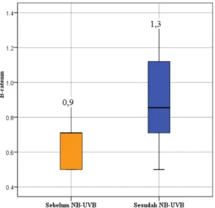
Table 3 Number of melanocyte stem cells in hair follicles that express β-catenin before and after irradiation of NB-UVB
β-catenin
Expression Minimum Maximum Median
Before NB-UVB 0.5 0.9 0.7
After NB-UVB 0.5 1.3 0.9
The number of melanocyte stem cells that express Β-Catenin before and after NB-UVB irradiation Cells in the hair follicles that show positive results (β-catenin+/Fzd4+) are characterized by blue nuclei, blue and brown cytoplasm. The number of melanocyte stem cells in hair follicles that express β-catenin before and after NB-UVB is shown in Table 3. The number of melanocyte stem cells expressing β-catenin
has significant increased after irradiation (p = 0.001).
Discussion
The search for various scientific evidence shows that some Wnt proteins are associated with differentiation of melanocyte stem cells into melanoblasts, which in turn will become melanocytes which lead to pigmentation.
Progression of vitiligo lesions after administration of therapy is characterized by the reappearance of pigmentation in vitiligo (repigmented) lesions. The success rate of
UVB therapy varies. The administration of NB-UVB in this study led to improvements in lesions characterized by a decrease in the extent of vitiligo lesions (p= 0.0001).
Perifollicular pattern of repigmentation in this study occurred in 72.2% of the sample. Yang et al. states that the pattern of perifollicular repigmentation in vitiligo lesions after administration of NB-UVB is present in 42.2% of patients1, Khullar et al. mention 68%2 and El-Zawahry et al. get a perifolicullar repigmentation pattern of 70%.2 The results of this study are similar to those obtained by El-Zawahry et al. This supports the theory of the role of hair follicles in the occurrence of perifollicular repigmentation after NB-UVB administration.
An important finding in this study was the expression of Wnt1 in vitiligo. Wnt1 is expressed in hair follicles, both before and after irradiation. The number of cells expressing Wnt1 increased after NB-UVB irradiation (p = 0.031). This shows that there is induction of Wnt1 with the administration of NB-UVB and suggests that Wnt1 plays a role in the perifollicular repigmentation of vitiligo.
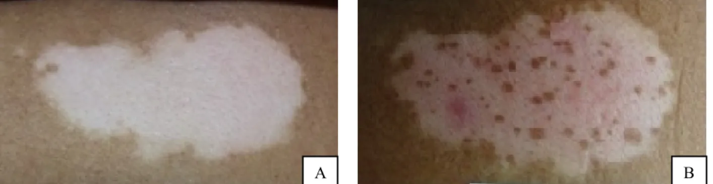
Figure 1 Vitiligo lesions before administration of NB-UVB (A) and after administration of NB-UVB (B). Red arrows indicate perifollicular repigmentation arising after administration of NB-UVB
Figure 2 Immunohistochemistry examination using antibodies to Wnt1 before (A) and after irradiation (B) NB-UVB with 400x magnification. The green arrow shows a positive result in the form of chromogen brown spots on the cytoplasm, while the red arrow shows a negative result
Figure 3 Number of cells expressing Wnt1 in hair follicles before and after irradiation of NB-UVB
Figure 4 Immunohistochemistry examination using antibodies to β-catenin and antibodies to Fzd4 before (A) and after NB-UVB (B) with 400x magnification. The green arrow shows a positive result in the form of a blue core, blue and brown cytoplasm, while the red arrow shows a negative result.
Rabbani's research on mouse hair follicles shows that melanocyte stem cells in the bulge region of the hair follicle do not produce Wnt protein. Epidermal stem cell markers (K15) are used to prove that Wnt proteins are produced by
epidermal stem cells that are around melanocyte stem cells, which then activate Wnt signals on melanocyte stem cells.12
A B
A B
Figure 5 Number of melanocyte stem cells expressing β-catenin before and after irradiation of NB-UVB
Yamada's study in mice showed that Wnt was expressed by epithelial lineages (hair follicle stem cells, hair follicle keratinocytes outside the hair follicles, and epidermal keratinocytes) after UVB exposure.13 Both studies showed that Wnt1 was excreted by cells in the niche of melanocyte stem cells and paracrine. Wnt1 expression in this study was examined without providing additional markers, either for melanocyte stem cells or for the epithelial lineage, so that no cell type that expresses the Wnt1 protein can be known.
The Wnt/ β-catenin pathway depends on the level of β-catenin in the cell. The amount of
β-catenin in the cytoplasm is normally maintained low through continuous degradation by the ubiquitin-proteasome system.14 Melanocyte stem cells that express β-catenin in this study were seen by immunohistochemistry using monoclonal antibodies to β-catenin and polyclonal antibodies to Fzd4 (β-catenin + / Fzd4 +) showed significant differences in
β-catenin expression before and after NB-UVB irradiation (p = 0.001).
The increase in expression of Wnt1 and
increased expression of β-catenin hair follicles in this study, accompanied by the emergence of perifollicular repigmentation with the administration of NB-UVB suggest the induction of intracellular Wnt/ β-catenin
pathway in vitiligo lesions. The Wnt/ β-catenin
pathway induction will be followed by binding of β-catenin to lymphoid enhancer-binding factor (LEF) transcription factors to then stimulate the expression of the MITF target gene, so that the melanocyte stem cells differentiate into melanoblasts, then melanocytes that migrate to the epidermis and actively do melanogenesis so that perifollicular repigmentation is formed. This mechanism still needs further investigation.
Conclusion
Increased expression of Wnt1 and increased expression of β-catenin hair follicles accompanied by the emergence of perifollicular repigmentation with the administration of NB-UVB suggests the induction of the intracellular Wnt/ β-catenin pathway in vitiligo lesions. These results can be used as a basis for further research so that the mechanism of perifolicular repigmentation becomes more clear and becomes the basis for developing therapy through exploration of substances that can induce Wnt and β-catenin so that melanocyte stem cell differentiation occurs leading to repigmentation.
References
1. Yang YS, Cho HR, Ryou JH & Lee MH. Clinical study of repigmentation patterns with either narrow-band ultraviolet B (NB-UVB) or 308 nm excimer laser treatment in Korean vitiligo patients, Int J Dermatol 2010; 49: 317–23.
Photoimmunol Photomed 2012; 28(2): 84-90.
3. Kumar YHK, Rao GRR, Gopal KVT, Shanti G & Rao KV. Evaluation of narrow-band UVB phototherapy in 150 patients with vitiligo, Indian J Dermatol Venereol Leprol 2009; 75: 162-66.
4. Girish PN, Narendra JS, Shetty RK, Shetty VH, Vasudevan OV, Sandhya I, Mallya U, Shetty N. Evaluation of narrow-band UVB phototherapy for vitiligo. J of Evolution of Medical and Dental Science 2013; 2: 8591-98.
5. Cui J, Shen L & Wang G. Role of hair follicle in the repigmentation of vitiligo, J O’Connell MP & Weeraratna AT. Hear the Wnt Ror: how melanoma cells adjust to changes in Wnt. Pigment Cell Melanoma Res 2009; 22: 724-39.
6. Chien AJ, Conrad WH & Moon RT, 2009. A Wnt survival guide: from flies to human disease. J Invest Dermatol 2009; 129(7): 1614-27.
7. Osawa M, 2009. Melanocyte stemcell, Downloaded 15 September 2013, <http://www.stembook.org>.
8. Yamada T, Hasegawa S, Inoue Y, Date Y, Arima M, Yagami A, Iwata Y, Abe M, Takahashi M, Yamamoto N, Mizutani H, Nakata S, Matsunaga K & Akamatsu H. Comprehensive analysis of melanogenesis and proliferation potent ial of melanocyte lineage in lentigo solaris. J Dermatol Sci 2014a; 73: 251-57.
9. Yamada T, Hasegawa S, Inoue Y, Date Y, Arima M, Yagami A, Iwata Y, Takahashi M, Yamamoto N, Mizutani H, Nakata S, Matsunaga K & Akamatsu H. Accelerated differentiation of melanocyte stem cells contributes to the formation of hyperpigmented maculae, Exp Dermatol, 2014b; 23: 652-58.
10. Khullar G, Kanwar AJ, Singh S & Parsad D. Comparison of efficacy and safety profile of topical calcipotriol ointment in combination with NB‐UVB vs. NB‐UVB alone in the treatment of vitiligo: a 24‐week prospective right–left comparative clinical trial, JEADV 2015; 29: 925-32.
11. Stromberg S, Bjorklund MG, Asplund A, Rimini R, Lundeberg J, Nilsson P, Ponten F, Olsson M. Transcriptional profiling of melanocytes from patients with vitiligo vulgaris. Pigment Cell Melanoma Res 2007; 21: 162-71.
12. Rabbani P, Takeo M, Chou W, Myung P, Bosenberg M, Chin L, Taketo MM & Ito M. Coordinated activation of Wnt in epithelial and melanocyte stem cells initiates pigmented hair regeneration. Cell 2011; 45: 941–55.
13. Yamada T, Hasegawa S, Inoue Y, Date Y, Yamamoto N, Mizutani H, Nakata S, Matsunaga K, Akamatsu H. Wnt/ β-catenin and Kit signaling sequentially regulate melanocyte stem cell differentiation in UVB-induced epidermal pigmentation. J Invest Dermatol 2013; 133: 2753–62. 14. Rubinfeld B, Albert I, Porfiri E, Fiol C,

![Methyl 3 amino 1 (2 chlorophenyl) 9 methoxy 5,6 dihydro 1H naphtho[2,1 b]pyran 2 carboxylate](data:image/gif;base64,R0lGODlhAQABAIAAAP///wAAACH5BAEAAAAALAAAAAABAAEAAAICRAEAOw==)