| INVESTIGATION
Human SOD1 ALS Mutations in a
Drosophila
Knock-In
Model Cause Severe Phenotypes and Reveal
Dosage-Sensitive Gain- and
Loss-of-Function Components
AslıSxahin,* Aaron Held,* Kirsten Bredvik,* Paxton Major,* Toni-Marie Achilli,†Abigail G. Kerson,* Kristi Wharton,* Geoff Stilwell,†and Robert Reenan*,1
*Department of Molecular Biology, Cellular Biology, and Biochemistry, Brown University, Providence, Rhode Island 02912, and †Department of Biology, Rhode Island College, Providence, Rhode Island 02908
ABSTRACT Amyotrophic Lateral Sclerosis (ALS) is the most common adult-onset motor neuron disease and familial forms can be caused by numerous dominant mutations of the copper-zinc superoxide dismutase 1 (SOD1) gene. Substantial efforts have been invested in studying SOD1-ALS transgenic animal models; yet, the molecular mechanisms by which ALS-mutant SOD1 protein acquires toxicity are not well understood. ALS-like phenotypes in animal models are highly dependent on transgene dosage. Thus, issues of whether the ALS-like phenotypes of these models stem from overexpression of mutant alleles or from aspects of the SOD1 mutation itself are not easily deconvolved. To address concerns about levels of mutant SOD1 in disease pathogenesis, we have genetically engineered four human ALS-causing SOD1 point mutations (G37R, H48R, H71Y, and G85R) into the endogenous locus ofDrosophila
SOD1 (dsod) via ends-out homologous recombination and analyzed the resulting molecular, biochemical, and behavioral phenotypes. Contrary to previous transgenic models, we have recapitulated ALS-like phenotypes without overexpression of the mutant protein.
Drosophilacarrying homozygous mutations rendering SOD1 protein enzymatically inactive (G85R, H48R, and H71Y) exhibited neuro-degeneration, locomotor deficits, and shortened life span. The mutation retaining enzymatic activity (G37R) was phenotypically indistinguishable from controls. While the observed mutant dsod phenotypes were recessive, a gain-of-function component was uncovered through dosage studies and comparisons with age-matched dsod null animals, which failed to show severe locomotor defects or nerve degeneration. We conclude that theDrosophilaknock-in model captures important aspects of human SOD1-based ALS and provides a powerful and useful tool for further genetic studies.
KEYWORDSALS; SOD1;Drosophila; motor neuron
A
MYOTROPHIC Lateral Sclerosis (ALS) is characterized by progressive loss of upper and lower motor neurons (MNs) leading to paralysis, and death in afflicted individuals within 3–5 years after diagnosis, on average. The mecha-nisms of disease pathogenesis leading to the exclusive demise of MNs remain unclear and there is no effective therapy. Over 50 genes have been linked to ALS, suggesting large geneticheterogeneity of the disease mirroring a diverse range of clinical presentations (Abel et al. 2012; Sreedharan and Brown 2013; Leblondet al.2014). Thefirst ALS-associated mutations were found in the superoxide dismutase 1 (SOD1) gene (Rosenet al.1993). SOD1 encodes a small protein of 153 amino acids (16 kDa), constitutes1% of the cytoplas-mic protein, and is expressed ubiquitously (Pardo et al.
1995). Functional SOD1 is a homodimer and catalyzes the conversion of superoxide radicals to hydrogen peroxide. Re-cent data suggest SOD1 also acts as a transcription factor and upregulates genes involved in oxidative stress response (Hu
et al.2009; Tsanget al.2014; Bunton-Stasyshynet al.2015). Strong evidence indicates that SOD1 mutations produce toxic gain-of-function properties at the protein level. The vast majority of the.150 SOD1 mutations identified in patients
Copyright © 2017 by the Genetics Society of America doi: 10.1534/genetics.116.190850
Manuscript received April 29, 2016; accepted for publication November 13, 2016; published Early Online December 13, 2016.
Supplemental material is available online atwww.genetics.org/lookup/suppl/doi:10. 1534/genetics.116.190850/-/DC1.
1Corresponding author: Department of Molecular Biology, Cellular Biology, and
show dominant inheritance patterns, and disease severity correlates with aggregation potential of mutant protein rather than loss of enzymatic activity (Abel et al. 2012; Sacconet al.2013).
Animal models containing mutant ALS-associated transgenes have been important tools for understanding disease pathogen-esis. To date, most transgenic models generated in multiple species express mutant human SOD1 (hSOD1) in a genetic background containing the endogenous wild-type SOD1 gene in rodents (Gurneyet al.1994; Trottiet al.1999; Kato 2008), in
Drosophila(Watsonet al.2008; Bahadoraniet al.2013) and in other model organisms (Joyceet al.2011). The majority of these models recapitulate characteristics of ALS, including progressive motor deficits, paralysis, MN degeneration, and early lethality (McGoldrick et al. 2013). However, ALS-like phenotypes in these animals are highly dependent on transgene expression levels and severity of phenotypes correlate with level of protein overexpression (Gurney et al. 1994; Alexander et al. 2004; Wanget al.2009a). Furthermore, overexpression of wild-type hSOD1 (hSOD1wt) is sufficient to recapitulate some ALS
phe-notypes such as mitochondrial dysfunction, axonal degenera-tion, and premature MN death (Jaarsma et al. 2000; Ezzi
et al.2007; Graffmoet al.2013). These results suggest mutant phenotypes are sensitive to gene dose and/or SOD1 protein levels. Extrapolating information gained from these disease models to human ALS is therefore difficult.
While mutations in other genes cause ALS, the pathways leading to SOD1-mediated toxicity are still poorly under-stood and it is unclear to what extent pathogenic mecha-nisms are shared between the various forms of familial ALS (fALS). Intriguingly, cytoplasmic SOD1 inclusions have been reported in ALS patients irrespective of SOD1 mutations (Gruzman et al. 2007; Bosco et al. 2010; Forsberg et al.
2010), strengthening the hypothesis that there may be a common mechanism for neurodegeneration in ALS and em-phasizing a critical role for SOD1 regarding the general pathogenesis of the disease. Uncovering how mutations in SOD1 ultimately lead to the dysfunction and the ultimate death of MNs may shed light on how ALS develops and progresses in all patients, regardless of sporadic or familial modes of transmission.
To create an ALS model in which mutant SOD1 protein is expressed at endogenous levels, we have used ends-out ho-mologous recombination (HR) as a gene replacement strategy to introduce relevant ALS-associated mutations at conserved residues of theDrosophila sod1(dsod) gene. Similar strategies have been used inflies to successfully model other diseases (Sun et al. 2012; Schutte et al. 2014). In this study, we showdsodG85R, H71Y, and H48R mutants exhibit recessive ALS-associated phenotypes including a shortened life span, progressive paralysis, and degeneration of MNs. We also dem-onstrate that aspects of these phenotypes represent a gain of function through comparisons with SOD1 null (dsodnull) alleles. UsingDrosophilaas a genetically tractable model or-ganism, we have developed an additional tool which can be used to uncover relevant ALS-related cellular responses.
Materials and Methods
Drosophila strains
Drosophilawere raised at a constant 25°, on standard
corn-meal molasses food and under 12 hr day/night cycles. The
dsodX-39anddsodG51Slines (n1 or n108) were ordered from Bloomington DrosophilaStock Center (BDSC) (stock num-bers 24492 and 24490, respectively). The names of the dSOD mutations are based on the human SOD1 amino acid num-bering system throughout this work. ThedsodX-16line was a kind gift from W. C. Orr from Wayne State University.
HR and CRISPR
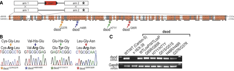
We performed ends-out HR to create three independent lines of
dsodWTLoxP,dsodG37R,dsodH48R,dsodH71Y, anddsodG85Rusing pre-viously reported methods (Staberet al.2011). Briefly, we used the ends-out targeting vector p[w25.2] that contains themini whitemarker (white+), a selectable red eye color,flanked byLoxP sites for subsequent removal by Cre recombinase (Figure 1). Ho-mology arm 1, corresponding to exon 1 ofdsod; and homology arm 2, corresponding to exon 2 ofdsod; were cloned and se-quenced in pTOPO (Life Technologies) and then shuttled into the multiple cloning sites of the vector to generate p[w25-dsod], which was then introduced into theDrosophilagenome by standardP-element transgenic methods (Genetic Services). Full targeting region coordinates were chromosome3L: 11,103,794–
11,108,715 (4922 nt). All targeting was done in a w1118
back-ground, as previously described, and performed in a wild-type chromosome3background (Gell and Reenan 2013). A full list of cloning, mutagenic, and sequencing primers can be found in Supplemental Material, Table S3. Targeting was performed to generate multiple independent targeting events that incorporate (G37R, H48R, H71Y, and G85R) or exclude engineered muta-tions (WTLoxP). All targeted animals have aLoxP“scar”of 72 nt. After generating the transgenicflies, white or mosaic-eyed females were collected from heat-shocked vials and then crossed with yw;ey-Flp,nocsco/CyO males and only red-eyed female progeny were selected for additional validation. Tar-geted alleles were validated by PCR amplification, using pri-mers outside the region of targeting and pripri-mers specific to the
white+marker. Following recombination, thewhite+minigene selection cassette was removed via genetic crosses, which in-troduced Cre recombinase. All targeted alleles were sequenced to verify that no unintended mutations were introduced. It is important to note here that all of the targeted alleles contain a natural polymorphism at the site 1013 having a C instead of an A, which leads to the N98K missense mutation in human amino acid numbering system (N96K in Drosophila dSOD numbering system), which is also referred asdsodfastallele in the literature (Phillipset al.1995). ThedsodG51Sallele that is generated through EMS mutagenesis does not contain this polymorphism, and it is thedsodslowallele.dsodfastanddsodslow alleles do not result in a phenotypic change inDrosophila, they are named after their differential mobility on a native poly-acrylamide gel (Leeet al.1981; Hudsonet al.1994; Phillips
backcrossed tow1118, a white-eyed genetic background, for five generations.Drosophilastocks were kept as heterozygotes using third chromosome balancers. Heterozygousdsodalleles that were used in the experiments were generated by crossing mutant lines to the wild-type lineWTLoxP.
To createdsodnullalleles, CRISPR/Cas9 was used to remove the complete dsod ORF (Genetivision, Houston, TX). Primers used to create target sequences were 59CTTCGACGAATTCGCAA GTAGAAT and 59AAACATTCTACTTGCGAATTCGTC for guide RNA 1 (gRNA1); and 59CTTCGGCATTTATTGGGGAATTCC and 59AAACGGAATTCCCCAATAAATGCC for gRNA2. Two indepen-dent lines were analyzed and reported herein.
Eclosion assay
For the eclosion percentages, we set up at least 12 vials of cohorts of 2–3 heterozygous males and virgin females at 25° on standard cornmeal molasses food and under 12 hr day/ night cycles. The parents were transferred to a new vial every day. The heterozygousdsodalleles were balanced over a third chromosome balancer:TM3,GFP,ser,w+(BDSC stock number 4534). When the progeny started eclosing, heterozygous progeny having red eyes due to the balancer were counted and homozygous progeny having white eyes due to lack of the balancer which eclosed or stuck in the pupal case were counted. The progeny carrying homozygous balancers died early in development due to a recessive lethal marker on the balancer chromosome. The progeny from each vial were counted until the number of homozygous mutants reached 200. Then, the eclosion percentages were calculated by (total number of eclosed homozygous adults)/(total number of ho-mozygous pupal cases)3100. The results of 12 vials were averaged and Fisher’s exact test was performed.
Survival assay
For the survival assay, parental flies were raised at 25° on standard cornmeal molasses food and under 12 hr day/night
cycles. The parental flies were collected from population-density-controlled broods (2–3 males and females in each vial) to avoid any confounding effects due to overcrowding. The parents were allowed to mate and lay eggs for 2 days before being transferred to fresh food twice. Three trials of survival analysis were performed for each genotype. For the heterozygous genotypes, to eliminate maternal effects on the
dsod mutant allele, in trial 1 the mutant allele was passed from the mother and in trial 2 the mutant allele was passed from the father. The third trial was either identical to either the trial 1 cross or the trial 2 cross. The offspring from these parents were collected over a period of 24 hr and sorted by sex. A total of 12 males and 12 females were kept in vials containing standard cornmeal molasses food. For each geno-type, multiple replicate vials were set up so the total sample size was 200–300 for each sex. Flies were transferred onto fresh food three times a week by blinded undergraduate re-searchers. The number of deaths was recorded. Survival as-says were either performed at 18 or 25°. Once all the flies were dead, log-rank tests were performed for statistical analysis.
H2O2feeding assay
All chemicals were delivered to Drosophilain instant food (Nutri-Fly Instant, Genesee, 66–118). Instant food is pre-pared based on the manufacturer recipe. For each vial, 2 g instant food was dissolved in 5 ml Milli-Q water or the chem-ical solution. For each bottle, 21 g instant food was dissolved in 50 ml Milli-Q water or the chemical solution.
A 2% concentration of hydrogen peroxide (H2O2) was
di-luted from 30% stock (Fisher Chemical H325-100) and de-livered to 1-day-posteclosion Drosophilain vials. Each vial contained 50 males or 50 females. Three trials of H2O2
sur-vival analysis were performed for each genotype and sex. As in survival analysis, for the heterozygous genotypes to elim-inate the maternal effects on thedsodmutant allele, in trial
one the mutant allele is passed from the mother and in trial two the mutant allele is passed from the father. Trial three was identical either to the trial one cross or the trial two cross. The number of deaths was recorded every 8 hr by blinded undergraduate researchers. Once all theflies were dead, log-rank test was performed for statistical analysis.
Larval motility assay (manual version)
The larval motility assay was performed as previously described in Batleviet al.(2010). Larvae were selected during the wandering third instar stage. They were washed in 13PBS and placed on a 1% agarose plate made with 0.5% TBE (100-315-mm petri dish) and were allowed 1 min to acclimate. The plate was placed on a 1-31-mm square grid and the larvae were allowed to crawl for 2 min. The total number of squares a larva crossed was counted by blinded undergraduate researchers. We considered a square to be crossed when the larvae’s posterior end crossed a line and the total number of squares crossed was counted. Dun-nett’s test following one-way ANOVA was used to compare the experimental group to the wild type.
Larval motility assay (computational version)
In the computational larval motility assay,five larvae were allowed to crawl on a 22-cm diameter dish filled with 1% agarose in H2O. The larvae were videotaped for 1.5 min and
videos were analyzed using a Matlab program that calculates total distance traveled for each larva. A total of.30 larvae were used per group. If two larvae collided, the data were discarded in case it altered behavior. Experiments used either midthird instar larvae or wandering third instar larvae. Wan-dering third instars were identified as fully grown larva that had exited the food but not inverted their anterior spiracles. Midthird instars were pulled from the food 24 hr before wandering. Tukey’s honest significant difference (HSD) test was used to calculate significance.
Adult climbing assay (negative geotaxis assays)
A total of 10 adult male or femaleflies at the appropriate age were placed into a vial without food and negative geotaxis assays were conducted in three trials with each group con-sisting of 20 vials for each sex and genotype. Theflies were gently tapped to the bottom of a vial and allowed to climb for 5 min. The number offlies reaching above a 75% mark (5 cm) of the total cylinder length in 5 min was recorded and the average calculated. Dunnett’s test following one-way ANOVA was used to compare the experimental group to the wild type.
Western blotting
For all the denaturing gels that are shown in the figures, protein samples were homogenized in Leammli buffer (Bio-Rad, Hercules, CA) andb-mercaptoethanol, and run out on a 4–20% gradient gel (Bio-Rad Mini-Protean-TGX, 456-1093). For the mutant tissue that shows trace amounts of dSOD protein on denaturing gels (dSODG85R/G85R, dSODH71Y/H71Y,
dSODG51S/G51S), various protein extraction solutions were
assayed,e.g., radioimmunoprecipitation assay buffer in
com-bination with proteinase inhibitor cocktails, but no significant improvements were observed. For adult tissue the abdomen region was discarded and for larval tissue intestines were re-moved. Unless otherwise specified, one adultfly or one larva was homogenized in 50ml sample buffer and a 10ml sample was run on 100 V. Samples were transferred to nitrocellulose membrane for 1 hr at 4°. For developmental time-point West-ern blots, the total protein amount was equalized between different samples via Coomassie Protein Assay using the man-ufacturer instructions (Thermo Scientific, 1856209). Rabbit polyclonal anti-dSOD antibody (a kind gift from W. C. Orr from Wayne State University) was used at 1:3000, and anti-actin (Millipore, Bedford, MA) was used at 1:50,000. Horse-radish peroxidase (HRP)-conjugated goat anti-mouse secondary antibody (Abcam ab5930) was used at 1:5000 and HRP-conjugated goat anti-rabbit secondary antibody (Jackson Immuno 111-0350144) was used at 1:10,000. Immunoreactive bands were visualized by ECL detection reagent (Genesee, Amersham ECL Reagent, 84-817). Nonsaturated bands were quantified on ImageJ (National Institutes of Health) and expressed as a ratio in relation to the internal reference actin. At least three biological replicates were quantified.
For high-salt buffer protein extraction experiments, the fol-lowing buffers have been tried: (i) 750 mM NaCl, 50 mM Tris-HCl (pH 7.5), 10 mM NaF, 5 mM EDTA; (ii) 750 mM NaCl, 50 mM Tris-HCl (pH 7.5), 5 mM EDTA, proteinase inhibitor cocktail (Roche), 0.1% Triton X-100; (iii) 5% SDS, 50 mM Tris-HCl (pH 7.5), 5 mM EDTA, 175 mM NaCl; (iv) 5% SDS, 50 mM Tris-HCl (pH 7.5), 5 mM EDTA, 175 mM NaCl, 8 M urea; (v) 750 mM NaCl, 50 mM Tris-HCl (pH 8.8), 10 mM NaF, 5 mM EDTA; (vi) 750 mM NaCl, 50 mM Tris, 10 mM NaF, 5 mM EDTA; and (vi) Bio-Rad ReadyPrep Protein Extraction Kit (Soluble/ Insoluble, 163-2083). The extraction conditions were similar to the methodology that was reported previously (Watsonet al.
2008). Briefly,five male adults (abdomen discarded) were ho-mogenized in 100ml high-salt buffer. Then, 30ml of the protein sample was removed and run as“total”protein sample. The rest of the protein sample was spun at 10,000gfor 30 min in 4°.
Then, 30 ml of the protein sample was removed and run as
“supernatant”protein sample. The pellet was resuspended in 100ml of high-salt buffer and spun at 10,000gfor 30 min at 4°
twice. Finally, the pellet was resuspended in 50 ml high-salt buffer and run as“pellet”sample.
The semi-denaturing detergent agarose gel electrophoresis assay was performed as described before in Halfmann and Lindquist (2008) and Cushman-Nicket al.(2013).
Native PAGE
human (ENZO, 80‐1642) or bovine (ENZO, ALX-202-022-UT50) SOD1 protein isolates were diluted in native sample buffer and 50 units were loaded to the well. The gel was blotted with 1:3000 dSOD antibody and the transfer condi-tions were the same as described in the previous Western blotting section. In addition, the same samples were run in SDS-PAGE denaturing gels, after addingb-mercaptoethanol.
SOD activity assay
For SOD activity assay, the same samples with native PAGE were used. Briefly, after running the gel for 5–8 hr, the gel was washed three times in distilled water. The gel was incubated for 15 min in 10 mg Nitro Blue Tetrazolium (NBT) (Sigma N5514-10TAB) and 4 mg riboflavin (Fisher BP167-50) solution. Then, the gel was incubated for 15 min in TEMED-water solution (10 ml TEMED in 10 ml distilled water in the dark). Finally, the gel was washed three times in distilled water and imaged on a white light box until the desired contrast was reached.
Leg muscle atrophy and nerve structure analyses
For bright field leg images, each leg was removed with tweezers on a silicon (Dow Corning, Sylgard 184 silicone elastomer kit) plate. The legs were incubated in Vectashield (Fisher NC9532821) overnight at 4°. The images were taken with a Carl Zeiss (Thornwood, NY) AX10 Imager M1.
dsodG85R/G85R full-leg lengths were calculated by tracing along the midline of the femur on ImageJ. The nerve integrity of 2-week-olddsodH71Y/H71Ylegs was evaluated either based on whether they maintained nerve branches or not. Addi-tional parameters including continuity of nerve bundle, nerve size, number of leg kinks, and lower leg morphology were scored blinded for genotypes on a scale of one, being the worst, tofive, being the best.
For muscle atrophy imaging, relevant dsod mutants were combined with a myosin heavy chain (mhc)-tau-GFP reporter
(BDSC: 38460):dsodWTLoxP/WTLoxP,mhc-tau-GFP;dsodG85R/G85R,
mhc-tau-GFP;dsodH71Y/H71Ylines were generated. Legs were dis-sected and fixed as described previously (Soler et al. 2004). Briefly, the full animal wasfixed in 4% paraformaldehyde for 5 hr at room temperature. Then, the legs were dissected and continued tofix more overnight at 4°. Images were taken at the confocal microscopy as described in the microscopy section.
Tunnel assay, immunohistochemistry, and microscopy
Adult or larval brains were dissected in PBS with 0.1% Tween 20 (PBTX) on a silicon (Dow Corning, Sylgard 184 silicone elastomer kit) plate. The tissue wasfixed with 4% paraformal-dehyde for 20 min at room temperature on the nuator. After three rounds of quick washes in PBTX, the tissue was blocked in PBTX with 5% normal goat serum. The primary antibodies used in this study were anti-Elav and anti-Repo (Developmental Studies Hybridoma Bank), both used at 1:200; Alexa Fluor 488 and 564 secondary antibodies were used at 1:200. DAPI (Invitrogen, Carlsbad, CA) was used at 1:1000.
For the terminal deoxynucleotidyl transferase (TdT) dUTP nick end labeling (TUNEL) assay, the CF-488 TUNEL kit
(Biotium) was used according to manufacturer instructions. After Elav and Repo staining performed as described above, the tissue was refixed with 4% paraformaldehyde for 20 min at room temperature on the nuator. After three rounds of quick washes in PBTX, the tissue was reblocked in PBTX with 5% normal goat serum. Each genotype was incubated in 10 ml TUNEL equilibrium buffer for 5 min. Then, the buffer was replaced with the enzyme solution (2 ml TdT enzyme in 100 ml TUNEL reaction buffer) and incubated for 2 hr at 37°in a humidifying chamber. Finally, the tissue was washed three times with PBST.
All tissue was mounted in Vectashield (Fisher NC9532821). All confocal Z-series images were obtained by LSM510 con-focal microscope. Images were contrast enhanced in Adobe Illustrator. Each image shown is a representative example of
n$5 unless otherwise reported.
Data availability
The full list ofDrosophilalines used in this study is found in Table S2. The authors state that all data necessary for confirming the conclusions presented in the article are rep-resented fully within the article .
Results
Generation of dsod mutant Drosophila alleles via HR
To developDrosophilamodels of fALS, we knocked in G37R, H48R, H71Y, and G85R point mutations into the endogenous
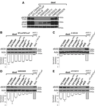
dsodlocus via HR. SOD1 is highly conserved between organ-isms; hSOD1 and dSOD differ at 49/153 residues, and 22 of these different residues possess a similar side-chain chemistry (Figure 1, A and B) (Bertiniet al.1998). The mutations we selected are highly conserved across species and have varying significance for the tertiary protein structure and enzymatic activity of the SOD1 protein in humans, and the patients carrying these mutations have different disease onsets and disease progression (Juneja et al. 1997; Rabe et al.2010; Weiset al.2011; Özo˘guzet al.2015) (Table S1). The target-ing and targeted mutations had no effect ondsodtranscript levels or splicing (Figure 1C). Because the HR technique leaves a 72-bp LoxP site within thedsod intron,dsodWTLoxP lines were analyzed as controls in these studies.
For mutant and control lines, we generated at least three independent targeting events. To remove any potential con-founding genetic background differences, one line for each mutation was backcrossed to w1118forfive generations. In
addition to the alleles we created by HR, we analyzed
dsodG51S, a previously reported line that carries a point mu-tation as a result of EMS mutagenesis (Campbellet al.1986; Phillipset al.1995).
dsodG85R, dsodH48R, and dsodH71Ymutants show
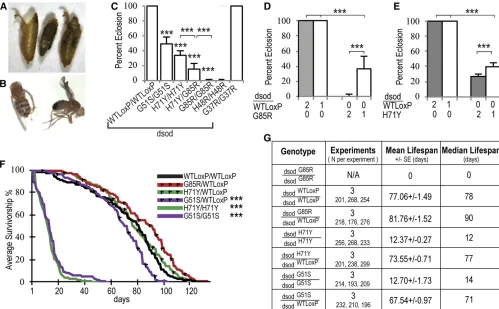
reduced viability and fertility
of these animals die with their heads everted from the pupal case and a characteristic shortening of the legs, in particular the metathoracic legs (Figure 2A andFile S1). We observed rare (1/1000) dsodG85R/G85R homozygotes that eclosed. These escapers live less than an hour and exhibit severe para-paresis, uncoordinated locomotion, leg muscle twitching, extreme proboscis pulsing, and seizures (File S2). Similar eclosion phenotypes were seen indsodH48R/H48Rhomozygous lines but slightly more adults eclose (2.3%), but they die within 24 hr. HomozygousdsodH71Y/H71Ymutants exhibit less severe eclosion abnormalities than dsodG85R/G85R homozy-gous adults (eclosion rate: 33.33 6 6.00%) (Figure 2C) and the majority displayed an obvious wrinkled-wing pheno-type (Figure 2B). Interestingly, after 2 weeks,dsodH71Y/H71Y exhibit a locomotor uncoordination phenotype similar to that of thedsodG85R/G85Rescaperflies (File S3). ThedsodG51Sallele that was generated via EMS mutagenesis, was also reported to display eclosion defects similar to those of dsodH71Y/H71Y (Staveleyet al.1991; Parkeset al.1998). Here, we confirm that dsodG51S/G51S animals exhibit lower eclosion rates
(48.48610.00%) (Figure 2C). We testedtrans-heterozygote
dsodG85R/H71Yflies and found eclosion rates intermediate be-tween the severe dsodG85R/G85R and dsodH71Y/H71Y alleles (15.00 67.00%) (Figure 2C). Moreover, the severity of the eclosion defects fordsodH71Y/H71YanddsodG85R/G85Rflies were dosage dependent (Figure 2, D and E). Reducing the mutant allele copy number by 50% by placing ALS-mutant alleles over a previously characterized null mutation partially rescued the eclosion phenotype (37.14 6 16.01% for dsodG85R/X-16 and 42.29 6 4.65% for dsodH71Y/X-16). However, eclosed adults exhibit an uncoordinated behavior and early death (File S4). These data suggest that both the G85R and H71Y mutations confer at least some part of their effects via a dosage-sensitive toxic gain of function.
Eclosed dsodH71Y/H71Y adults exhibit a severely reduced life span (12.3760.27 days), similar to that ofdsodG51S/G51S homozygotes (12.7060.97 days) (Figure 2, F and G). The viable adults ofdsodH71Y/H71Yare also infertile. Heterozygous combinations of mutantdsodalleles withdsodWTloxPas well as
dsodG37R/G37R homozygotes showed no life-span reduction
Figure 2 Targeted mutations cause eclosion defects and shorten life span. (A) A representative image of dsodG85R/G85R. (B) The wrinkled-wing
phenotype of eclosed adultdsodH71Y/H71Yflies. (C) HomozygousdsodG51S/G51S,dsodH71Y/H71Y,dsodH48R/H48R, anddsodG85R/G85Rflies do not eclose
in expected Mendelian ratios. The eclosion rates of heterozygoteflies anddsodG37R/G37Rflies are normal. Error bars are SEM.***P,0.0001, Fisher’s
exact test. Eclosion defects are dosage sensitive for (D) dsodG85R and (E) dsodH71Y mutants. Single-copydsod mutants were analyzed astrans
-heterozygotes withdsodX-16, an allele that deletes thefirst exon and promoter region. Bars are average eclosion rates from 12 different vials with
at least 200 homozygous mutant progeny. Error bars are SEM.***P,0.0001, Fisher’s exact test. (F) Life spans were analyzed fordsodmutants and controldsodWTloxP/WTLoxPflies and survivorship was plotted over time. Survival curves represent an average of three life-span trials.***P,0.0001,
log-rank test. (G) Statistical comparisons of life-span analysis show ecloseddsodH71Y/H71Yflies have a severely shortened life span, similar todsodG51S/G51S.
under standard laboratory conditions (Figure 2, F and G, and Figure S1).
Because SOD1 functions to convert superoxide radicals to H2O2 and water, we treateddsod mutants with H2O2 and
assessed survival to monitor response to oxidative stress (Fridovich 1986). Treating the heterozygous adults (dsodH71Y/WTLoxPanddsodG85R/WTLoxP) with a superoxide rad-ical (O22) generator did not alter survival when compared to
wild-type dsodWTLoxP/WTLoxPcontrols (Figure S2). However, homozygousdsodH71Y/H71Yflies were extremely sensitive to oxidative stress, consistent with previous results regarding the dsodG51S/G51S homozygous allele (Parkes et al. 1998; Kirbyet al.2008) (Figure S2, A and B). Finally,dsodG37R/G37R homozygotes respond similarly todsodWTLoxP/WTLoxPcontrols (Figure S2, C and D).
Severe locomotion defects in dsodG85Rand dsodH71Y
mutants
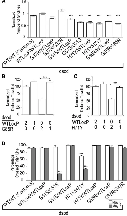
Motor dysfunction is one of thefirst apparent symptoms in ALS patients (Pasinelli and Brown 2006). We therefore analyzed locomotion indsodmutants by examining crawling behavior in larvae and climbing behavior in adults. During late third instar larval stages, dsodH71Y/H71Y, dsodG85R/G85R, and
dsodG51S/G51Shomozygotes crawled at almost half the rate of wild-type larvae (Figure 3A). When we reduced the mutant copy number by half, the crawling defect was suppressed completely; again supporting the notion of dominant gain-of-function toxicity (Figure 3, B and C). Homozygous
dsodG37R/G37R were indistinguishable from dsodWTLoxP/WTLoxP controls (Figure 3A). In keeping with a gain of function, het-erozygousdsodH71Y/WTLoxPanddsodG85R/WTLoxPlarvae showed crawling defects relative to wild type; indicating a dominant component for this phenotype at this life stage (Figure 3A). Because dsodH71Y/H71Y and dsodG51S/G51S adults survive for reasonable lengths of time, we assessed locomotion in adults through a negative geotaxis climbing assay. Homozygous
dsodH71Y/H71Y and dsodG51S/G51S lines show an almost-complete loss of climbing ability within 1 week of eclosion (Figure 3D for males andFigure S3A for females). All het-erozygous combinations with dsodWTLoxP/WTLoxP as well as
dsodG37R/G37R homozygotes were indistinguishable from
dsodWTLoxP/WTLoxPflies throughout course of the assay (Figure 3 andFigure S3). Thus, in the adult stage, heterozygotes for the severe alleles appear to be recessive for locomotor defects.
Muscle atrophy and denervation in dsodG85Rand
dsodH71Ymutants
Denervation of muscles by MNs in the limbs of patients is a characteristic symptom of ALS. At the terminal stages,
dsodG85R/G85Ranimals struggle in the pupal case while usu-ally failing to eclose (File S1). Theseflies look morphologi-cally wild type except for the fact that their legs are shorter when compared to wild-type flies (Figure 4, A and B, and Figure S4). In addition, it was clear from visual inspection of the few escaper flies thatdsodG85R/G85R and 2-week-old
dsodH71Y/H71Y animals drag their metathoracic legs (leg 3)
while walking (File S2andFile S3). We determined the over-all health scores of legs from dsodG85R/G85R flies based on nerve health, lower leg structure, and kink severity of the femur based on a scale of 1–5 (worst to best) (Materials and Methods, andFigure S5). IndsodG85R/G85Rflies, leg 3 was the most severely affected, however leg 2 and leg 1 were not as healthy as a wild-type control legs. Wild-type legs scored
Figure 3 Locomotion defects are apparent indsodlarvae and adults. (A) Larval crawling ability is measured manually in single-blinded assays counting the total number of squares each third larva traveled in 2 min. Error bars are SEM.dsodmutant larvae display crawling defect in a dosage-dependent manner.N.40, Dunnett’s test,***P,0.001. Computational measure of (B)dsodG85R/G85Rand (C)dsodH71Y/H71Ylarval
crawling behavior in a dosage-dependent manner. Tukey’s HSD test,
***P,0.001,N.33. (D) Adult climbing ability is significantly reduced in homozygousdsodG51S/G51SanddsodH71Y/H71Yflies within a week of
eclosion compared to dsodWTLoxP/WTLoxP. Heterozygous dsodWTLoxP/G85R
and dsodWTLoxP/H71Yflies show normal climbing ability when assessed
within a 7-week period after eclosion. In all adult climbing tests in this
higher than 4.5, whereasdsodG85R/G85Rscores were 2.87 for leg 1, 2.5 for leg 2, and 1.40 for leg 3.
Next, we used anmhc-tau-GFPintrinsicfluorescent muscle reporter to investigate the muscle structure and condition of
dsod mutants (Figure 4C and Figure S4). In dsodG85R/G85R flies, the muscle appeared to be undergoing severe atrophy and the MNs surrounding the leg appear degenerated (Figure 4A andFigure S4). IndsodH71Y/H71Y, the muscle structure and leg length were normal at eclosion (Figure 4, C and D). How-ever, leg nerves lost efferent branches by 2 weeks in 13 out of 15 legs when theseflies were near the terminal stage of their life span (Figure 4D).
MNs of dsod mutants do not undergo cell death and gliosis
Since MN death is a feature of ALS, we examined whether MNs died in our mutantflies (Figure 5). To visualize MNs, we used a nuclear GFP protein (UAS-nlsGFP) that was expressed un-der an MN-specific driver (ok371-GAL4) as well as staining nuclear DNA with DAPI. We examined both third instar larval brains and adult ventral nerve cord (specifically the T1/T2 region of the thoracic ganglia) of dsodG85R/G85Rand dsodH71Y/H71Y and did not observe any MN cell body loss (Figure 5). An-other signature of ALS, gliosis in the neuronal samples, was also not seen in larval central nervous system, adult brain, or adult ventral nerve cord as observed by staining for the glial cell marker,Repo, between mutants and controls (Figure S6). We also found no evidence for an increase in TUNEL staining
in mutants, further strengthening the argument for the ab-sence of MN death in third instar larval brains and adult ventral nerve cord of ALS-mutant Drosophilaat the stages assessed (Figure S6).
Protein levels and dismutase activity of mutant dsod animals
In ALS patients, hSOD1 missense mutations variously alter protein folding, protein stability, enzymatic activity, and metal-binding properties; while others maintain wild-type SOD1-like properties (Valentine et al. 2005). hSOD1G85R
lacks enzymatic activity while hSOD1G37R displayed 150%
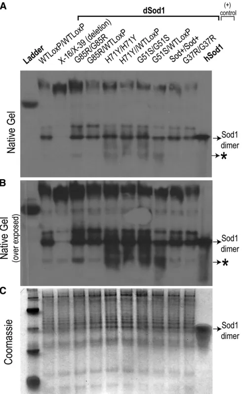
activity of wild-type SOD1 protein based on previous studies, while enzymatic activity has not been characterized for H48R or H71Y mutations (Borchelt et al.1994) (Table S1). With our allelic series, we asked whether the ALS-like phenotype severity correlates with the possible loss of superoxide scav-enging. We measured the state of dSOD dimerization and enzymatic activity via a native gel-based SOD activity assay (Figure 6). Based on the native gel, homozygous
dsodH71Y/H71Y, dsodG85/G85R, dsodH48R/H48R, dsodG51/G51S, and heteroallelicdsodX16/X39control adults exhibited no de-tectable SOD1 activity whiledsodG37/G37R, and all heterozy-gotes, were indistinguishable from wild-type Canton-S and
dsodWTLoxP/WTLoxP(Figure 6).
To determine dSOD protein levels indsodmutants, we used denaturing and native polyacrylamide gels to quantify protein amounts based on immunoreactivity with a dSOD antibody.
Figure 4 dsodmutant adults show progressive degeneration of the leg MNs. (A) Bright-field image of the MN bundle in the femur of the metathoracic (leg 3) leg exhibit deformation indsodG85R/G85R.N= 10. Bar, 0.2 mm. (B) Average leg length quantification ofdsodWTLoxP/WTLoxP,dsodH71Y/H71Y, and
dsodG85R/G85Rlegs in microns. AlldsodG85R/G85Rlegs are significantly shorter than wild type.N= 10. Error bars are SEM.***P,0.001, one-tailed
student’st-test. (C) Leg degeneration is progressive indsodH71Y/H71Yadults. Bright-field andfluorescent-microscope images of the same metathoracic
leg of 1-day-old and 14-day-old adults:mhc-tau-GFP;dsodWTLoxP/WTLoxPandmhc-tau-GFP;dsodH71Y/H71Y.N= 10. Bar, 0.2 mm. (D) Quantification of legs
Similar to SOD1 enzymatic assays, the null dsodX-16/X-39,
dsodH71Y/H71Y,dsodG85R/G85R,dsodH48R/H48R, anddsodG51S/G51S homozygotes also did not reveal any SOD1 protein immuno-reactivity by SDS-polyacrylamide denaturing gel (Figure S7 andFigure S8A). These data are consistent with reports of reduced SOD1 protein amount in the nontransgenic mouse model harboring the mSOD1D83G/D83Gmutation (Joyceet al.
2015). Because mutant SOD1 forms insoluble aggregates in ALS patients and model animals, we then examined if the undetectable dSOD protein on SDS-PAGE gel is in insoluble protein fractions by extracting protein using six different
high-salt or urea protein extraction solutions. All of these ex-traction methods failed to reveal detectable dSOD protein in the insoluble fraction via SDS-PAGE (a representative gel is shown inFigure S8). Semidenaturing detergent agarose gel electro-phoresis also failed to reveal higher molecular size SOD1-positive inclusions (a representative gel is shown inFigure S9).
Because the SOD1 activity assay is based on native gel electrophoresis, we performed native gel Western blot anal-ysis to assess whether immunoreactive dSOD dimers are present. Surprisingly, the native polyacrylamide gel results revealed that dSOD dimers are present in all mutants on a nondenaturing gel at levels approaching the wild-type con-trols (Figure 7). Moreover, in the native gel immunoblot, dSOD positive protein species with altered motilities were detected in homozygous mutants and heterozygotes. These additional protein bands could represent misfolded dSOD or destabilized dimers yielding monomeric dSOD (Rakhitet al.
2007; Auclairet al.2010; Liuet al.2012). It is an intriguing observation that the faster migrating species of dSOD immu-noreactivity seen in homozygotes, which comprises substan-tial amounts of dSOD immunoreactivity, is greatly diminished in heterozygotes. This observation hints at potential mutant: wild-type subunit interactions to form dimeric complexes.
Next, we attempted to discern whether the lack of detect-able dSOD on an SDS-PAGE denaturing gel is a general technical artifact, or if it is related to the progression of disease pathology. To assess if ALS mutations affect dSOD protein stability or accessibility to extraction in earlier stages as dramatically as in adults, we measured protein levels of dSOD during the adult stage from all mutant lines by SDS-PAGE (Figure 8A). As previously observed in the endogenous point-mutant mSOD1D83R/D83Rmouse,dsodH71Y/H71Yand
dsodG85R/G85Rexhibited a significant reduction in amounts of dSOD protein. Intriguingly, in the previously designated“null ordsod2/2”alleledsodG51S(other names:n108orn1allele),
Figure 5 Absence of MN cell body loss indsodG85R/G85Rmutants. (A)
The nuclear GFP protein (UAS-nlsGFP) is expressed under the ok371 MN-specific driver (ok371-GAL4). The MN regions that are shown in the following pictures are highlighted in red squares. DAPI is used to label all nuclei. MNs and DAPI staining in dsodG85R/G85R(B) third instar larval
brains (N= 5) and (C) adult ventral nerve chord (N= 5) do not exhibit MN loss. Bar, 50mm.
Figure 6 Superoxide dismutase activity is diminished in severedsod mu-tants. (A) SOD1 dismutase activity was assessed by NBT after native-PAGE. Homoyzgotes dsodG85R/G85R, dsodH71Y/H71Y, dsodG51S/G51S,
dsodH48R/H48Rshow dramatically reduced enzyme activity compared to
dsodG37R/G37R, heterozygous combinations, and dsodWTloxP/WTloxP
we observed protein expression, suggesting that thedsodG51S point mutation did not result in a protein null expression as previously reported (Campbell et al. 1986; Staveley et al.
1991; Phillips et al. 1995; Parkes et al.1998; Kirby et al.
2008; O’Keefeet al.2011; Mishraet al.2014).
Next, we asked whether lack of immunoreactivity in SDS-PAGE gels is consistent throughout development or whether it is stage specific. Detection of dSOD protein from homozygous dsodH71Y/H71Yand dsodG85R/G85Ris found in L1 larvae and
declines gradually during development, indicating possible maternal contribution which reduces during larval develop-ment (Figure 8, B, D, and E). Maternally derived dSOD pro-tein is only detectable in L1 larvae, albeit at reduced levels compared to wild-type L1 larvae (Figure 8, B and C).
Toxicity associated with the dsodG85Rallele is
dosage sensitive
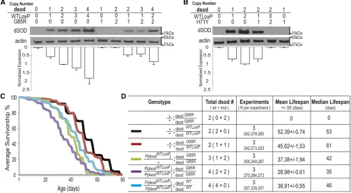
To determine to what extent mutant dsod alleles produce dosage-sensitive phenotypes, we increaseddsodcopy number and assessed phenotypes. We used the original HR cassette containing the entire wild-type dsodlocus to increase gene dose. Transformed lines containingP[dsodWTLoxP]show dSOD protein levels are comparable to the endogenousdsodlocus by Western blot analysis (Figure 9, A and B). Flies carrying four copies of dSODwt (P[dsodWTLoxP]/P[dsodWTLoxP]; dsodWTLoxP
/dsodWTLoxP) showed a reduced life span compared to two copy dSODwtcontrols (Figure 9, C and D; Figure S10, A and B;
38.9160.55 days for males and 49.8061.25 for females
vs.52.3960.74 days for males and 61.9560.85 for females). We further assessed the effect on phenotype of altering the copy number of mutantdsodrelative to wild-typedsod. Ani-mals containing either one or two extra doses of wild-type dsod (P[dsodWTLoxP]/P[dsodWTLoxP]; dsodG85R/dsodG85R or
P[dsodWTLoxP]/+;dsodG85R/dsodG85R) showed dose-dependent suppression of dsodG85R/dsodG85Rlethality at eclosion (Figure S10C). One dose ofdsodwtpartially rescued viability to70%,
while two doses rescued eclosion to wild-type levels. However, flies carrying two extra wild-type dsod alleles in a dsodG85R
background (P[dsodWTLoxP]/P[dsodWTLoxP]; dsodG85R/dsodG85R) showed a 40% decrease in life span relative to controls carrying four wild-typedsodalleles (28.986 0.61 days for males and 39.3761.32 days for females, compared to 52.3960.74 days for males and 61.9560.85 days in females) (Figure 9, C and D; Figure S10, A and B). In all cases,flies carrying four copies of
dsod failed to show eclosion or progressive locomotor defects regardless of whether four wild-type copies or two wild-type and two mutant copies were present (Figure S10, C, D, and E). Nevertheless, there was a general rapid loss of locomotor function in the days preceding death forP[dsodWTLoxP]/
P[dsodWTLoxP];dsodG85R/dsodG85Rflies (File S5).
Complete loss-of-function mutations at dsod support a gain-of-function toxicity component for phenotypic ALS alleles
Because mutantdsodalleles exhibit predominantly recessive phenotypes and lack enzymatic activity by native gel analysis,
we compared ourdsodmutants withsodnulllines to determine whether any components of the phenotype might be due to loss of function. To analyzesodnull, we assessed phenotypes using a heteroallelic combination of previously reported null mutations (X-16/X-39) (Staveley et al. 1991; Parkes et al.
1998; Missirlis et al. 2003). A heteroalleleic combination was used because homozygoussodX-16/X-16orsodX-39/X-39
al-leles showed that numerous downward-modifying mutations had clearly accumulated in the stock, severely reducing eclo-sion rates and viability. Extensive backcrossing of these stocks did not alleviate this problem. To overcome this obstacle, we generated new sodnull mutations via CRISPR/cas9 that re-moved the dsod ORF (see Materials and Methods) (Figure 10A). Two lines were characterized,dsodnull21anddsodnull26,
which had similar eclosion ratios as originally reported for
dsodX-16anddsodX-39alleles, which displayed complete infertil-ity (Mockettet al.2003) similar todsodH71Y/H71Y(Figure 10B). In contrast todsodH71Y/H71Y,dsodnull21/null21anddsodnull26/null26
homozygotes exhibit less severe climbing and life-span defects (Figure 10, C and D). dsodnull21/null21anddsodnull26/null26
dis-played median life spans of 11 and 14 days respectively, and climbing defects were only apparent in 2-week-old adults. In contrast,dsodH71Y/H71Ydisplayed a median life span of 7 days at
25°and the 50% of animals alive at this time point displayed a complete loss of climbing ability (Figure 10, File S6, andFile S7). There were not sufficient numbers of SOD1 nullflies left at 30 days to perform climbing assays. These data confirm that, while lack of dSOD does confer deleterious phenotypes in Dro-sophila, ALS-causing mutations cause more severe phenotypes, produce mutant proteins, and likely act through a combination of both loss-of-function mechanisms (superoxidase activity) and a toxic gain of function whose mechanism is unknown.
Discussion
In this study, we used HR inDrosophilato model fALS muta-tions found in human disease:dsodG37R,dsodH48R,dsodH71Y,
dsodG85R, and a wild-type control dsodWTLoxP. Generating multiple lines and assessing consistency of phenotypes is im-portant in HR methodology because the recombinases and
enzymes used (SceI endonuclease, FLP, and Cre recombi-nase) can potentially introduce unintended mutations in the genome that may affect downstream analysis of the tar-geted alleles (O’Keefeet al.2007). We also minimized any undesired background effects by backcrossing thefinal engi-neered lines to a common genetic background stock,w1118. Thus we show that the ALS-related phenotypes described in this study result from point mutations in the endogenous
dsodlocus. Similar to the genetic architecture of ALS patients, these mutations are within the context of the endogenous
dsod gene and mutant dsod alleles used in this study are not overexpressed at either transcript or protein levels. Here, we show that homozygousdsodG85R/G85R,dsodH48R/H48R, and
dsodH71Y/H71YALS-associated mutations confer eclosion de-fects, reduced survival, and locomotor defects in larvae and adults; potentially stemming from neuronal degeneration and retraction from adult muscles.
In humans, 95% of ALS-associated SOD1 mutations confer disease via dominant inheritance (Sacconet al.2013); how-ever, some heterogeneity exists. For example, the hSOD1D90A
mutation shows mostly dominant inheritance patterns but is recessive in some Scandinavian populations (Andersenet al.
1995, 1997). Of the dsod alleles we created, dsodG85R,
dsodH48R, anddsodH71Ydisplayed prominent ALS-like pheno-types only when made homozygous. While these results are
Figure 8 Expression of mutant dSOD is diminished in third instar larvae. (A) Immunodetection of dSOD protein is reduced dramatically in dsodG85R/G85R,
dsodH71Y/H71Y, anddsodG51S/G51Sadults compared
to wild-type Canton-S ordsodWTLoxP/WTLoxPcontrols.
Upon overexposure, a trace amount of protein is visible. Five maleflies were homogenized for this blot. dSOD protein levels were assessed throughout development and (B) constant dSOD levels were observed in dsodWTloxP/WTloxP; whereas (C) dSOD
protein is seen only in L1 for null transheterozygotes dsodX-39/X-16, representing maternal contribution. In
(D)dsodG85R/G85Rand (E)dsodH71Y/H71Ylines, dSOD
discrepant with the genetic pattern of inheritance for human fALS in general, mice carrying an endogenous mSOD1D83G
point mutation (Joyce et al. 2015) created by N-ethyl-N-nitrosourea (ENU) mutagenesis display predominantly reces-sive phenotypes. These phenotypes include progresreces-sive motor and behavioral deficits and loss of muscle force due to degeneration of lower and upper MNs (Joyceet al.2015). Interestingly, SOD1null mice do not display this phenotype,
providing strong evidence that ALS-mutant SOD1 can confer both loss of SOD1 enzymatic activity, and a toxic gain of func-tion (Joyceet al.2015). We observed very similar results in
dsodmutantflies. Most phenotypes we observed were reces-sive yet clearly more severe thansodnull/nullmutants. These endogenous SOD1 mutants in mice and flies demonstrate that ALS-like phenotypes are achievable in model organisms without the necessity for transgenic overexpression of the mutant allele. In addition, 73% of reported cases of SOD1 mutant canines develop degenerative myelopathy (Zeng
et al. 2014) and exhibit ALS-like symptoms only when mu-tant alleles were homozygous.
There are several possible explanations for the apparent recessive nature for our observed phenotypes. First, given the importance of mutant dSOD1 protein dosage in accelerating the ALS-like phenotype in transgenic SOD1-mediated ALS models (Acevedo-Arozena et al. 2011), it is possible that
heterozygotes may develop symptoms later in life, or would only display symptoms if they lived well beyond their normal life span. Mice, dogs, and fruitflies have relatively short life spans compared to humans. Since ALS is a late-onset disease in humans (55 years of age in average), we speculate that extending the life span of heterozygous fruit flies and non-transgenic SOD1 animal models beyond their normal life span might be required to cause more dramatic, late-onset ALS-like phenotypes. Alternatively, there may be some sec-ond site modifier or genetic background differences present in model organisms that in wild-type form partially suppress pathogenesis, similar to what is proposed in Scandinavian populations.
A second explanation for lack of dominant inheritance in animal models involves dosage of mutant dSOD protein. The amount of disease-causing mutant protein may be insufficient to cause cellular toxicity in one dose, or one dose of wild-type dSOD confers a novel protective effect in the short life span of animal models. We addressed this possibility in several ways. We observed that lowering mutantdsodG85RordsodH71Ygene copy number by half in dsodG85R/X-16mutants rescued eclo-sion (Figure 2, D and E) and larval locomotion defect (Figure 3, B and C). However, adultdsodG85R/X-16flies still exhibited very uncoordinated behavior (File S4). Therefore, expressing only one copy ofdsodG85Ris clearly less severe than the two
Figure 9 dsodG85Rallele causes survival defects through gain of toxic function in a dosage-sensitive manner. (A) dSOD protein levels are gene dose
dependent fordsodWTloxPalleles but not for mutantdsodG85Ror (B)dsodH71Yby denaturing SDS-PAGE analysis. (C) Average life span of three trials of
males of transgenic lines expressing extradsodon the second chromosome and controls.dsodG85R/G85Rflies die in the pupal case and do not survive to
adulthood. (D) Summary statistics for life-span analysis. log-rank test was used.dsodG85R/dsodWTLoxPanddsodWTLoxP/dsodWTLoxPare not significantly
different from each other.P,0.0001P[dsodWTLoxP]/P[dsodWTLoxP]; dsodG85R/dsodG85R, P[dsodWTLoxP]/1;dsodG85R/dsodG85RandP[dsodWTLoxP]/P[dsodWTLoxP];
doses of a homozygote, yet confers much of the severity of the ALS-like phenotype. As such, the phenotypes we see do not result strictly from loss of dSOD activity, but from some toxic gain of function which wild-type dSOD can counter via a protective effect, potentially by dimerization with mutant dSOD.
While the majority of the mutant phenotypes we observed were recessive, heterozygousdsodG85R/WTLoxPanddsodH71Y/WTLoxP late third instar larvae show locomotor defects. This transient phenotype might stem from the differential gene expression milieu or neuronal physiology between the larval stage and the adult stages. The molecular mechanisms underlying this discrepancy remain to be determined. It is also possible that further experiments with altered environmental conditions such as altered diet, stress levels, or pharmacological inter-ventions could reveal an ALS-like phenotype for Drosophila
heterozygousdsodmutantflies.
Another observation suggesting that our ALS-like symp-toms are likely due to a toxic gain of function of mutant dSOD proteins are provided by the transgenic expression of wild-typedsodfrom an engineered locus expressing normal dSOD levels. In this case, doses of wild-type dSOD protein only
partially rescueddsodG85R/G85Rphenotypes (Figure 9), lead-ing to shortened life span. Similar to human patients devel-oping late-onset ALS, as well as exhibiting a short progression quickly followed by death, P[dsodWTLoxP]/P[dsodWTLoxP];
dsodG85R/dsodG85Rflies exhibited locomotor deficits for about a day before dying early (55 days posteclosion) (File S5), which is comparable to ALS patients losing locomotor func-tion over a 1–2 year span before the need for respiratory support (Benditt and Boitano 2008). Thus, we observe that in flies that are “effective” heterozygotes, albeit with two doses each of wild-type and G85Rdsod, a shortened life span and apparent precipitous loss of locomotor function occurs; arguing again for a toxic gain of function. These flies with four copies ofdsod(two wild-type copies and two G85R mu-tant copies) may be most similar to modeling the heterozy-gote state found in ALS patients with SOD1 mutations.
Even excess dosage of wild-type dSOD appeared to be slightly deleterious in our study. Four copies of wild-typedsod
(P[dsodWTLoxP]/P[dsodWTLoxP];dsodWTLoxP/dsodWTLoxP) exhibited a shortened life span compared to two-dose wild-typedsodWTLoxP
/dsodWTLoxPflies. This shortening of life span was milder com-pared to when two G85R alleles were expressed. Previous
Figure 10 Phenotype comparison betweendsodH71Y/H71Yand additionaldsodnullalleles supports gain-of-function toxicity for ALS-associated dsod point
mutations. (A) Two newdsodnull lines were generated by removing thedsodORF using CRISPR/cas9,dsodnull21anddsodnull26, resulting in no dSOD
protein product on SDS-PAGE. (B)dsodnull21/null21anddsodnull26/null26lines had similar eclosion ratios asdsodH71Y. Error bars are SEM.***P,0.0001,
Fisher’s exact test. (C)dsodnull21anddsodnull26lines still retain climbing ability by 2 weeks compared todsodH71Y/H71Y. Error bars are SEM.***P,
0.001, one-way ANOVA followed by Dunnett’spost hoctest. (D) Life span ofdsodnull21/null21anddsodnull26/null26lines are significantly shorter than
studies agree with thisfinding and underscore the importance of SOD1 protein dosage. Upon overexpression of wild-type hSOD1 in mice, wild-type SOD1 can acquire an abnormal conformation and lead to ALS-like symptoms such as mitochondrial dysfunc-tion, axon degeneradysfunc-tion, premature MN death, and SOD1 ag-gregation (Jaarsma et al. 2000; Graffmo et al. 2013). The overexpression of hSOD1 also caused locomotion deficits in Dro-sophila(Watsonet al.2008). So, while copies of wild-type dSOD might be protective through dimerization with the mutant dSOD or through the restoration of cellular damage induced by oxida-tive stress due to loss of enzymatic activity, our study agrees with others that elevated levels of normal SOD1 can be deleterious; emphasizing the need to use models with near physiological levels of SOD1 protein.
A consensus has not been reached regarding the nature of disrupted cellular pathways in ALS pathogenesis. Although most of the SOD1 transgenic animals exhibit similar cellular responses such as apoptosis and glia activation, it is not known what proportion of these responses may stem from overex-pression of the mutant protein. As one hSOD1G85Rtransgenic
mouse model suggests, in later stages there may be SOD1 protein aggregates in MNs and glial cells (Wang et al.
2009b). Homozygous SOD1 mutant dogs also exhibited mis-folded SOD1 species (Awano et al. 2009; Wininger et al.
2011; Crispet al.2013; Zenget al.2014). On the other hand, in SOD1 mutant patient induced pluripotent stem cell mod-els, insoluble SOD1 species were detected only after inhibit-ing the proteasome (Kiskinis et al. 2014) or using ultrasensitive methods such as immunogold staining fol-lowed by electron microscopy analysis (Chen et al. 2014). Homozygous mSOD1D83G/D83Gmice lack SOD1-positive
pro-teinaceous inclusions, retain no detectable dismutase activity, and show very little detectable SOD1 protein by denaturing SDS-PAGE (Joyceet al.2015). A lack of protein aggregation in endogenous SOD1 mouse mutants suggests that the SOD1 protein aggregation is not required for ALS pathogenesis; however, we have not determined the state of aggregation indsodmutants.
Previously publishedDrosophilaALS models recapitulate some ALS-like phenotypes such as locomotion deficits, glio-sis, reduced synaptic transmission, SOD1 protein aggrega-tion, and mitochondrial defects; however, these models are based on overexpression of hSOD1 (Watson et al. 2008; Bahadoraniet al.2013). Our data in HR-generateddsod mu-tants agrees with some aspects of this previously published work including a lack of MN loss, the presence of climbing deficits, and a sensitivity to oxidative stress (Watsonet al.
2008; Bahadoraniet al.2013). Our preliminary results sug-gest that the cell bodies of MNs remain intact even at the terminal stages ofdsodG85R/G85R. Lack of MN death despite neuromuscular deficits is not surprising because ALS is con-sidered a“dying back”disorder in which muscle denervation precedes the death of the MN cell body (Dadon-Nachumet al.
2011). This phenomenon of neuromuscular-junction degen-eration preceding MN cell body death was observed in trans-genic mutant hSOD1-expressing mouse models (Kato 2008),
a nontransgenic mouse model (Joyce et al.2015), and an early autopsy of an ALS patient who unexpectedly died from unrelated causes (Fischer et al. 2004). Neurodegenerative processes do appear to come into play in the later stages of a dsod mutant approaching adulthood in Drosophila. The
dsodG85R/G85Rpharate adults fail to eclose and show short-ened leg phenotypes (Figure 2 and Figure 4). Accompanying this phenotype, the leg nerves appear degenerated, with leg 3 exhibiting a distinctly more severe phenotype than leg 2 and leg 1 (File S1andFile S2). IndsodH71Y/H71Y, all legs look wild type upon eclosion. However, by day 14, 87% of leg 3 nerves have lost most of the side branches diverging from the main nerve (Figure 4 andFile S3). In short, there appears to be a degenerative process involving at least MNs that correlates with phenotypic severity and is progressive, and yet, does not appear to be the result of MN cell death.
Our study also addresses loss-of-function and gain-of-toxic-function questions at a protein level. The little or no detectable dismutase activity in homozygousdsodG85R/G85R,
dsodH71Y/H71Y,dsodH48R/H48R, anddsodG51S/G51Smutant lines suggests a loss of SOD1 enzymatic activity in these mutants. While we cannot rule out low levels of enzymatic activity below levels of detection, SOD1 activity seen within these mutants is clearly diminished compared to either heterozy-gous or homozyheterozy-gousdsodWTLoxP/WTLoxP. To assess protein lev-els of mutant dSOD proteins, we used standard SDS-PAGE followed by immunoblot with antibodies to dSOD. For phe-notypic mutants (G85R, H48R, H71Y, and G51S) we ob-served very little dSOD protein (Figure 7 and Figure S7).
dsodG37Rflies showed normal levels of dSOD protein in addi-tion to being active enzymatically (Figure 6 and Figure 7). In parallel with ourfindings, SOD1 mutants show dramatically reduced protein amounts on denaturing SDS-PAGE gels in mice homozygous for an endogenous mSOD1D83Gpoint
mu-tation (10% of wild type in homozygotes) (Joyce et al.
2015) and patient-induced pluripotent stem cell-derived MNs (Chenet al.2014; Kiskinis et al.2014). These results have been explained by the instability of mutant SOD1 and its subsequent degradation (Kabuta et al. 2006). It has been previously reported that mutant SOD1, especially SOD1G85R,
has a decreased half-life when compared to wild-type SOD1 (Borcheltet al.1994; Farret al.2011). This is in agreement with current literature suggesting that hSOD1G85Rmonomers
are unable to form dimers with each other or with mSOD1 and hSOD1 (Wanget al.2009b).
heterozygotes, again, roughly normal amounts of dSOD pro-teins are seen, but the anomalous fast migrating band is less intense. We interpret this as dimer formation between mutant and wild-type dSOD proteins. Further experiments will be nec-essary to confirm this speculation. Nevertheless, the data strongly suggest that all of the phenotypic mutants character-ized in this study produce substantial amounts of dSOD pro-tein, which is a prerequisite for generating a toxic gain of function. In addition, the data suggest a potential mechanism (dimerization) for alleviating phenotypes caused by homozy-gosity of toxic mutations. Recent research has shown that small, soluble monomers of SOD1, rather than insoluble ag-gregates, are likely to be the cytotoxic species causing neuro-degeneration (Rakhitet al.2007; Auclairet al.2010; Liuet al.
2012).
Lastly, our newly generated dsodnull mutations confirm results from other studies in a conclusive manner (Reaume
et al.1996; Huanget al.1997; Hoet al.1998; Matzuket al.
1998; Yoshidaet al.2000; Sacconet al.2013).dsodnull mu-tants display eclosion defects, shortened life spans and infer-tility (Figure 10). However, their life span is not nearly as short asdsodH71Y/H71Y homozygotes, and they do not show any locomotor deficits until much later in life. All of our phenotypic mutants (H48R, G85R, and H71Y) as homozy-gotes show undetectable levels of dSOD activity in gel assays, yet have much more severe phenotypes than the homozygote
dsodnull/nullmutants. We conclude from this that the pheno-types we observe in animals are a combination of both loss-and gain-of-function components. Recently, it has been strongly suggested that dominantly inherited human SOD1-based ALS is modified additionally by the loss of function of SOD1 activity, as most patients with mutations demonstrate anin vivoloss of activity (Sacconet al.2013). Further studies will be necessary to untangle the loss- and gain-of-function contributions to disease phenotype, but our studies raise an interesting conjecture that the mutation G37R, which shows SOD1 activity but no phenotype as homozygotes, may be dominated by the gain-of-function component of the pro-posed mechanism. Thus, these mutations may be useful if under appropriate conditions or treatments, a phenotype can be revealed in heterozygotes.
OurDrosophilamodel provides a rich, fast, and efficient system complementary to rodent model organisms for addressing mechanisms associated with human SOD1 muta-tions causing ALS and for elucidating the dosage-sensitive results of SOD1-mediated ALS, as well as unraveling the con-tributions of loss and gain of function to mutant phenotypes. UsingDrosophilato model ALS provides a unique system in which to assess effects on conserved molecular pathways fundamental to neuronal function and dysfunction in ALS. Moreover, the severe phenotypic consequences of some of these mutations provide an excellent motivation for unbiased forward genetic suppressor screens to identify genes that, when mutated, can reverse the effects of these human dis-ease-causing mutations; potentially serving as the foundation for novel therapeutic approaches acting through such genes.
Acknowledgments
We thank Cynthia Staber for critical reading of the manu-script. We are grateful to William C. Orr for the gift of antibody raised against dSOD. This work was supported by the National Institutes of Health (GM-068118 to K.W., T32 DK-060415 to A.H.), the Judith and Jean Pape Adams Foundation to K.W., the Institutional Development Award Network for Biomedical Research Excellence from the National Institute of General Medical Sciences of the National Institutes of Health under grant number P20 GM-103430 to G.S., and the Amyotrophic Lateral Sclerosis Association #2279 to R.R. Sequencing analysis was con-ducted at a Rhode Island National Science Foundation Experimental Program to Stimulate Competitive Research (EPSCoR) facility, the Genomics and Sequencing Center, supported in part by the National Science Foundation EPSCoR cooperative agreement #EPS-1004057. Stocks obtained from the Bloomington Drosophila Stock Center (National Institutes of Health P40 OD-018537) were used in this study.
Literature Cited
Abel, O., J. F. Powell, P. M. Andersen, and A. Al-Chalabi, 2012 ALSoD: a user-friendly online bioinformatics tool for amyotrophic lateral sclerosis genetics. Hum. Mutat. 33: 1345– 1351.
Acevedo-Arozena, A., B. Kalmar, S. Essa, T. Ricketts, P. Joyceet al., 2011 A comprehensive assessment of the SOD1G93A low-copy transgenic mouse, which models human amyotrophic lat-eral sclerosis. Dis. Model. Mech. 4: 686–700.
Alexander, G. M., K. L. Erwin, N. Byers, J. S. Deitch, B. J. Augelli
et al., 2004 Effect of transgene copy number on survival in the G93A SOD1 transgenic mouse model of ALS. Brain Res. Mol. Brain Res. 130: 7–15.
Andersen, P. M., P. Nilsson, V. Ala-Hurula, M. L. Keränen, I. Tarvainen
et al., 1995 Amyotrophic lateral sclerosis associated with homo-zygosity for an Asp90Ala mutation in CuZn-superoxide dismutase. Nat. Genet. 10: 61–66.
Andersen, P. M., P. Nilsson, M. L. Keränen, L. Forsgren, J. Hägglund
et al., 1997 Phenotypic heterogeneity in motor neuron disease patients with CuZn-superoxide dismutase mutations in Scandi-navia. Brain 120: 1723–1737.
Auclair, J. R., K. J. Boggio, G. A. Petsko, D. Ringe, and J. N. Agar, 2010 Strategies for stabilizing superoxide dismutase (SOD1), the protein destabilized in the most common form of familial amyotrophic lateral sclerosis. Proc. Natl. Acad. Sci. USA 107: 21394–21399.
Awano, T., G. S. Johnson, C. M. Wade, M. L. Katz, G. C. Johnson
et al., 2009 Genome-wide association analysis reveals a SOD1 mutation in canine degenerative myelopathy that resembles amyotrophic lateral sclerosis. Proc. Natl. Acad. Sci. USA 106: 2794–2799.
Bahadorani, S., S. T. Mukai, J. Rabie, J. S. Beckman, J. P. Phillips
et al., 2013 Expression of zinc-deficient human superoxide dis-mutase in Drosophila neurons produces a locomotor defect linked to mitochondrial dysfunction. Neurobiol. Aging 34: 2322–2330.
Batlevi, Y., D. N. Martin, U. B. Pandey, C. R. Simon, C. M. Powers