ABSTRACT
WEIGER, MICHAEL CHRISTIAN. Integration of Soluble and Adhesive Signals during Fibroblast Migration. (Under the direction of Jason M. Haugh.)
Cell migration is found in everyday biological processes including embryogenesis,
cancer metastasis, and wound healing. During wound healing, fibroblasts must detect and
transform soluble (platelet-derived growth factor) and adhesive (fibronectin) stimuli into
directed movement in order to rapidly invade the wound to facilitate repair.
In fibroblasts and many other cells, activation of the phosphoinositide 3-kinase
(PI3K) pathway is required for certain motility processes. PI3Ks generate specific 3' PI lipid
products which act as membrane second messengers, and the spatial pattern of 3' PI density
in the membrane is thought to control the directionality of membrane protrusion and cell
migration. Using a specific 3' PI lipid fluorescent-biosensor, PI3K signaling and changes in
the contact area of spreading and randomly migrating fibroblasts were monitored using total
internal reflection fluorescence (TIRF) microscopy.
We report that PI3K is spontaneously and locally activated during cell spreading in a
manner that is uncoupled from classical integrin-mediated pathways and feedback from the
actin cytoskeleton. Inhibition of PI3K impairs efficient cell spreading, while disruption of
microtubules causes contact area contraction giving rise to distinct and dynamic protrusion
events that are PI3K dependent. We speculate that the interplay between motile forces, such
as contraction, and PI3K signaling might constitute a positive feedback loop.
During random migration, we observe that fibroblasts exhibit either periods of
more transient. However, in both instances PI3K frequently localized to areas of active,
membrane protrusion. Using a novel image analysis framework, we find that PI3K signaling
quantitatively correlate with the velocity of migration. We also report that random migration
is mediated by retraction events and PI3K-coupled protrusions. These results suggest that
PI3K might influence a cell’s intrinsic polarity before extracellular stimulation.
Finally, we present the development of two TIRF-compatible techniques designed to
probe the effects of PDGF gradients and substrate compliance on PI3K signaling dynamics
Integration of Soluble and Adhesive Signals during Fibroblast Migration
by
Michael Weiger
A dissertation submitted to the Graduate Faculty of North Carolina State University
in partial fulfillment of the requirements for the Degree of
Doctor of Philosophy
Chemical Engineering
Raleigh, North Carolina
March 2008
APPROVED BY:
_______________________________ ______________________________
Jason M. Haugh Robert M. Kelly
(Chair of Advisory Committee)
________________________________ ________________________________
BIOGRAPHY
Michael Weiger was born in Colorado Springs, CO. He grew up with his parents John and
Janet, his younger sister Sarah, and the family’s two golden retrievers in sight of the majestic
Rocky Mountains. Throughout his life, Michael was very active and always curious about
how things worked. During his early schooling, he became fascinated with math and
science, while also enjoying art classes. In high school, Michael furthered his studies of
math and science, and thanks to some incredible teachers, took particular interest in math,
physics, chemistry and biology. He still pursued his artistic interest in drawing and sculpting
classes, and quenched his seemly unlimited amount of energy by participating in competitive
swimming and basketball. With his desire to pursue science, and not leave Colorado,
Michael enrolled at Colorado State University were he studied to become a chemical
engineer. Attending Colorado State University was an unforgettable and very valuable
experience. The summer of his junior year, Michael participated in a research experience for
undergraduates (REU) program and studied bioremediation. This taste of research solidified
his desire to attend graduate school and kindled his excitement for biological research.
Michael began graduate school at North Carolina State University in the fall of 2002. He
joined Dr. Haugh’s laboratory to learn how everyday biological processes such as wound
healing and cancer metastasis are regulate by a complex communication system between
cells and their environment. It was also during his graduate career that Michael met his
ACKNOWLEDGMENTS
My experience at North Carolina State University has been full of truly great and memorable
experiences. Successfully pursuing a Ph.D. in the Department of Chemical and
Biomolecular Engineering is one of the greatest challenges I have ever faced, and it required
the help and support from many people in my life. I would first like to thank my advisor, Dr.
Jason Haugh for his guidance and support throughout my research endeavors. Although I
still have much to learn, he taught me a lot about research and what it takes to manage a
successful laboratory. I would also like to acknowledge Dr. Kelly for all his support, advice,
and encouragement. I want to convey my deepest thanks to Dr. Ian Schneider who was not
only an excellent teacher and council, but also a great friend. I want acknowledge Dr. Glenn
Walker and Adrian O’Neill for teaching me how to fabricate microfluidic devices. I would
also like to thank all the students and post docs in the Haugh and Kelly laboratory’s for their
helpful discussions on topics related to research, but also for providing support during the
ups and downs of research. I want to extend a sincere thank you to Chun-Chao Wang,
Kristen (Krupa) Comfort, and Adam Melvin for all their support in topics research related
and unrelated. In particular, I want to thank Chun-Chao for his help with the blots in Chapter
2 and Matej Krajcovic for all his help with the microfluidics device. I was also very lucky to
work with an excellent undergraduate student, John Rhoden, who helped with the spreading
analysis in Chapter 2. I want to extend a warm thanks to Dr. Jan Genzer and Dr. Julie
Willoughby for their enthusiastic support and teaching me about polymers and life. Finally, I
years, especially my fiancée, Kathy who has been by my side, in the lab and in life, providing
TABLE OF CONTENTS
LIST OF FIGURES...x
LIST OF TABLES...xii
CHAPTER 1: Cell Migration at the Cellular and Molecular Level 1.1 MOTIVATION ...1
1.2 BACKGROUND ...2
1.2.1 Wound healing...2
1.2.2 Platelet-derived growth factor (PDGF)...4
1.2.3 PDGF receptors...5
1.2.4 PDGF receptor-mediated signaling...6
1.2.5 Phosphoinositide 3-kinase (PI3K) ...8
1.2.6 Roles of 3’ phosphoinositides...10
1.2.7 PI3K and chemotaxis ...12
1.2.8 Integrin receptors ...13
1.2.9 Integrin signaling ...15
1.2.10 Focal adhesions...17
1.2.11 Migration: Convergence of integrin and growth factor receptor signaling ...18
1.3 APPROACH ...23
CHAPTER 2: Spatiotemporal dynamics of phosphoinositide 3-kinase and relation to fibroblast spreading on adhesive surfaces
2.1 ABSTRACT...39
2.2 INTRODUCTION ...40
2.3 MATERIALS AND METHODS...42
2.3.1 Cell culture and reagents...42
2.3.2 Cell spreading experiments...43
2.3.3 TIRF microscopy ...44
2.3.4 Immunoblotting...45
2.3.5 Image and Data Analysis ...45
2.4 RESULTS ...47
2.4.1 PI3K is activated in a dynamic fashion during fibroblast spreading ...47
2.4.2 PI3K activation during fibroblast spreading on polylysine is not accompanied by classical integrin-mediated signaling...50
2.4.3 PI3K signaling is required for efficient spreading of fibroblasts...51
2.4.4 Inhibition of actin polymerization uncouples PI3K signaling from the spreading response...53
2.4.5 Dissociation of microtubules induces transient, PI3K-dependent protrusion events...54
2.5 DISCUSSION ...56
CHAPTER 3: Spatiotemporal dynamics of phosphoinositide 3-kinase and relation to random fibroblast migration
3.1INTRODUCTION ...63
3.2 MATERIALS AND METHODS...65
3.2.1 Cell migration experiments...65
3.2.2 TIRF microscopy ...66
3.2.3 MATLAB and MetaMorph image analysis ...66
3.2.4 Signaling vector analysis ...70
3.3 RESULTS ...72
3.3.1 Fibronectin promotes enhanced fibroblast movement during random migration...72
3.3.2 PI3K signaling is localized in protruding lamellipodia during random migration ....74
3.3.3 PI3K signaling correlates with cell direction and velocity ...75
3.3.4 Fibroblast movements coincide with 3’ PI-enriched protrusions or retractions of trailing membrane structures...80
3.4 DISCUSSION ...87
3.5 REFERENCES ...91
CHAPTER 4: Development and implementation of a TIRF-compatible microfluidic device for studying fibroblast chemotaxis 4.1 INTRODUCTION ...95
4.2 MATERIALS AND METHOD...98
4.2.2 Soft lithography processing ...100
4.2.3 Poly(dimethylsiloxane) molding and device construction...101
4.2.4 Long term gradient-migration experiments ...103
4.2.5 Fluorescence microscopy...105
4.3 RESULTS and DISCUSSION...106
4.3.1 Generation of tunable PDGF gradients...106
4.3.2 Imaging PI3K signaling in the microfluidic device...109
4.3.3 Minimizing bubbles: the major technical challenge ...111
4.4 REFERENCES ...114
CHAPTER 5: Development and implementation of a TIRF-compatible polymeric surface coating with tunable elastic modulus for studying PI3K signaling in fibroblasts 5.1 INTRODUCTION ...117
5.2 MATERIALS AND METHODS...120
5.2.1 Fabrication of Poly(vinylmethyl)siloxane substrates...120
5.2.2 Cell adhesion and proliferation on polymeric substrates...122
5.2.3 Cell spreading on PVMS using TIRF Microscopy...123
5.3 RESULTS AND DISSCUSSION...124
5.3.1 Fibronectin coated PVMS supports fibroblast adhesion and is TIRF compatible...124
5.3.2 The rate of fibroblast spreading is affect by substrate compliance...126
5.4 REFERENCES ...131
APPENDIX A ...134
APPENDIX B...135
APPENDIX C...140
LIST OF FIGURES
FIGURE 1.1 Wound healing events ...3
FIGURE 1.2 PDGF and PDGF receptor types ...5
FIGURE 1.3 PDGFβ receptor and recruited molecules...8
FIGURE 1.4 Type IA phosphoinositide 3-kinase: enzyme, substrate, and product ...10
FIGURE 1.5 Integrin structure and function ...14
FIGURE 1.6 Integrin mediated signaling through FAK...16
FIGURE 1.7 Integrin and PDGF receptor mediated migration ...19
FIGURE 1.8 Rho family of GTPases ...21
FIGURE 1.9 Cell migration is a cyclic crawling process...23
FIGURE 1.10 Experimental Setup: TIRF Microscopy...25
FIGURE 2.1 PI3K activation during fibroblast spreading on adhesive surfaces ...49
FIGURE 2.2 PI3K activation in the presence or absence of integrin signaling ...50
FIGURE 2.3 PI3K is required for efficient spreading in a subpopulation of fibroblasts ...52
FIGURE 2.4 Disrupting actin polymerization halts spreading but not PI3K signaling...54
FIGURE 2.5 Dissociation of microtubules during spreading elicits dynamic, PI3K-dependent motility processes ...55
FIGURE 3.1 K-means segmentation and vector analysis...69
FIGURE 3.2 Fibronectin supports increased fibroblast motility ...73
FIGURE 3.3 PI3K activation dynamics in randomly migrating fibroblasts...75
FIGURE 3.4 PI3K signaling shows a positive correlation with cell direction ...77
FIGURE 3.6 PI3K signaling and cell persistence times correspond ...79
FIGURE 3.7 Changes in PI3K signaling foretell of changes in cell movement...81
FIGURE 3.8 Retraction of the trailing edge contributes to fibroblast migration ...83
FIGURE 3.9 Disorganization of PI3K signaling corresponds to minimal cell movement...84
FIGURE 3.10 Retraction and PI3K-coupled protrusions correlate with fibroblast movements ...86
FIGURE 4.1 Microfluidics design...100
FIGURE 4.2 Microfluidic device fabrication ...103
FIGURE 4.3 Step gradients are observed at the start of the gradient channel...108
FIGURE 4.4 Down-channel gradient shapes tuned by inlet flowrates...109
FIGURE 4.5 Dual color imaging of both PDGF gradients and PI3K signaling...110
FIGURE 4.6 Introduction of air bubbles disrupts gradient profile and TIRF field ...111
FIGURE 5.1 Generating UV curable thiol-terminated PVMS networks ...121
FIGURE 5.2 Tuning elastic modulus using differential UV exposure masks...122
FIGURE 5.3 TIRF compatible PVMS substrates support PI3K signaling and cell spreading...126
FIGURE 5.4 PVMS substrates enhance fibroblast spreading rate ...127
LIST OF TABLES
CHAPTER 1
Cell Migration at the Cellular and Molecular Level
1.1 MOTIVATION
Cell proliferation, survival, and migration involve a complex orchestration of a dynamic
network of intracellular signaling molecules. Interactions between extracellular cues such as
growth factors and extracellular matrix (ECM) proteins with their respective cell-surface
receptors mediate such cell behavior and function. Cell migration is a crucial cell response
impacting a multitude of physiological processes. Abnormal migration during processes
such as the immune response, embryogenesis and wound healing can lead to pathological and
developmental diseases.
During wound healing, aggregated blood platelets form a clot and secrete soluble growth
factors, such as platelet derived growth factor (PDGF), which stimulate several cell types to
migrate toward the wound site at various rates. Additionally, the ECM protein fibronectin
accumulates at the wound sight providing a provisional matrix upon which invading cells can
migrate. Dermal fibroblasts, the last to enter the wound and mediate tissue repair and wound
closure, are stimulated by fibronectin and PDGF. PDGF activates the phosphoinositide (PI)
3-kinase (PI3K) intracellular signaling pathway causing the generation of 3’ PI lipids at the
cell membrane which mediate localized membrane protrusion in the direction of the highest
PDGF concentration. This intracellular molecular compass guides the fibroblast up the
PDGF gradient and into the wound site.
Controlling the extracellular environment at the micron to sub-micron level and using
develop a more quantitative understanding, at the molecular level, of these signaling
pathways that regulate directed cell migration; potentially elucidating targets for therapeutic
treatment to enhance wound healing while minimizing scaring.
1.2 BACKGROUND
1.2.1 Wound healing
Wound healing is a highly organized and collaborative effort among several different cell
types (Martin, 1997; Pollard, 2002; Singer and Clark, 1999). Shortly after the wounding
event, the blood plasma proteolytic enzyme thrombin cleaves fibrinogen to form fibrin at the
wound site. Fibrin polymers and plasma fibronectin form a cross-linked mesh that traps
blood platelets from ruptured blood vessels, forming a provisional matrix clot that prevents
further blood loss (Pollard, 2002). As depicted in Fig. 1, platelets in the clot release growth
factors such as epidermal growth factor (EGF), platelet-derived growth factor (PDGF),
insulin-like growth factor 1 (IGF-1) and transforming growth factor β1(TGF-β1) (Deuel et
al., 1991; Martin, 1997; Singer and Clark, 1999). These growth factors serve to stimulate
cell proliferation, chemotaxis (directional cell movement up a biochemical gradient) and
ultimately enhance tissue repair (Bhora et al., 1995; Deuel et al., 1991; Pierce et al., 1991).
Chemotactic gradients of these soluble molecules recruit neutrophils, monocytes, and
Macrophages
Fibroblasts EGF
IGF-1
PDGF
TGF-β1 Fibronectin
(Hours) (Minutes)
(Days)
Platelets &Neutrophils Recruited Cells Blood Clot
FIGURE 1.1 Wound healing events. After the initial wounding event, a blood clot is formed and gradients of soluble growth factors like EGF, IGF-1, TGF-β1, and PDGF and gradients of immobilized ECM proteins like Fn are formed at the wound site. These molecules direct platelets, neutrophils, macrophages and fibroblasts to the wound where they clean and repair the affected tissue.
Within minutes these cells migrate into the wound site and proceed to remove foreign
particles and bacteria. Neutrophils also release interleukin 1 (IL-1) which in turn stimulates
macrophages to secret a second wave of TGF-β1, β2, and PDGF further stimulating the
recruitment of other cell types, primarily fibroblasts (Martin, 1997; Singer and Clark, 1999).
PDGF is a potent stimulus that diffuses from the wound site into the surrounding dermis and
stimulates quiescent fibroblasts to proliferate and migrate toward the wound (Deuel et al.,
1991; Seppa et al., 1982b). Fibroblast migration toward the wound is also mediated by
several ECM proteins including fibrin, fibrinogen, thrombospondin and fibronectin (Fn), a
key component for directed fibroblast migration and tissue repair (Grinnell et al., 1981;
Postlethwaite et al., 1981). A combination of plasma Fn and cell secreted Fn, increases
stimulated fibroblasts can migrate (Clark, 1990; Couchman et al., 1990). The increase in
soluble PDGF and immobilized Fn mediates the expression of Fn-specific surface adhesion
receptors, integrins, which facilitate cell migration onto the new provisional matrix (Clark,
1990; Gailit and Clark, 1996; Xu and Clark, 1996). Approximately 4 days after the initial
wounding event, fibroblasts enter the wound site and begin synthesis, deposition and
remodeling of the ECM in order to support tissue growth and wound contraction (Deuel et
al., 1991; Martin, 1997; Singer and Clark, 1999). Fibroblasts secret Fn and type I and III
collagen, gradually replacing the Fn rich provisional matrix and then using this new matrix
scaffold to arrange themselves across the wound in order to mediate tissue contraction
(Clark, 1990; Martin, 1997). Proper wound healing requires migrating fibroblasts to spatially
sense soluble factors such as PDGF in their surrounding environment.
1.2.2 Platelet-derived growth factor (PDGF)
Platelet derived-growth factor (PDGF) is a major contributor to fibroblast mitogenic and
chemotactic activity during wound healing (Heldin and Westermark, 1999; Seppa et al.,
1982b). It is secreted by many cell types, but platelets and macrophages appear to be the
primary sources of PDGF during wound healing (Heldin and Westermark, 1999; Kaplan et
al., 1979; Shimokado et al., 1985). PDGF is a family of cationic polypeptide chains bound
together by disulfide bonds forming homo or heterodimers (Heldin and Westermark, 1999).
Currently, the four known PDGF chains include isoforms A, B, C, and D. Isoforms C and D
chains are synthesized as precursor molecules that undergo proteoytic processing and are
Li et al., 2000). The PDGF dimer isoforms most relevant during wound healing and directed
migration are AA, AB, and BB. It has been shown that these isoforms elicit different
chemotactic affects on different cell types (Siegbahn et al., 1990). They all have been shown
to mediate chemotactic responses; however, PDGF- BB has been shown consistently to
specifically mediate a strong chemotactic response in several fibroblast cell lines (Hosang et
al., 1989; Seppa et al., 1982a; Siegbahn et al., 1990). Fibroblasts detect these soluble
molecules via PDGF receptors expressed on their surface (Heldin and Westermark, 1999).
α α α β β β PDGF-AA PDGF-AB PDGF-BB Plasma Membrane Tyrosine Kinase Domains (TK) TK TK TK TK TK TK TK TK TK TK TK TK A A A
A B B
B B B B B B
Intracellular
Extracellular
Receptor Type
FIGURE 1.2 PDGF and PDGF receptor types. PDGF primarily exists as 3 distinct dimers, AA, AB, and BB; however, there are only two types of PDGF receptors α and β. PDGF receptors contain intrinsic kinase domains that become activated upon PDGF-dependent receptor dimerization. PDGF-AA induces αα receptor dimers, PDGF AB induces αα and αβ receptor dimers, and PDGF-BB induces all three dimer combinations αα,
αβ, and ββ.
1.2.3 PDGF receptors
PDGF receptors are receptor tyrosine kinases consisting of extracellular ligand binding
Vandergeer et al., 1994). There are two structurally similar receptors: the 170 kDa α
-receptor and the 180 kDa β-receptor. The α-receptor binds both A and B PDGF chains. The
β-receptor binds only the B chain with high affinity (Claesson-Welsh, 1994; Heldin and
Westermark, 1999; Seifert et al., 1989). The PDGF molecules (AA, AB, and BB) bind to
their respective receptors with different specificities and induce dimerization of two PDGF
receptors, illustrated in Fig. 1.2. PDGF-AA induces αα receptor dimerization, PDGF-AB
induces αα and αβ, and PDGF-BB induces all three possible combinations αα, αβ and ββ
(Bishayee et al., 1989; Claesson-Welsh, 1994; Heldin et al., 1989). Ligand-induced
dimerization juxtaposes the intracellular portions of the PDGF receptors, facilitating
phosphorylation of specific tyrosine residues in trans between the two receptors (Emaduddin
et al., 1999; Heldin et al., 1989). The phosphorylation of the Tyr 857 residue is important in
regulating the intrinsic kinase activity of the PDGF β receptor (Claesson-Welsh, 1994). The
human PDGF β phoshorylation sites Tyr 740, Tyr 751 and Tyr 771 are especially important
in regulating the PDGF mediated cellular mitogenic, survival and chemotactic responses
(Heldin et al., 1998; Kashishian et al., 1992). Thus the phosphorylation of the PDGF
receptors serves two main functions: to increase the catalytic efficiencies of the intrinsic
kinase domains of the receptor and create docking sites for signal transduction molecules
containing Src homology (SH2) domains and/or phosphotyrosine binding (PTB) domains
1.2.4 PDGF receptor-mediated signaling
After ligand induced dimerization, several cytosolic enzymes containing SH2 domains are
recruited to activated receptors. As shown in Fig. 1.3, each signaling molecule binds to
unique phosphorylated tyrosine residues in the intracellular tail of the PDGF receptor. These
signaling molecules mediate several important downstream signaling events that ultimately
affect DNA synthesis, cell survival, chemotactic response, cell polarity and migration
(Claesson-Welsh, 1994; Fantl et al., 1989; Goldschmidt-Clermont et al., 1991; Heldin and
Westermark, 1999; Schlessinger, 2000). For example, the binding of the Grb2/Sos1 complex
to the PDGF receptor localizes it near membrane-bound Ras, a small GTPase that triggers
cell proliferation. The guanine nucleotide exchange factor (GEF) Sos1 facilitates the
conversion of Ras-GDP (inactive form) to Ras-GTP (active form). Ras-GTPase activating
proteins (GAPs) also associate with the PDGF receptor and provide further regulation of
GTP-bound proteins (Kashishian et al., 1992). Activated Ras initiates the MAP Kinase
cascade which leads to the activation and regulation of transcription factors as well as the
activation of PI3K (Heldin et al., 1998; RodriguezViciana et al., 1997). The tyrosine kinase
Src is also recruited by PDGF receptors to the membrane. Binding of this kinase to the
PDGF receptor along with extrinsic kinase and phosphatase activity leads to Src activation.
Activated Src also appears to be an important mediator in PDGF induced mitogenic and
migrational responses (Heldin and Westermark, 1999). The lipase PLC-γ is also recruited and
upon binding to the PDGF receptor is phosphorylated, which enhances its enzymatic activity
(Ronnstrand 1999, Kundra 1994). PLC-γ catalyzes the conversion of phosphatidylinositol
(DAG). The regulation of PI(4,5)P2 metabolism by PLC-γ is important in several biological
processes including cell migration and is mediated by both activated PDGF receptors and the
lipid products of PI3K (Kundra et al., 1994; Piccolo et al., 2002). PI3K is a prominent
PDGFβ-recruited enzyme that is an important mediator of cell survival and directed
migration.
TK TK
-Y579- -Y581-
TK TK
-Y716- -Y740- -Y751- -Y763- -Y771- -Y775- -Y778- -Y857- -Y1009- -Y1021- β β
PLC-γ1 RasGAP
Grb2 SOS
Src
PI-3 Kinase
FIGURE 1.3 PDGFβ receptor and recruited molecules. Once PDGFβ receptors bind to PDGF-BB and dimerize, their intrinsic tyrosine kinase domains phosphorylate several intracellular tyrosine residues in the adjacent receptor. These phosphorylated tyrosine residues promote the recruitment of cytosolic molecules such as the Grb2/Sos complex, Src, RasGAP, PLC-γ1 and notably PI3K which takes part in several signaling pathways that regulate cell survival, chemotaxis, and migration.
1.2.5 Phosphoinositide 3-kinase (PI3K)
The phosphoinositide 3-kinases (PI3Ks) are lipid kinases that catalyze the specific addition
of a phosphate group to the 3-position of the inositol ring of phosphoinositides via ATP
(3’ PIs) constitute less than ~ 0.25% of the total lipid in eukaryotic cell membranes, making
their production a key regulatory switch for signaling (Rameh and Cantley, 1999). PI3K
members are capable of producing four different lipid products: singly phosphorylated
PI(3)P, doubly phosphorylated PI(3,4)P2 and PI(3,5)P2 and triply phosphorylated PI(3,4,5)P3
or PIP3. The PI3K members are grouped into three classes according to their preferential
lipid substrate (Rameh and Cantley, 1999; Vanhaesebroeck and Waterfield, 1999). The
Class I PI3Ks and their 3’ PI products have been show to be key mediators of cell polarity
and chemotaxis in the slime mold Dictyostelium discoideum, neutrophils, and fibroblasts
(Weiner, 2002). Class I PI3Ks are cytosolic heterodimeric molecules consisting of catalytic
and regulatory subunits. Class I PI3Ks are further subdivided into class IA and class IB.
Class IB PI3Ks are recruited and activated by heterotrimeric G-protein-coupled receptors in
Dictyostelium and neutrophils (Parent and Devreotes, 1999; Rickert et al., 2000). Stimulated
tyrosine kinases and tyrosine kinase receptors such as PDGF receptors in fibroblasts, recruit
and activate class IA PI3Ks (Coughlin et al., 1989; Hawkins et al., 1992b; Vanhaesebroeck
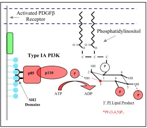
and Waterfield, 1999). During PDGF stimulation, activated PDGF β receptors recruit
cytosolic class IA PI3K via two SH2 domains located in its 85kDa regulatory subunit (p85)
(Fig. 1.4). A 110 kDa catalytic domain (p110) associated with the p85 subunit gives the
enzyme its kinase activity (Cooper and Kashishian, 1993; Escobedo et al., 1991; Kazlauskas
and Cooper, 1990; McGlade et al., 1992). PI3K binds to activated PDGF receptors via its
SH2 domains thereby inducing a conformational change that regulates the activity of its
catalytic domain (Shoelson et al., 1993). The PDGF mediated recruitment of PI3K brings the
phosphorylation of PI(4,5)P2 and generates PI(3,4,5)P3, which can be subsequently
dephosphorylated to PI(3,4)P2 by SHIP, a 5’phosphospatase (Hawkins et al., 1992a; Klippel
et al., 1996; Parent, 2002). These lipid products have been implicated as important second
messengers in cell survival, proliferation, cytoskeletal organization and chemotaxis (Czech,
2000; Rickert et al., 2000; Van Haastert and Devreotes, 2004).
O
OO O
C C C
OH OH OH OH P P P P 1 2 3 4 5 6 Type IA PI3K
p110 p85
SH2 Domains
ATP ADP
Activated PDGFβ
Receptor
3’ PI Lipid Product
Phosphatidylinositol
*PI (3,4,5)P3
FIGURE 1.4 Type IA phosphoinositide 3-kinase: enzyme, substrate, and product. Activated PDGFβ
receptors serve as scaffolds to recruit PI3K to the plasma membrane. PI3K is activated when its SH2 domains engage the Tyr 740 and 751 residues on the PDGFβ receptor. Once at the membrane and activated, PI3K phosphorylates its lipid substrate, phosphatidylinositol (4,5)-bisphosphate, at the D3 position producing phosphatidylinositol (3,4,5)-trisphosphate.
1.2.6 Roles of 3’ phosphoinositides
The 3’ PI lipid products PI(3,4)P2 and PIP3 mediate the recruitment of cytosolic signaling
molecules containing pleckstrin homology (PH) domains. The PH domains of various
molecules engage specific phosphoinositide lipids on the inner leaflet of the plasma
protein that is recruited to the membrane by PI(3,4)P2 and PIP3 is the serine/threonine kinase
Akt/protein kinase B (PKB), an important mediator of cell survival (Franke et al., 1997;
Klippel et al., 1997). Akt/PKB activation is regulated by lipid binding and phosphorylation
by either phosphoinositide-dependent protein kinase-1 (PDK1) or integrin-linked kinase
(ILK). Activated Akt/PKB phosphorylates BAD (BCL-2 family member) preventing it from
initiating apoptosis and promoting cell survival (Klippel et al., 1997; Stupack and Cheresh,
2002; Watton and Downward, 1999). The PI(4,5)P2 lipase PLC-γ also contains a PH
domain. In cells stimulated with PDGF, PLC-γ translocates to the leading edge of a
migrating cell in a PI3K dependent manner implying its membrane localization is regulated
in part by 3’PI lipids, namely PI(3,4,5)P3 (Falasca et al., 1998; Rameh and Cantley, 1999).
This selective localization allows PLC-γ to regulate the local PI(4,5)P2 levels. PI(4,5)P2 is an
important regulator of F-actin assembly and other cytoskeleton components that regulate
actin polymerization such as profilin (Goldschmidt-Clermont et al., 1991; Zigmond, 1996).
Thus PI(4,5)P2 lipid turnover is indirectly regulated by 3’PI lipids through PLC-γ and is
dependent on PDGF stimulation. The 3’ PI lipid products of PI3K also play a direct and vital
role in cell migration, serving as key mediators in the regulation of membrane protrusions
and as a molecular ‘compass’ for directed migration in the presence of a chemoattractant
gradient (Chung et al., 2001; Parent and Devreotes, 1999; Van Haastert and Devreotes, 2004;
Weiner, 2002). PDGF stimulated fibroblasts exhibit PI3K dependent membrane ruffling, an
event caused by the rearrangement of the actin cytoskeleton (Wennstrom et al., 1994a;
Wymann and Arcaro, 1994). This physiological response is mediated by PI3K dependent
HooshmandRad et al., 1997; Welch et al., 2003). Fibroblasts treated with both synthetic and
isolated PIP3 exhibit membrane ruffling and chemotaxis further supporting the involvement
of PI3K and PIP3 in directed movement (Derman et al., 1997).
1.2.7 PI3K and chemotaxis
During wound healing, fibroblasts translate spatial differences in PGDF concentration into
directed movements that will propel them toward the wound. This ability to sense spatial
chemoattractant differences involves localized receptor activation and asymmetric
amplification of the signal (Devreotes and Janetopoulos, 2003; Van Haastert and Devreotes,
2004). PI3K has been implicated as a key mediator in the localization and amplification of
signaling cues from both G-protein coupled receptors (Dictyostelium and neutrophils) and
PDGF receptors (fibroblasts) (Firtel and Chung, 2000; Haugh et al., 2000; Parent et al., 1998;
Rickert et al., 2000). A gradient of chemoattractant causes an asymmetric activation of
surface receptors and higher localized recruitment of PI3K to the region of cell membrane
exposed to the largest concentration of stimuli (Rickert et al., 2000) (Devreotes and
Zigmond, 1988; Parent et al., 1998). Localized PI3K activity leads to localized production of
PIP3 and PI(3,4)P2 (Chung et al., 2001; Haugh et al., 2000). Using green fluorescent proteins
(GFP) fused to the PH domain of Akt, the rates of lipid accumulation under chemoattractant
stimulus can be monitored in living cells (Haugh et al., 2000; Hein and Tsien, 1996; Meili et
al., 1999; Servant et al., 2000). This polarization of lipid products asymmetrically activates
the Rho GTPases Rac and Cdc42, which respectively mediate the formation of membrane
of the cell by the actions of lipid phosphatases such as PTEN(3’) and SHIP(5’) (Di
Cristofano and Pandolfi, 2000; Rickert et al., 2000; Van Haastert and Devreotes, 2004).
Fibroblasts not expressing PTEN show increased protrusion and motility mediated by Rac
and Cdc42 GTPases which suggests that lipid phosphatases regulate their activity by
affecting lipid turnover (Liliental et al., 2000; Parent, 2002). The actions of these lipid
phosphatases minimize the accumulation of 3’PIs in regions other than the leading edge of
the cell. This generates a signaling asymmetry and drives membrane extension in the
direction of the highest level of chemoattractant. PI3K has specifically been singled out as
the primary mediator of 3’ PI lipid production because treating chemotaxing cells with
specific PI3K inhibitors (Wortmannin and LY294002) completely blocks lipid localization,
membrane protrusions, and chemotaxis (Haugh et al., 2000; Hawkins et al., 1995;
HooshmandRad et al., 1997; Wymann and Arcaro, 1994). In order for stimulated cells to
successfully migrate to the source of the chemoattractant, the lamellipodia must form stable
attachments with the substratum while the rear or uropod of the cell must detach from the
surface, processes tightly regulated by integrin adhesion receptors.
1.2.8 Integrin receptors
Integrins are a diverse class of surface receptors essential during cellular adhesion, spreading,
and migration (Danen and Yamada, 2001; Hynes, 1992; Schwartz et al., 1995). Their
versatility makes them important mediators in many important physiological processes
including cell proliferation, matrix assembly, cell survival and chemotaxis (Aplin et al.,
composed of α and β subunits that are non-covalently bound together in the cell membrane.
Currently there are 24 dimers that form in combinations of the known 18 α and 8 β subunits
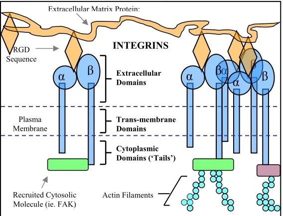
(Berman et al., 2003). Each subunit consists of a glycosylated extracellular domain, a
hydrophobic transmembrane domain, and a short cytoplasmic domain (Berman et al., 2003;
Schwartz et al., 1995). The various integrin heterodimers exhibit affinity for one or more
ECM proteins such as Fn, collagen, laminin, and vitronectin. As depicted in Fig. 1.5,
integrins provide a physical link between these matrix proteins and the intracellular
cytoskeleton components of a cell, the machinery responsible for generating cell motility
(Hynes, 1992). These integrin adhesion sites are populated with several molecules
participating in intracellular signaling.
α β
α β α β α β
Extracellular Matrix Protein:
Plasma Membrane
Extracellular Domains
Trans-membrane Domains
Cytoplasmic Domains (‘Tails’)
Actin Filaments RGD
Sequence
Recruited Cytosolic Molecule (ie. FAK)
INTEGRINS
1.2.9 Integrin signaling
Integrin signaling events can be divided into two categories: inside-out and outside-in
signaling. The assembly of a Fn matrix by fibroblasts involves inside-out regulation of
integrin α5β1 affinity (Christopher et al., 1997; Damsky and Ilic, 2002). Fn contains a cell
recognition amino-acid sequence Arg-Gly-Asp (RGD) that is recognized by α5β1 and αvβ3
integrins. Initial binding of the integrin is followed by inside-out signals which activate and
enhance the affinity of the integrin for Fn allowing fibroblasts to manipulate and assemble
the Fn (Christopher et al., 1997; Damsky and Ilic, 2002; Schwartz et al., 1995). This change
in integrin affinity is mediate by a cation (Mg2+ and Mn2+) dependent mechanism, the binding of cytoskeleton proteins such as talin to the integrin cytoplasmic tail or
phosphorylation (Edelman, 1985; Schwartz et al., 1995). These external influences cause
structural changes and/or a change in the interaction between α and β subunits that ultimately
increase or decrease an integrin’s affinity for its ECM substrate (Liddington and Ginsberg,
2002). In certain cell types, PI3K has also been suggested to regulate integrin activation in
an inside-out dependent manner (Kolanus and Seed, 1997). Integrins also mediate cell
survival and cell migration via outside-in signaling processes (Damsky and Ilic, 2002;
Stupack and Cheresh, 2002). Upon engagement with ECM proteins, integrins cluster
together into focal contacts and promote the recruitment of cytosolic signaling molecules to
these newly formed adhesion sites (Clark and Brugge, 1995; Damsky and Ilic, 2002;
Schwartz et al., 1995). Fibroblast survival has been shown to be dependent on Fn anchorage
and can be mediated through two distinct signaling pathways. The C-Jun NH2-terminal
after activation by focal adhesion kinase (FAK) and p130Cas in Fn anchored fibroblasts
(Almeida et al., 2000). Alternatively, the α5β1 and αVβ3 integrins promote cell survival on
Fn via the PI3K-Akt pathway which is also mediated by FAK (Matter and Ruoslahti, 2001).
Cellular adhesion to Fn also promotes other important mitogenic signal transduction cascades
(Ras-Raf-MEK-Erk) and controls progression through the G1 phase of the cell cycle (Danen
and Yamada, 2001; Mettouchi et al., 2001). Integrin engagement to Fn can augment and
enhance growth factor dependent signaling (Schwartz, 1997). For example, fibroblasts
plated on Fn show enhanced PDGF dependent activation of Ras and DNA synthesis (DeMali
et al., 1999). Integrins mediate signaling by their ability to recruit other signaling molecules.
The majority of integrin mediated signaling events involve the tyrosine kinase FAK, which
has been implemented as a central player in integrin signaling (Parsons et al., 2000;
Schlaepfer and Hunter, 1998).
N
Focal Adhesion Kinase
C Y397
PI-3 Kinase
Y925
Src
Grb2 SOS
Integrin Clustering
FAK
1.2.10 Focal adhesions
ECM induces integrin clustering into small, transient focal complexes at the leading edge of
the lamellipodia, while larger clusters of integrins form strong adhesions in the underlying
body of the cell (Clark et al., 1998; Parsons et al., 2000). Several important signaling
molecules have been observed to associate with focal adhesions including FAK, c-Src, PI3K,
and PLC-γ (Plopper et al., 1995). FAK co-localizes with integrins at cell-substratum contact
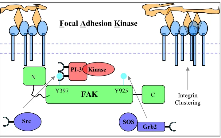
sites. As shown in Fig. 1.6, the recruitment of FAK to these integrin clusters is mediated by
its N-terminal domain which interacts with the short cytoplasmic domains of the β integrins
(Parsons et al., 2000; Schlaepfer and Hunter, 1998; Schwartz et al., 1995). The integrin
mediated localization induces FAK autophosphorylation at the conserved Tyr 397 residue
(Burridge et al., 1992; Schlaepfer and Hunter, 1998). It should also be noted that cells
cultured on Fn, but not poly-D-lysine show this increase in FAK phosphorylation (Burridge
et al., 1992) (Guan et al., 1991; Kornberg et al., 1992). Phosphorylation of Tyr 397 mediates
the recruitment of Src which fully activates FAK by phosphorylating its other Tyr residues.
FAK controls the recruitment of a diverse group of cytosolic adaptors, kinases, and
cytoskeleton molecules containing both SH2 and SH3 (a proline rich recognition site)
domains (Parsons et al., 2000; Schlaepfer et al., 1999; Schwartz et al., 1995). FAK recruits
and binds the adaptor Grb2 which binds the Ras activating GEF Sos (Schlaepfer et al., 1994;
Schlaepfer et al., 1999). Activated Ras can mediate the activation of PI3K dependent
membrane ruffling (RodriguezViciana et al., 1997). FAK has also been reported to directly
associate with and activate PI3K in fibroblasts plated on Fn through Tyr 397 (Chen et al.,
integrin-dependent accumulation of PI3K lipid products and activation of PI3K-dependent
Akt/PKB (King et al., 1997). Fibroblasts cultured on Fn also show an increase in the level of
PI3K substrate PI(4,5)P2 compared to cells held in suspension (McNamee 1993). Even
growth factors have a greatly reduced ability to activate the Akt/PKB survival pathway via
PI3K in cells held in suspension (Khwaja et al., 1997). The process of suspended cells
spreading on surface substratum, such as Fn, requires integrin-FAK mediated activation of
PI3K (Meng and Lowell, 1998). Consistent with the fact that activated PI3K can mediate
cell migration, fibroblasts not expressing FAK show rounded morphology and defective
migration in the presence of Fn (Sieg et al., 1999). In addition, PI3K specific inhibitors
(LY294002 and wortmannin) inhibit FAK promoted cell migration on Fn (Reiske et al.,
1999). Combined, these results strongly suggest that integrin engagement of Fn leads to a
FAK dependent recruitment and activation of PI3K.
1.2.11 Migration: Convergence of integrin and growth factor receptor signaling
Integrins are catalytically inactive, but their ability to recruit signaling and cytoskeletal
proteins allows them to mediate biochemical and biophysical events during cell migration.
ECM induced integrin clustering into focal complexes and focal adhesions promote the
recruitment of cytoskeletal proteins talin and α-actinin, which link integrins to actin filaments
and stress fibers. These proteins, along with integrins themselves, promote recruitment of
important signaling molecules such as FAK, Src, and PI3K. The 3’ PI lipid products
generated by PI3K along with PLC-γ regulated PI(4,5)P2 promote localized actin
Ridley et al., 2003). Motor proteins like myosin I and II can generate tension through the
integrin linked ECM and actin network allowing the cell to contract and crawl forward. Both
integrins and growth factors are important mediators of cell migration. As described above,
PDGF stimulated fibroblasts initiate chemotaxis in a PI3K dependent manner (Seppa et al.,
1982a; Wennstrom et al., 1994b). The engagement of PDGF receptors and integrins with
PDGF-BB and Fn respectively promote 3’ PI lipid production and membrane extension
(Hawkins et al., 1992b; King et al., 1997).
PDGFβ Receptors
Integrins (α5β1 & αvβ3)
Cortical Actin Filaments
PDGF-
BB 3’ PI Lipids PI3K
Fibronectin (Fn)
Source (Wound)
Source (Wound)
Fibroblast
In Fig. 1.7, the PDGF-PI3K signaling system regulates the direction and persistence of the
membrane protrusions while α5β1 and αvβ3 integrins mediate the attachment to Fn in addition
to stimulating PI3K (Clark and Brugge, 1995; Hynes, 1992). The Rho family of GTPase
proteins (Rac, Cdc42, and Rho) are the central regulators of membrane protrusions
(Lauffenburger and Horwitz, 1996; Ridley et al., 2003). The activity of the Rho family
proteins is regulated by cycling through guanosine triphosphate (GTP) or guanosine
diphosphate (GDP) bound states as illustrated in Fig. 1.8. The intrinsic GTPase activity of
these proteins, in conjunction with guanine nucleotide exchange factors (GEFs) and GTPase
activating proteins (GAPs), regulates the conversion between GTP (active) and GDP
(inactive) bound states (Bray, 2001; Ridley et al., 1992). The GTPase Rac is activated by
GEFs which are themselves activated in a PI3K dependent manner. For example, the Rac
GEF Vav contains a PH domain that upon engagement with 3’PI lipids frees the catalytic
domain of Vav, which facilitates Rac-GDP conversion to Rac-GTP (Das et al., 2000; Fukata
et al., 2003). The asymmetric activation and localization of PI3K and lipid phosphatases
leads to a signaling polarization. Rac GEFs are locally activated by PI3K products at the
leading edge of the cell (Small et al., 2002). Here activated Rac mediates the formation of an
extending, dendritic network of cross-linked actin filaments that produce a protruding
lamellipodium, a broad, flat, sheet-like membrane protrusion (illustrated in Fig. 1.8). The
formation of a lamellipodium signifies the establishment of morphological polarity and
defines the leading edge of the cell (Lauffenburger and Horwitz, 1996; Small et al., 2002;
Zigmond, 1996). Cdc42 regulates the formation of filopodia which are composed of bundles
1.8). Filopodia are thought to serve as a cell’s initial sensory probe which allows the cell to
detect changes in chemoattractant or ECM (Bray, 2001), and will often evolve into the
lamellipodia. Actin polymerization drives the formation of these protruding structures and is
regulated by numerous molecules. Notably, the Arp2/3 complex and proteins from the
WASP family (Wiskott-Aldrich Syndrome protein) regulate actin polymerization in a Rac
and Cdc42 dependent manner (Calderwood et al., 2000; Ridley et al., 2003; Small et al.,
2002). Rho is responsible for the formation of stress fibers and focal adhesions (Fig. 1.8)
which allow motor proteins like myosin II to generate forward movement through stress fiber
contraction (DeMali et al., 2003; Ridley et al., 2003).
Rac Cdc42 Rho
GTP GDP
GTP GDP
GTP GDP
Lamellipodia Filopodia Stress Fibers & Focal Adhesions
Figure 1.8 Rho family of GTPases - The Rho family proteins cycle between ‘active’ GTP-bound and
‘inactive’ GDP-bound states through the actions of GTP-promoting GEFs, GDP-promoting GAPs, and/or their intrinsic GTPase activity. Rac, Cdc42 and Rho play vital roles in cell migration by mediating the formation of lamellipodia, filopodia, stress fibers and focal adhesions respectively.
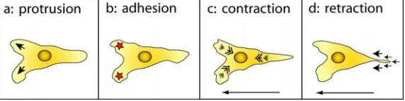
In a migrating cell, the asymmetric and localized activation of both Rac (front) and Rho
contraction and finally release (Fig. 1.9). The activity of Rac and Cdc42 exhibit a biphasic
dependence on substratum (Fn) concentration while Rho activation is saturable at high
substratum concentration (Fn) (Cox et al., 2001). This signaling phenomenon is
supplemented by the finding that cell speed is a biphasic function of the strength of cell
attachment, which is dependent on the substratum concentration, integrin expression and
affinity (Lauffenburger and Horwitz, 1996; Palecek et al., 1997). Thus, cells show maximal
migration speed at intermediate substratum concentrations which is proportional to the
strength of attachment and number of integrin-ECM complexes (Palecek et al., 1997)
(Dimilla et al., 1991). Growth factor stimulation can increase or decrease the migration
speed of fibroblasts depending on the ECM surface concentration (Maheshwari et al., 1999;
Ware et al., 1998). Thus, growth factor receptors and integrins are tightly coupled to
efficient cell migration (Woodard et al., 1998). Growth factors such as EGF disrupt cell
adhesion and disassemble focal adhesions (Maheshwari et al., 1999). PDGF stimulated
fibroblasts also show disassembly of focal adhesion and rapid relocation of β1 integrins
(Ahlen et al., 2004). These observations suggest that gradients of growth factors ‘prime’
cells for directed migration by first mediating the removal of established adhesions through
actin rearrangement and then directing the formation of new Rac-dependent membrane
extensions in the direction of increasing stimuli. Integrins in these new extensions attach to
ECM proteins and establish cytoskeleton linkages while also enhancing growth factor
mediated signaling. Together the synergistic signals generated from growth factor receptors
FIGURE 1.9 Cell migration is a cyclic crawling process. On two dimensional surfaces, cells execute crawling-like motions to generate forward movement. First cells extend a lamellipodia that then forms integrin-dependent adhesions with the ECM coated surface; next motor proteins mediate contraction through the actin cytoskeletal network which relocates the nuclear region and other organelles to the leading edge; and finally this contraction mediates uropod release and the cycle repeats.
1.3 APPROACH
The primary technique used in this study is called total internal reflection
fluorescence (TIRF) microscopy, which was developed by Dan Axelrod in the early 1980s as
a method to image the separation between cell membranes and the underlying substratum
(Axelrod, 1989; Axelrod, 2003). With the development of fusion proteins that couple a
fluorescent reporter protein to specific molecular binding domains, the application of TIRF in
the field of cell biology has grown. Now, TIRF is a well characterized microscopy technique
that exclusively excites these fluorescent probes in the contact region of living cells cultured
on glass surfaces (Steyer and Almers, 2001; Toomre and Manstein, 2001). In TIRF, an
excitation wavelength generated by a laser is directed into a prism that is optically coupled to
a glass coverslip, using immersion oil with the same refractive index as glass. The prism
directs the incoming beam such that it approaches the glass/aqueous interface at an incident
angle greater than the critical angel (θcritical), which can be calculated using the medium
refractive indices and Snell’s Law. An incoming beam with an incidence angle θ that is
greater than θcritical is totally internally reflected. This creates an evanescent wave that
intensity I (energy) that decays exponentially with perpendicular distance z from the prism
surface according to the following equations:
⎟ ⎠ ⎞ ⎜ ⎝ ⎛ − = d z I
I oexp (1.1)
(
)
21 2 2 2 2 1 sin
4 n n
d − = θ π λ (1.2)
where Io is the intensity of the wave at z = 0 defining the glass-buffer interface, n1 and n2 are
the refractive indices of glass (~1.52) and buffer/cell cytosol (~1.33) respectively, d is the
characteristic decay depth, and λ is the excitation wavelength of the incoming beam in the
visible light regime. The evanescent wave typically penetrates roughly 100 nm into the
adjacent medium or buffer as measured from the glass interface (Axelrod, 1981; Burmeister
et al., 1998). TIRF allows us to monitor and make quantitative measurements of intracellular
signaling events occurring at the plasma membrane because the excitation field selectively
excites fluorescent probes in a small volume near the contact region of the cell. As a result,
TIRF minimizes cellular photodamage, photobleaching of fluorescent probes, and cellular
auto-fluorescence while still maintaining a significantly larger signal to noise ratio, compared
to confocal microscopy (Axelrod, 2003; Steyer and Almers, 2001; Toomre and Manstein,
Glass Cover-Slip Teflon Ring Buffer Chamber Objective Pipette or MicroPipette Evanescent Wave Immersion Oil Laser Beam
λ = 488nm or 442nm 514nm Prism CCD Camera Metamorph Imaging Software TOP VIEW SIDE VIEW
FIGURE 1.10 Experimental Setup: TIRF Microscopy – Illustration of the experimental setup for imaging and analyzing fibroblasts in the presence of either soluble or immobilized stimuli or both.
The TIRF setup, illustrated in Fig. 1.10, consists of a Zeiss upright microscope equipped with
three water immersion objectives (10X, 20X, and 40X). The excitation sources include a
tunable wavelength Argon laser with a 60 mW maximum at both 488 nm (GFP) and 514 nm
(YFP), a HeCd laser (Melles Griot) with 120 mW maximum at 442 nm, and a 100 mW
diode-pumped 561 nm line (Crystalaser, Reno, NV) was recently added to excite red emitting
fluorescent probes. Shutter controllers regulates the exposure times of the excitation beams.
The microscope is fitted with an emission filter wheel and controller as well as emission
filters compatible with GFP, YFP, CFP, and Texas Red excitation and emission wavelengths.
Digital images are acquired using a CCD camera (Hamamatsu) and analyzed with
Metamorph Imaging software. Samples are prepared by securing a Teflon ring, coated with
an accessible buffer well. The coverslip is then mounted on a custom made, x-y directional
motorized stage. A prism directly beneath the stage is optically coupled to the glass
coverslip via immersion oil (Zeiss). We have the option of conducting experiments at 37oC using a heated Plexiglas chamber that securely fits around the microscope stage while still
allowing access to the buffer chamber.
TIRF microscopy allows for quantitative fluorescence imaging of the spatial and
temporal dynamic of fluorescent probes at the cell membrane. Using the 3’ PI binding
specificity of the PH domain of Akt coupled to a fluorescent protein, we are able to monitor
the patterns of PI3K activity during cell spreading and migration under a variety of
environmental stimuli. With this technique and the development of complementary
experimental and computational methods, we hope to address several questions associated
with fibroblast motility:
1. Does integrin mediated adhesion promote PI3K activation, and is its activation
localized and coincident with fibroblast motility? (Chapter 2)
2. In some cell types, the cytoskeleton influences PI3K signaling through a positive
feedback loop, but is the same true for fibroblasts? (Chapter 2) Chapter 2 presents
work that has been recently submitted for publication.
3. During random migration on adhesive surfaces, is PI3K active and if so what is its
possible function as it pertains to fibroblast movement? (Chapter 3) In this Chapter 3,
we present a novel analysis framework to quantify and relate PI3K signaling and
4. Microfluidic devices are being employed to study directed migration, but can they be
integrated with TIRF microscopy to study fibroblast chemotaxis? (Chapter 4)
Chapter 4 presents the methodologies for fabrication and the initial implementation of
a microfluidic device for studying fibroblast chemotaxis.
5. How are mechanical signals from the extracellular environment integrated to
influence the direction of cell migration? Can experimental techniques be developed
to assess whether or not PI3K, which is intimately involved in chemotaxis, plays a
role in transducing information about substrate rigidity? (Chapter 5) In Chapter 5, we
present methods for generating a novel polymeric material, and preliminary results
1.4 REFERENCES
Ahlen, K., P. Ring, B. Tomasini-Johansson, K. Holmqvist, K.E. Magnusson, and K. Rubin. 2004. Platelet-derived growth factor-BB modulates membrane mobility of beta(1) integrins. Biochemical and Biophysical Research Communications. 314:89-96.
Almeida, E.A.C., D. Ilic, Q. Han, C.R. Hauck, F. Jin, H. Kawakatsu, D.D. Schlaepfer, and C.H. Damsky. 2000. Matrix survival signaling: From fibronectin via focal adhesion kinase to c-Jun NH2-terminal kinase.
Journal of Cell Biology. 149:741-754.
Aplin, A.E., A.K. Howe, and R.L. Juliano. 1999. Cell adhesion molecules, signal transduction and cell growth.
Current Opinion in Cell Biology. 11:737-744.
Axelrod, D. 1981. Cell-Substrate Contacts Illuminated by Total Internal-Reflection Fluorescence. Journal of Cell Biology. 89:141-145.
Axelrod, D. 1989. Total internal reflection fluorescence microscopy. Methods Cell Biol. 30:245-70.
Axelrod, D. 2003. Total internal reflection fluorescence microscopy in cell biology. Methods Enzymol. 361:1-33.
Bergsten, E., M. Uutela, X.R. Li, K. Pietras, A. Ostman, C.H. Heldin, K. Alitalo, and U. Eriksson. 2001. PDGF-D is a specific, protease-activated ligand for the PPDGF-DGF beta-receptor. Nature Cell Biology. 3:512-516.
Berman, A.E., N.I. Kozova, and G.E. Morozevich. 2003. Integrins: Structure and signaling. Biochemistry-Moscow. 68:1284-1299.
Bhora, F.Y., B.J. Dunkin, S. Batzri, H.M. Aly, B.L. Bass, A.N. Sidawy, and J.W. Harmon. 1995. Effect of Growth-Factors on Cell-Proliferation and Epithelialization in Human Skin. Journal of Surgical Research. 59:236-244.
Bishayee, S., S. Majumdar, J. Khire, and M. Das. 1989. Ligand-Induced Dimerization of the Platelet-Derived Growth-Factor Receptor - Monomer-Dimer Interconversion Occurs Independent of Receptor Phosphorylation. Journal of Biological Chemistry. 264:11699-11705.
Bray, D. 2001. Cell Movements: from molecules to motility. Garland Publishing Taylor and Francis Group, New York.
Burridge, K., C.E. Turner, and L.H. Romer. 1992. Tyrosine Phosphorylation of Paxillin and Pp125(Fak) Accompanies Cell-Adhesion to Extracellular-Matrix - a Role in Cytoskeletal Assembly. Journal of Cell Biology. 119:893-903.
Calderwood, D.A., S.J. Shattil, and M.H. Ginsberg. 2000. Integrins and actin filaments: Reciprocal regulation of cell adhesion and signaling. Journal of Biological Chemistry. 275:22607-22610.
Chen, H.C., P.A. Appeddu, H. Isoda, and J.L. Guan. 1996. Phosphorylation of tyrosine 397 in focal adhesion kinase is required for binding phosphatidylinositol 3-kinase. Journal of Biological Chemistry. 271:26329-26334.
Chen, H.C., and J.L. Guan. 1994. Association of focal adhesion kinase with its potential substrate phosphatidylinositol 3-kinase. Proc Natl Acad Sci U S A. 91:10148-52.
Christopher, R.A., A.P. Kowalczyk, and P.J. McKeownLongo. 1997. Localization of fibronectin matrix assembly sites on fibroblasts and endothelial cells. Journal of Cell Science. 110:569-581.
Chung, C.Y., S. Funamoto, and R.A. Firtel. 2001. Signaling pathways controlling cell polarity and chemotaxis.
Trends in Biochemical Sciences. 26:557-566.
Claesson-Welsh, L. 1994. Platelet-derived growth factor receptor signals. J Biol Chem. 269:32023-6.
Clark, E.A., and J.S. Brugge. 1995. Integrins and Signal-Transduction Pathways - the Road Taken. Science. 268:233-239.
Clark, E.A., W.G. King, J.S. Brugge, M. Symons, and R.O. Hynes. 1998. Integrin-mediated signals regulated by members of the Rho family of GTPases. Journal of Cell Biology. 142:573-586.
Clark, R.A.F. 1990. Fibronectin Matrix Deposition and Fibronectin Receptor Expression in Healing and Normal Skin. Journal of Investigative Dermatology. 94:S128-S134.
Cooper, J.A., and A. Kashishian. 1993. In vivo Binding-Properties of Sh2 Domains from GTPase-Activating Protein and Phosphatidylinositol 3-Kinase. Molecular and Cellular Biology. 13:1737-1745.
Couchman, J.R., M.R. Austria, and A. Woods. 1990. Fibronectin-cell interactions. J Invest Dermatol. 94:7S-14S.
Cox, E.A., S.K. Sastry, and A. Huttenlocher. 2001. Integrin-mediated adhesion regulates cell polarity and membrane protrusion through the Rho family of GTPases. Molecular Biology of the Cell. 12:265-277.
Czech, M.P. 2000. PIP2 and PIP3: Complex roles at the cell surface. Cell. 100:603-606.
Damsky, C.H., and D. Ilic. 2002. Integrin signaling: it's where the action is. Current Opinion in Cell Biology. 14:594-602.
Danen, E.H.J., and K.M. Yamada. 2001. Fibronectin, integrins, and growth control. Journal of Cellular Physiology. 189:1-13.
Das, B., X.D. Shu, G.J. Day, J. Han, U.M. Krishna, J.R. Falck, and D. Broek. 2000. Control of intramolecular interactions between the pleckstrin homology and Db1 homology domains of Vav and Sos1 regulates Rac binding. Journal of Biological Chemistry. 275:15074-15081.
DeMali, K.A., E. Balciunaite, and A. Kazlauskas. 1999. Integrins enhance platelet-derived growth factor (PDGF)-dependent responses by altering the signal relay enzymes that are recruited to the PDGF beta receptor. Journal of Biological Chemistry. 274:19551-19558.
DeMali, K.A., K. Wennerberg, and K. Burridge. 2003. Integrin signaling to the actin cytoskeleton. Current Opinion in Cell Biology. 15:572-582.
Derman, M.P., A. Toker, J.H. Hartwig, K. Spokes, J.R. Falck, C.S. Chen, L.C. Cantley, and L.G. Cantley. 1997. The lipid products of phosphoinositide 3-kinase increase cell motility through protein kinase C. J Biol Chem. 272:6465-70.
Deuel, T.F., R.S. Kawahara, T.A. Mustoe, and G.F. Pierce. 1991. Growth-Factors and Wound-Healing - Platelet-Derived Growth-Factor as a Model Cytokine. Annual Review of Medicine. 42:567-584.
Devreotes, P., and C. Janetopoulos. 2003. Eukaryotic chemotaxis: distinctions between directional sensing and polarization. J Biol Chem. 278:20445-8.
Devreotes, P.N., and S.H. Zigmond. 1988. Chemotaxis in Eukaryotic Cells - a Focus on Leukocytes and Dictyostelium. Annual Review of Cell Biology. 4:649-686.
Di Cristofano, A., and P.P. Pandolfi. 2000. The multiple roles of PTEN in tumor suppression. Cell. 100:387-390.
Edelman, G.M. 1985. The Cell in Contact: adhesions and junctions as morphogenetic determinants. John Wiley and Sons, New York. 303-333 pp.
Emaduddin, M., S. Ekman, L. Ronnstrand, and C.H. Heldin. 1999. Functional co-operation between the subunits in heterodimeric platelet-derived growth factor receptor complexes. Biochemical Journal. 341:523-528.
Escobedo, J.A., D.R. Kaplan, W.M. Kavanaugh, C.W. Turck, and L.T. Williams. 1991. A Phosphatidylinositol-3 Kinase Binds to Platelet-Derived Growth-Factor Receptors through a Specific Receptor Sequence Containing Phosphotyrosine. Molecular and Cellular Biology. 11:1125-1132.
Falasca, M., S.K. Logan, V.P. Lehto, G. Baccante, M.A. Lemmon, and J. Schlessinger. 1998. Activation of phospholipase C gamma by PI 3-kinase-induced PH domain-mediated membrane targeting. Embo Journal. 17:414-422.
Fantl, W.J., J.A. Escobedo, and L.T. Williams. 1989. Mutations of the Platelet-Derived Growth-Factor Receptor That Cause a Loss of Ligand-Induced Conformational Change, Subtle Changes in Kinase-Activity, and Impaired Ability to Stimulate DNA-Synthesis. Molecular and Cellular Biology. 9:4473-4478.
Fantl, W.J., D.E. Johnson, and L.T. Williams. 1993. Signaling by Receptor Tyrosine Kinases. Annual Review of Biochemistry. 62:453-481.
Firtel, R.A., and C.Y. Chung. 2000. The molecular genetics of chemotaxis: sensing and responding to chemoattractant gradients. Bioessays. 22:603-615.
Franke, T.F., D.R. Kaplan, L.C. Cantley, and A. Toker. 1997. Direct regulation of the Akt proto-oncogene product by phosphatidylinositol-3,4-bisphosphate. Science. 275:665-668.
Frech, M., M. Andjelkovic, E. Ingley, K.K. Reddy, J.R. Falck, and B.A. Hemmings. 1997. High affinity binding of inositol phosphates and phosphoinositides to the Pleckstrin homology domain of RAC protein kinase B and their influence on kinase activity. Journal of Biological Chemistry. 272:8474-8481.
Fukata, M., M. Nakagawa, and K. Kaibuchi. 2003. Roles of Rho-family GTPases in cell polarisation and directional migration. Current Opinion in Cell Biology. 15:590-597.
Gailit, J., and R.A.F. Clark. 1996. Studies in vitro on the role of alpha v and beta 1 integrins in the adhesion of human dermal fibroblasts to provisional matrix proteins fibronectin, vibronectin, and fibrinogen.
Journal of Investigative Dermatology. 106:102-108.
Grinnell, F., R.E. Billingham, and L. Burgess. 1981. Distribution of Fibronectin During Wound-Healing Invivo.
Journal of Investigative Dermatology. 76:181-189.
Guan, J.L., J.E. Trevithick, and R.O. Hynes. 1991. Fibronectin Integrin Interaction Induces Tyrosine Phosphorylation of a 120-Kda Protein. Cell Regulation. 2:951-964.
Haugh, J.M., F. Codazzi, M. Teruel, and T. Meyer. 2000. Spatial sensing in fibroblasts mediated by 3' phosphoinositides. J Cell Biol. 151:1269-80.
Hawkins, P.T., A. Eguinoa, R.G. Qiu, D. Stokoe, F.T. Cooke, R. Walters, S. Wennstrom, L. Claesson-Welsh, T. Evans, M. Symons, and et al. 1995. PDGF stimulates an increase in GTP-Rac via activation of
phosphoinositide 3-kinase. Curr Biol. 5:393-403.
Hawkins, P.T., T.R. Jackson, and L.R. Stephens. 1992a. Platelet-Derived Growth-Factor Stimulates Synthesis of Ptdlns(3,4,5)P3 by Activating a Ptdlns(4,5)P2 3-Oh Kinase. Nature. 358:157-159.
Hawkins, P.T., T.R. Jackson, and L.R. Stephens. 1992b. Platelet-derived growth factor stimulates synthesis of PtdIns(3,4,5)P3 by activating a PtdIns(4,5)P2 3-OH kinase. Nature. 358:157-9.
Hein, R., and R.Y. Tsien. 1996. Engineering green fluorescent protein for improved brightness, longer wavelengths and fluorescence resonance energy transfer. Current Biology. 6:178-182.
Heldin, C.H., A. Ernlund, C. Rorsman, and L. Ronnstrand. 1989. Dimerization of B-Type Platelet-Derived Growth-Factor Receptors Occurs after Ligand-Binding and Is Closely Associated with Receptor Kinase Activation. Journal of Biological Chemistry. 264:8905-8912.
Heldin, C.H., A. Ostman, and L. Ronnstrand. 1998. Signal transduction via platelet-derived growth factor receptors. Biochimica Et Biophysica Acta-Reviews on Cancer. 1378:F79-F113.
Heldin, C.H., and B. Westermark. 1999. Mechanism of action and in vivo role of platelet-derived growth factor.
Physiological Reviews. 79:1283-1316.
HooshmandRad, R., L. ClaessonWelsh, S. Wennstrom, K. Yokote, A. Siegbahn, and C.H. Heldin. 1997. Involvement of phosphatidylinositide 3'-kinase and Rac in platelet-derived growth factor-induced actin reorganization and chemotaxis. Experimental Cell Research. 234:434-441.
Hosang, M., M. Rouge, B. Wipf, B. Eggimann, F. Kaufmann, and W. Hunziker. 1989. Both Homodimeric Isoforms of Pdgf (Aa and Bb) Have Mitogenic and Chemotactic Activity and Stimulate
Hynes, R.O. 1992. Integrins - Versatility, Modulation, and Signaling in Cell-Adhesion. Cell. 69:11-25.
Kaplan, D.R., F.C. Chao, C.D. Stiles, H.N. Antoniades, and C.D. Scher. 1979. Platelet Alpha-Granules Contain a Growth-Factor for Fibroblasts. Blood. 53:1043-1052.
Kashishian, A., A. Kazlauskas, and J.A. Cooper. 1992. Phosphorylation Sites in the Pdgf Receptor with Different Specificities for Binding Gap and Pi3 Kinase Invivo. Embo Journal. 11:1373-1382.
Kazlauskas, A., and J.A. Cooper. 1990. Phosphorylation of the Pdgf Receptor Beta-Subunit Creates a Tight-Binding Site for Phosphatidylinositol-3 Kinase. Embo Journal. 9:3279-3286.
Khwaja, A., P. RodriguezViciana, S. Wennstrom, P.H. Warne, and J. Downward. 1997. Matrix adhesion and Ras transformation both activate a phosphoinositide 3-OH kinase and protein kinase B/Akt cellular survival pathway. Embo Journal. 16:2783-2793.
King, W.G., M.D. Mattaliano, T.O. Chan, P.N. Tsichlis, and J.S. Brugge. 1997. Phosphatidylinositol 3-kinase is required for integlrin-stimulated AKT and Raf-1/mitogen-activated protein kinase pathway activation.
Molecular and Cellular Biology. 17:4406-4418.
Klippel, A., W.M. Kavanaugh, D. Pot, and L.T. Williams. 1997. A specific product of phosphatidylinositol 3-kinase directly activates the protein 3-kinase Akt through its pleckstrin homology domain. Mol Cell Biol. 17:338-44.
Klippel, A., C. Reinhard, W.M. Kavanaugh, G. Apell, M.A. Escobedo, and L.T. Williams. 1996. Membrane localization of phosphatidylinositol 3-kinase is sufficient to activate multiple signal-transducing kinase pathways. Molecular and Cellular Biology. 16:4117-4127.
Kolanus, W., and B. Seed. 1997. Integrins and inside-out signal transduction: converging signals from PKC and PIP3. Current Opinion in Cell Biology. 9:725-731.
Kornberg, L., H.S. Earp, J.T. Parsons, M. Schaller, and R.L. Juliano. 1992. Cell-Adhesion or Integrin Clustering Increases Phosphorylation of a Focal Adhesion-Associated Tyrosine Kinase. Journal of Biological Chemistry. 267:23439-23442.
Kundra, V., J.A. Escobedo, A. Kazlauskas, H.K. Kim, S.G. Rhee, L.T. Williams, and B.R. Zetter. 1994.
Regulation of Chemotaxis by the Platelet-Derived Growth-Factor Receptor-Beta. Nature. 367:474-476.