Cancer Management and Research 2018:10 2563–2571
Cancer Management and Research
Dove
press
submit your manuscript | www.dovepress.com 2563
O R i g i n a l R e s e a R C h
open access to scientific and medical research
Open Access Full Text Article
e2F1 induces lsinCT5 transcriptional activity and
promotes gastric cancer progression by affecting
the epithelial-mesenchymal transition
Peng Qi,1–3
Wan-run lin,4
Meng Zhang,1–3
Dan huang,1–3
shu-juan ni,1–3
Xiao-li Zhu,1–3
Qian-ming Bai,1–3
Wei-qi sheng,1–3
Xiang Du,2
Xiao-yan Zhou1–3
1Department of Pathology, Fudan
University shanghai Cancer Center, shanghai, China; 2institute
of Pathology, Fudan University, shanghai, China; 3Department of
Oncology, shanghai Medical College, Fudan University, shanghai, China;
4Department of Pathology, University
of Texas southwestern Medical Center, Dallas, TX, Usa
Background: Long noncoding RNAs (lncRNAs) have been shown to play important regulatory
roles in human cancer. We previously verified that the lncRNA long stress-induced noncoding transcript 5 (LSINCT5) is overexpressed in gastric cancer (GC) cells and closely correlated with cell proliferation and patient prognosis. However, whether aberrant LSINCT5 expression has an important effect on GC progression is unclear, and the potential mechanisms remain unknown. In GC, E2F1 expression is also aberrant, but the biological functions of E2F1 are controversial, and the correlation between E2F1 and lncRNAs remains unknown.
Materials and methods: Expression of LSINCT5 was analyzed in metastatic GC tissues compared with nonmetastatic tissues using quantitative real-time PCR (qRT-PCR) assays. Gain and loss of function approaches were used to investigate the biological role of LSINCT5 in GC cell migration and invasion. A computational screen of LSINCT5 promoter was conducted to search for transcription factor-binding sites. LSINCT5 promoter activities were examined by ChIP and luciferase reporter assays. qRT-PCR and western blotting assays were performed to detect the expression of multiple EMT markers in cells in which LSINCT5 was overexpressed or knocked down.
Results: An integrated quantitative analysis revealed that LSINCT5 was significantly
over-expressed in metastatic GC tissues. Forced LSINCT5 expression promoted cell migration and invasion, whereas loss of LSINCT5 function decreased cell migration and invasion. Mechanistic investigations showed that LSINCT5 is a direct transcriptional target of E2F1. Moreover, LSINCT5 overexpression was found to play an important role in the epithelial-to-mesenchymal transition by regulating the expression of E-cadherin, N-cadherin, vimentin, and matrix metalloproteinase-2. Conclusion: These data suggest that E2F1-mediated activation of LSINCT5, a regulator of cell
migration and invasion, constitute the mechanistic link between the E2F1-mediated pathway and lncRNA that regulates cell migration and invasion. Thus, LSINCT5 may be a target for new GC therapies.
Keywords: E2F1, LSINCT5, metastasis, gastric cancer
Introduction
Gastric cancer (GC) is one of the major health problems in the world.1 Although the
global mortality rates from GC have decreased over the past several decades, mortality remains extremely high in many countries, where many patients are diagnosed with advanced stage disease and have very poor survival.2 However, traditional prognostic
assessments based on histological differentiation and the TNM stage are unable to indicate a tumor’s metastatic status or predict the clinical outcome of a patient. As a result, there is a growing need to explore novel prognostic biomarkers for assessing the metastasis process.
Correspondence: Xiao-yan Zhou Department of Pathology, Fudan University shanghai Cancer Center, 270 Dong an Road, shanghai 200032, China Tel +86 216 417 5590 ext 8330 Fax +86 216 417 0067 email 13524324387@163.com
Journal name: Cancer Management and Research Article Designation: Original Research Year: 2018
Volume: 10
Running head verso: Qi et al
Running head recto: E2F1 induces LSINCT5 expression and promotes GC progression DOI: http://dx.doi.org/10.2147/CMAR.S171652
Cancer Management and Research downloaded from https://www.dovepress.com/ by 118.70.13.36 on 20-Aug-2020
For personal use only.
Dovepress
Qi et al
The key role of the epithelial–mesenchymal transition (EMT) in cancer cell metastasis was elucidated in the past decade.3 This biological process is characterized by the loss of
epithelial markers and the acquisition of mesenchymal mark-ers, and E-cadherin, N-cadherin, and vimentin are considered the most important molecular markers of the EMT. More recently, several studies have implicated long noncoding RNAs (lncRNAs), which are key RNA molecules >200 nucleotides in length, in tumorigenesis, cancer progression, and metastasis.4,5
For instance, our research groups have demonstrated that PVT1 enhances GC cell proliferation and invasion in a FOXM1-mediated manner.6 Moreover, the lncRNA ncRAN mediates
colorectal cancer (CRC) cell migration and invasion and might be a novel prognostic indicator, specifically, a biomarker, for the early detection of metastasis in CRC.
Although a small number of lncRNAs have been function-ally characterized, little is known regarding most lncRNAs.7
Long stress-induced noncoding transcript 5 (LSINCT5), which is ~2.6-kb long, is an intergenic lncRNA located at Chr 5p. Previous experiments showed that LSINCT5 expression was 7- and 10-fold higher in primary breast tumor tissues and breast cancer cell lines than in corresponding normal benign breast tissue and normal cell lines, respectively, and that the knockdown of LSINCT5 with antisense oligos decreased the proliferation of breast cancer cell lines.8 In our previous study,
we verified that LSINCT5 is overexpressed in GC cells and closely related to cell proliferation and patient prognosis.9
However, whether aberrant LSINCT5 expression exerts an important effect on GC progression remains unclear, and the potential mechanisms are unknown.
E2F transcription factors are key participants in numerous cellular events, such as the control of the cell cycle, DNA syn-thesis, and nuclear transcription. In mammals, 8 members of the E2F family have been identified, and these are divided into 4 groups based on homology: E2F1 to 3, E2F4 and 5, E2F6, E2F7 and 8. The transcriptional activity of E2Fs is predominantly regulated by the Rb protein family (Rb, p107 and p130),10,11 and
the activation of E2F1 to 3 is central to the regulation of cell cycle-related genes.12 E2F1 is the most thoroughly investigated
member of the E2F family with respect to human malignancies and plays pivotal roles in tumor progression by modulating the expression of both coding and noncoding transcripts.13,14
Inter-estingly, E2F1 has been reported to act as either an oncogene or a tumor suppressor to modulate tumorigenesis depending on the cell context.13,15,16 Emerging evidence has revealed that E2F1
has an important effect on GC progression, but its biological functions remain debated.17–19
Here, we demonstrate a novel pathway involving E2F1 and LSINCT5 in GC cell progression. In this study, we found
the following: 1) LSINCT5 is overexpressed in metastatic GC tissues and has a significant effect on GC cell migration and invasion in vitro; 2) E2F1 induces the transcriptional activa-tion of LSINCT5; and 3) LSINCT5 forces cancer progression by affecting EMT. This study advances our understanding of the role of lncRNAs, such as LSINCT5, as regulators of GC pathogenesis and facilitates the development of lncRNA-directed diagnostics and therapeutics.
Materials and methods
Plasmids and Rna interference
The pMax-E2F1 expression vector was purchased from Addgene (plasmid #16007) (Cambridge, MA, USA), and siRNAs specific for E2F1 (#AM16708) and LSINCT5 (#4392420) were chemically synthesized (Invitrogen, Carls-bad, CA, USA). For the construction of luciferase reporter vectors, the core promoter of the LSINCT5 gene (−1,400 to
+100 relative to the transcription start site of the LSINCT5 gene) and regions containing binding site deletions were subcloned into the pGL3 basic firefly luciferase reporter. All the polymerase chain reaction (PCR) products were verified by DNA sequencing. Cell transfection was performed using Lipofectamine 2000 according to the manufacturer’s instruc-tions (Invitrogen).
Cell lines
The human gastric adenocarcinoma cancer cell lines MGC803, BGC823, MKN45, AGS, and SGC7901, and the normal gastric epithelium cell line GES-1 were obtained from the Chinese Academy of Sciences Committee on Type Culture Collection Cell Bank (Shanghai, China). The cells were cultured in Roswell Park Memorial Institute 1640 medium (Invitrogen) or DMEM (HyClone, Logan, UT, USA). All media were supplemented with 10% fetal bovine serum (FBS) and 2 mM L-glutamine (Invitrogen), and all the cells were cultured at 37°C in a 10% CO2 atmosphere.
Tissue samples and clinical data collection
In this study, 71 patients underwent primary GC resection at the Fudan University Shanghai Cancer Center.9 The study
was approved by the Ethics Committee on Human Research of the Fudan University Shanghai Cancer Center. Written informed consent was obtained from all the patients.
Rna preparation and quantitative
real-time PCR (qRT-PCR)
Total RNA was extracted with the TRIzol reagent (Invitro-gen, Grand Island, NY, USA), and qRT-PCR analyses were
Cancer Management and Research downloaded from https://www.dovepress.com/ by 118.70.13.36 on 20-Aug-2020
Dovepress e2F1 induces lsinCT5 expression and promotes gC progression
conducted according to the manufacturer’s instructions (Takara, Dalian, China). The results were normalized to the expression of glyceraldehyde-3-phosphate dehydrogenase (GAPDH) using the comparative 2–ΔΔCt method. Each sample
was assayed in triplicate, and the primer sequences are listed in Table S1.
luciferase reporter assay
Cells were transfected with appropriate plasmids in 24-well plates for 48 hours, collected, and lysed for luciferase assays. The relative luciferase activity was obtained by normalizing the activity of firefly luciferase to that of Renilla luciferase.
Wound healing assay
The wound healing assay was used to evaluate the tumor cell motility capacity. Briefly, 3×10⁵ cells were seeded in 6-well
plates, cultured overnight, and transfected with the control, pcDNA-LSINCT5,9 or si-LSINCT5. When the cells reached
~85% confluence, the cell layer was scratched with a sterile plastic tip, washed twice, and cultured in complete medium. Wound gaps were observed for 48 hours, and images were captured every 24 hours at 100× magnification, then the migration rate was calculated accordingly. The data were summarized based on 3 independent assays.
Cell invasion and migration assay
The upper sides of MilliCell (12-mm diameter with 8-µm pores) chambers (Millipore, Billerica, MA, USA) were pre-coated with Matrigel (BD Biosciences, Bedford, MA, USA). A total of 5×10⁴ cells in serum-free medium were
added to the upper compartment in medium supplemented with 0.1% FBS, and the chambers were placed into 24-well plates with medium containing 10% FBS. After incubation for an appropriate time at 37°C in 5% CO2, the cells remain-ing on the upper membrane were removed with a cotton swab, and the invaded cells on the lower membrane surface were stained with 4% paraformaldehyde and crystal violet. The invasive activity was quantified by counting 9 random fields (at 200× magnification) per chamber. For each experimental point in each assay, mean values were obtained from at least 3 individual chambers. The migration assay was performed using the same protocol as that used for the invasion assay with the exceptions that no Matrigel was used and the cells were allowed to permeate for 12 hours.
Chromatin immunoprecipitation (ChiP)
ChIP assays were performed using an EZ-ChIPTM Kit
(Millipore) according to the instruction manual. E2F1 anti-body was obtained from Cell Signaling Technology (Danvers,
MA, USA). The immunoprecipitated DNA was quantified by qPCR using SYBR Green Master Mix (Takara). ChIP data are presented as percentages relative to the input DNA.
Western blotting analysis
Cells were collected and lysed using radioimmunoprecipitation assay protein extraction reagent (Beyotime, Beijing, China) supplemented with a protease inhibitor cocktail and phenyl-methylsulfonyl fluoride (Roche, Pleasanton, CA, USA). The protein concentration was determined using a Bio-Rad protein assay kit (Bio-Rad, Hercules, CA, USA). Approximately 50 µg of protein extract was separated on 10% sodium dodecyl sulfate-polyacrylamide gel electrophoresis gels, transferred to nitrocellulose membranes (Sigma, St. Louis, MO, USA) and incubated with specific antibodies. Enhanced chemilu-minescence chromogenic substrate was used to visualize the bands, and the band intensities were quantified by densitom-etry (Quantity One software; Bio-Rad) using GAPDH as the control. Antibodies (1:1,000 dilution) against E-cadherin and N-cadherin were purchased from BD (BD Biosciences), and antibodies against vimentin, matrix metalloproteinase (MMP)-2 and MMP-9 were purchased from Cell Signaling Technology.
statistical analysis
All data analyses were performed using SPSS statistical software (Version 20.0; SPSS Inc., Chicago, IL, USA). The significance of the differences between groups was evaluated using Student’s t-test or chi-squared test, as appropriate. The Pearson correlation test was used to investigate the correlation between E2F1 and LSINCT5 mRNA expression. Two-sided
P-values were calculated, and a statistical significance level of 0.05 was chosen.
Results
lsinCT5 is overexpressed in metastatic
GC tissues and exhibits a significant
effect on gC cell migration and invasion
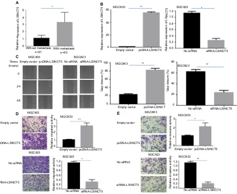
Our first goal was to investigate whether LSINCT5 expression is altered in metastatic GC tissues compared with nonmeta-static tissues. Using GAPDH as the normalization control, we found that LSINCT5 expression was significantly higher in metastatic GC tissues than in nonmetastatic GC tissues (Figure 1A; P<0.05).
Because our previous study validated an association between LSINCT5 and GC cell growth,9 we further
investi-gated the biological role of LSINCT5 in GC cell migration and invasion using gain- and loss-of-function approaches. LSINCT5 was overexpressed by transfecting MGC803 cell
Cancer Management and Research downloaded from https://www.dovepress.com/ by 118.70.13.36 on 20-Aug-2020
Dovepress
Qi et al
lines, which have relatively low LSINCT5 expression, with the pcDNA-LSINCT5 vector, and chemically synthesized small interfering RNAs (siRNAs) were used to knockdown LSINCT5 expression in BGC823 cells, which have relatively high LSINCT5 expression. The ectopic expression and depletion of LSINCT5 in these cells was confirmed by qRT-PCR (Figure 1B). A wound healing assay showed that cell migration was promoted in pcDNA-LSINCT5-transfected MGC803 cells compared with control cells (Figure 1C), and BGC823 cells exhibited a slower scratch closure rate (inhibi-tion of migra(inhibi-tion) after the knockdown of LSINCT5 than of control siRNA (Figure 1C). We then investigated the invasion and migration of GC cells through Matrigel and transwell assays, respectively. As shown in Figure 1D, E, MGC803 cell migration and invasion were promoted following the
upregulation of LSINCT5 expression, and reduced LSINCT5 expression significantly hindered the migration and invasion of BGC823 cells compared with the control. These results suggest that LSINCT5 may be closely related to GC cell migration and invasion.
e2F1 upregulates lsinCT5 expression in
gC cells
To explore the role of E2F1 in human GC progression, we examined the E2F1 expression level in gastric tumor tissues (Ts) and paired adjacent normal tissues (ANTs) and found that the E2F1 mRNA levels were 3.2-fold higher in Ts than in ANTs (Figure 2A). In addition, we determined that E2F1 was more highly expressed in metastatic tumor tissues than in nonmetastatic tissues (Figure 2B). We next examined the
5 60 1.2
1.0 0.8 0.6 0.4 0.2 0.0 80 80 100 60 60 40 40
Gap closure (%
)
Gap closure (%
) 20 20 0 0 MGC803 MGC803 MGC803 MGC803 MGC803 MGC803 MGC803 Empty vector Empty vector Empty vector Empty vector Empty vector BGC823 BGC823 BGC823 BGC823 1.2 1.0 0.8 0.6 0.4 0.2 0.0 ** ** pcDNA-LSINCT5 pcDNA-LSINCT5 pcDNA-LSINCT5 pcDNA-LSINCT5 pcDNA-LSINCT 3
Relative invasive activity
Relative invasive activity
1.2 1.0 0.8 0.6 0.4 0.2 0.0
Relative migrated activity
Relative migrated activity
2 1 0 3 4 2 1 0 * * BGC823 BGC823 BGC823 40 20 0 Empty vector Empty vector Times (hours) 0 24 48 pcDNA-LSINCT5 pcDNA-LSINCT5 Nc-siRNA * siRNA-LSINCT5 Nc-siRNA Nc-siRNA siRNA-LSINCT5 siRNA-LSINCT5 Nc-siRNA Nc-siRNA Nc-siRNA Nc-siRNA siRNA-LSINCT5 siRNA-LSINCT5 siRNA-LSINCT5 siRNA-LSINCT5 Without metastasis
n=21 With metastasisn=50
* ** ** ** A B E D C 4
Relative expression of L
SINCT
5
Relative expression of L
SINCT 5 Relative expression of LSINCT 5 3 2 1 0
Figure 1 effects of lsinCT5 on gC cell migration and invasion.
Notes: (A) qRT-PCR analysis of the lsinCT5 expression levels in metastatic and nonmetastatic gC tissues. (B) qRT-PCR analysis of the lsinCT5 expression levels following the transfection of MgC803 cells with empty vector or pcDna-lsinCT5 (left panel) or following the transfection of BgC823 cells with scrambled siRna or si-lsinCT5 (right panel). (C) Wound healing assays were performed to investigate the migratory ability of gC cells. (D, E) Transwell assays were used to investigate changes in the migratory and invasive abilities of gC cells. all the experiments were performed in triplicate. Bars: sD; *P<0.05 and **P<0.01.
Abbreviations: gC, gastric cancer; qRT-PCR, quantitative real-time polymerase chain reaction; nC, negative control.
Cancer Management and Research downloaded from https://www.dovepress.com/ by 118.70.13.36 on 20-Aug-2020
Dovepress e2F1 induces lsinCT5 expression and promotes gC progression
E2F1 expression levels in 1 normal gastric epithelium cell line (GES-1) and in several GC cell lines and demonstrated that E2F1 expression was significantly higher in the GC cell lines than in the GES-1 cell line (Figure 2C).
Previous data have shown that E2F1 promotes cancer progression by activating the transcription of downstream target coding and noncoding genes. To ascertain whether LSINCT5 is regulated by E2F1 expression in GC, we per-formed a ChIP assay using E2F1 antibody and to quantify the immunoprecipitated DNA, and the results revealed that endogenous E2F1 could bind to the LSINCT5 promoter, in untreated GC cells or increased or reduced E2F1 expression
GC cells (Figure 2D). Then, we cloned the human LSINCT5 promoter fragment (nucleotides −1,400 to +100) into a pGL3 vector and performed luciferase activity assays and found that E2F1 overexpression induced LSINCT5 transcriptional activity (Figure 2E), which suggests that E2F1 participates in the transcriptional regulation of LSINCT5. To further confirm this notion, we examined the LSINCT5 core pro-moter region for transcription factor-binding sites using online software programs (Jaspar: http://jaspar.genereg. net/, ChIPBase: http://deepbase.sysu.edu.cn/chipbase/, and TFSEARCH: http://www. cbrc.jp/research/db/TFSEARCH. html) and identified four predicted tandem E2F1-binding sites
6 5
4
3
2 1
0
5
4 3 2
1
0
5 *
** E2F1 construct
Relative luciferase activity
(fireflyrenila ration)
4 3 2 1
0
8
6
Relative E2F1 expression (∆Ct
)
Relative LSINCT5 expression (∆Ct) 4
4 6
2
2 0
0
H
WT E1-del E2-del E3-del E4-del
F
GES-1
MGC803 AG S
MKN 45
BGC823SGC790 1 Without metastasis
n=21 With metastasisn=50 **
*
* * * *
**
Relative expression of E2F1
Relative enrichment over input (%
)
Relative luciferase activity
Relative expression of E2F1 Relative expression of E2F1
A
D E
G
B C
4
2
0
ANTs n=71
120 lgG E2F1
Control E2F1 construct E2F1 siRNA **
* **
**
** 90
60
30
0
0.8 Empty vector
E2F1 construct
pGL3-Basic
BGC823 MGC803
Relative expression of LSINCT
5
Relative expression of LSINCT
5 3 1.5
2
1
0
1.0
0.5
0.0
Nc-siRNA
Empty vector pMax-E2F1 siRNA-E2F1
** **
pGL3-LSINCT5 0.6
0.4
0.2
0.0 Ts
Figure 2 e2F1 upregulates lsinCT5 expression in gC cells.
Notes: qRT-PCR analysis of the e2F1 expression levels in gC tissues and paired anTs (A) and in metastatic and nonmetastatic gC tissues (B). (C) qRT-PCR analysis of e2F1 expression in a series of human gC cell lines and a normal gastric epithelial cell line (ges-1). (D) ChiP assays using e2F1 antibody demonstrated endogenous e2F1 binding to the lsinCT5 gene promoter, and ectopic expression or siRna knockdown increased or reduced e2F1 enrichment at the lsinCT5 promoter, respectively. (E) a dual-luciferase reporter assay was performed through cotransfection of the lsinCT5 promoter fragment (pgl3-lsinCT5) and an e2F1-overexpression construct. (F) We examined the LSINCT5 core promoter region for transcription factor binding sites, and identified four tandem putative E2F1-binding sites. Reporter assays were conducted in cells transfected with various lsinCT5 promoter constructs in which different e2F1-binding elements were deleted (WT, del). luciferase activity is presented relative to that of the pgl3 vector (a promoter-less vector). (G) qRT-PCR analysis of the lsinCT5 expression levels following the transfection of MgC803 and BgC823 cells with a pMax-e2F1 expression vector and siRna-e2F1, respectively. (H) Analysis of the relationship between LSINCT5 and E2F1 mRNA levels (ΔCt value) in GC tissues. Bars: SD; *P<0.05 and **P<0.01.
Abbreviations: anTs, adjacent normal tissues; Ts, tumor tissues; ChiP, chromatin immunoprecipitation; del, deletion; gC, gastric cancer; qRT-PCR, quantitative real-time polymerase chain reaction; WT, wild type; nC, negative control.
Cancer Management and Research downloaded from https://www.dovepress.com/ by 118.70.13.36 on 20-Aug-2020
Dovepress
Qi et al
at the following regions: −1,156 to −1,149 bp (E1), −1,039 to −1,032 bp (E2), −1,030 to −1,023 bp (E3) and −993 to −986 bp (E4) (Figure 2F). We subsequently deleted the putative binding sites individually and performed additional reporter assays to validate this aforementioned finding. Deletion of the E1-binding motif significantly impaired the ability of E2F1 to activate LSINCT5 transcription, suggesting that special binding motifs are required for the E2F1-mediated regulation of LSINCT5 transcription (Figure 2F).
We then determined whether E2F1 can mediate LSINCT5 overexpression through both gain- and loss-of-function approaches. The results showed that the ectopic expression or siRNA knockdown of E2F1 increased or reduced E2F1 enrichment to the LSINCT5 promoter, respectively (Figure 2D), and up- or downregulated LSINCT5, respectively, in GC cells (Figure 2G). In addition, the correlation between E2F1 and LSINCT5 gene transcription in GC tissue samples was assessed, and the results revealed that LSINCT5 expression was positively correlated with E2F1 mRNA expression (Pear-son R=0.685, P<0.001) (Figure 2H). These results suggest that E2F1 is a transcription factor that activates LSINCT5 transcription and upregulates its expression.
lsinCT5 impacts the eMT in gC cells
EMT has been shown to be critically important in the early events of tumor cell metastatic dissemination by inducing cell motility and allowing cells to acquire invasion potential. We hypothesized that LSINCT5 influences the malignant pheno-type, partly by regulating the EMT. qRT-PCR and Western blotting assays were performed to detect the expression of EMT markers in cells in which LSINCT5 was overexpressed
or knocked down. Our results revealed that the upregula-tion of LSINCT5 expression led to increased N-cadherin, vimentin, and MMP-2 expression and reduced E-cadherin expression (Figure 3A). In contrast, the downregulation of LSINCT5 expression induced E-cadherin expression and decreased N-cadherin, vimentin, MMP-2, and MMP-9 expression (Figure 3B). Moreover, the results of a Western blotting analysis showed that LSINCT5 downregulation stimulated the E-cadherin protein expression and reduced N-cadherin, vimentin, and MMP-2 protein expression in GC cells, whereas LSINCT5 upregulation yielded the opposite effects (Figure 3C). These results support our hypothesis that LSINCT5 influences the malignant cell phenotype by regulating the EMT.
Discussion
Accumulating evidence has shown that lncRNAs play impor-tant roles in cancer pathophysiology and could provide a new avenue for elucidating the biology of this disease.20 Xu et al
revealed that the FENDRR inhibits the metastatic behavior of GC cells.21 The lncRNA TINCR has been reported to
promote GC proliferation by accelerating KLF2 mRNA degradation,22 and we previously demonstrated that TUSC7 is
a p53-regulated tumor suppressor that acts in part by repress-ing miR-23b in GC.23 Therefore, the identification of
GC-associated lncRNAs may provide missing information related to the well-known oncogenic and tumor suppressor network.
In our previous study, we primarily focused on the clini-cal significance and cell proliferation function of aberrant LSINCT5 expression.9 Because high LSINCT5 expression is
associated with a metastatic tumor phenotype, we presumed
2.5 2.0
1.5 1.0
0.5 0.0
2.5 2.0
1.5
1.0
0.5 0.0
E-cadherinN-cadherin Vimentin MMP-2 MMP-9 E-cadherin
E-cadherin
N-cadherin
N-cadherin
Vimentin
Vimentin
MMP-2
MMP-2 MMP-9
GAPDH
MMP-9
Relative mRNA
expression
Relative mRNA
expression
A B C
Empty vector Nc-siRNA MGC803 BGC823
Vector LSINCT5 si-LSINCT5
siRNA-LSINCT5 pcDNA-LSINCT5
** **
** *
* * *
**
Nc-siRNA
Figure 3 Overexpression of lsinCT5 promotes gC cell invasion and metastasis by affecting the eMT.
Notes: (A, B) qRT-PCR analysis of e-cadherin, n-cadherin, vimentin, MMP-2, and MMP-9 expression following the transfection of MgC803 cells with empty vector or pcDna-lsinCT5 or the transfection of BgC823 cells with scrambled siRna or si-lsinCT5. (C) Western blot analysis of e-cadherin, n-cadherin, vimentin, MMP-2, and MMP-9 expression in MgC803 cells transfected with pcDna-lsinCT5 and BgC823 cells transfected with si-lsinCT5. all the experiments were performed in triplicate. Bars: sD; *P<0.05 and **P<0.01.
Abbreviations: eMT, epithelial–mesenchymal transition; gC, gastric cancer; MMP, matrix metalloproteinase; qRT-PCR, quantitative real-time polymerase chain reaction; nC, negative control.
Cancer Management and Research downloaded from https://www.dovepress.com/ by 118.70.13.36 on 20-Aug-2020
Dovepress e2F1 induces lsinCT5 expression and promotes gC progression
that LSINCT5 plays a significant role in the metastasis pro-cess. In the current study, we initially revealed that LSINCT5 was overexpressed in metastatic GC tissues. Functional in vitro studies indicated that ectopic LSINCT5 expression significantly promoted cell migration and invasion, whereas the knockdown of LSINCT5 expression impeded these biological processes. These findings indicate that LSINCT5 exerts a marked influence on the progression and metastasis of GC cells and might be a novel metastatic marker for GC.
Previous findings have suggested that E2F protein fam-ily members play important roles during the development of human malignancies.15 E2F1, which has been reported
to be upregulated in GC,19 is a key regulator of the G1/S
phase transition,24 but its functional role in GC progression
remains debated. In the current study, we found that E2F1 expression was significantly higher in GC tissues compared with corresponding noncancerous tissues. Specifically, E2F1 expression was significantly higher in metastatic tumor tis-sues than in nonmetastatic tistis-sues. Our results indicate that E2F1 expression may have significant predictive value for the early detection of metastasis in patients with GC.
To explore the mechanism underlying the induction of high LSINCT5 expression, we performed a computational analysis of the LSINCT5 promoter region using several computational algorithms. Because E2F1 plays pivotal roles in tumor development by modulating the expression of both coding and noncoding transcripts,13,14 we focused
on the prediction of E2F1-binding sites in the LSINCT5 promoter region. A ChIP combined with luciferase reporter assay analysis indicated that E2F1 bound to approximately −1,156 to −1,149 bp in the LSINCT5 promoter region and specifically activated LSINCT5 transcription. These results indicate that LSINCT5 may be a downstream effector of E2F1 that can control GC cell migration and invasion.
The EMT plays crucial roles in cancer initiation, pro-gression, and particularly metastasis.3,25,26 Accumulating
data have revealed that lncRNAs regulate tumor metastasis by affecting the EMT.27,28 The hallmarks of the EMT are
a loss of E-cadherin expression and aberrant N-cadherin and vimentin expression. It is well known that MMPs play key roles in the invasion and metastasis of many types of carcinomas. To explore the potential mechanism through which LSINCT5 contributes to GC invasion and metastasis, we determined the levels of metastasis markers following LSINCT5 overexpression or downregulation. The upregula-tion of LSINCT5 expression reduced E-cadherin expression and increased N-cadherin, vimentin, and MMP-2 expres-sion, whereas the downregulation of LSINCT5 increased
E-cadherin expression and decreased N-cadherin, vimentin, MMP-2, and MMP-9 expression. Our results indicate that LSINCT5 mediates increases in the migration and invasion of GC cells and possibly affects the EMT. The EMT, a central differentiation process, allows tissue remodeling during the early stages of embryogenesis and is also implicated in the promotion of cancer cell migration and tumor metastasis. Thus, as important and extensive regulators of the EMT, lncRNAs might be suitable candidates for cancer treatment. Recent years have shown progress in molecular therapeutics targeting key drivers of cancer progression,29 and LSINCT5,
an important regulator of the EMT, shows promise as a drug target. Drugs that regulate LSINCT5 expression have the potential to be used for clinical application, and clinical tests or assays should be developed to test such therapeutics.
Conclusion
In summary, our results showed that LSINCT5 is markedly upregulated in metastatic GC tissues. Moreover, upregu-lated LSINCT5 may be a downstream effector of E2F1 that promotes GC cell migration and invasion by affecting the EMT. However, the molecular mechanisms through which LSINCT5 regulates the EMT require further investigation.
Acknowledgments
The work was supported by the National Natural Science Foundation of China (81502429, 81472220, 81470353), the Science and Technology Commission of the Shang-hai Municipality (15495810300), the ShangShang-hai Hospital Development Center Emerging Advanced Technology Joint Research Project (SHDC12014105), the Shanghai Key Developing Disciplines (2015ZB0201), the Shanghai Science and Technology Development Fund (No. 15ZR1407400), and Shanghai Municipal Commission of Health and Family Planning (2015ZB0204).
Disclosure
The authors report no conflicts of interest in this work.
References
1. Torre LA, Bray F, Siegel RL, et al. Global cancer statistics, 2012. CA
Cancer J Clin. 2015;65(2):87–108.
2. Patru CL, Surlin V, Georgescu I, Patru E. Current issues in gastric cancer
epidemiology. Rev Med Chir Soc Med Nat Iasi. 2013;117(1):199–204.
3. Thiery JP, Acloque H, Huang RY, Nieto MA. Epithelial-mesenchymal
transitions in development and disease. Cell. 2009;139(5):871–890.
4. Qi P, du X. The long non-coding RNAs, a new cancer diagnostic and
therapeutic gold mine. Mod Pathol. 2013;26(2):155–165.
5. Shen XH, Qi P, Du X. Long non-coding RNAs in cancer invasion and
metastasis. Mod Pathol. 2015;28(1):4–13.
Cancer Management and Research downloaded from https://www.dovepress.com/ by 118.70.13.36 on 20-Aug-2020
Dovepress
Qi et al
6. Xu MD, Wang Y, Weng W, et al. A Positive Feedback Loop of lncRNA-PVT1 and FOXM1 Facilitates Gastric Cancer Growth and Invasion.
Clin Cancer Res. 2017;23(8):2071–2080.
7. Guil S, Esteller M. Cis-acting noncoding RNAs: friends and foes. Nat
Struct Mol Biol. 2012;19(11):1068–1075.
8. Silva JM, Boczek NJ, Berres MW, Ma X, Smith DI. LSINCT5 is over expressed in breast and ovarian cancer and affects cellular proliferation.
RNA Biol. 2011;8(3):496–505.
9. Xu MD, Qi P, Weng WW, et al. Long non-coding RNA LSINCT5 predicts negative prognosis and exhibits oncogenic activity in gastric
cancer. Medicine. 2014;93(28):e303.
10. Chen HZ, Tsai SY, Leone G. Emerging roles of E2Fs in cancer: an exit
from cell cycle control. Nat Rev Cancer. 2009;9(11):785–797.
11. Burkhart DL, Sage J. Cellular mechanisms of tumour suppression by
the retinoblastoma gene. Nat Rev Cancer. 2008;8(9):671–682.
12. Bertoli C, Skotheim JM, de Bruin RA. Control of cell cycle transcription
during G1 and S phases. Nat Rev Mol Cell Biol. 2013;14(8):518–528.
13. Tarangelo A, Lo N, Teng R, et al. Recruitment of Pontin/Reptin by E2f1
amplifies E2f transcriptional response during cancer progression. Nat
Commun. 2015;6:10028.
14. Sun M, Nie F, Wang Y, et al. LncRNA HOXA11-AS Promotes Pro-liferation and Invasion of Gastric Cancer by Scaffolding the
Chro-matin Modification Factors PRC2, LSD1, and DNMT1. Cancer Res.
2016;76(21):6299–6310.
15. Engelmann D, Pützer BM. The dark side of E2F1: in transit beyond
apoptosis. Cancer Res. 2012;72(3):571–575.
16. Yamasaki L, Bronson R, Williams BO, Dyson NJ, Harlow E, Jacks T. Loss of E2F-1 reduces tumorigenesis and extends the lifespan of
Rb1(+/-)mice. Nat Genet. 1998;18(4):360–364.
17. Xie Y, Wang C, Li L, Ma Y, Yin Y, Xiao Q. Overexpression of
E2F-1 inhibits progression of gastric cancer in vitro. Cell Biol Int.
2009;33(6):640–649.
18. Xie Y, Yin Y, Li L, Ma Y, Xiao Q. Short interfering RNA directed against
the E2F-1 gene suppressing gastric cancer progression in vitro. Oncol
Rep. 2009;21(5):1345–1353.
19. Zhang X, Ni Z, Duan Z, et al. Overexpression of E2F mRNAs associated with gastric cancer progression identified by the
transcrip-tion factor and miRNA co-regulatory network analysis. PLoS One.
2015;10(2):e0116979.
20. Xu MD, Qi P, du X. Long non-coding RNAs in colorectal cancer:
implications for pathogenesis and clinical application. Mod Pathol.
2014;27(10):1310–1320.
21. Xu TP, Huang MD, Xia R, et al. Decreased expression of the long non-coding RNA FENDRR is associated with poor prognosis in gastric cancer and FENDRR regulates gastric cancer cell metastasis by affecting
fibronectin1 expression. J Hematol Oncol. 2014;7:63.
22. Xu TP, Liu XX, Xia R, et al. SP1-induced upregulation of the long noncoding RNA TINCR regulates cell proliferation and apoptosis
by affecting KLF2 mRNA stability in gastric cancer. Oncogene.
2015;34(45):5648–5661.
23. Qi P, Xu MD, Shen XH, et al. Reciprocal repression between TUSC7
and miR-23b in gastric cancer. Int J Cancer. 2015;137(6):1269–1278.
24. Hallstrom TC, Nevins JR. Balancing the decision of cell proliferation
and cell fate. Cell Cycle. 2009;8(4):532–535.
25. de Craene B, Berx G. Regulatory networks defining EMT during cancer
initiation and progression. Nat Rev Cancer. 2013;13(2):97–110.
26. Guo F, Parker Kerrigan BC, Yang D, et al. Post-transcriptional regulatory network of epithelial-to-mesenchymal and mesenchymal-to-epithelial
transitions. J Hematol Oncol. 2014;7:19.
27. Yuan JH, Yang F, Wang F, et al. A long noncoding RNA activated by
TGF-β promotes the invasion-metastasis cascade in hepatocellular
carcinoma. Cancer Cell. 2014;25(5):666–681.
28. Liu F, Yuan JH, Huang JF, et al. Long noncoding RNA FTX inhibits hepatocellular carcinoma proliferation and metastasis by binding MCM2
and miR-374a. Oncogene. 2016;35(41):5422–5434.
29. Liu LK, Jiang XY, Zhou XX, Wang DM, Song XL, Jiang HB. Upregu-lation of vimentin and aberrant expression of E-cadherin/beta-catenin complex in oral squamous cell carcinomas: correlation with the
clini-copathological features and patient outcome. Mod Pathol. 2010;23(2):
213–224.
Cancer Management and Research downloaded from https://www.dovepress.com/ by 118.70.13.36 on 20-Aug-2020
Dovepress
Cancer Management and Research
Publish your work in this journal
Submit your manuscript here: https://www.dovepress.com/cancer-management-and-research-journal Cancer Management and Research is an international, peer-reviewed open access journal focusing on cancer research and the optimal use of preventative and integrated treatment interventions to achieve improved outcomes, enhanced survival and quality of life for the cancer patient. The manuscript management system is completely online and includes
a very quick and fair peer-review system, which is all easy to use. Visit http://www.dovepress.com/testimonials.php to read real quotes from published authors.
Dove
press
e2F1 induces lsinCT5 expression and promotes gC progression
Supplementary material
Table S1 Primers used in this study
Qualitative real-time polymerase chain reaction primers lsinCT5: 5′-TTCggCaagCTCCTTTTCTa-3′ (forward) and 5′-gCCCaagTCCCaaaaagTTCT-3′ (reverse);
gaPDh: 5′-CCgggaaaCTgTggCgTgaTgg-3′ (forward) and 5′-aggTggaggagTgggTgTCgCTgTT-3′ (reverse); e2F1:5′-TgCagagCagaTggTTaTgg-3′ (forward) and 5′-agaTgaTggTggTggTgaCa-3′ (reverse).
Cancer Management and Research downloaded from https://www.dovepress.com/ by 118.70.13.36 on 20-Aug-2020