Scholarship@Western
Scholarship@Western
Electronic Thesis and Dissertation Repository
12-12-2014 12:00 AM
Role of Integrin-linked kinase in epidermal integrity and barrier
Role of Integrin-linked kinase in epidermal integrity and barrier
function
function
Samar Sayedyahossein
The University of Western Ontario Supervisor
Dr Lina Dagnino
The University of Western Ontario
Graduate Program in Physiology and Pharmacology
A thesis submitted in partial fulfillment of the requirements for the degree in Doctor of Philosophy
© Samar Sayedyahossein 2014
Follow this and additional works at: https://ir.lib.uwo.ca/etd Part of the Cell Biology Commons
Recommended Citation Recommended Citation
Sayedyahossein, Samar, "Role of Integrin-linked kinase in epidermal integrity and barrier function" (2014). Electronic Thesis and Dissertation Repository. 2640.
https://ir.lib.uwo.ca/etd/2640
This Dissertation/Thesis is brought to you for free and open access by Scholarship@Western. It has been accepted for inclusion in Electronic Thesis and Dissertation Repository by an authorized administrator of
(Thesis format: Integrated Article)
by
Samar Sayedyahossein
Graduate Program in Physiology and Pharmacology
A thesis submitted in partial fulfillment of the requirements for the degree of
Doctor of Philosophy
The School of Graduate and Postdoctoral Studies The University of Western Ontario
London, Ontario, Canada
ii
Abstract
Integrin-linked kinase (ILK) is a β integrin adaptor protein that translates extracellular
stimuli to intracellular signaling events. ILK plays a role in actin cytoskeleton dynamics
and cell adhesion. The structure and function of the epidermis is highly dependent on
cell-cell adhesion and cell-basement membrane interactions. The mechanisms whereby
ILK contributes to epidermal integrity are poorly understood. Using a mouse model of
epidermis-restricted Ilk gene inactivation, I observed that ILK loss causes abnormal
morphology and presence of intra-epidermal and epidermal-dermal microblisters in
embryos as early as E17.5. ILK-deficient epidermis is also characterized by abnormal
localization or/and absence of adherens junctions, tight junctions and desmosomes. These
are structures that maintain the barrier properties of the epidermis. Ca2+ is an important
inducer of cell-cell junctions and differentiation in epidermal keratinocytes. In the
absence of ILK, cultured keratinocytes are unable to properly respond to Ca2+, due to an
impaired activation of the RhoA GTPase.
I further investigated the barrier function of the epidermis against Staphylococcus aureus,
a major component of the skin microbiota. Using cultured keratinocytes, I investigated
the role of integrin-linked kinase (ILK) in epidermal S. aureus invasion. ILK-deficient
mouse keratinocytes internalized bacteria 2- to 4-fold less efficiently than normal cells.
The reduced invasion by live S. aureus of ILK-deficient cells was restored in the
presence of exogenous, constitutively active Rac1. Thus, Rac1 functions downstream
iii
Another function of the epidermis is protection against UV radiation. Phagocytic
melanosome uptake by epidermal keratinocytes is a central protective mechanism against
damage induced by ultraviolet radiation. I have examined the role of ILK in regulation of
phagocytosis, and shown that ILK-deficient cells exhibit severely impaired capacity to
engulf fluorescent microspheres in response to stimulation of the keratinocyte growth
factor (KGF) receptor or the protease-activated receptor-2. KGF promoted activation of
Rac1 and formation of pseudopodia in ILK-expressing, but not in ILK-deficient cells.
Rac1-deficient keratinocytes also showed substantially impaired phagocytic ability,
underlining the importance of ILK-dependent Rac1 function for particle engulfment. In
summary, my data suggest a key role for ILK in activation of small GTPases and
regulation of actin dynamics during phagocytosis by keratinocytes.
Keywords:
Integrin-linked kinase, epidermal barrier, junction, small GTPases,iv
Co-Authorship Statement
This dissertation is prepared in an integrated article format. Manuscripts, which have
been previously published, or finalized for submission, are presented with some
adjustments in Chapters 2, 3 and 4.
Chapter 1: Role of integrins and their associated proteins in phagocytosis. Authors: S.
Sayedyahossein and L. Dagnino. A part of this review was used in chapter 1 of this
thesis. The draft of the review was written by Samar Sayedyahossein and closely edited
and modified by Dr Lina Dagnino. This manuscript was published in the journal of
International Review in Cell and Molecular Biology (Int. Rev. in Cell. Mol. Bio) 2013;
302:321-354.
Chapter 2: Integrin linked kinase is required for epidermal integrity and barrier function
Authors: S. Sayedyahossein, A. Rudkouskaya, S. X. Xu, J. K. McCormick and L.
Dagnino. The experimental works were conducted by Samar Sayedyahossein (Figure
2.1E, Figure 2.3A-E and G, Figure 2.4A, B, D and Figure 2.5) and Alena Rudkouskaya
(Figure 2.1A-D, F and G, Figure 2.2A-C) under the guidance of advisor Dr. Lina
Dagnino. Measurement of TNF-α and flow cytometry analysis was done by Stacey. X.
Xu under the supervision of Dr John K. McCormick. The draft of this manuscript was
written by Samar Sayedyahossein. Modifications were carried out under the close
supervision of Lina Dagnino. The final version of this article is under submission.
Chapter 3: Staphylococcus aureus keratinocyte invasion is mediated by
v
McGavin, J. K. McCormick and L. Dagnino. The experimental works were conducted by
Samar Sayedyahossein under the guidance of advisor Dr Lina Dagnino. Stacey X. Xu
helped in preparation of GFP-tagged S. aureus for invasion assays, under the supervision
of Dr John K. McCormick. Ex vivo invasion assays were conducted with the help of
Alena Rudkouskaya. Dr Martin McGavin made the GFP plasmid that was incorporated in
S. aureus. The draft of this manuscript was written by Samar Sayedyahossein with the
help of Stacey X. Xu in the introduction and discussion. Modifications were carried out
under the close supervision of Dr. Lina Dagnino. The final version of this article is
accepted for publication by the FASEB Journal October 2014.
Chapter 4: Essential role of integrin-linked kinase in regulation of phagocytosis in
keratinocytes. Authors: S Sayedyahossein, L Nini, TS Irvine, and L Dagnino. The
experimental works were conducted by Samar Sayedyahossein under the guidance of
advisor Dr Lina Dagnino. Lylia Nini helped with the experiments in Figure 4.1D, and
Figure 4.5A, B. Tames Irvine helped with the experiments in Figure 4.6. The draft of this
manuscript was written by Samar Sayedyahossein and revised by Dr Lina Dagnino. This
vi
Dedication
To those whom I have learned from during my life,
vii
Acknowledgments
I would like to express my deep gratitude to Professor Lina Dagnino for her tremendous
guidance, encouragement, and support. The completion of this dissertation would not
have been possible without her incomparable assistance and invaluable effort.
I gratefully thank my advisors Dr. John DiGugliemo, Dr. Sung kim and Dr Marco Prado
for their impressive advice and help. Also, I would like to express my special
appreciation to Dr. John DiGugliemo for his invaluable advice and constructive
comments in this thesis. I gratefully thank Alena Rudkouskaya and Stacey Xu for being
such great collaborators in this work. Also, I thank Dr John McCormick for his help with
bacterial experiments. I also would like to appreciate the assistance of Judith Sholdice,
Karen Nygard, Nicole Bechard, Meera Karajgikar, Dr Richard Gardiner, Alireza Khazaei
and Dr Kristin Chadwick.
I would also thank all my colleagues in Dagnino’s Lab (past and present) for their
support, cooperation and helpful discussion specifically, Michelle Im, Randeep Sing,
Bradley Jackson and Tames Irvine. Michelle, Meera and Randeep, I will never forget our
amazing tea times and I will miss that without any doubt. In particular, I acknowledge my
great friends: Sahar Samimi, Khaereh Sepanjnia, Salma Bahreinian, Maryam Sharif and
Maryam Adeli for their love, friendship and support. I would also acknowledge my
colleagues at Isfahan Medical Student Research Center (IMSRC) for instilling the spirit
viii
I would also thank Maysam, for his unconditional love and support throughout these
years.
Words are not enough to express my gratitude towards my family and especially my
grandparents for their devotion and endless support. My unique and special parents, Baba
Ali and Maman Leila, my only and “the one” brother, Amirsalar, without all you have
done for me, this would not be possible.
And my special and tremendous appreciation goes to Aaram, who was with me in the
ix
Table of Contents
Abstract ... ii
Co-Authorship Statement ... iv
Dedication ... vi
Acknowledgments ... vii
Table of Contents ... ix
List of Tables ... xiv
List of Figures ... xv
List of Appendices ... xvii
List of Abbreviations, Symbols, Nomenclature ... xviii
Chapter 1 ... 1
1 General Introduction and Literature Review ... 1
1.1 The skin and the epidermis ... 1
1.2 The epidermal architecture and constituent cell types ... 1
1.3 Epidermal structure and its relationship to integrity and permeability barrier function ... 4
1.3.1 Cell-Cell Junctions ... 7
1.3.2 Role of actin and microtubules in cell-cell junctions ... 9
1.4 Epidermal polarity ... 10
1.5 Integrin-modulated signaling and its role in the epidermis ... 11
1.5.1 Integrin-linked kinase ... 13
1.6 Epidermal permeability barrier: protection against microorganisms ... 14
1.6.1 Bacterial invasion of basal keratinocytes and other epithelial cell types .. 16
x
1.7.1 Phagocytosis ... 19
1.7.2 Role of phagocytosis in epidermis maintenance and homeostasis ... 26
1.8 Rationale for study ... 26
1.9 References ... 28
Chapter 2 ... 41
2 Integrin-linked kinase is required for epidermal integrity and barrier function ... 41
2.1 Introduction ... 41
2.2 Materials and methods ... 42
2.2.1 Mouse strains ... 42
2.2.2 Cell culture and transfections ... 43
2.2.3 Immunohistochemistry and microscopy ... 43
2.2.4 Bacterial Strains ... 45
2.2.5 Ex vivo bacterial invasion assay ... 46
2.2.6 Immunoblot analysis ... 47
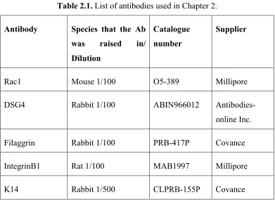
2.2.7 Antibodies ... 47
2.2.8 Measurement of cytosolic Ca2+ ... 49
2.2.9 Measurement of GTP-bound RhoA levels ... 49
2.2.10 Statistical analysis ... 50
2.3 Results ... 50
2.3.1 Alterations in architecture and integrity in ILK-deficient epidermis ... 50
2.3.2 Inflammatory responses in ILK-deficient epidermis ... 52
2.3.3 Inactivation of Ilk leads to abnormal cell-cell junctions and loss of polarity in the epidermis ... 60
xi
2.3.5 Impaired differentiation responses to extracellular Ca in the absence
of ILK ... 68
2.4 Discussion ... 78
2.5 References ... 85
Chapter 3 ... 89
3 Staphylococcus aureus keratinocyte invasion is mediated by Integrin-linked-kinase and Rac 1 ... 89
3.1 Introduction ... 89
3.2 Materials and methods ... 91
3.2.1 Mouse strains ... 91
3.2.2 Reagents and antibodies ... 92
3.2.3 Plasmids and bacteria ... 93
3.2.4 Cell culture, transfections and adenovirus transduction ... 94
3.2.5 Fluorescence-activated cell sorting (FACS) ... 95
3.2.6 Heat-killed bacterial phagocytosis assays ... 96
3.2.7 Bacterial attachment assays ... 97
3.2.8 Confocal and fluorescence microscopy ... 97
3.2.9 Immunoblot analysis ... 98
3.2.10 Measurement of secreted TNF-α ... 98
3.2.11 Gentamicin protection/bacterial invasion assays ... 98
3.2.12 Ex vivo bacterial invasion assays ... 100
3.2.13 Data analysis ... 100
3.3 Results ... 101
3.3.1 Phagocytosis of heat-killed S. aureus by primary keratinocytes ... 101
3.3.2 Invasion of mouse keratinocytes by live S. aureus ... 102
xii
3.3.4 Role of Rac1 in S. aureus invasion of keratinocytes ... 112
3.3.5 Involvement of β1 integrin in S. aureus internalization ... 116
3.3.6 Invasion of ILK-deficient epidermis by S. aureus ... 116
3.4 Discussion ... 121
3.5 References ... 130
Chapter 4 ... 135
4 Essential Role of Integrin-Linked Kinase in Regulation of Phagocytosis in Keratinocytes ... 135
4.1 Introduction ... 135
4.2 Materials and methods ... 137
4.2.1 Mouse strains ... 137
4.2.2 Cell culture, transfections and adenoviral infections ... 138
4.2.3 Plasmids and adenoviruses ... 138
4.2.4 Antibodies and reagents ... 139
4.2.5 Phagocytosis and macropinocytosis assays ... 139
4.2.6 Measurement of Rac1 activity ... 140
4.2.7 Confocal and fluorescence microscopy, and immunoblot analysis ... 141
4.2.8 Transmission Electron microscopy ... 141
4.3 Results ... 142
4.3.1 Phagocytosis in primary mouse keratinocytes ... 142
4.3.2 ILK is an essential downstream mediator of keratinocyte phagocytosis 147 4.3.3 ILK is necessary for KGF-induced formation of probing filopodia ... 148
4.3.4 Role of ILK in KGF receptor signaling ... 152
xiii
4.4 Discussion ... 166
4.5 References ... 172
Chapter 5 ... 176
5 Discussion ... 176
5.1 Summary ... 176
5.2 ILK and epidermal integrity ... 178
5.3 ILK and epidermal barrier function ... 179
5.4 Future directions ... 182
5.5 References ... 186
Appendices ... 189
Appendix A. Copyright releases. ... 189
xiv
List of Tables
xv
List of Figures
Figure 1.1. Schematic representation of epidermal stratified epithelium. ... 6
Figure 1.2. Signaling pathways leading to actin polymerization during phagocytosis. ... 23
Figure 2.1. ILK-deficient epidermis exhibits abnormal morphology and defective integrity. ... 54
Figure 2.2. Lack of ILK results in inflammatory environment in the epidermis. .... 58
Figure 2.3. ILK-deficiency results in abnormal organization of cell-cell junctions in the epidermis. ... 64
Figure 2.4. Impaired barrier function in the absence of ILK. ... 70
Figure 2.5. Defective response to increased extracellular calcium in the absence of ILK. ... 74
Figure 2.6. Proposed model for the role of ILK in CaR signaling. ... 84
Figure 3.1. Phagocytosis of heat-killed S. aureus by keratinocytes. ... 104
Figure 3.2. Invasion of keratinocytes by S. aureus. ... 108
Figure 3.3. Impaired invasion of ILK-deficient keratinocytes by S. aureus. ... 111
Figure 3.4. TNF-α secretion and MAPK activation in ILK-deficient keratinocytes co-cultured with S. aureus. ... 115
Figure 3.5. Role of Rac1 in keratinocyte invasion by S. aureus. ... 118
xvi
Figure 3.7. Ex-vivo invasion of ILK-deficient epidermis by S. aureus. ... 123
Figure 4.1. Induction of phagocytosis in keratinocytes by KGFR and PAR-2 stimulation. ... 146
Figure 4.2. Phagocytosis in ILK-deficient cells. ... 150
Figure 4.3. Induction of pseudopods and ERK1/2 phosphorylation by KGF. ... 154
Figure 4.4. Formation of Rac1-GTP by KGF receptor stimulation. ... 157
Figure 4.5. Role of Rac1 in phagocytosis. ... 160
Figure 4.6. Phagocytosis in Rac1-deficient cells. ... 162
Figure 4.7. Impaired phagocytosis in integrin β1-deficient cells. ... 165
xvii
List of Appendices
xviii
List of Abbreviations, Symbols, Nomenclature
Abbreviations
AJs adherens junctions (AJs)
α-SMA α-smooth muscle actin 7-AAD 7-aminoactinomycin D
β-gal β-galactosidase
BMPs bone morphogenetic proteins BSA bovine serum albumin CFU colony forming unit CaR Ca2+-sensing receptor
Dsg4 desmoglein 4
ECM extracellular matrix
EDTA ethylenediaminetetraacetic acid EGF epidermal growth factor
EMEM Eagle’s minimum essential medium FAK focal adhesion kinase
FGF fibroblast growth factor
FSS forward scatter
GAPDH glyceraldehyde 3-phosphodehydrogenase GFP green fluorescent protein
HEPES N-(2-hydroxyethyl)piperazine-N'-ethanesulfonic acid ILK integrin-linked kinase
K14 Keratin14
KGF keratinocyte growth factor
KGFR keratinocyte growth factor receptor MAPK mitogen-activated protein kinase MOI multiplicity of infection
MRSA methicillin-resistant S. aureus
xix
ORP1L oxysterol-binding protein related protein PAR-2 protease-activated receptor-2
PAMPs pathogen-associated molecular patterns PBS phosphate-buffered saline
PFA paraformaldehyde
PI3K phosphatidyl inositol 3-kinase
PIPK1α phosphatidylinositol 4-phosphate 5-kinase 1α PLCγ phospholipase C-γ
PRRs pathogen recognition receptors RILP Rab-interacting lysosomal protein
S. aureus Staphylococcus aureus
SD standard deviation
SDS-PAGE denaturing polyacrylamide gel electrophoresis SEM standard error of the mean
SG Stratum Granulosum
SSC side scatter
TNF-α tumour necrosis factor-alpha TLRs Toll-like receptors
Chapter 1
1
General Introduction and Literature Review
1.1
The skin and the epidermis
The skin is the largest organ of the body, with a surface area of approximately 1.8 m2 (1).
The skin is composed of three main layers: the epidermis, the dermis and the hypodermis.
The uppermost layer of the skin is the epidermis, which adheres to a basement membrane
that separates it from the underlying dermis. The dermis is mostly composed of
fibroblasts and extracellular matrix proteins. Epidermal appendages, including hair
follicles and sebaceous glands, extend from the surface of the epidermis, and invaginate
into the dermis. The hypodermis or subcutaneous adipose tissue is found underneath the
dermis (2).
1.2
The epidermal architecture and constituent cell types
The epidermis is a stratified squamous epithelium with essential protective functions, and
constitutes a barrier between the organism and its environment. The epidermis prevents
water and electrolyte loss, resists mechanical stress, serves as protection against UV
skin epithelium derives from a single-cell layer neuroectoderm. Early in development,
Wnt signalling is activated in the neuroectoderm, preventing responses to fibroblast
growth factor (FGF), which induces these early cells to follow a neuronal fate. In
response to Wnt signalling, the ectoderm expresses bone morphogenetic proteins
(BMPs), which promote epithelial formation (4). The epidermis at this stage of
development consists of a single layer of multipotent epithelial cells, which proliferate
and expand in response to activation of epidermal growth factor (EGF) receptors (5). In
humans, the primary epidermal layer matures towards the end of pregnancy, whereas in
mice, this takes place just after mid-gestation, around 15 days post-coitum. The epidermis
is a stratified epithelium, composed of keratinocytes at different stages of differentiation.
Basal keratinocytes form the innermost layer, and exhibit high proliferative capacity.
Asymmetric division in basal keratinocytes leads to the formation of the spinous layer of
the epidermis, where cells differentiate. The spinous layer is rich in desmosomes and
intermediate filaments, which contribute to the flattened morphology of these cells
(Figure 1.1). Spinous keratinocytes further differentiate and move outwards, giving rise
to the granular layer. Granular keratinocytes lose their nuclei, and produce electron-dense
keratohyalin granules, which accumulate in their cytoplasm, giving these cells their
granular appearance. In mice, the epidermis contains three granular layers, termed SG1,
SG2 and SG3. SG1 and SG2 are characterized by the presence of tight junctions, which
contribute to the formation of an impermeable envelope (6). Granular keratinocytes
secrete impermeable, lipid- and antimicrobial peptide-containing granules termed
lamellar bodies. These organelles concentrate at the interface between the granular and
outermost cornified layer is composed of terminally differentiated keratinocytes, which
are tightly linked to each other through corneodesmosomes. The latter are degraded by
proteolytic enzymes, leading to desquamation of the corneocytes at the surface of the
epidermis. The lipids and proteolytic enzymes in the cornified layer also contribute to the
degradation of microbial organelles (8). The epidermis is characterized by the presence of
a concentration gradient of extracellular Ca2+, which modulates differentiation programs
in keratinocytes, and the formation of the epidermal permeability barrier (9).
Extracellular Ca2+ levels are low in the basal layer, and increase progressively towards
the granular layer. The level of extracellular Ca2+ decreases again in the cornified layer
(10). Ca2+ tightly regulates the expression of various epidermal differentiation markers.
Specifically, whereas basal keratinocytes express keratin 14 (K14) and keratin 5 (K5),
cells in the spinous layer express the differentiation markers, K1 and K10. In the
outermost granular layer, cells begin to express markers of cornification, such as
involucrin, loricrin and filaggrin. Profilaggrin is a precursor of mature filaggrin, and is
expressed in the granular layer too. Filaggrin binds to the intermediate filaments and
contributes to the tight packing and flattened appearance of keratinocytes in this layer
(11). Cells in the cornified layer express structural proteins, such as involucrin and
loricrin, that are cross-linked via transglutaminase I to form the cornified envelope (12).
In addition to keratinocytes, other cell types are found in the epidermis, including
melanocytes and immune cells. Melanocytes produce melanin, which provides
pigmentation to the skin and hair. Melanin is packaged into organelles termed
melanosomes, which are transferred to neighbouring keratinocytes. In human epidermis,
(13). In adult mouse epidermis, melanocytes are primarily found associated with the hair
follicle (14).
The immune cells found in the epidermis contribute to adaptive immunity. Specifically,
γδ+ T cells in the suprabasal layers protect the epidermis from microbial invasion (15).
Langerhans cells are dendritic cells that originate from lymphoid progenitors in the bone
marrow (16). Little is known about how Langerhans cells home to the epidermis,
although it has been demonstrated that transforming growth factor-β1 (TGF-β1) is
essential for their survival (17). Langerhans cells migrate to draining lymph nodes, and
present self and non-self antigens from the epidermis to the immune cells, including
antigens from commensal and pathogenic microorganisms (18). Antigen presentation by
antigen-presenting cells gives rise to adaptive immune responses and lymphocyte
migration into the skin to support effective immune responses.
1.3
Epidermal structure and its relationship to integrity and
permeability barrier function
As mentioned above, Ca2+ is an important modulator of keratinocyte differentiation. In
cultured primary epidermal keratinocytes, increasing the extracellular Ca2+ concentration
from ∼0.03 mM to >0.1 mM (hereafter termed “Ca2+ switch”) induces differentiation.
Epidermal injury and barrier disruption transiently abolish the Ca2+ gradient in the
epidermis (19). In cultured keratinocytes, an increase in extracellular Ca2+ concentration,
which stimulates Ca2+-sensing receptor (CaR), leads to the delivery of CaR and
Figure 1.1. Schematic representation of epidermal stratified epithelium.
This, in turn results in the formation of complexes that contain CaR, E-cadherin,
phosphoinositide 3-kinase (PI3K) and RhoA (20). The translocation of E-cadherin and
desmoplakin to the cell membrane is the initial step in the formation of intercellular
junctions, and occurs immediately after the Ca2+ switch. Actin rearrangements at this
stage are essential for junction stability (21). The Ca2+ switch also initiates
phosphoinositide metabolism in a RhoA- and PLCγ-dependent manner, generating
phosphatidylinositol- 4, 5 bisphosphate and diacylglycerol, which serve as second
messengers for the activation of differentiation pathways (22). Diacylglycerol activates
PKC and is necessary for the sustained increase in intracellular Ca2+ levels, which is
necessary for normal keratinocyte differentiation (23).
1.3.1
Cell-Cell Junctions
The maintenance of epidermal structure occurs by virtue of the ability of keratinocytes to
adhere to each other. Intercellular adhesion in the epidermis is achieved through adherens
junctions (AJs), tight junctions (TJs), and desmosomes.
1.3.1.1
Adherens junctions
These are the first junctions that are formed in keratinocytes in response to increase in
extracellular Ca2+ levels. They are composed of E-cadherin, and are linked with the actin
cytoskeleton. In epithelial cells, adherens junctions can be visualized as a continuous
ribbon along cell borders. E- cadherin molecules assembled in adherens junctions bind to
actin filaments via α-, and β-catenin. Of note, the classical cadherins are important
1.3.1.2
Tight junctions
Tight junctions function as gates in the epidermis, to modulate diffusion of small soluble
molecules. Tight junctions contain occludin, claudin and junctional adhesion molecules
called JAMs, which are connected to F-actin through ZO-1, ZO-2 and ZO-3 (24). ZO-1
appears to work as an organizing adaptor protein that contributes to occludin targetting to
the TJs. Occludin participates in maintenance of epidermal barrier function (24, 25).
There is also evidence supporting a role of ZO-1 in transcriptional regulation. Indeed,
ZO-1 can translocate into the nucleus in subconfluent epithelial and mesenchymal cells in
culture. The nuclear translocation of ZO-1 regulates epithelial proliferation, and it
inversely correlates with the maturation of cell-cell junctions (26). In simple renal and
intestinal epithelial cells, the tight junctions separate lipids from protein components in
bilayer membranes (27). This aids in the establishment of apical-basal cell polarity,
although this clear separation of cell domains is not obvious in epidermal keratinocytes.
Another function of tight junctions concerns innate and adaptive immunity. For example,
Langerhans cells extend protrusion through tight junctions to probe for and capture
antigens (28), which is a key step in the initiation of adaptive immune responses. The
tight junctions in the epidermis and other stratified epithelia modulate paracellular
permeability.
1.3.1.3
Desmosomes
It has been hypothesized that desmosomes are passively formed between adherens
junctions. E-cadherin-containing adherens junctions basically function to bring the
molecules to engage and form desmosomes (29). Desmosomes are desmoglein- and
desmocollin-containing structures that physically link the intermediate filaments to the
plasma membrane. Desmogleins and desmocollins are members of the cadherin
superfamily, which play a critical role in cell-cell adhesion. Desmoglein is necessary for
the establishment of normal cell-cell junctions in keratinocytes. In individuals who suffer
from pemphigus, autoantibodies against desmogleins are produced. These antibodies
interfere with the formation of cell-cell contacts, and lead to the loss of skin integrity,
which is manifested by the presence of blisters as well as skin colonization by pathogens
(30). Several desmoglein isoforms are expressed in the epidermis (31). Desmoglein 1 and
3 are mainly expressed in the basal layer, and desmoglein 4 is expressed in the granular
layer as well as in the hair follicles. The expression of different desmoglein isoforms and
their redundant function is important to preserve epidermal integrity and barrier function
(32). As mentioned above, normal actin cytoskeleton dynamics are also necessary for the
maintenance of intercellular junctions.
1.3.2
Role of actin and microtubules in cell-cell junctions
Actin filaments are polymers of globular actin, and play key roles in many fundamental
processes in epithelial cells. The mechanical forces that push or pull intracellular
organelles, and produce cell movements are generated through actin filament
rearrangements. Actin filaments have two ends: the barbed end to which actin monomers
are recruited and actin polymerization takes place. Simultaneously, at the opposite end,
actin is depolymerized, to generate monomers that can be used at the barbed end. Actin
Actin-myosin interactions are involved in the assembly and disassembly of apical
junctions in simple epithelial tissue (34). The importance of microtubules in cell
morphology, polarity and differentiation is well established (35). Microtubules are
arranged at the apical aspect of basal keratinocytes and upon differentiation, they adopt a
cortical arrangement, in the vicinity of cell-cell junctions (36). Many signaling
molecules, such as Ca2+ and cAMP, exert their effects through their modulation of
F-actin and microtubules in keratinocytes (37).
1.4
Epidermal polarity
One of the fundamental properties of the epidermis is polarity, which is essential for
barrier function (38). Polarity is defined as differential positioning and asymmetric
distribution of intracellular organelles, membrane proteins, RNA and lipids. As a result of
impaired or absent polarity, cells lose their orientation and may become unresponsive to
environmental cues (39). Although the mechanisms that regulate cell polarity are not
fully understood, it is well established that cell-cell adhesions are important contributors,
together with small GTPases, to the cytoskeleton, vesicular transport system and polarity.
The PAR complex, which is composed of PAR3, PAR6 and atypical PKC (aPKC), also
modulates polarity in the epidermis (38, 40). In mammalian stratified epithelia, aPKC is
located to the apical aspect of basal keratinocytes (41). The mechanisms whereby PAR
complexes localize apically have been studied in culture. Upon Ca2+ stimulation, RhoA
moves to the cell membrane. As mentioned earlier, in keratinocytes, E-cadherin
translocation to the membrane initiates the formation of cell-cell contacts through
junctions mature with time, and promote the subsequent formation of tight junctions.
Following the initiation of adherens junction formation, aPKC translocates to the plasma
membrane (43). The activation of the PAR polarity complexes is dependent on RhoA,
Rac1 and CDC42 (44). Rac1 and its activator Tiam1 interact with PAR complexes at the
early stages of adherens junction formation, resulting in aPKC activation, maintenance of
high intracellular Ca2+, differentiation and establishment of polarity in basal keratinocytes
(45).
Cell-ECM interactions also determine the establishment of cell polarity, partially through
localization of ECM components (46). In the epidermis, basal keratinocytes adhere to the
basement membrane through hemidesmosomes, which localize exclusively to the basal
aspect of these cells. Hemidesmosomes are complex structures, composed of α6β4
integrins, which link the basement membrane to intracellular keratin network, collagen
XVII. In cultured keratinocytes, α6β4 integrin-based hemidesmosomes and α3β
1-containing focal adhesions link the extracellular matrix, respectively, to the intermediate
filament and intracellular actin network.
1.5
Integrin-modulated signaling and its role in the epidermis
Integrins are a family of transmembrane proteins that mediate cell adhesion and
signaling. Their biological importance is emphasized by their presence in all multicellular
animals. Integrins are important for cell adhesion and migration during embryonic
development, for tissue maintenance and regeneration, host immune functions and
homeostasis (47). They modulate multiple cellular functions, many of them related to
extracellular environment cues into intracellular signaling responses through their
interactions with cytoplasmic adaptor proteins. Integrins exist as heterodimeric
complexes composed of one α and one β subunit. Although there are eighteen α- and
eight β-subunits known in mammals, only 24 heterodimeric combinations have been
identified to-date (48). Integrins bind extracellular matrix substrates, and are broadly
divided into three subfamilies, depending on whether they bind to collagen,
fibronectin/vitronectin or laminin. Integrins can adopt an inactive, low-affinity state.
Binding to ligands or mechanical stimuli trigger conformational changes in the integrin
cytoplasmic domain (49), integrin clustering and interactions with a vast number of
cytoplasmic proteins. This results in the activation of downstream signaling cascades
(50). Integrins can be activated bidirectionally through processes initiated by extracellular
factors (termed “outside-in” signaling) or by cytoplasmic proteins (“inside-out”
activation). Outside-in signalling involves formation of integrin complexes with various
cellular proteins, which link and allow integrins to modulate the cytoskeleton, cellular
spreading and migration, as well as survival and proliferation (51). Proteins such as focal
adhesion kinase (FAK) or Src-family kinases, Rho GTP-binding proteins, paxillin,
integrin-linked kinase (ILK), and vinculin (47) become activated as a result of outside-in
signaling, and trigger a variety of cellular responses. In contrast to outside-in processes,
inside-out activation occurs when intracellular stimulators, such as talin, ILK or kindlin,
bind to the integrin β subunit tail, inducing conformational changes that promote integrin
1.5.1
Integrin-linked kinase
Integrin-linked kinase is a ubiquitous adaptor protein that modulates many integrin-
dependent processes, such as adhesion, polarization and migration in keratinocytes (53,
54). ILK interacts with many proteins in addition to β1 and β3 integrins, including
ELMO2 (55), PINCH (56-58), α- and β-parvin (59-62), paxillin (63), and IQGAP1 (64).
ILK is composed of three main domains: An N-terminal region with five ankyrin repeats,
a central pleckstrin-homology-like (PH-like) domain, and a C-terminal pseudokinase
domain (65). PINCH interacts with ILK through the ankyrin repeats (66), and
phosphoinositides may interact with the PH-like domain. The pseudokinase domain
mediates interaction with other adaptor proteins, such as paxillin and parvins (66).
Although conflicting evidence exists regarding the kinase activity of ILK (67, 68), recent
work has shown compelling evidence that ILK is a pseudokinase with no intrinsic
phosphorylation capacity (69-71). ILK is essential for embryo implantation (72) and cell
migration (73, 74), and constantly shuttles into and out of the nucleus (75, 76).
Significantly, ILK is central in many processes that require actin cytoskeletal remodeling,
such as development of front-rear polarity, forward movement and cell movements
during tissue repair in vivo (53, 55, 76, 77). ILK is also required for hair follicle
morphogenesis (78, 79). In the dermis, ILK is necessary for transducing signals that are
involved in injury-induced repair mechanisms (80). Although a growing body of research
has been focused on the role of ILK in epidermal functions, whether it also participates in
1.6
Epidermal permeability barrier: protection against
microorganisms
The skin is exposed to a large number of microorganisms, ranging from innocuous
commensals to harmful pathogens, and must form a physical and a biological barrier to
prevent their entry into the organism. Staphylococcus aureus (S. aureus) is a
Gram-positive, highly virulent human pathogen (81). S. aureus is one of the most common
causes of skin, blood stream, lower respiratory tract and soft tissue infections in Canada,
the United States, Europe, Latin America and the Western Pacific (82). This
microorganism is able to penetrate into the dermis, causing ulcerations and wounds,
which can progress to osteomyelitis, pneumonia, endocarditis and sepsis (83). A large
percentage of hospital infections are caused by exceptionally virulent methicillin-resistant
S. aureus (MRSA) strains. Persistent colonization and infections of S. aureus have been
attributed to ‘hidden reservoirs’ of these bacteria in non-professional phagocytes,
including keratinocytes (84). S. aureus skin colonization has also been linked to atopic
dermatitis, a condition characterized by impaired epidermal barrier function (85). Thus,
disruption of the normal skin barrier contributes to increased susceptibility to constant
microbial presence and abnormal immuneresponses.
The first line of protection against pathogens in the skin is the cornified layer, which is
composed of fully differentiated, anuclear keratinocytes rich in keratin filaments.
Underneath the corneocytes, the granular and spinous layers produce polar lipids,
phospholipids and glycosphingolipids, which are stored in the lamellar bodies right
the granular cell membrane, and secrete their contents into the intercellular spaces. These
lipids are arranged along the cell surface, to form a protective film. Major lipid classes in
this envelope are ceramides, free fatty acids and cholesterol. The lipid components of the
skin are increased in response to barrier breaches, injury or infection (87). Some diseases
with impaired barrier function are linked to defective lipid modification or lamellar body
content (88).
Epidermal keratinocytes also produce bactericidal peptides, which induce the formation
of pores in the cell wall (89). Defensins and cathelicidins are the principal antimicrobial
peptides in the skin (90). There are four β-defensin forms, constitutively expressed in
differentiated keratinocytes (89). β-defensin 2 is stored in lamellar bodies in the spinous
and granular layers. In addition, β-defensins stimulate Ca2+ mobilization from
intracellular sources, and trigger cytokine and chemokine secretion by keratinocytes (91).
If microorganisms survive the effects of antimicrobial peptides and gain access to deeper
epidermal layers, they encounter a third line of defense, constituted by the Langerhans
cells. Langerhans cell activation and migration to nearby lymph nodes depends on TNF-α
production by epidermal keratinocytes exposed to pathogens (28). This is the first step in
the adaptive immune responses against microorganisms that have invaded the skin.
Finally, an additional protective mechanism against microorganisms that have breached
the upper epidermal layers is provided through the phagocytic capacity of basal
1.6.1
Bacterial invasion of basal keratinocytes and other epithelial
cell types
Bacteria have developed mechanisms to adhere and invade host cells, to escape
antibody-mediated immune responses and to achieve long-term intracellular persistence and
replication (93). Pathogenic bacteria express proteins termed invasins, which mediate
adhesion to and internalization by the host cell. Notably, fibronectin-binding integrins are
prime targets for bacterial invasins (94). S. aureus also expresses surface-associated
adhesins that mediate bacterial attachment to epithelial cells via integrins or their ligands,
such as fibronectin and collagen (95). Fibronectin-binding proteins (FnBPs) are adhesins
that mediate attachment to and invasion of epithelial cells via α5β1 integrins (96). FnBPs
are necessary and sufficient for bacterial adhesion to and engulfment by host cells, for
tissue colonization and development of mastitis, endocarditis and wound infections (93,
97, 98). Extracellular adhesion protein (Eap) also plays a role in keratinocytes invasion
(99). Given its association with integrins, ILK is a logical candidate to participate in the
interactions between bacteria and host cells. For example, Streptococcus pyogenes is able
to attach to and invade various mammalian cell types through integrin-mediated
processes. Loss of ILK in epithelial cell lines established from the epidermis, the larynx
and the cervix results in decreased bacterial adherence and internalization (100, 101).
Similarly, renal fibroblast cell lines engineered from ILK-deficient tissues fail to support
attachment and invasion of Streptococcus pneumonia (102). The molecular mechanisms
involved in integrin-mediated S. aureus uptake have been extensively characterized.
Fibronectin-coated S. aureus can associate with β1 integrins, inducing their clustering
accumulation of focal adhesion proteins, such as tensin and zyxin, adjacent to the bound
S. aureus, as well as activation of Src and focal adhesion kinases (FAK) (103, 104). FAK
is necessary for internalization, but not for binding of S. aureus to the cell surface in
Chinese Hamster Ovary (CHO) cells. This kinase facilitates bacterial uptake by
phosphorylating cortactin, which results in reorganization of the actin cytoskeleton.
Bacterial internalization appears to occur through formation of plasma membrane
invaginations, rather than formation of pseudopodia surrounding the bacteria, in a process
that may involve formation of lipid rafts (94).
S. pyogenes produces a different type of FnBP that mediates binding to endothelial cells,
and induces Rac1 activation and uptake that resembles phagocytosis (105). Streptococcus
pneumonia colonizes cells in the upper respiratory tract, and is an etiological agent of
severe infections, such as sinusitis, otitis media, pneumonia, septicemia and meningitis
(106). S. pneumonia produces several factors that interact with integrins in epithelial and
endothelial host cells (102). S. pneumonia can bind to vitronectin, which in turn mediates
interactions with αVβ3 integrins in nasopharyngeal epithelial cells and in microvascular
endothelial cells (102). This interaction causes formation of cell membrane protrusions
reminiscent of filopodia, followed by engulfment. Bacterial invasion of pulmonary
epithelial cells requires F-actin remodeling, in a manner dependent on ILK,
phosphatidylinositol-3-kinase (PI3K) and protein kinase B (Akt) R. Similarly, in HaCaT
cells, a human keratinocyte line, and in HEp2 human larynx epithelial cells,
Streptococcus pyogenes bound to plasminogen/plasmin can interact with α5β1 and α1β1
integrins, respectively (100). This interaction triggers bacterial internalization and cell
of ILK, AKT and PI3K. Under these circumstances, internalization actually protects the
bacteria against killing by macrophages, and is associated with development of mild
superficial infections of the skin and naso-pharynx mucous membranes as well as severe
infections (100, 101). S. aureus was deemed to be an extracellular pathogen, but recent
findings demonstrated that it can invade epidermal keratinocytes, evading immune
response and establishing an intracellular reservoir (84). Thus, bacterial internalization by
keratinocytes is required for invasion and intracellular persistence of S. aureus (107).
1.7
Interactions of the epidermis with the environment:
Protective mechanisms against UV radiation
The epidermis is constantly subjected to exposure to UV radiation. Protection of cellular
proteins and DNA against UV damage is mainly provided by the pigment melanin (108).
Melanin is produced in melanocytes, and packaged into lysosome-like organelles termed
melanosomes (109). Melanosomes are transferred from melanocytes to neighboring
keratinocytes via 1) direct transfer, 2) release of melanosomes by melanocytes followed
by keratinocyte phagocytosis, and/or 3) cytophagocytosis of melanocytes by
keratinocytes (110). An early event triggered in response to UV exposure in keratinocytes
is the activation of growth factor receptors (111). Activation of Protease-activated
Receptor-2 (PAR2) and keratinocyte growth factor (KGF) receptors are involved in
melanosome uptake by keratinocytes (112). Indeed, KGF enhances phagocytic capacity
of these cells, promoting membrane ruffle formation and protrusions (113). Following
engulfment, melanosomes are transported to perinuclear regions in these cells, serving as
motor proteins, such as dynein, as well as coordinated actin cytoskeleton rearrangements
(115). Phagocytosis has been considered to be the predominant mechanism of
melanosome uptake (116).
1.7.1
Phagocytosis
Phagocytosis is the engulfment of particles > 0.5-µm in diameter (117, 118). Although
much research has focused on the phagocytic properties of immune cells for degradation
of pathogens, other cell types, including endothelial, epithelial, neuronal and
mesenchymal cells, are capable of engulfing particles via phagocytosis. These cell types
are termed “non-professional” phagocytes to denote their somewhat lesser ability to take
up particles, compared to “professional” phagocytic immune cells.
Phagocytosis is essential for innate and adaptive immune responses. Phagocytosis by
non-professional phagocytes is important for tissue homeostasis and remodeling.
Irrespective of the cell type and nature of a particle involved, phagocytosis occurs
through a series of steps that include particle recognition, particle binding to the cell
surface, engulfment, and disposition (117).
1.7.1.1
Integrins as phagocytic receptors
Interference with integrin binding or activation of cytoplasmic effectors inhibits particle
engulfment (119). In mammalian immune cells, phagocytosis is mediated by αMβ2,
αVβ3, αVβ5 and/or α6β1 integrins (120-122). In non-professional phagocytes, αVβ5
and α2β1 integrins have been implicated in particle engulfment (123, 124). To-date, all
activation of Rho GTPases and actin polymerization. Although multiple types of particles
and receptors participate in phagocytosis, the mechanisms associated with engulfment
converge on the activation of Rho family GTPases, which direct F-actin remodeling
beneath particles bound to the cell surface for subsequent internalization (125) (Figure
1.2). In immune cells, phagocytosis via the complement receptor integrin αMβ2, also
termed CR3, proceeds through activation of RhoA (117, 126), whereas Fcγ
-receptor-mediated phagocytosis involves activation of Cdc42 and Rac1 (127). Morphologically,
αMβ2-mediated phagocytosis was thought not to involve formation of pseudopods, but
rather particles appeared to sink into the plasma membrane (128, 129). More recent
studies have challenged this notion, and reported the formation of membrane protrusions
around particles being engulfed in response to αMβ2 stimulation. These observations
suggest the existence of common elements between the two phagocytic pathways (130,
131). Further, comprehensive analyses of Rho GTPase involvement in αMβ2- and Fcγ
-mediated phagocytosis in macrophages have established that the former requires Vav3,
RhoA and RhoG, whereas the latter is dependent on Cdc42, Rac2 and RhoG (132, 133).
Thus, common elements involved in particle internalization appear to exist, irrespective
of the initial trigger. However subsequent aspects of phagocytosis, including F-actin
remodeling and activation of diverse signaling cascades, may diverge depending on the
phagocytic stimulus. In addition, activation of the small GTPase Rap1 in response to
stimulation of various growth factor receptors or bacterial factors is a potent inside-out
activating signal for αMβ2 integrins and enhances phagocytic uptake (134).
Rho GTPases are essential regulators of actin reorganization. Cortical actin
driving force for ingestion. Particle engulfment mediated by integrins in non-immune
phagocytic cells is also critically dependent on Rho GTPase activity. For example, shed
photoreceptor outer segments bind to integrin αVβ5 in retinal pigmented epithelial cells,
resulting in activation of Rac1, but not of RhoA or Cdc42, and triggering phagocytic
internalization (135).
1.7.1.2
Receptors involved in particle recognition and binding
Particle recognition and binding occur when receptors on the plasma membrane of the
phagocytic cell recognize specific ligands or components on the surface of the particle. In
most cases, particle recognition is followed by receptor clustering and formation of
protrusions around the particle, in a zipper-like process. This causes the cell membrane to
extend and wrap itself around the particle, forming a structure termed “phagocytic cup”,
which seals and leads to internalization (127, 136, 137). If the particle is a
microorganism, recognition can occur through the interaction between particle-specific
proteins and receptors on the surface of the phagocytic cell (83, 138, 139). Keratinocytes
probe for pathogen-associated molecular patterns (PAMPs) of microorganisms through
their Toll-like receptors (TLRs), mannose receptors and NOD-like receptors collectively
called pathogen recognition receptors (PRRs). Once a pathogen is recognized, its
interaction with phagocytic receptors expressed on the keratinocyte surface leads to actin
cytoskeleton remodelling, which causes formation of protrusions around the phagocytic
Figure 1.2. Signaling pathways leading to actin polymerization during phagocytosis.
Particles can also become coated by factors found in the circulation or in the extracellular
matrix, such as IgG or fibronectin, respectively. Particles opsonized in this manner bind
to a variety of receptors, including Fcγ in immune cells, and integrins in both professional
and non-professional phagocytes (137, 140, 141). Once a particle has been recognized
and bound by phagocytic cell surface receptors, the latter cluster at the attachment site,
stimulating the activation of a wide array of phagocytic signaling cascades. Irrespective
of the nature of these signals, they all result in local polymerization of actin and
membrane remodeling, which precede particle ingestion. Many aspects of phagocytosis
share similarities with cell migration on two-dimensional substrates and often involve
integrin activation, especially in non-professional phagocytes.
1.7.1.3
Phagosome closure and particle engulfment
Internalization occurs after the tips of pseudopods surrounding the particle meet and fuse,
through mechanisms that likely involve contractile events modulated by non-muscle
myosins (142). Before phagosomes can pinch off the plasma membrane, actin
polymerization halts. In the case of particle internalization triggered by αMβ2 integrin
stimulation, production of phosphatidylinositol-3,4,5-trisphosphate at the phagocytic cup
is required (143). In this manner, a sequence of F-actin assembly and disassembly,
necessary for particle binding and engulfment, occurs. Shortly after sealing of the
phagocytic vesicle and detachment from the plasma membrane has occurred, localized
actin polymerization is triggered once again. These newly formed actin filaments
participate in mediating the movement of phagosomes to the cell interior (131, 144).
1.7.1.4
Phagosome maturation and particle disposition
Following internalization, phagosomes containing particles undergo a series of
transitional steps collectively termed “maturation”. Maturation is accompanied by a
gradual decrease in the pH of nascent phagosomes due to the fusion with acidic
lysosomes (145). An exception to this pathway appears to be the engulfment of
melanosome-containing vesicles in epidermal keratinocytes. In these cells, the plasma
membrane surrounding the phagocytosed melanosomes is targeted to perinuclear regions,
rather than to lysosomes, where they protect nuclear DNA from UV radiation (146).
Early during phagosome maturation, the small GTPase Rab5 is recruited to the
phagosome in a dynamin-dependent fashion (147). At this stage, Rab5 is activated and
recruits a variety of proteins, which mediate the subsequent association of Rab7 with the
maturing phagosome (142, 145). Recruitment of Rab7 happens simultaneously with
dissociation of Rab5, and is essential to prepare the phagosome for lysosomal fusion.
Although the precise mechanisms have yet to be elucidated, active Rab7 may mediate
recruitment of Rab-interacting lysosomal protein (RILP) and oxysterol-binding protein
related protein (ORP1L). These two factors, in turn, serve as adaptors to bind dynein and
regulate microtubule minus end-directed transport of the phagosome to the
microtubule-organizing centre, where lysosomes are found (148). The fusion of phagosomes to
lysosomes containing multiple degradative enzymes is associated with pronounced
intravesicular acidification, resulting in fragmentation and digestion of the phagosomal
1.7.2
Role of phagocytosis in epidermis maintenance and
homeostasis
Melanin provides photoprotection to the skin. UVB irradiation results in the upregulation
of melanogenic enzymes in melanocytes, concomitantly with increased expression and
activation of PAR-2 and KGF receptors in keratinocytes. These are physiological routes
that stimulate melanosome uptake in epidermal cells (113, 149, 150).
In the skin, UVB irradiation also causes activation and expression of early growth
response genes and growth factors. In particular, mesenchymal cells in the dermis
produce KGF, which acts in a paracrine manner on keratinocytes, (113, 151, 152). KGF
induces activation of Src and phospholipase C-γ, which is required for phagocytosis.
1.8
Rationale for study
Integrin and integrin adaptor proteins participate in a large number of keratinocyte
functions. ILK is an important integrin adaptor protein, and plays multiple roles in the
epidermis, including keratinocyte survival, forward migration and development of
front-rear polarity (53, 79). ILK is also necessary for normal hair follicle morphogenesis and
hair follicle stem cell contribution to the regenerating epidermis (54, 153). However, the
role of ILK in maintaining epidermal architecture and whether it contributes to the
permeability barrier acquisition has not been investigated. I hypothesized that ILK plays
pleiotropic functions in the epidermis, and is necessary for proper epidermal barrier
function. These hypotheses were tested by fulfilling the following aims:
2) To determine the role of ILK in bacterial invasion in keratinocytes.
3) To examine the role of ILK in melanosome-like particle phagocytosis in
1.9
References
1. Grice, E. A., and Segre, J. A. (2011) The skin microbiome. Nature reviews. Microbiology 9, 244-253
2. Simpson, C. L., Patel, D. M., and Green, K. J. (2011) Deconstructing the skin: cytoarchitectural determinants of epidermal morphogenesis. Nat Rev Mol Cell Biol 12, 565-580
3. Proksch, E., Brandner, J. M., and Jensen, J. M. (2008) The skin: an indispensable barrier. Exp Dermatol 17, 1063-1072
4. Stern, C. D. (2005) Neural induction: old problem, new findings, yet more questions. Development 132, 2007-2021
5. Atit, R., Conlon, R. A., and Niswander, L. (2003) EGF signaling patterns the feather array by promoting the interbud fate. Dev Cell 4, 231-240
6. Furuse, M., Hata, M., Furuse, K., Yoshida, Y., Haratake, A., Sugitani, Y., Noda, T., Kubo, A., and Tsukita, S. (2002) Claudin-based tight junctions are crucial for the mammalian epidermal barrier: a lesson from claudin-1-deficient mice. J Cell Biol 156, 1099-1111
7. Braff, M. H., Di Nardo, A., and Gallo, R. L. (2005) Keratinocytes store the antimicrobial peptide cathelicidin in lamellar bodies. J Invest Dermatol 124, 394-400
8. Meckfessel, M. H., and Brandt, S. (2014) The structure, function, and importance of ceramides in skin and their use as therapeutic agents in skin-care products.
Journal of the American Academy of Dermatology 71, 177-184
9. Bikle, D. D., Xie, Z., and Tu, C. L. (2012) Calcium regulation of keratinocyte differentiation. Expert review of endocrinology & metabolism 7, 461-472
10. Cornelissen, L. H., Oomens, C. W., Huyghe, J. M., and Baaijens, F. P. (2007) Mechanisms that play a role in the maintenance of the calcium gradient in the epidermis. Skin research and technology : official journal of International Society for Bioengineering and the Skin 13, 369-376
11. Sandilands, A., Sutherland, C., Irvine, A. D., and McLean, W. H. (2009) Filaggrin in the frontline: role in skin barrier function and disease. J Cell Sci 122, 1285-1294
12. Henry, J., Toulza, E., Hsu, C. Y., Pellerin, L., Balica, S., Mazereeuw-Hautier, J., Paul, C., Serre, G., Jonca, N., and Simon, M. (2012) Update on the epidermal differentiation complex. Frontiers in bioscience 17, 1517-1532
14. Nowak, J. A., Polak, L., Pasolli, H. A., and Fuchs, E. (2008) Hair follicle stem cells are specified and function in early skin morphogenesis. Cell stem cell 3, 33-43
15. Girardi, M. (2006) Immunosurveillance and immunoregulation by gammadelta T cells. J Invest Dermatol 126, 25-31
16. Romani, N., Holzmann, S., Tripp, C. H., Koch, F., and Stoitzner, P. (2003) Langerhans cells - dendritic cells of the epidermis. APMIS : acta pathologica, microbiologica, et immunologica Scandinavica 111, 725-740
17. Borkowski, T. A., Letterio, J. J., Farr, A. G., and Udey, M. C. (1996) A role for endogenous transforming growth factor beta 1 in Langerhans cell biology: the skin of transforming growth factor beta 1 null mice is devoid of epidermal Langerhans cells. J Exp Med 184, 2417-2422
18. Hemmi, H., Yoshino, M., Yamazaki, H., Naito, M., Iyoda, T., Omatsu, Y., Shimoyama, S., Letterio, J. J., Nakabayashi, T., Tagaya, H., Yamane, T., Ogawa, M., Nishikawa, S., Ryoke, K., Inaba, K., Hayashi, S., and Kunisada, T. (2001) Skin antigens in the steady state are trafficked to regional lymph nodes by transforming growth factor-beta1-dependent cells. International immunology 13, 695-704
19. Mauro, T., Bench, G., Sidderas-Haddad, E., Feingold, K., Elias, P., and Cullander, C. (1998) Acute barrier perturbation abolishes the Ca2+ and K+ gradients in murine epidermis: quantitative measurement using PIXE. J Invest Dermatol 111, 1198-1201
20. Xie, Z., Singleton, P. A., Bourguignon, L. Y., and Bikle, D. D. (2005) Calcium-induced human keratinocyte differentiation requires src- and fyn-mediated phosphatidylinositol 3-kinase-dependent activation of phospholipase C-gamma1.
Mol Biol Cell 16, 3236-3246
21. Vasioukhin, V., Bauer, C., Yin, M., and Fuchs, E. (2000) Directed actin polymerization is the driving force for epithelial cell-cell adhesion. Cell 100, 209-219
22. Xie, Z., and Bikle, D. D. (1999) Phospholipase C-gamma1 is required for calcium-induced keratinocyte differentiation. J Biol Chem 274, 20421-20424
23. Tu, C.-L., Chang, W., and Bikle, D. D. The Calcium-Sensing Receptor-Dependent Regulation of Tu, C.-L., Chang, W., and Bikle, D. D. The Calcium-Sensing Receptor-Dependent Regulation of Cell-Cell Adhesion and Keratinocyte Differentiation Requires Rho and Filamin A. J Invest Dermatol 131, 1119-1128
24. Mitic, L. L., and Anderson, J. M. (1998) Molecular architecture of tight junctions.
25. Dragsten, P. R., Blumenthal, R., and Handler, J. S. (1981) Membrane asymmetry in epithelia: is the tight junction a barrier to diffusion in the plasma membrane?
Nature 294, 718-722
26. Benezra, M., Greenberg, R. S., and Masur, S. K. (2007) Localization of ZO-1 in the nucleolus of corneal fibroblasts. Invest Ophthalmol Vis Sci 48, 2043-2049
27. Lee, D. B., Jamgotchian, N., Allen, S. G., Abeles, M. B., and Ward, H. J. (2008) A lipid-protein hybrid model for tight junction. American journal of physiology. Renal physiology 295, F1601-1612
28. Ouchi, T., Kubo, A., Yokouchi, M., Adachi, T., Kobayashi, T., Kitashima, D. Y., Fujii, H., Clausen, B. E., Koyasu, S., Amagai, M., and Nagao, K. (2011) Langerhans cell antigen capture through tight junctions confers preemptive immunity in experimental staphylococcal scalded skin syndrome. J Exp Med 208, 2607-2613
29. Boggetti, B., and Niessen, C. M. (2012) Adherens junctions in mammalian development, homeostasis and disease: lessons from mice. Sub-cellular biochemistry 60, 321-355
30. Amagai, M. (2002) Pemphigus as a paradigm of autoimmunity and cell adhesion.
The Keio journal of medicine 51, 133-139
31. Elias, P. M., Matsuyoshi, N., Wu, H., Lin, C., Wang, Z. H., Brown, B. E., and Stanley, J. R. (2001) Desmoglein isoform distribution affects stratum corneum structure and function. J Cell Biol 153, 243-249
32. Amagai, M., and Stanley, J. R. (2012) Desmoglein as a target in skin disease and beyond. J Invest Dermatol 132, 776-784
33. Shen, L., and Turner, J. R. (2005) Actin depolymerization disrupts tight junctions via caveolae-mediated endocytosis. Mol Biol Cell 16, 3919-3936
34. Haviv, L., Gillo, D., Backouche, F., and Bernheim-Groswasser, A. (2008) A cytoskeletal demolition worker: myosin II acts as an actin depolymerization agent. Journal of molecular biology 375, 325-330
35. Vladar, E. K., Bayly, R. D., Sangoram, A. M., Scott, M. P., and Axelrod, J. D. (2012) Microtubules enable the planar cell polarity of airway cilia. Current biology : CB 22, 2203-2212
36. Lechler, T., and Fuchs, E. (2007) Desmoplakin: an unexpected regulator of microtubule organization in the epidermis. J Cell Biol 176, 147-154