T H E DEVELOPMENT OF DOMINANT RUMPLESSNESS I N CHICK EMBRYOS
EDGAR ZWILLING
Slorrs Agricultural Experiment Station, Shrrs, Connecticut
Received June 2 5 , 1942
TUDY of a genetic variant usually concerns three fairly distinct prob-
S
lems. The first is, necessarily, that of defining its nature-its morpho- logical or physiological effect upon the organism. The second deals with the mechanism by which it is transmitted from one individual to others. The third problem involves the study of the development of the variant in order to determine when, where, and how the normal ontogeny is modified to produce the final effect.The first two problems for a mutation which causes a rumpless condition in chickens have been analyzed (DUNN 1925; LANDAUER 1928; DUNN and LANDAUER 1925, 1934, 1936). The mutation results in a loss of caudal vertebrae and attendant tail structures and is transmitted as a simple dominant (Rp). The question as to when and how the tail is reduced or lost has as yet received no unequivocal answer. LIBON (1911) and D u TOIT
(1913) studied the development of rumplessness in chicks and arrived a t opposing conclusions. The former claimed that there is a degeneration of the normally formed tail during the ninth and subsequent days of em- bryonic life. The latter, on the other hand, showed fairly clearly that the tails of the chicks which he studied were reduced in size as early as the fourth day of development. Unfortunately his material did not extend to stages sufficiently early to give information concerning the cause of this reduction.
The present report is an attempt to supply an answer to the remaining problem for the mutation analyzed by D u " and LmDAmR-namely, when is the tail lost or reduced in the Rumpless chicks and what is the mechanism by which it is lost?
MATERIAL AND METHODS
Eggs for this work were obtained from two matings of the dominant Rumpless stock a t the STORRS EXPERIMENT STATION. One of these was a mating of an heterozygous male by heterozygous females
(Rp+
X R p + ) ;the other was a mating of a normal Leghorn male by heterozygous females (++ XRp+). The latter mating was used because of the low fertility resulting when two Rp animals mate. Most of the eggs were laid in trap- nests so that their pedigrees were known. Eggs from normal Leghorn matings were used for comparison. Optimum incubation conditions were obtained through the use of a large forced draft incubator.
The eggs were opened a t desired stages (hours of incubation stated in the text are for total time in the incubator), and the embryos were fixed in either Bouin’s or Kleinenberg’s solutions. They were then stained with borax-carmine and cleared in cedar-wood oil. Each embryo was ex- amined after it had been cleared; somites were counted, the caudal end observed, and an outline sketch was made by tracing around a projection of the animal. After this, the embryos were embedded and sectioned a t
7, IO, 12, or 15p depending on the stage. Transverse and sagittal sections
were used. Some were then restained with alum-haematoxylin and counter- stained with orange-G.
Since the Rp stock used was one in which modifiers towards the normal were present (resulting in intermediate rumpless conditions, ’ DUNN and LANDAUER, 1934, 1936), a number of animals were allowed to develop to hatching. The rumps were stained with alizarin red S and cleared by a modified Spalteholtz technique in order that the degree of rumplessness of the skeleton might be studied and the range of variation for this condition be available for comparison with the embryo material.
OBSERVATIONS-GROSS
Observations were made on 378 embryos from the Rp matirigs and 84 from the normal Leghorn stock. These confirm
Du
TOIT’S description for embryos of four days and older. Embryos which will give rise to Rumpless chicks may be recognized by the absence of a tail. The one significant contribution that we can make in this regard arises from the action of the genetic modifiers mentioned above. Although these were presumably pres- ent inDu
TOIT’S animals, he probably had insufficient cases to demon- strate them; one of his 1 2 rumpless embryos does show what seems to be an intermediate condition.At the end of the fourth day of incubation, tail development ranges from nearly normal to complete absence. A continuous series of reduction has been found. The tail may vary from a slightly shorter and blunter than normal appendage to a‘ small rounded mass a short distance pos- terior to the hind limbs (fig. 2). As D u TOIT pointed out, there is a reduc- tion in the number of somites: normal embryos a t this age (and later) may have from 47 to 5 2 or more somites.
Du
TOIT’S rumpless embryos (four to eleven days) had from 33 to 36 somites. Among our embryos (fourth and fifth days) which could be classified by gross examination as showing some degree of rumplessness the range was from 32 to 43 somites. Some of the higher grade intermediates may have escaped detection.RUMPLESSNESS IN CHICKS 643 The relation between the anterior axial structures and the appendages was normal even in the extreme cases. Thus, somite #19 was opposite the fore-limb bud and somite #31 was opposite the hind-limb bud. There was no distortion of the axial pattern other than that found in the tail.
Table I gives the data for the distribution of the different classes of embryos (four and five days) for the two crosses. These data include those found for the various classes among newly hatched chicks. The same parents are involved in the two groups. The absence of extreme Rp cases
TABLE I
_ _ ~
CROSS NORMAL INTERMEDIATE* EXTREME
Embryos (4 & 5 day)
+ + X R P + I 7 22 1 0
+ + X R P + 26 3 1 0
RP+ XRP+ I O I 3 8
Chicks
RP+ X@+ I 2 16 6
* All grades
in the newly hatched chicks of the
+
+
XRp+ mating probably is due to poor sampling. The number of cases involved is too small to justify any conclusions concerning segregation ratios, but when the total Rp popula- tion (regardless of grade) is compared with the normals, the expected ratios are roughly obtained.The tail of three days embryos is not very long; growth in length is just beginning. Therefore no distinction between Rp and normal embryos, based on size difference of the tails, can be made. In some embryos which had been incubated for 70 hours or more the distal region of the nerve cord was distended and considerably vesiculated; similar to the four day embryos in which the pouching was associated with tail reduction. The pouching was sufficiently marked to be clearly seen in transparent and, in a few cases, in opaque specimens. It was not until the embryos were sec- tioned that this feature could be linked with others to indicate the first grossly visible evidence of the onset of the Rp condition. Pouching of the nerve cord was seen in younger embryos, even normal ones. It was not until the end of the third day, however, that, in embryos destined to be Rp, the pouching became so pronounced as to be definitely unlike any of the normals.
DU TOIT has classified one 7 2 hour embryo as rumpless. His conclusion
ber between Rp and normal embryos could be detected a t the end of the third day of incubation. In 31 embryos from the Rumpless matings, which had been incubated for 70-74 hours, the range was from 28-38 somites, with 21 of them having either 33,34, or 35 somites. I n 24 normal Leghorn
embryos, incubated for the same length of time, the range was from 27-37 somites, with ten embryos having 33, 34, or 35 somites. If anything, this indicates a slightly greater number of somites for the Rp stock. This is probably due to sampling. A clearer indication that the somite reduction has not begun by the end of the third day is seen from the analysis of sec- tions. The embryos of this age whose somite number is small do not neces- sarily have the other features which are considered diagnostic for the Rp condition, whereas some of those with the greatest somite counts may have these features.
Embryos of two and two and one-half days of incubation could not be distinguished from the normal by gross examination.
OBSERVATIONS-SECTIONS
In his micro-anatomical studies Du TOIT found that his rumpless em- bryos varied from the normal in four features. The most obvious was a reduction in somite number. In addition the nerve cord was very irregular and pouched at its termination, the notochord was very thick and bent ventrally a t the distal end, and both ended abruptly without merging with an indifferent mass of cells; the indifferent mass was not found a t all.
TABLE 2
AGE R p ?dATINGS LEGHORNS
5 days
100-105 hours 90- 99 hours 80- 89 hours
70- 79 hours 60- 69 hours 50- 59 hours 40- 49 hours
30- 39 hours
Total 15 I5 I5 37 37 7 0 I1 2 0
-
I57 2 0 3 23 0 0 I 1 I 4 44-
Embryos from our collection were sectioned to see whether or not they showed the same structural abnormalities as Du TOIT’S and to extend the observations in order to discover the reasons for the abnormalities. For this study 157 embryos from the Rumpless and 44 from the Leghorn col- lection were selected. Table 2 shows the age groups into which these em-
numbers of the three conditions (normal, intermediate, and extreme) were sectioned. The earlier embryos had to be taken at random.
Fourth and
jfth
daysSince the tail structure of embryos of the fourth and fifth days of incu- bation proved to be very similar, they will be treated together.
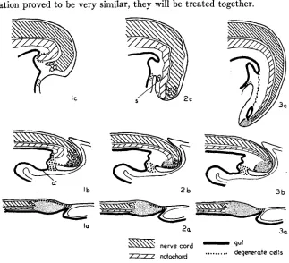
FIGURE I.-Diagrammatic sagittal sections illustrating the important morphological events in the development of Rumpless embryos. I, extreme rumplessness; 2, intermediate rumplessness;
3, normal; a, latter part of second day of incubation; b, latter part of third day; c, latter part of fourth day; s, abortive somite; a’, allantoic diverticulum. Fine stippling indicates the indifferent tail mass. Small circles in distal portion of the nerve cords represent multiple canals.
(This description does not differ from that of Du TOIT.) In our material the distortion of the nerve cord was usually much less marked in the extreme Rp embryos, where the distal neural tissue frequently appeared eroded, than in the intermediates. In two of the more normal intermediates some of the indifferent tail tissue was present and was full of necrotic cells (these will be discussed below). In one case the embryo looked normal superficially, but the nerve cord was irregular.
Notochord: As in Du TOIT’S material, it was found that the notochord of Rumpless embryos was very thick and blunt a t the distal end and stodped abruptly without merging with the indifferent mass. Normally the noto- chord tapers gradually and loses itself in this mass (fig. I-3c). In our ma- terial, too, the notochord bent ventrad sharply a short distance before terminating (fig. I-IC). In four of our extreme cases this bending was so marked that the notochord actually pushed into the cloacal portion of the gut (fig. 6 ) ; the gut epithelium was pushed before it. Invariably the distal tip of the notochord was surrounded by pycnotic and some degenerate cells.
Somites: The somites of the tail were quite normal, except for a decrease in number, until near the region of nerve cord distortion. Here, frequently, were several small vesicles, undoubtedly abortive somites, which were either above, below, or to one side of the nerve cord and notochord (fig.
or many. Some greatly enlarged cells with as many as a dozen granules have been seen. These definitely resemble macrophages, but may occur in stages where macrophages supposedly do not. Nuclei are present in these cells; they may appear normal in cells with one or two globules or shriveled in those with many. When globules occur only in occasional cells (the usual situation) only one is present in each cell. Less frequently there are large foci of degenerating cells. In such foci it is often difficult to see individual cells (except a t the periphery); all that can be seen is a large mass of chromophilic granules (fig. 3 ) . Either the cells are so closely packed that cell boundaries cannot be distinguished or the cells have broken down and extruded the granules.
While a thorough search for degenerating cells in regions anterior to the tail was not made in the present material, many were observed in both normal Leghorn embryos and embryos from the Rumpless matings. In four and five day embryos from both sources such isolated cells were ob- served in the nerve cord (more found in posterior regions, but many seen anteriorly), brain, notochord, somites, ganglia, dermatome, scleratome, and mesenchyme. Light concentrations of degenerate cells were frequently encountered in the dorsal part of the distal nerve cord, in the Wolffian ducts a t the point where they enter the cloaca, in the hind-gut, in the mesoderm around the base of the allantois, in the central portion of the post-anal gut, in the hind-limb buds (in several cases they were seen in the center of the mesoderm, in one in the distal mesoderm and ectoderm), and in the indifferent cell mass of the tail tip. This latter focus is not present in either the intermediate or extreme Rumpless embryos since the indif- ferent mass is not present. In these, scattered degenerate cells, together with true pycnotic cells, were found around the distal tip of the blunt notochord. As mentioned previously, in the two intermediates in which some of the indifferent mass remained, the mass was full of degenerate cells. I n several intermediates the distal part of the nerve cord (the dis- torted region) had concentrations of these cells.
Third day. 68-76 hours
nerve cords merged with the cells of the indifferent mass. As may be seen in table 3, a certain amount of distortion occurs normally in the distal region of the nerve cord. Of 23 normal Leghorn embryos in this age group,
13 had multiple canals. None were so markedly irregular as the extreme cases found in the embryos from the Rp matings.
Notochord: The notochord in embryos from the Rp matings in most cases
did not differ from the normal condition. At this stage the notochord normally becomes somewhat thicker where it merges with the indifferent mass. In several of the Rumpless embryos the notochord was even thicker than normal and ended abruptly, without merging with the indifferent mass. The notochords of these embryos (fig. I-Ib) bent sharply and came
TABLE 3
CONCENTRATION OF DEG. CELLS IN INDIFF.
TAIL MASS CONCENTRATION OF DEG.
CONDITION OF NO. OF CELLS IN N.C.
NERVE CORD CASES
NONE MILD EXTREME NONE MILD EXTREME
R p matings
No distortion 9 9 0 0 4 5 0
Slight distortion 2 2 17 3 2 6 14 2
Extreme distortion 17 I 4 I 2 0 2 15
No distortion IO 8 2 0 4 6 0
Slight distortion I 3 IO I 0 9 2 0
Extreme distortion o 0 0 0 0 0 0
Leghorn
into contact with the bulge which separates the hind-gut from the allantois; they are undoubtedly fore-runners of such cases as the four embryos, de- scribed in the previous age group, in which the notochords pushed into the cloacae.
Somites: Abnormalities of the somites were not observed a t this age. Apparently somite formation was still going on or had just stopped, since some of the embryos had as many somites as were found in later extreme
Rp embryos and none had so many as the less extreme intermediates and normals. The range of somite numbers, as previously indicated, was almost identical with that for normal Leghorns of the same age group.
Gut: There was no post-anal gut in embryos of this age. The hind-gut was a blind sac which was in contact with the mesenchyme of the proximal portion of the tail-bud.
RUMPLESSNESS IN 649
mesenchyme of various parts of the body, liver diverticula, the distal ends of the Wolffian ducts, the distal end of the notochord, and, very con- sistently, in the sero-amnionic raphe. Large concentrations of degenerating cells were encountered only in the tail region, and these are of marked interest for the present study.
The allantoic diverticulum a t this stage is a shallow trough of endo- dermal epithelium which is directed somewhat downward and anteriorly. Posteriorly the gut epithelium is continuous with that of the hind-gut a t the region where the sub-caudal pocket pushes forward and forms an anteriorly directed bulge of all tissues. At this point the mesenchyme sur- rounding the allantois and hind-gut makes contact with the mesenchyme of the proximal portion of the tail-bud. Here, too, the gut is closest to the ectoderm and forms the so-called cloacal membrane or anal plate; and here the ectoderm of the amnion is continuous with that of the tail-bud. The tissues, especially the mesoderm, of this cloacal membrane region were full of degenerating cells. All of the mesoderm around the base of the allantois,
at the cloacal membrane, and ventral to the hind-gut had many cells which were full of chromophilic globules. These cells extended into the mesoderm of the proximo-ventral portion of the tail-bud. Many were found in the endodermal cells of the hind-gut and the base of the allantois; and many were found in the ectoderm at the mid-line of the region where the amnionic ectoderm joins the body ectoderm. The lumen of the hind-gut was fre- quently full of degenerating cells. The above description applies to all embryos, both from the Rp matings and the Leghorn stock (fig. 1-1, 2,3 b).
There was variation in the degree of concentration of such cells in both stocks, but more embryos showing extreme concentrations were found in the group from the Rp matings.
ventral line near the base of the tail was actually eroded away, and de- generate cells seemed to be extruding from this spot (fig. 7). Bending and thickening of the notochord was observed only among these extreme cases. The least extreme concentrations were found in embryos in which the nerve cord was undistorted.
After this description was written, the author was informed of a discus- sion of the role of degenerating cells in cloaca formation in Gallinaceous birds by BOYDEN (1922). The latter’s description is almost identical with the present. Two foci of phagocytic cells (BOYDEN identifies them as macro- phages) are described: one a t the region of the cloacal membrane, asso- ciated with the formation of the fenestrations which precede the formation of the bursa of Fabricius; the other a t the peripheral region of the indiffer- ent tail tissue. I n reference to the latter I quote (page 168) : “The proximal half, on the other hand, is degenerating. Some of it may contribute to the mesenchyme of the tail, but most of it, as indicated by the presence of in- numerable phagocytes gorged with pycnotic nuclei, is undergoing resorp- tion.” The present work differs only in relation to the number of de- generating cells found in the indifferent mass of normal embryos, but this is undoubtedly due to a difference in relative quantities since the author’s figure I-3b is almost identical with BOYDEN’S (1936) figure 5.
SI-59 hours
Nerve cord: Multiple canals were encountered a t the distal tips of the nerve cords in nearly all embryos of this age from both groups. All 11 normal Leghorns had them, as did 35 of the 37 embryos from the Rp mat- ings. This condition varied, in both stocks, from two to five or six small canals. There was no additional distortion of the nerve cord; the region of multiple canals was just slightly thicker than more anterior regions of the cord.
Notochord: There was no difference in the notochords of the two stocks. Two or three chromophilic granules were seen a t the distal tip of nearly every notochord.
Somites: Somites were normal in both groups. Their number varied from
2 0 to 2 8 in the Rp group and from 20 to 29 in the normal Leghorn embryos.
.
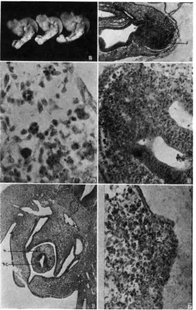
FIGURE 2 . 9 2 4 hr. embryos showing normal, intermediate and extreme rumpless tails. FIGURE 3.-71) hr. R p embryo. Sagittal section (oblique) showing extreme concentration
FIGURE 4.-Oil immersion photograph of cells containing chromophilic globules.
FIGURE 5.-704 hr. Rp embryo showing the presence of degenerating cells in the distal portion of the nerve cord, which is double a t this level, but has as many as eight canals more anteriorly.
FIGURE 6.-Transverse section through the cloacal region of a four day extreme Rumpless embryo. The nerve cord (n.c.) and notochord (n) are in the cloaca.
FIGURE 7.-754 hr. R p embryo. Sagittal section (high power) through the mid-ventral tail region showing the degenerate cells being extruded through the ectoderm.
distal tip of which is in contact with the other layers. The tail fold will form anterior to this point and anterior to the endodermal pouch.'
Degenerating cells: In embryos from both of our stocks cells in the region of fusion, described above, had many chromophilic granules. This is true for all three layers: the mesoderm had the most, the endoderm fewer, and the ectoderm the least. In virtually every embryo some of these cells (seemingly scattered from this focus) were present a t the distal tip of the tail thickening. The concentration (amount and extent) of such cells in this region was variable (fig. I-ra, b, c). Twelve of the 37 embryos from the Rp matings had more extensive concentrations than were observed in any of the normals; and in seven of these the granules were found in the adjacent nerve cord.
Second day. 30-49 hours
Earlier embryos were studied to determine any difference in the dis- tribution of chromophilic granules. Embryos in this age group do not differ from the normal in other features. In normal embryos as young as 15 somites chromophilic granules were present at the posterior end of the remnant of the primitive streak. The earliest Rp embryos studied exten- sively were of the 16-somite stage; three of nine such embryos had chromo- philic granules extending throughout the primitive streak and into the posterior portion of the nerve cord. In one case the concentration of granules was considerable. A few granules were found at the posterior region of one 12-somite embryo, while none were found in the one eight- somite embryo examined.
RECAPITULATION
Our observations lead us to the following synthesis of the genesis of dominant'rumplessness: the tails are reduced or missing because of an early destruction of the material from which this structure is formed. The destruction, evidence of which is furnished by the presence of de- generating cells full of chromophilic granules, is begun some time toward the end of the second day of incubation and reaches its height by the end of the third day (when, in normal embryos, the most active growth in length of the tail is initiated). Sections of affected embryos at this stage reveal that the entire indifferent mass, the source of material for the pos-
1 The nature of the endodermal pouch has been the occasion of some disagreement. LILLIE
(1919) and others hold that it is the hind-gut. Recently GRUENWALD (1941) claims that it is a
tenor extension of the caudal structures, is in a condition of extreme degeneration and that the nerve cord may be in the same condition. Variations in the amount of tissue involved and the degree of degeneration have been encountered and may be correlated with variations in the final condition. Gross effects of this degeneration are found toward the end of the fourth day when all degrees of reduction of the tail may be ob- served. It is during this day that the greatest growth in length of the tail occurs normally. The most obvious internal effect of the absence of the indifferent mass is the failure of the somites to form. The notochord and the nerve cord are shortened and end abruptly. The former is thick and blunt and the latter is distorted at the distal end. The fact that the nerve cord is less irregular in the extremes than in the intermediates of the fourth day may be explained by an actual sloughing off of cells in the former; some of the nerve cords in extreme cases have a very eroded appearance. This is substantiated by the existence of greater irregularity of the nerve cord and the presence of numerous degenerate cells within the neural tissue of embryos diagnosed as extreme Rumpless at the end of the third day.
DISCUSSION
Various developmental roles have been ascribed to degenerating cells. According to ERNST (1926) they are associated with processes of fusion, separation, or dissolution of tissues. PETER (1936) doubts that they have any morphogenetic function. In regard to the scattered cells containing chromophilic granules PETER may be correct, but when one encounters masses of degenerating cells consistently in the same region and a t the same time in development (for example, cloacal region of the chick embryo during the third day), one suspects that they may be concerned in local morphogenesis (BOYDEN 1918, 1922). Regardless of their part in normal development, it is quite evident that degenerating cells play a considerable role in the genesis of the Rumpless condition. The facts that they occur in great concentration a t a critical time, that there is evidence of their being extruded from the body, and that the tissue in which they are con- centrated (indifferent cell mass) is lacking in older stages all point to this. conclusion.
The presence of degenerating cells is undoubtedly the result of some previous activity which results in cell death, but the nature of the under- lying process is obscure as yet.
particular, is shorter than in Archaeornithes. It is conceivable that the degenerating primitive-streak mass in the tail of the chick embryo repre- sents a persistence of material once utilized in tail-building but now super- fluous.” The condition found in our Rumpless embryos may result from an exaggeration of the destruction of “superfluous” tissue so that it in- cludes more or less of the material involved in normal tail formation. Some mechanisms for restricting marked developmental degenerations (such as occur in insect and amphibian metamorphosis) undoubtedly exist, and it is not a t all impossible that a mutation may change the protective processes so that they would be less effective. The modifiers found in our Rumpless stocks could be factors favoring the restricting mechanisms.
Du TOIT (1913) has emphasized the distorted nerve cords and thickened notochords in his rumpless embryos. We feel certain that they are sec- ondary results of the destruction of the indifferent tail mass. Multiple canals in the caudal end of the nerve cord are common in embryos of birds and mammals. SCHUMACHER (1927) claims that this occurrence is sufficiently frequent that it cannot be regarded as a pathological condition but must be a normal manifestation of the manner in which the posterior portion of the nerve cord forms. HOLMDAHL (1933) has pointed out that the posterior part of the nerve cord arises from a solid mass of cells (in- different tail mass or “Rumpfschwanzknospe”) instead of forming from a plate of tissue. According to SCHUMACHER, it is typical for canals which form from solid masses to appear as several smaller canals, discrete a t first, then merging after some time. I n our material multiple canals were encountered frequently among both the Rumpless embryos and those from the normal Leghorn stock. Additional distortion and irregularity were found only in older (third and fourth days) embryos from the Rump- less stock and were associated with degenerating cells in the indifferent cell mass. It is probable that the later distortion of the nerve cord is not causally dependent on the initial condition of multiple canals but may be superimposed upon it. It probably results from an increase in cell number a t this region (for mitotic activity is high) in the absence of sufficient space for orderly expansion. The thickening of the notochord is probably due to a similar sequence of events.
Rumplessness in chicks has been described as probably being due to a growth retardation a t a critical period (DANFORTH 1932). This is definitely not the case for the dominant Rumpless animals. Their growth is normal throughout development, and the tail abnormality is already established a t the time a t which DANFORTH applied his temperature treatment.
RUMPLESSNESS IN CHICKS 655
LEY 1935; GLUECKSOHN-SCHOENHEIMER 1938a, 1938b, 1940). The first of these results is a short-tailed condition in the heterozygotes ( T + ) . The homozygotes ( T T ) die in utero a t about 11 days. A striking feature of these embryos is the presence of numerous chromophilic granules in the posterior region shortly before death. CHESLEY was uncertain whether these were secondary results of a moribund condition or whether they were the cause of the degeneration. The other two mutants (t' and to)
are expressed when combined with the T gene and yield only one type of surviving offspring, the tailless mice (Ttl and TtO). The tot0 embryos die shortly after implantation, and the t'tl embryos die before implantation. It is interesting that genes having such similar results (tail reduction) in two different animals produce their effects by such dissimilar means. For, the tail reduction in surviving mice is effected by the degeneration of either part of or the entire tail after it had formed normally until about the I Ith day of embryonic development. At this time a constriction occurs at a given region of the tail, and all of the tissues distal to this degenerate and form a filament which eventually is sloughed off. The most obviously abnormal structure in all of the heterozygotes is the notochord; this struc- ture is interpreted by the quoted authors as probably playing a causal role in the etiology of tail reduction.
Tail reduction due to the dominant gene Rp differs from the above in that: (I) There is no lethal action in the homozygotes. All of the gene effect appears to be localized. ( 2 ) The presumptive and not the definitive tail tissue degenerates, and the distal somites do not form. (3) The noto- chord does not have a causal role; its deformity (as well as that of the nerve cord) is a secondary result of the absence of its formative material.
SUMMARY
A study of dominant Rumpless embryos reveals that tail reduction is completed by the end of the fourth day of development. This confirms Du TOIT'S findings.
At this time there is a variation in the amount of tail reduction that corresponds with a similar variation (probably the result of genetic modi- fiers) found in adults.
Sections of earlier embryos show that the tail reduction results from a degeneration of the presumptive tail tissue. Degeneration is initiated to- ward the close of the second day and is most pronounced during the third day of development.
ACKNOWLEDGEMENT
656
LITERATURE CITED
BOYDEN, E. A., 1918 Vestigial gill filaments in chick embryos with a note on similar structures in reptiles. Amer. J. Anat. 23: 205-236.
1922 The development of the cloaca in birds, with special reference to the origin of the bursa of kabricius, the formation of the urodeal sinus, and the regular occurrence of a cloacal fenestra. Amer. J. Anat. 30: 163-202.
1936 A laboratory atlas of the pig embryo. Philadelphia: Wistar Institute Press.
CHESLEY, P., 1935 Development of the short-tailed mutant in the house mouse. J. Exp. Zoo].
DANFORTH, C. H., 1932 Artificial and hereditary suppression of sacral vertebrae in the fowl.
DUNN, L. C., 1925 The inheritance of rumplessness in the domestic fowl. J. Hered. 16: 127-134.
DUNN, 1,. C., and W. LANDAUER, 1925 Two types of rumplessness in domestic fowls. J. Hered. 16: 151-160.
1934 The genetics of the rumpless fowl with evidence of a case of changing dominance. J. Genet. 29: 217-243.
1936 Further dataon genetic modification of rumplessness in the fowl. J. Genet. 33: 401-
40.5.
Du TOIT, P, J., 1913 Untersuchungen uber das Synsacrum und den Schwanz von Gullus domes- 7'3: 429-459-
Proc. Soc. Exp. Biol. N. Y.30: 143-145.
ticus. Jena. Z. Natunv. 49: 1-164.
ERNST, M., 1926 m e r Untergang von Zellen wahrend der normalen Entwicklung bei Wir- beltieren. Z. Anat. U. Entwgesch. 79: 228-262.
GLUECKSOHN-SCHOENHEIMER, S., 1938a Time of death of lethal homozygotes in the T (Bra- chyury) series of the mouse. Proc. Soc. Exp. Biol. N. Y. 39: 267-268.
1938b The development of two tailless mutants in the house mouse. Genetics 23: 573- 584.
1940 The effect of an early lethal (to) in the house mouse. Genetics 25: 391-400.
Normal and abnormal detachment of body and gut from the blastoderm in the chick embryo, with remarks on the early development of the allantois. J. Morph. 69: GRUENWALD, P., 1941
82-rzc. - v --*-
HOLMDAHL, D. E., 1933 Die meifache Bildungsweise des zentralen Nervensystems bei den Wirbeltieren. Arch. Entwmech. Org. 129; 206-254.
LANDAUER, W., 1928 The morphology of intermediate rumplessness in the fowl. J. Hered. 19: 453-467.
LIBON, G., 1911 Ansichten uber das Vorkommen, die Abstammung und den Entstehung des schwanzlosen Haushuhnes. Dissertation, Bem (Zootech. Instit. d. Univ. Bem).
LILLIE, F. R., 1919 The development of the chick. New York: Henry Holt and CO.
PETER, K., 1936 Untersuchungen uber Zelluntergang in der Embryogenese. Z. Anat. U. Entw-
SCHUYACHER, S., 1927 ffber die sogenannte Vervielfachung des Medullarrohres (bzw. des gesch. 105: 409-428.