Methodologies for Foetal Phonography Signals
Processing - A Survey
Trushna Kamdi 1, Naziya Pathan 2, Shyam Dubey3
M. Tech. (CSE), Scholar, Department of CSE, NUVA COET, Nagpur, Maharashtra, India1
Assistant Professor, Department of CSE, NUVA COET, Nagpur, Maharashtra, India2
Assistant Professor, Department of CSE, NUVA COET, Nagpur, Maharashtra, India2
ABSTRACT: Foetal phonocardiography is a continuous non-invasive, low-cost and accurate monitoring technique for analysing the heartbeat rate and takes care of foetal well-being. It involves picking up, through a highly sensitive microphone, sonic vibrations from the heart which are then converted into electrical energy and fed into a galvanometer and is recorded for further analysis. In this paper, we review state of the art in foetal cardiac signal de-noised extraction and analysis. Due to the long-aged history of the problem and the rich literature contribution of the various authors in the field; it is not possible to cover all the existing methods in their details. Also, due to the complexity of the problem, many of the existing methods have used a combination of approaches, some of which have been borrowed from other contexts. Therefore, In this paper, we review a selection of the available literature with special focus on the most significant ones, which have been specifically developed for the problem of interest.
KEYWORDS:Phonocardiograph, Foetal Heart Rate, Phonography, Phonocardiograph.
I. INTRODUCTION
Electronic foetal monitoring (EFM) is a method for examining the condition of an unborn in the uterus by noting any unusual changes in its heart rate [1]. The EFM is either performed late in pregnancy or continuously during labour to ensure the normal delivery of a healthy baby. This method was originally introduced in the 1960s and 1970s, with the hope that it would help physicians diagnose foetal hypoxia, or lack of oxygen and abnormal heart rate in the unborn baby.
Current methods for EFM rely heavily on Ultrasound Techniques. This has two important disadvantages; firstly, there are significant chances that long-term exposure to ultrasound may harm the unborn, and secondly, the ultrasound machines and its trained operators prove to be an expensive proposition hence could not be utilized for a longer period of time [2]. Because of these reasons, the existing instruments are not suitable for carrying out the frequent and long-term recording of a foetal heart sound, which is highly recommended in high-risk pregnancies [3].
Foetal phonocardiography is a continuous non-invasive, low- cost and accurate monitoring technique for analysing the heartbeat rate and takes care of foetal well-being [4]. It involves picking up, through a highly sensitive microphone, sonic vibrations from the heart which are then converted into electrical energy and fed into a galvanometer, where they are recorded on paper. The procedure is most useful when there is evidence of heart murmurs or unusual heart sounds, such as gallops that are difficult to discern by the human ear. Most recordings are made through an externally applied microphone but intra-cardiac recordings, made through a phono catheter, are also possible.
Despite all these advantages of fPCG, this technique is not popular with the obstetricians because of its poor signal-to-noise ratio (SNR) at the time of recording [7]. The fPCG signals recorded from the maternal abdominal surface are contaminated by various unwanted signals which can prohibit their automated analysis. These factors can be categorized as [8]:
• respiration sounds (lung mechanics), • patient movements,
• small movements of the stethoscope ("shear noises"), • acoustic damping through the bones and tissues, and • external noises from the environment.
There are some other typical disturbances in the case of foetal phonocardiography: • acoustic damping of forewaters and maternal tissues,
• acoustic noises produced by the foetal movements, • noises of the maternal digestive system, and • sounds of the maternal heart.
II. REVIEWOF DIFFERENT TECHNIQUES
Foetal phonocardiograph is contaminated with various unwanted signals due to the reasons listed in the previous chapter. Hence this technique requires robust signal-processing to extract the foetal heart sound signals [9]. There have been significant efforts to develop long-term monitoring methods based on fPCG technique. These methods have projected different types of sensors and filtering schemes, as well as different numbers of channels, for this purpose.
Most of the existing phonocardiograph processing methods concern only with the diagnostic analysis of heart sounds without an adequate emphasize on the denoising of the records. Existing methods usually apply digital band-pass filters (most commonly IIR-filters of FFT-based filtering) as a simple denoising method. The cut-off frequencies of the filters are determined by empirical observations, and commonly the pass band lies between 30 and 200 Hz [10-14].
The main difficulty in dealing with the fPCG signals is their extreme variability and necessity to operate on a case-to-case basis [15]. Another important aspect of these signals is that the information of interest is often a combination of features that are well localized temporally or spatially. This requires the use of analytical methods sufficiently versatile to handle events that can be at opposite extremes in terms of their time-frequency localization.
A significant number of studies found in the literature report the implementation of the wavelet transforms for the extraction of the foetal heart rate. In particular, in the work of [16,17], a wavelet transform is applied in order to extract the wavelet-based features of the foetal cardiac signal so that the normal cases can be easily distinguished from the abnormal ones. So far, the wavelet analysis has proved to be one of the most successful techniques for the analysis of signals at multiple scales and has rendered many successful applications in the area of biomedical signal processing [18].
Another method to monitor foetal heart rate patterns is by studying the foetal heart rate variability. It has been shown that heart rate variability is an important marker for the foetal well-being [19]. Various methods have been therefore used to analyse the foetal heart rate variability, in particular concerning variables from both the time and the frequency domain [20].
principles of the BSS methods but make additional use of prior information about the autocorrelation property of the FECG from the noisy free measurements [30].
To cover up this brief review on the foetal cardiogram extraction methods, see also [30, 31], listed below are the methods used in the literature, such as:
• singular value decomposition (SVD) [32] • matched filtering [33]
• adaptive neuro-fuzzy inference systems [34] • dynamic neural networks [35]
• temporal structure [36] • fuzzy logic [37] • frequency tracking [38] • polynomial networks [39]
• multi-reference adaptive noise cancellation [24] • real-time signal processing [40]
• time-frequency analysis [41]
All the above techniques have been successfully applied to the extraction of the foetal cardiac component, but a maximum of them are used in the perspective of processing ECG and not PCG signals. The following sections cover the review of processing of fPCG using signal processing and pattern recognition techniques.
III.SIGNALPROCESSINGANDCOMPUTATIONALLEARNINGTECHNIQUES
Signal processing methods enable to uncover the structure of the signal and to extract features which compactly represent the properties of the raw data. Feature extraction is usually a preceding step of a classification or regression task, which is used to infer application-specific information from the input signals. Feature selection can utilize domain-specific knowledge for choosing features with physical meaning (e.g. frequency, time interval, energy), or alternatively, may be automatic, relying on general properties of the signals. Domain-specific features used for heart sound analysis include, for example, dominant frequencies of the signal’s spectrum [66], the bandwidth of the dominant frequencies, mean and total spectral energy, and the intensity ratio of S1 and S2 [67]. In the model-based signal analysis, the parameters of the model are a natural set of features. For example, Guo et al. used the coefficients of 12-order all-pole system as features [68]. Bentley et al. used a search scheme to select an optimal subset of features from a larger set, extracted using discrete wavelet transform [69]. They showed that this feature set provides better performance than morphological features extracted from CWD in classifying the condition of native and artificial heart valves. The application of identifying degenerated artificial valves from features of their closure sounds was addressed using different types of classifiers, including K-nearest neighbour (KNN), Gaussian-Bayes and neural networks. Reported results, which seem to be optimistic, indicate the high accuracy of 89%-98% in detecting degeneration of different types of artificial heart valves.
Unsupervised learning using morphological cluster analysis has been applied before to biomedical imaging modalities such as magnetic resonance imaging [70], as well as to electrocardiogram signals [71]. To the best of the knowledge of this thesis, there are no previous studies that have applied cluster analysis on the morphology of heart sounds.
The following section describes the different methods of data acquisition and methods proposed for the processing of phonocardiograph for denoised extraction of the foetal heart sound signals in the literature.
IV.PHONOCARDIOGRAPHYWAVELETSANALYSIS
The wavelet transform is a frequently used tool for the denoising of audio signals [8]. The choice of wavelet family, mother wavelet and de-noising algorithm greatly affects the accuracy of the wavelet analysis of the signal [72].
V. DENOISINGALGORITHMS
Three steps are required for de-noising of signals: Decomposition, thresholding and reconstruction. The first and last steps are performed with the selection of suitable wavelet family and mother wavelet. The thresholding step is the selection of threshold level for noising of the signals. The thresholding algorithms commonly employed for de-noising of the fPCG signals are —
Universal threshold Minimax threshold Rigorous SURE threshold
VI.OVERVIEW OF NOISE CANCELLATION
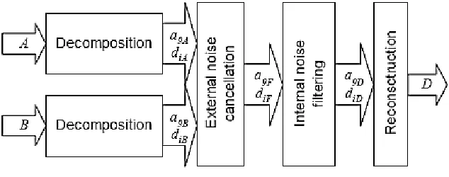
Fig.1. Block diagram of the wavelet-based denoising system.
Figure 2.1 illustrates the structure of the proposed wavelet based denoising method in [8]. It consisting of four major steps:
1. wavelet-based decomposition of both recorded signal channels A and B,
2. cancelling of external noises using the adaptive cross channel thresholding of the signal coefficients, 3. adaptive thresholding of the coefficients resulted in Step 2 in order to eliminate the internal noises, and 4. reconstruction of the D denoised signal from the threshold coefficients of Step 3.
VII. WAVELET-BASEDDECOMPOSITIONANDRECONSTRUCTION
In the following section, the various wavelet thresholding techniques and their comparison are discussed. Peer review of the wavelet based algorithm for noise cancelation is also discussed.
VIII. 2.4.3.1UNIVERSALTHRESHOLD
The universal threshold de-noising algorithm is also known as VisuShrink. It uses a fixed threshold form given by
(1)
where ‘n’ denotes the length of the signal and σ is the standard deviation. This threshold was determined in an optimal context for soft thresholding with random Gaussian noise. This scheme is very easy to implement but typically provides a threshold level larger than other thresholding algorithms, therefore resulting in smoother reconstructed data. Also, such estimation does not take into account the content of the data but only depends on the data size.
IX.2.4.3.2MINIMAXTHRESHOLD
The minimax threshold de-noising algorithm consists of an optimal threshold that is derived from minimizing the constant term in an upper bound of the risk involved in the estimation of the signal. This threshold level depends on the noise and signal relationships in the input data, and it is given by
where λn’ is determined by a minimax rule such that the maximum risk of estimation error across all locations of the data is minimized.
X. 2.4.3.3RIGOROUSSURETHRESHOLD
The de-noising algorithms described previously use global thresholds, that is, the computed threshold is applied to all wavelet coefficients. The rigorous SURE threshold algorithm describes a scheme that uses a threshold value λj at each resolution level ‘j’ of the wavelet coefficients. This algorithm is also known as SureShrink, and it uses the Stein’s unbiased risk estimate (SURE) criterion to get an unbiased estimate.
XI.2.4.3.4CROSSCHANNELTHRESHOLD
The cancellation of the external noises in [8] is a two-channel problem. A novel method was developed which utilizes the adaptive cross-channel thresholding of the wavelet transformed signal coefficients. The dyadic wavelet transformed of a signal f(t) on the i-th dyadic scale can be written as:
(3)
Where ψ is is the orthogonal mother wavelet, and:
(4)
The discrete version of the dyadic wavelet transform can be based on digital filter banks [14]. Let us define:
(5)
(6)
where i ≥ 0. For a given filter x[n], xi[n] denotes the filter obtained by inserting 2i-1 zeros between every filter
coefficient (“holes algorithm”). a0[n] is the original signal, and its wavelet-based decomposition at the i+1th scale
is:
where a defines the approximated signal and d the signal details at the given scale; h and g are biorthogonal filters, corresponding to the selected mother wavelet. The reconstruction of the signal is done by the inverse wavelet transform, which is at the i-th scale:
(7)
where and ĥ and ĝ are the dual filters of h and g, respectively. In the case of biorthogonal filters: ĝ=h and ĥ=g.
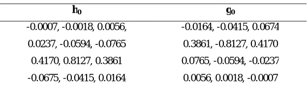
Table I: Base Coefficients of the Equivalent Filter Bank
h0 g0
-0.0007, -0.0018, 0.0056, -0.0164, -0.0415, 0.0674
0.0237, -0.0594, -0.0765 0.3861, -0.8127, 0.4170
0.4170, 0.8127, 0.3861 0.0765, -0.0594, -0.0237
-0.0675, -0.0415, 0.0164 0.0056, 0.0018, -0.0007
It uses the Coiflet-wavelet with 4 moments equal to zero (Coiflet-2) as the mother wavelet. The two channels were wavelet decomposed up to the 9th order (i = 0, 1 ... 8), resulting in two sets of coefficients: a9A, d9A, d8A, ..., d1A, and
similarly: a9B, d9B, ... d1B, where the A and B letters in the indices correspond to the recorded channels.
XII. 2.4.3.5ADAPTIVECROSS-CHANNELCANCELLATIONOFEXTERNALNOISES
The task of the external noise cancellation is to construct a new set of coefficients: a9F, d9F, ...,d1F, which can be
reconstructed (inverse wavelet transformed) as the heart sound signal free of external noises. The following algorithm is proposed in [11] to obtain these coefficients:
a9F = a9A,
if | diA[n] | > max(μ, λ) then diF[n] = diA[n] - diB[n],
else diF[n] = 0
where i = 1, 2, ... 9; n = 1, 2, ..., dim(diA), and λ = 0.5 max( | diA | ), μ = 2 max( | diB| ).
Here, the detail coefficients diA of the abdominal channel are adaptively thresholded (μ and λ), whereby the details
diB of the external channel is adaptively subtracted from diA.
XIII. 2.4.3.6INTERNAL NOISE CANCELLATION
Another algorithm was designed which works in the frequency band 35 Hz < f < 200 Hz and adaptively threshold
previously resulted set of coefficients a9F, d9F, ..., d1F:
a9D = 0, d9D = 0
if | diF[n] | > λ then diD[n] = diF[n],
else diD[n] = 0
where i = 1, 2, ... 8; n = 1, 2, ..., dim(diF), and λ = 0.5 μ, where μ is the average of the a9F.
The resulted coefficient set a9D, d9D,…...,d1D is used at the reconstruction of the phonocardiographic signal free of
noises.
It should be noted that this second thresholding cannot remove all internal noises. Disturbances still remain in the reconstructed signal; however, the overall noise cancellation performance is far better than in the case of simple band-pass filtering.
XIV. 2.4.4 ACOMPARATIVEANALYSISOF DE-NOISINGALGORITHMS
inexpensive data-recording module (DRM) [73]. The signals were recorded with a sampling frequency of 8000 Hz, 16-bit resolution, and saved in a personal computer for further processing. Fig.2 shows the waveform of one of these fPCG signals.
Fig.2 Waveform of the fPCG Signal
All the three de-noising algorithms are simulated in Matlab® for de-noising of the fPCG signals. The mean squared error (MSE) is used to evaluate the performance of the presented approach for the selection of appropriate de-noising algorithm. It can be obtained using the following expression:
(8)
where ‘n’ denotes the length of the signal, ‘s’ represents the original signal and se is the estimated signal obtained from the de-noised wavelet coefficients. Figure 2.3 is a test signal generated by adding simulated random noise in the original fPCG signal, which is shown in Figure 2.2. This simulated random noise is analogous to the noise produced because of maternal organ sounds, foetal movement effects and ambient noise.
The wavelet analysis of this signal is performed with five levels of decomposition using fourth-order Coilets wavelet. This selection of mother wavelet is random and based on the fact that it possesses all the properties needed for analysis of the fPCG signals. All the three algorithms with soft or hard thresholding rule are applied for de-noising of the fPCG signal. The authors of [72] show the comparisons of the fPCG signal obtained using these optimal wavelet functions.
Figure 2.3 fPCG Signal with additive noise
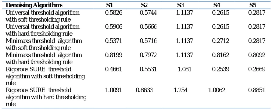
Finally, in [72] the efficacy of the method is evaluated using fPCG signals (S1-S5) recorded from five different subjects. All the de-noising algorithms are applied for de-noising of these fPCG signals using selected mother wavelets, and the respective results generated are depicted in the tabular form [Table II] below.
Table II: Comparison of De-Noising Algorithms
Denoising Algorithms S1 S2 S3 S4 S5
Universal threshold algorithm with soft thresholding rule
0.5826 0.5744 1.1137 0.2615 0.2817
Universal threshold algorithm with hard thresholding rule
0.5906 0.5666 1.1137 0.2615 0.2817
Minimaxs threshold algorithm with soft thresholding rule
0.5371 0.5716 1.1137 0.2712 0.2817
Minimaxs threshold algorithm with hard thresholding rule
0.8199 0.7972 1.1137 0.8162 0.8092
Rigorous SURE threshold algorithm with soft thresholding rule
0.4661 0.5531 1.081 0.2539 0.2669
Rigorous SURE threshold algorithm with hard thresholding rule
1.0091 0.8633 1.254 1.0062 0.8851
Table II shows a comparison among three de-noising algorithms with soft or hard thresholding rule. The rigorous SURE threshold algorithm with soft thresholding rule yields the best estimation with considerably smaller MSE as compared to other algorithms for de-noising of fPCG signals.
XV. 2.4.5 BLIND SOURCE SEPARATION AND PHONOCARDIOGRAPHY
Recently, blind source separation by Independent Component Analysis (ICA) has received attention because of its potential applications in signal processing such as in speech recognition systems, telecommunications and medical signal processing. The goal of ICA is to recover independent sources given only sensor observations that are unknown linear mixtures of the unobserved independent source signals. In contrast to correlation-based transformations such as Principal Component Analysis (PCA), ICA not only decorrelates the signals (2nd-order statistics) but also reduces higher-order statistical dependencies, attempting to make the signals as independent as possible.
There have been two different fields of research considering the analysis of independent components. On the one hand, the study of separating mixed sources observed in an array of sensors has been a classical and difficult signal processing problem. The seminal work on blind source separation is by Jutten, Herault and Guerin (1988) where they proposed an adaptive algorithm in a simple feedback architecture. The learning rule was based on a neuromimetic approach and was able to separate simultaneously unknown independent sources. This approach has been explained and further developed by Jutten and Herault (1991), Comon (1991), Karhunen and Joutsensalo (1993), Cichocki and Moszczynski (1992) and others. Furthermore, Comon (1994) introduced the concept of independent component analysis and proposed cost functions related to the minimization of mutual information between the sensors.
analysis, and the blind separation of sources. Bell and Sejnowski (1995) were the first explaining the blind source separation problem from an information-theoretic viewpoint and applying them to separation and deconvolution of sources. Their adaptive methods are more plausible from a neural processing perspective than the cumulant-based cost functions proposed by Comon. A similar adaptive but 'non-neural' method for source separation has been proposed by Cardoso and Laheld (1996).
Other algorithms have been proposed from different approaches: The maximum likelihood estimation approach was first proposed by Gaeta and Lacoume (1990), negentropy maximization approach by Girolami and Fyfe (1996), nonlinear PCA algorithm developed by Karhunen and Joutsensalo (1994) and Oja (1995). Lee, Girolami and Sejnowski (1997) give a unifying framework to the source separation problem by explaining the relation of the different algorithms to each other. The derived learning rule is optimized when the natural gradient (Amari, 1997) or relative gradient (Cardoso and Laheld, 1996) is used.
The originally proposed algorithm by Bell and Sejnowski (1995) was suited to separate super-Gaussian sources. To overcome this limitation other techniques have been developed that were able to simultaneously separate sub- and super-Gaussian sources. Pearlmutter and Parra (1996) derive a generalized ICA learning rule from the maximum likelihood estimation where they explicitly model the underlying source distribution which was assumed to be fixed in the original algorithm. Although the density estimation was burdensome and required a sufficient amount of data, the algorithm was able to separate a fairly large number of the source with a wide range of distributions. A simpler and highly effective algorithm has been proposed by Girolami and Fyfe (1996) from the negentropy maximization approach. They used an extended exploratory projection pursuit network with inhibitory lateral connections that could separate sub- and super-Gaussian sources. Lee, Girolami and Sejnowski (1997) derive the same learning rule from the infomax approach preserving the simple architecture and showing superior convergence speed.
Extensive simulations have been performed to demonstrate the power of the learning algorithm. However, instantaneous mixing and unmixing simulations are toy problems, and the challenge lies in dealing with real world data. Makeig et al. (1996) applied the original infomax algorithm to EEG and ERP data showing that the algorithm can extract EEG activations and isolate artifacts. Jung et al. (1997) show that the extended infomax algorithm is able to linearly decompose EEG artifacts such as line noise, eye blinks, and cardiac noise into independent components with sub- and super-Gaussian distributions. McKeown (1997) has used the extended ICA algorithm to investigate task-related human brain activity in fMRI data. By determining the brain regions that contained significant amounts of specific temporally independent components, they were able to specify the spatial distribution of transiently task-related brain activations. Other potential applications may result from exploring independent features in natural images. Bell and Sejnowski (1997) suggest that independent components of natural scenes are edge filters. The filters are localized, mostly oriented and similar to Gabor like filters. The outputs of the ICA filters are sparsely distributed. Bartlett and Sejnowski (1997) and Gray, Movellan and Sejnowski (1997) demonstrate the successful use of the ICA filters as features in face recognition tasks and lipreading tasks respectively.
Since ICA is restricted and relies on several assumptions researchers, have started to tackle a few limitations of ICA. One obvious but non-trivial extension is the nonlinear mixing model. In (Hermann and Yang, 1996; Lin and Cowan, 1997; Pajunen, 1997) nonlinear components are extracted using self-organizing-feature-maps (SOFM). Other researchers (Burel, 1992; Lee, Koehler and Orglmeister, 1997; Taleb and Jutten, 1997; Yang, Amari and Cichocki, 1997) have used a more direct extension to the previously presented ICA models. They include certain flexible nonlinearities in the mixing model, and the goal is to invert the linear mixing matrix as well as the nonlinear mixing. Other limitations such as the under-determined problem in ICA, i.e. having fewer sensors than sources and noise models in the ICA formulation are subject to current research efforts.
ICA is a fairly new and a generally applicable method to several challenges in signal processing. It reveals a diversity of theoretical questions and opens a variety of potential applications. Successful results in EEG, fMRI, speech recognition and face recognition systems indicate the power and optimistic hope in the new paradigm. Literature review reveals that there is very little work done on processing phonocardiography; especially foetal phonocardiograph processing using BSS and still have a wide scope, in the perspective of foetal heart rate monitoring and foetal wellbeing.
In the preceding paragraph, the research work, using blind source separation as a signal processing tool for processing the foetal heart signals are discussed.
Vivek Nigam [74] provides an algorithm to non-invasively estimate the phonocardiogram of an individual foetus in a multiple fetus pregnancies. A mixture of foetal phonocardiograms is modelled by a generalized pure delayed mixing model. Mutual independence of foetal phonocardiograms is assumed to apply blind source separation based techniques to extract the foetal phonocardiograms from their mixtures. The performance of the algorithm is verified through simulation results and on experimental data obtained from a phantom that is used to simulate a twin pregnancy. Bruno AZZERBONI [75] presented a new approach where they have used a recently developed Wavelet-ICA method, based on the joint use of Wavelet Analysis and Wavelet-ICA, to fECG extraction, in order to improve the extraction performance. V. Vigneron in [76], apply BSS to fECG extraction and showed that (i) exploiting the ECG non-stationarity could improve source separation, (ii) wavelets seems a well-suited and promising method for extracting the foetal PQRST complexes. Further investigations will include (i) a quantitative comparison of the performance of BSS algorithms and focus in particular on wavelet denoising for improving PQRST complex extraction, (ii) a qualitative evaluation (by physicians) of the foetal PQRST extracted by this method, especially in pathological cases. By projecting single-channel signal into a higher dimension, the multi-channel technique can be employed. In [77] a blind-source separation method using an SVD of the spectrogram, followed by an iterative application of ICA on both the spectral and temporal representations of the ECG signals is presented. To define the decomposition problem, they make both an assumption of statistical independence of the heart-beat occurrences, and a similar assumption on the basis vectors they used to represent the spectral slices of the individual signals. This is a reasonable assumption in this domain because each signal is a repeating fPCG complex. The method they developed is likely to be applicable to other problem domains defined by the need to separate nearly periodic sources from noisy mixtures into a higher dimension; the multi-channel technique can be employed.
XVI.CONCLUSION
REFERENCES
[1] N. Malik, C. Raghunandan, N. Madan, “Foetal Heart-rate patterns in early labour in low and high risk pregnancies & its correlation with perinatal outcome,” Journal of Indian Medical Association, Vol. 100(11), pp. 646-650, Nov. 2002.
[2] T. Kupka, J. Jezewski, A. Matonia, K. Horoba, J. Wrobel, “Timing events in Doppler ultrasound signal of fetal heart activity,” 26th Annual International Conference of the IEEE on Engineering in Medicine and Biology Society, Vol. 1, pp. 337-340, 2004.
[3] V. Chourasia, A. Mittra, “Passive Acoustic Signal Acquisition System for Non-Invasive Fetal Heart Sound Monitoring Applications,” The Internet Journal of Medical Technology, Vol. 5(1), 2009.
[4] R. K. Freeman, and T. J. Garite, “Fetal Heart Rate Monitoring,” Williams and Wilkins, pp. 7-17, 1981
[5] P. Várady, L. Wildt, Z. Benyo, and A. Hein, “An Advanced method in fetal phonocardiography,” Computer Method and Program in Biomedicine, Vol. 71, pp. 283-96, 2003.
[6] J. Chen, K. Phua, Y. Song, and L. Shue, “A portable phonocardiographic fetal heart rate monitor,” Proceedings of the International Symposium of the IEEE on Circuits and System, pp. 2141-4, 2006.
[7] V. Padmanabhan, R. Fischer, J. L. Semmlow, and W. Welkowitz, “High sensitivity PCG transducer for extended frequency applications,” Proceedings of the Annual International Conference Of the IEEE on Engineering in Medicine and Biology Society, Images of the Twenty First Century, Vol. 1, pp. 57-8, 1989.
[8] P. Várady, "Wavelet-based Adaptive Denoising of Phonocardiographic Records,” 23rd Annual Conference – IEEE/EMBS, 2001.
[9] F. Kovacs, Cs. Horvath, M. Torok, and G. Hosszu, “Long-term Phonocardiographic fetal home monitoring for telemedicine system,” 27th Annual International Conference of the IEEE on Engineering in Medicine and Biology Society, pp. 3946-9, 2005.
[10] J. C. Wood, D. T. Barry, “Time-Frequency Analysis of the First Heart Sound,” IEEE EMBS Magazine, pp. 144-151., 1995.
[11] J. M. Bentley, P. M. Grant, J. T. E. McDonnel, “Time-Frequency and Time-Scale Techniques for the Classification of Native and Bioprosthetic Heart Valve Sounds,” IEEETrans. Biomed. Eng., Vol. 45. pp. 125-128., 1998.
[12] J. Ritola, S. Lukkarinen, “Comparison of Time-Frequency Distributions in the Heart Sounds Analysis,” Medical & Biological Engineering & Computing, Vol 34.,supplement 1., part 1., pp. 89-90., 1996.
[13] H. Liang, S. Lukkarinen, I. Hartimo, “Heart Sound Segmentation Algorithm Based on Heart Sound Envelogram,” in Proc. Computers in Cardiology, Vol. 24., 1997.
[14] F. Kovács, M. Török, I. Habermajer, “A Rule-Based Phonocardiographic Method for Long-Term Fetal Heart Rate Monitoring,” IEEE Trans. Biomed. Eng., Vol. 47, pp. 124-130., 2000.
[15] J. Nagal, “New diagnostic and technical aspects of fetal phonocardiography,” European Journal of Obstetrics, Gynecology and Reproductive Biology, Vol.23, pp. 295-303, 1986.
[16] Barros A. K., Mansour A., Ohnishi N., “Removing artifacts from electrocardiographic signals using independent component analysis,” Neurocomputing 22, 13, 173, 1998.
[17] Spyridou, K. K., Hadjileontiadis L. J., “Analysis of Fetal Heart Rate in Healthy and Pathological Pregnancies Using Wavelet-based Features,” Proceedings of the 29th Annual International Conference of the IEEE EMBS Cit´e Internationale, Lyon, France, 2007.
[18] G. Vasios, A. Prentza, D. Blana, E. Salamalekis, P.Thomopoulos, D Giannaris, and D.Koutsouris, “Classification of fetal heart rate tracings based on wavelet-transform and self-organizing-map neural networks,” Proceedings of the 23th Annual International Conference of the IEEE on Engineering in Medicine and Biology Society, Vol. 2, pp. 1633-6, 2001.
[19] Van Leeuwen P., Lange S., Bettermann H., Groenemeyer D., Hatzmann W., “Fetal heart rate variability and complexity in the course of pregnancy,” Early Human Development 54, 259, 1999.
[20] Karin J., Hirsch M., Akselrod S., “An estimate of fetal autonomic state by spectral analysis of fetal heart rate fluctuations,” Pediatr Res, 34, 134, 1993.
[21] Cichocki A., Amari S., “Adaptive Blind Signal and Image Processing: Learning Algorithms and Applications,” Wiley, 2002.
[22] Jafari M. G., Chambers J. A., “Adaptive noise cancellation and blind source separation, in: Proceedings of the Fourth International Symposium on Independent Component Analysis and Blind Signal Separation,” ICA2003, Japan, 627, 2003.
[23] Jafari M. G., Chambers J. A., “Fetal electrocardiogram extraction by sequential source separation in the wavelet domain,” IEEE Trans. Biomed. Eng. 52, 3, 390, 2005.
[24] Zarzoso V., Nandi A. K., “Noninvasive fetal electrocardiogram extraction blind separation versus adaptive noise cancellation,” IEEE Trans. Biomed. Eng., 48, 1, 12, 2001.
[25] Zhang Z.-L., Yi Z., “Extraction of temporally correlated sources with its application to non-invasive fetal electrocardiogram extraction,” Neurocomputing 69, 894, 2006.
[26] Ananthanag K. V. K. , Sahambi J. S., “Investigation of blind source separation methods for extraction of fetal ECG,” in: IEEE CCECE. Canadian Conference on Electrical and Computer Engineering, Montreal, 3, 3, 2021, 2003.
[27] Barros A. K., Cichocki A., “Extraction of specific signals with temporal structure,” Neural Comput. 13, 9, 1995, 2001.
[28] Cichocki A., Thawonmas R., Amari S., “Sequential blind signal extraction in order specified by stochastic properties,” Electron. Lett. 33,1, 64, 1997.
[29] Cruces-Alvarez S. A., Cichocki A., Amari S., “From blind signal extraction to blind instantaneous signal separation: criteria, algorithm, and stability,” IEEE Trans. Neural Networks, 15, 4, 859, 2004.
[30] Li Y., Yi Z., “An algorithm for extracting fetal electrocardiogram,” Neurocomputing, doi:10.1016/j.neucom., 2007.
[32] Kanjilal P. P., Palit S., Saha G., “Fetal ECG extraction from single channel maternal ECG using singular value decomposition,” IEEE Trans. Biomed. Eng. 44, 1, 51, 1997.
[33] Pieri J. F., Crowe J. A., Hayes-Gill B. R., Spencer C. J., Bhogal K, James D. K., “Compact long-term recorder for the transabdominal foetal and maternal electrocardiogram,” Med Biol Eng Comput, 39,118, 2001.
[34] Assaleh K., Al-Nashash H., “A Novel Technique for the Extraction of Fetal ECG Using Polynomial Networks,” IEEE Trans Biomed Eng, 2005, 52, 1148, 2002.
[35] Camps-Valls G., Martinez-Sober M., Soria-Olivas E., Guerrero-Martinez J., Calpe-Maravilla J., “Foetal ECG recovery using dynamic neural networks,” Artificial Intelligence in Medicine, 31, 197, 2004.
[36] Barros A. K., Cichocki A., “Extraction of specific signals with temporal structure,” Neural Comput. 13, 9, 1995, 2001.
[37] Azad K. A. K., “Fetal QRS Complex Detection from Abdominal ECG: A Fuzzy approach,” in Proc. IEEE Nordic Signal Processing Symposium (NORSIG), Sweden, 275, 2000.
[38] Barros A. K., “Extracting the fetal heart rate variability using a frequency tracking algorithm,” Neurocomputing, 2002.
[39] Assaleh K., “Extraction of fetal electrocardiogram using adaptive neurofuzzy inference systems,” IEEE Trans Biomed Eng, 2007.
[40] Ibrahimy M. I., Ahmed F., Mohd Ali M. A., Zahedi E., “Real-Time Signal Processing for Fetal Heart Rate Monitoring,” IEEE Trans Biomed Eng, 2003.
[41] Karvounis E. C., Tsipouras M., Fotiadis D. I., Naka K. K., “An Automated Methodology for Fetal Heart Rate Extraction from the Abdominal Electrocardiogram,” to appear in IEEE Transactions on Information Technology in Biomedicine, 2008.
[42] Jacobs, J. E., K. Horikoshi, and M. L. Petrovick, “Feasibility of Automated Analysis of Phonocardiogram,” Journal of the Audio Engineering Society, 1969. Vol. 17(1): pp. 49-54.
[43] McKusick, V. A., “On cardiovascular sound: further observations by means of spectral phonocardiography,” Circulation, 1955. Vol. 11(6): pp. 849-70.
[44] Yoganathan, A. P., “Use of the fast Fourier transform in the frequency analysis of the second heart sound in normal man,” Med BiolEng, 1976. Vol. 14(4): pp. 455-60.
[45] Yoganathan, A.P., “Use of the fast Fourier transform for frequency analysis of the first heart sound in normal man,” Med BiolEng, 1976. Vol. 14(1): pp. 69-73.
[46] Karpman, L., “Sound envelope averaging and the differential diagnosis of systolic murmurs,” Am Heart J, 1975. Vol. 90(5): pp. 600-6. [47] Beyar, R., “Heart-sound processing by average and variance calculation- physiologic basic and clinical implications,” IEEE Trans Biomed
Eng, 1984. Vol. 31(9): p. 591-6.
[48] Gerbarg, D.S., “Analysis of phonocardiogram by a digital computer,” Circ Res, 1962. Vol. 11, pp. 569-76.
[49] Sarkady, A.A., R.R. Clark, and R. Williams, “Computer analysis techniques for phonocardiogram diagnosis,” Comput Biomed Res, 1976. Vol. 9(4): pp. 349-63.
[50] Liang, H., S. Lukkarinen, and I. Hartimo, “Heart Sound Segmentation Algorithm Based on Heart Sound Envelogram,” in Computers in Cardiology. 1997. p. 105- 108.
[51] Nigam, V. and R. Priemer, “Accessing heart dynamics to estimate durations of heart sounds. Physiological Measurement,” 2005. Vol. 26: pp. 1005-1018.
[52] Iwata, A., “Algorithm for detecting the first and the second heart sounds by spectral tracking,” Med BiolEngComput, 1980. Vol. 18(1): pp. 19-26.
[53] Joo, T.H., “Pole-zero modeling and classification of phonocardiograms,” IEEE Trans Biomed Eng, 1983. Vol. 30(2): pp. 110-8. [54] Wang, W., “Analysis of the first heart sound using the matching pursuit method,” Med BiolEngComput, 2001. Vol. 39(6): pp. 644-8. [55] Sava, H., P. Pibarot, and L.G. Durand, “Application of the matching pursuit method for structural decomposition and averaging of
phonocardiographic signals,” Med BiolEngComput, 1998. Vol. 36(3): pp. 302-8.
[56] Durand, L.G., “an Evaluation of FFT-based and modern parametric method for the spectral analysis of bioprosthetic valve sounds,” IEEE Trans Biomed Eng, 1986. Vol. 33(6): pp. 572-8.
[57] Akay, Y.M., “Noninvasive acoustical detection of coronary artery disease: a comparative study of signal processing methods,” IEEE Trans Biomed Eng, 1993. Vol. 40(6): pp. 571-8.
[58] Gamero, L.G. and R. Watrous, “Detection of the First and Second Heart Sound Using Probabilistic Models,” in IEEE Engineering in Medicine and Biology Society. 2003. pp. 2877-80.
[59] Gill, D., N. Gavriely, and N. “Intrator, Detection and Identification of Heart Sounds Using Homomorphic Envelogram and Self-Organizing Probabilistic Model,” in Computers in Cardiology. 2005. pp. 957-960.
[60] Lin, Z. and J.D. Chen, “Advances in time-frequency analysis of biomedical signals,” Crit Rev Biomed Eng, 1996. Vol. 24(1): pp. 1-72. [61] Wood, J.C. and D.T. Barry, “Quantification of first heart sound frequency dynamics across the human chest wall,” Med BiolEng Comput,
1994. pp. S71-8.
[62] Wood, J.C., A.J. Buda, and D.T. Barry, “Time-frequency transforms: a new approach to first heart sound frequency dynamics,” IEEE Trans Biomed Eng, 992. Vol. 39(7): pp. 730-40.
[63] Chen, D., “Time-frequency analysis of the first heart sound. Part 2: An appropriate time-frequency representation technique,” Med BiolEngComput, 1997. Vol. 35(4): pp. 311-7.
[64] Obaidat, M.S., “Phonocardiogram signal analysis: techniques and performance comparison,” J Med EngTechnol, 1993. Vol. 17(6): pp. 221-7.
[65] Livanos, G., Ranganathan, N., Jiang, J., “Heart sound analysis using the S transform,” in Computers in Cardiology 2000. pp. 587-590. [66] Stein, P.D., “Frequency spectra of the first heart sound and of the aortic component of the second heart sound in patients with degenerated
[67] Durand, L.G., “Comparison of pattern recognition methods for computer assisted classification of spectra of heart sounds in patients with a porcine bioprosthetic valve implanted in the mitral position,” IEEE Trans Biomed Eng, 1990. Vol. 37(12): pp. 1121-9.
[68] Guo, Z., “Artificial neural networks in computer-assisted classification of heart sounds in patients with porcine bioprosthetic valves” Med BiolEngComput, 1994. Vol. 32(3): pp. 311-6.
[69] Bentley, P.M., P.M. Grant, and J.T. McDonnell, “Time-frequency and time-scale techniques for the classification of native and bioprosthetic heart valve sounds,” IEEE Trans Biomed Eng, 1998. Vol. 45(1): pp. 125-8.
[70] Wismüller, A., Lange, O., Dersch, D. R., Leinsinger, G. L., Hahn, K., Pütz, B., and Auer, D, “Analysis of Biomedical Image Time-Series,” Int. J. Comput. Vision, 2002. Vol. 46(2): pp. 103-128.
[71] Cuesta-Frau, D., J. C. Pérez-Cortés, and G. Andreu-García, “Clustering of electrocardiograph signals in computer-aided Holter analysis,” Computer Methods and Programs in Biomedicine, 2003. 72(3): pp. 179-196.
[72] Vijay Chaurasia, A. K. Mitra, “A Comparative Analysis of De-Noising Algorithms for Fetal Phonocardiographic Signals”, IETE Journal of Research, Vol 55, Issue 1, Jan-Feb 2009, pp. 10-15.
[73] V. S. Chaurasia and A. K. Mitra, “Development of Data Acquisition Module for a Non-invasive Fetal Monitoring Syatem”, International Journal of Biomedical Signal Processing-2008.
[74] Vivek Nigam, Roland Priemer, “Generalized Blind Delayed Source Separation Model for Online Non-invasive Twin-fetal Sound Separation: A Phantom Study”, J. Medical Systems, 2008, Vol. 32(2), pp. 123-135.
[75] Bruno Azzerboni, Fabio La Foresta, Nadia Mammone, and Francesco Carlo Morabito, “A new approach based on wavelet-ica algorithms for fetal electrocardiogram extraction”, In Proceedings of the 13th European Symposium on Artificial Neural Networks (ESANN 2005), pp. 193-198, 2005.
[76] V. Vigneron, A. Paraschiv-Ionescu, A. Azancot, O. Sibony,C. Jutten, “Fetal Electrocardiogram Extraction Based On Non-Stationary ICA And Wavelet Denoising”, Proceedings of ISSPA 2003.