The Identification of Pcl1-Interacting Proteins That Genetically Interact With
Cla4 May Indicate a Link Between G
1Progression and Mitotic Exit
Megan E. Keniry,
1Hilary A. Kemp,
2,3David M. Rivers
3,4and George F. Sprague, Jr.
5Department of Biology and Institute of Molecular Biology, University of Oregon, Eugene, Oregon 97403-1229
Manuscript received August 28, 2003 Accepted for publication November 19, 2003
ABSTRACT
In budding yeast, Cla4 and Ste20, two p21-activated kinases, contribute to numerous morphogenetic pro-cesses. Loss of Ste20 or Cla4 individually confers distinct phenotypes, implying that they regulate different processes. However, loss of both proteins is lethal, suggesting some functional overlap. To explore the role(s) of Cla4, we and others have sought mutations that are lethal in acla4⌬strain. These mutations define⬎60 genes. Recently, both Ste20 and Cla4 have been implicated in mitotic exit. Here, we identify a genetic interaction betweenPHO85, which encodes a cyclin-dependent kinase, andCLA4. We further show that the Pho85-coupled G1cyclins Pcl1 and Pcl2 contribute to this Pho85 role. We performed a two-hybrid screen with Pcl1. Three Pcl1-interacting proteins were identified: Ncp1, Hms1, and a novel ATPase dubbed Epa1. Each of these proteins interacts with Pcl1 in GST pull-down experiments and is specifically phosphorylated by Pcl1•Pho85 complexes.NCP1, HMS1, andEPA1also genetically interact withCLA4.
Like Cla4, the proteins Hms1, Ncp1, and Pho85 appear to affect mitotic exit, a conclusion that follows from the mislocalization of Cdc14, a key mitotic regulator, in strains lacking these proteins. We propose a model in which the G1Pcl1•Pho85 complex regulates mitotic exit machinery.
C
ELLULAR morphogenesis in budding yeast re- Chenet al. 1997; Martin et al. 1997; Bi et al. 2000). quires the essential, small Rho-like GTPase Cdc42. Two of these effectors, Cla4 and Ste20, are members This molecule is required at many steps during morpho- of the p21-activated kinase (PAK) family of signaling genesis, from bud site selection to cytokinesis (Adams molecules. Both Cla4 and Ste20 physically interact withet al.1990;JohnsonandPringle1990;Johnson1999; and are regulated by Cdc42 (Leberer et al. 1992;
Richmanet al.1999;Gulliet al.2000;Kozminskiet al. Cvrckovaet al.1995). In addition, both Cla4 and Ste20 2000;Richmanand Johnson2000;Gladfelteret al. contribute to cellular morphogenesis (Leberer et al.
2002). The ability of Cdc42 to function at numerous 1992;Cvrckovaet al.1995).
points during the budding process implies that its activity PAK kinases are conserved among eukaryotic species is regulated and that it derives specificity in some manner. (Manseret al.1994;Bagrodiaet al.1995;Creasyand Two classes of proteins directly regulate the activity of Chernoff 1995; Martin et al. 1995). This family of Cdc42 by modulating its GDP/GTP bound state. The gua- kinases regulates mitogen-activated protein (MAP) ki-nine nucleotide exchange factor Cdc24 promotes activa- nase signaling, cell cycle progression, and cellular mor-tion of Cdc42 whereas the GTPase activating factors Rga1, phogenesis. In budding yeast, Cla4 and Ste20 perform Rga2, and Bem3 promote its inactivation (Bender and both distinct and overlapping cellular tasks. Cla4 was
Pringle 1989; Adams et al. 1990; Gladfelter et al. initially identified in a mutant screen for genes required
2002;Smithet al.2002). In addition, Cdc42 has an array for viability in the absence of Cln1 and Cln2 (two G
1
of effector molecules that are able to perform subsets
cyclins), suggesting a functional connection to G1
pro-of its morphogenetic functions (Benton et al. 1997;
gression (Cvrckovaet al.1995). Cla4 is also required for septin function during bud formation (Hollyand
Blumer1999;Gulliet al.2000;Longtineet al.2000;
1Present address:Institute for Cancer Genetics, College of Physicians
Boseet al.2001;Gladfelteret al.2002). Ste20, on the
and Surgeons, Columbia University, New York, NY 10032.
2Present address:Howard Hughes Medical Institute, Division of Basic other hand, was identified as a component of the mating
Science, Fred Hutchinson Cancer Research Center, 1100 Fairview pathway and functions upstream of the MAP kinase Ave. N., Seattle, WA 98109.
cascade (Lebereret al. 1992). Ste20 was subsequently
3These authors contributed equally to this work.
shown to signal upstream of two other MAP kinase
cas-4Present address:Wellcome Trust/Cancer Research, UK Institute for
Developmental Biology, Tennis Court Rd., Cambridge CB2 1QR, cades, one involved in filamentation and the other in
United Kingdom.
growth on high salt (Roberts andFink1994; Mosch
5Corresponding author:Department of Biology and Institute of
Molec-et al.1996; Roberts et al. 1997; O’Rourke and Her-ular Biology, University of Oregon, Eugene, OR 97403-1229.
E-mail: [email protected] skowitz1998;Raittet al.2000). In addition to these
MATERIALS AND METHODS
distinctive functions, Cla4 and Ste20 are thought to
func-tion redundantly in at least one instance (Cvrckovaet al. Yeast manipulations:Strains used in this study are listed in
1995). This interpretation follows from the observation Table 1. Standard media and yeast manipulations were used (Sambrooket al.1989;Burkeet al.2000).
that the loss of either Cla4 or Ste20 is viable, but the
Plasmid construction:To create pSL2805, YJR072C/EPA1
loss of both proteins leads to a block in cell cycle
progres-was cloned into YEp351 using recombination-based
subclon-sion. These double mutants are able to replicate their
ing as described previously (Ma et al. 1987). In brief, the
DNA but fail to direct bud growth properly and to un- YEp351 plasmid was cleaved usingBamHI, gel purified, and dergo anaphase efficiently (Cvrckovaet al.1995). Con- then transformed into yeast along with PCR products con-taining sequence homologous to YEp351 and to the gene of
sistent with these results, both Cla4 and Ste20 have
re-interest.YJR072C/EPA1was amplified using the primers
(5⬘-cently been shown to contribute to mitotic exit (Hofken
CAG CTA TGA CCA TGA TTA CGA ATT CGA GCT CGG
andSchiebel2002;Chiroliet al.2003). TAC CCG GCA ATC TTC ATA TGC AAA CCC-3⬘) and (5⬘-To investigate the roles of Cla4, mutations were identi- GTG CCA AGC TTG CAT GCC TGC AGG TCG ACT CTA GAG GAT CGA GCT CTA AAT CTG TTG GCC-3⬘). Plasmids
fied that, like ste20⌬, are lethal in the absence of Cla4
used to express maltose-binding fusion proteins were as
fol-(cla4⌬;MitchellandSprague2001). Remarkably, at
lows.YJR072C/EPA1andNCP1were subcloned into the
bacte-least 62 genes are individually required for viability
un-rial expression vector pMAL-c2G (New England Biolabs,
Bev-der this condition. The molecular mechanisms responsi- erly, MA) at the XbaI site, thus generating pSL2814 and ble for these synthetic genetic interactions are poorly pSL2816, respectively. TheYJR072C/EPA1gene was amplified using the primers (5⬘-GCG CTC TAG AAT GAG TCT CAG
understood. To shed light on these mechanisms, we
CAC AAT CAT-3⬘) and (5⬘-GCG CTC TAG AGG CCA AAA
have chosen individual mutations for careful
character-CTG TTT TGC CGG-3⬘), which include engineeredXbaI sites
ization. One such mutation led to the identification of at the 5⬘and 3⬘ ends for cloning purposes. TheNCP1gene
PHO85 as being required for viability in the absence was amplified using the primers (5⬘-GCG CTC TAG AAT GCC
GTT TGG AAT AGA CAA-3⬘) and (5⬘-GCG CTC TAG AGG
of Cla4.PHO85encodes a nonessential cyclin-dependent
ATT TGA CGT GAA GAA CGG-3⬘), which include engineered
kinase involved in many cellular processes including
phos-XbaI sites at the 5⬘and 3⬘ ends for cloning purposes.HMS1
phate metabolism and cell cycle progression (Toh-eet al.
was subcloned into the EcoRI site of pMAL-c2G to obtain
1988;Huanget al.1996;O’Neillet al.1996;Timblin pSL2815. TheHMS1 gene was amplified using the primers
et al.1996;Tennysonet al.1998;McBrideet al.2001; (5⬘-CCC GGA ATT CAT GCC AAA TTT TCA AAA ACC-3⬘)
and (5⬘-CCC GGA ATT CCT TCC AAG CTG TTC TGG CGG-CarrollandO’Shea2002).PHO85derives specificity
3⬘), which include engineeredEcoRI sites at the 5⬘and 3⬘ends
by coupling with specific cyclins that direct interactions
for subcloning. pEG-GST and pEG-GST-PCL1were kind gifts
with particular substrates (Huanget al.1998;Tennyson
of M. Snyder. TheseURA3-based plasmids express either GST
et al.1998;Wanget al.2001b). We found that the loss or GST-Pcl1 under a galactose-regulated promoter. To make of Pho85, or the simultaneous loss of two Pho85 G1 the Pcl1 bait, pSL2796,PCL1was cloned into theBamHI site of pGBDU-C(1) using recombination-based subcloning.PCL1
cyclin partners, Pcl1 and Pcl2, is lethal when combined
was amplified by PCR using the primers (5⬘-AAA GGT CAA
with the loss of Cla4, consistent with previous
observa-AGA CAG TTG ACT GTA TCG CCG GAA TTC CCC ATG
tions (LenburgandO’Shea2001;Huanget al.2002). TGT GAA TAC AGC AAG GCT-3⬘) and (5⬘-TTT TCA GTA To explore the significance of the genetic interactions TCT ACG ATT CAT AGA TCT CTG CAG GTC GAC AAA CCC between CLA4 and PCL1, we performed a two-hybrid ATG TTG ACT CAT GAT-3⬘). Two-hybrid plasmids expressing fusions to the Gal4 activation domain discovered during the
screen using Pcl1 as the bait. We identified three
inter-two-hybrid screen are described in detail below. Briefly, they
acting proteins: Ncp1 (an NADP-cytochrome P450
re-were as follows: AD-Yjr072c/Epa1, pSL2793; AD-Ncp1, pSL2794;
ductase), Hms1 (a transcription factor), and Yjr072C and AD-Hms1, pSL2795. The empty library plasmid, pGAD-[an essential putative ATPase of unknown function, C1, has been described previously (Jameset al.1996). The four which we have dubbed Epa1 (essentialPcl1-interacting low-copy LEU2-based plasmids containing PHO85, pSL2820, pSL2821, pSL2822, and pSL2823, were isolated from the p366
ATPase;LorenzandHeitman 1998;Venkateswarlu
library (ATCC). Low-copy ELP2-LEU2, pSL2825, was made
et al.1998;BairochandApweiler2000)]. These
two-by recombination-based subcloning into the BamHI site of
hybrid interactions were validated by glutathioneS-trans- pRS315. The emptyLEU2vector is pRS315. TheCLA4-URA3 ferase (GST) pull-down experiments and by in vitroki- plasmid, pSL2674 (also named pRS316ADE8CLA4in previous
publications), has been described previously (Gietz et al.
nase assays that demonstrated the ability of Pcl1•Pho85
1992). All plasmids generated during the course of this study
complexes to specifically phosphorylate these three
were confirmed by DNA sequencing.
Pcl1-interacting proteins.NCP1,HMS1, andEPA1were
Yeast two-hybrid screen:A yeast two-hybrid screen was
per-individually shown to genetically interact withCLA4. Epa1 formed using the Phil James strains and reagents (James et shows sequence similarity to minD, a bacterial septation al.1996). ThePCL1“bait,” pSL2796, was introduced into the yeast strain PJ69-4A (Gietzet al.1992). The subsequent strain
regulator, suggesting a potential role for Epa1 during
was transformed with the genomic libraries C1,
Y2HL-mitotic exit. Pho85, Hms1, and Ncp1 are required for
C2, and Y2HL-C3; 6⫻106, 5⫻105, and 1⫻106transformants
the proper localization of Cdc14, itself a member of the were screened from each library, respectively (Gietz et al. mitotic exit network. Hence, we propose that Pcl1•Pho85 1992). Transformants were initially screened for the ability to
grow on medium lacking histidine and supplemented with
TABLE 1
Yeast strains used in this study
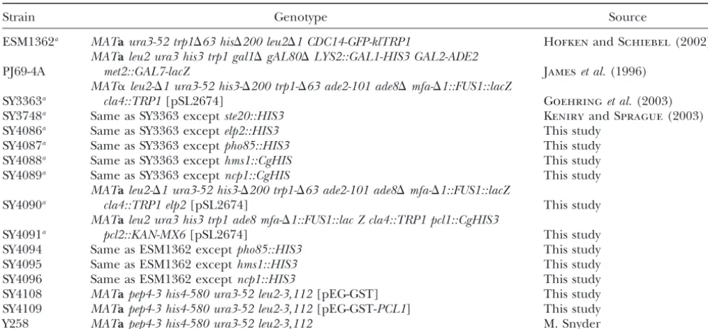
Strain Genotype Source
ESM1362a MATaura3-52 trp1⌬63 his⌬200 leu2⌬1 CDC14-GFP-klTRP1 HofkenandSchiebel(2002)
MATaleu2 ura3 his3 trp1 gal1⌬gAL80⌬LYS2::GAL1-HIS3 GAL2-ADE2
PJ69-4A met2::GAL7-lacZ Jameset al.(1996)
MAT␣leu2-⌬1 ura3-52 his3-⌬200 trp1-⌬63 ade2-101 ade8⌬mfa-⌬1::FUS1::lacZ
SY3363a cla4::TRP1[pSL2674] Goehringet al.(2003)
SY3748a Same as SY3363 exceptste20::HIS3 KeniryandSprague(2003)
SY4086a Same as SY3363 exceptelp2::HIS3 This study
SY4087a Same as SY3363 exceptpho85::HIS3 This study
SY4088a Same as SY3363 excepthms1::CgHIS This study
SY4089a Same as SY3363 exceptncp1::CgHIS This study
MATaleu2-⌬1 ura3-52 his3-⌬200 trp1-⌬63 ade2-101 ade8⌬mfa-⌬1::FUS1::lacZ
SY4090a cla4::TRP1 elp2[pSL2674] This study
MATaleu2 ura3 his3 trp1 ade8 mfa-⌬1::FUS1::lac Z cla4::TRP1 pcl1::CgHIS3
SY4091a pcl2::KAN-MX6[pSL2674] This study
SY4094 Same as ESM1362 exceptpho85::HIS3 This study
SY4095 Same as ESM1362 excepthms1::HIS3 This study
SY4096 Same as ESM1362 exceptncp1::HIS3 This study
SY4108 MATapep4-3 his4-580 ura3-52 leu2-3,112[pEG-GST] This study SY4109 MATapep4-3 his4-580 ura3-52 leu2-3,112[pEG-GST-PCL1] This study
Y258 MATapep4-3 his4-580 ura3-52 leu2-3,112 M. Snyder
aDerivatives of YPH499 and YPH500 (S288C;SikorskiandHieter1989).
4.8 mm3AT. A total of 1200 positives were identified in the incubated with glutathione-Sepharose (Amersham Pharmacia) for 1 hr with gentle agitation. The GST complexes were washed primary screen. These transformants were then tested for the
ability to grow on medium lacking adenine; 56 activated both several times with IP buffer and then incubated with 2g of bacterially purified fusion protein for 2 hr on ice. The GST reporter genes. Only 3 of these still activated the reporter
genes after plasmid rescue and retransformation into the PJ69- complexes were rewashed several times with IP buffer, and the final pellets were suspended in 30l of 2⫻Laemmli buffer 4A strain containing thePCL1bait. These 3 library plasmids
required the PCL1 bait to activate the reporter genes. Se- (Laemmli 1970), boiled for 5 min, centrifuged for 1 min, and then resolved on SDS-PAGE gels. Western analysis was quence analysis of the 3 positives revealed three unique
activa-tion domain fusions. Positive 1, pSL2793, had theGAL4activa- performed as described below.
Kinase assays:GST fusion protein expression was induced tion domain fused to theYJR072C/EPA1coding sequence at
position 529. Positive 284, pSL2794, had theGAL4activation in strains containingGAL-promoter-driven GST-Pcl1 or GST alone as described above. Cells were then harvested, sphero-domain fused to theNCP1coding sequence at position 517.
Positive 686, pSL2795, had theGAL4activation domain fused plasted (Bowerset al. 2000), and lysed in IP buffer for 15 min. Following centrifugation for 5 min at 13,000 rpm, lysates to theHMS1coding sequence at position 183.
Bacterial protein purification: Maltose-binding protein were incubated with glutathione-Sepharose for 1 hr with gen-tle agitation. GST complexes were washed once with IP buffer, (MBP) fusions, MBP-Epa1, MBP-Hms1, and MBP-Ncp1
(ex-pressed from pSL2814, pSL2815, and pSL2816, respectively), once with RIPA buffer (150 mmNaCl, 1% NP40, 0.5% DOC, 0.1% SDS, and 50 mmTris-HCl, pH 8.0), and twice with kinase were individually expressed inEscherichia coli(gold cells) and
purified over separate amylose columns (SmithandJohnson reaction buffer (50 mmTris-HCl, pH 7.5; 40 mmmagnesium chloride; 1 mmdithiothreitol; 0.5 mmsodium orthovanadate; 1988). MBP alone was obtained from New England Biolabs.
GST pull-down experiments:To confirm a physical interac- 5 g aprotinin/ml; and 5 g leupeptin/ml). Kinase assays were carried out in 30l of kinase buffer containing 1m tion between Pcl1 and Epa1, Hms1, or Ncp1, GST pull-down
experiments were performed. Plasmids expressingGAL-pro- MBP-Hms1, MBP-Ncp1, MBP-Epa1, or MBP alone and ATP (1000 Ci/mol) for 30 min at 30⬚. These reactions were termi-moter driven GST-Pcl1 or GST alone (pEG-GST-PCL1 and
pEG-GST, respectively, both generous gifts from M. Snyder), nated with 30l of 2⫻Laemmli buffer followed by 5 min of boiling. Terminated reactions were then subjected to electro-were transformed into yeast (ESM1362) to obtain strains
SY4108 and -4109, respectively. Cells were grown to midlog phoresis through an 8% polyacrylamide gel and transferred to nitrocellulose. Phosphorylated proteins were detected using phase in selective medium lacking uracil and containing 2%
raffinose as the carbon source. GST fusion protein expression a STORM 860 phosphodetector system (Amersham Biosci-ences, Piscataway, NJ) and quantified using Imagequant V1.11 was induced by growing cultures for 4 hr in 2% galactose.
Cells were then harvested, converted to spheroplasts (Bowers (Molecular Dynamics, Sunnyvale, CA). Blots were then sub-jected to Western analysis as described below (see Western et al.2000), and lysed in IP buffer (50 mmTris, pH 8.0; 1 mm
EDTA; 50 mmNaCl; 1% NP40; 5g of aprotinin/ml; 5g analysis), except blots were developed using ECL plus (Amer-sham, Arlington Heights, IL), detected using the STORM 860 of antipain/ml; 5 g of leupeptin/ml; 5 g of pepstatin
A/ml; and 1 mmphenylmethylsulfonyl fluoride) for 15 min. scanner, and quantified using Imagequant V1.11 software.
electrophoresis through an 8% polyacrylamide gel and trans-ferred to nitrocellulose. To detect GST, the blots were probed with a 1:200 dilution of polyclonal GST antibody (Molecular Probes, Eugene, OR) and then with a 1:3000 dilution of Bio-Rad (Richmond, CA) goat anti-rabbit IgG horseradish peroxi-dase conjugate. To detect MBP fusions, the blots were probed with a 1:10,000 dilution of polyclonal MBP antibody (New England Biolabs) and subsequently with a 1:3000 dilution of Bio-Rad goat anti-rabbit antibodies. Proteins were visualized using ECL plus.
ATPase assays:NADH-based indirect ATPase assays were performed as described previously (Tsunodaet al.2001).
Bac-terially purified proteins—maltose-binding protein alone, Figure1.—PHO85is a low-copy suppressor of theelp2cla4⌬
MBP-Ncp1, and MBP-Epa1—were individually added to 1 ml lethality. The ability ofPHO85-containing plasmids to restore of reaction mixture containing 25 mmTris, pH 7.5, 25 mm viability to anelp2cla4⌬strain is shown. The growth phenotype KCl, 2 mmMgCl2, 5 mmKCN, 2 mmphosphophenolpyruvate, of an elp2cla4⌬[CLA4-URA3] strain was cotransformed with 5 mmATP, 0.5 mmNADH, 30 units ofl-lactic acid dehydroge- one of the indicated low-copyLEU2-marked plasmids: empty nase, and 30 units of pyruvate kinase. These reactions pro- vector,ELP2-containing plasmid, and four different plasmids ceeded at 30⬚for 3 min. The absorbance at 340 nm was fol- containing genomic DNA that spans thePHO85 locus. The lowed spectrophotometrically during the 3-min incubation; strains were replica plated to either selective medium or selec-changes in absorbance reflect ATP hydrolysis. Each sample tive medium containing 0.1% 5-FOA. The ability to grow on was assayed in triplicate. 5-FOA indicates the ability to lose the CLA4-URA3 plasmid
Cdc14 localization:Cdc14 localization was performed essen- and thus to suppress theelp2cla4⌬lethality. Strains and plas-tially as previously described (Hofkenand Schiebel2002). mids used were as follows: elp2cla4⌬ [CLA4-URA3] strain, Briefly, yeast strains containingCDC14-GFP(ESM1362, a gen- SY4090;CLA4-URA3 plasmid, pSL2674; emptyLEU2-marked erous gift from E. Schiebel) and strains additionally deleted vector, pRS315; ELP2-containing LEU2-marked plasmid, forpho85⌬,hms1⌬, orncp1⌬(SY4111-4113) were synchronized pSL2825; andPHO85-containingLEU2-marked plasmids 1–4, with␣-factor, released from G1arrest, and grown at 10⬚. Cells pSL2820, pSL2821, pSL2822, and pSL2823.
were observed using a Zeiss Axioplan II microscope with No-marski optics or differential interference contrast optics with a fluorescence microscopy filter. The fraction of cells exhibiting
mids to suppress an elp2 allele suggested that PHO85
nucleolar Cdc14-GFP was quantitated. A total of 100 cells were
andCLA4genetically interact. Indeed, we found that,
observed for each sample and all samples were analyzed in
triplicate. like elp2⌬cla4⌬ mutants, the pho85⌬cla4⌬ double
mu-tants were inviable (Figure 2). In addition, we found that simultaneous loss of the Pho85 cyclin molecules
RESULTS Pcl1 and Pcl2 was lethal in the absence of Cla4, consis-tent with previous observations (Figure 2;Lenburgand
PHO85is required for viability in the absence ofCLA4:
O’Shea2001;Huanget al.2002). The contribution of Cla4 during cell cycle progression
Pcl1 interacts with Ncp1, Hms1, and Yjr072C/Epa1:
remains poorly understood. To gain insight into this
Cla4 is required for normal bud formation and for mi-cellular role, mutations were identified that, like the
totic exit (Cvrckovaet al.1995;HofkenandSchiebel
loss of STE20, were lethal in the absence of CLA4
2002). Given that Pho85 or Pcl1 and Pcl2 together are (MitchellandSprague2001). Several alleles ofelp2
required for viability in the absence of Cla4, we consid-(a transcription elongation factor) were identified
ered that the targets of Pcl1•Pho85 might be involved (MitchellandSprague2001). In the process of
clon-ing ELP2, four low-copy PHO85-containing plasmids that bypassed the elp2cla4⌬ lethal phenotype were ob-tained (Figure 1). The basis of the ELP2 and CLA4
synthetic interaction is under investigation and has been described elsewhere; here we focus onPHO85 ( Goeh-ringet al.2003).PHO85encodes a nonessential cyclin-dependent kinase involved in many cellular tasks includ-ing phosphate metabolism and cell cycle progression
Figure2.—PHO85orPCL1andPCL2together are required
(Toh-e et al. 1988; Huang et al. 1996, 1998, 2002;
for viability in the absence ofCLA4.PHO85,ELP2, or PCL1 O’Neill et al. 1996;Timblin et al. 1996; Tennysonet andPCL2together were deleted in acla4⌬strain containing al. 1998; Carroll et al. 2001; Lenburg and O’Shea a CLA4-URA3 plasmid. These strains were replica plated to
2001;McBrideet al.2001;Wanget al.2001a,b;Carroll either rich medium or selective medium lacking uracil and containing 0.1% 5-FOA. The inability of particular strains to
and O’Shea 2002). This kinase derives specificity by
grow on 5-FOA demonstrates the requirement of the
CLA4-coupling with cyclin molecules; its role in G1progression
URA3plasmid for viability. Strains and plasmids were as
fol-is mediated by its association with the G1 cyclins, Pcl1 lows:cla4⌬[CLA4-URA3], SY3363;cla4⌬elp2⌬[CLA4-URA3],
and Pcl2 (Huanget al.1998;Wilsonet al.1999;Wang SY4086;cla4⌬pho85⌬[CLA4-URA3], SY4087;cla4⌬pcl1⌬pcl2⌬ [CLA4-URA3], SY4091; andCLA4-URA3plasmid, pSL2674.
plas-in one or more Cla4-mediated processes. As one means to identify targets of Pcl1•Pho85 complexes, we per-formed a two-hybrid screen using Pcl1 as the bait. We screened 6 million library plasmids using the Phil James two-hybrid system (Jameset al.1996) and found three proteins that interact with Pcl1: Ncp1, Hms1, and Yjr072c (Figure 3). Ncp1 is an NADP-cytochrome P450 reductase involved in ergosterol biosynthesis (O’Neill
et al. 1996). Hms1 is an Myc-like transcription factor involved in filamentous growth (Jaspersenet al.1998). Yjr072C is an essential, novel, presumptive ATPase of previously unknown function, which we have termed Epa1 (see below).
As one means to validate the physical interaction of Pcl1 with Epa1, Hms1, and Ncp1, we performed GST pull-down experiments using GST-Pcl1 and GST alone. GST-Pcl1 or GST alone was purified and subsequently incubated with 1mof bacterially expressed and puri-fied maltose-binding protein fusions to Epa1, Hms1, or Ncp1. We found that samples containing GST-Pcl1 pulled down MBP-Epa1, MBP-Hms1, and MBP-Ncp1, but failed to pull down MBP alone (Figure 4). The GST alone samples failed to pull down any of the fusion proteins (Figure 4). Therefore, GST-Pcl1 specifically in-teracts with maltose-binding fusions of Epa1, Hms1, and Ncp1.
Figure3.—Pcl1 interacts with Epa1, Ncp1, and Hms1 by
Pcl1•Pho85 complexes phosphorylate bacterially
puri-yeast two-hybrid. The ability of Pcl1 to interact with Epa1,
fied Epa1, Hms1, and Ncp1:The ability of Epa1, Hms1,
Ncp1, and Hms1 by yeast two-hybrid is shown. Strains of the
and Ncp1 to physically interact with Pcl1 suggests that PJ69-4A background containing a plasmid encoding the Gal4 these may be targets of Pcl1•Pho85 kinase. We tested the DNA-binding domain fused to Pcl1 (Pcl1 bait) and a library plasmid (encoding the Gal4 activation domain fusion alone
ability of GST-Pcl1 purified from yeast to phosphorylate
or fused to one of the following proteins: Epa1, Ncp1, or
the following bacterially expressed and purified substrates:
Hms1) were replica plated to medium lacking both uracil and
MBP-Epa1, MBP-Hms1, MBP-Ncp1, and MBP
(maltose-leucine. To assay for two-hybrid interaction, cells were also
binding protein alone). We found that Pcl1•Pho85 com- replica plated to medium additionally lacking histidine supple-plexes phosphorylated MBP-Epa1, MBP-Hms1, and MBP- mented with 4.8 mm3AT and to medium additionally lacking adenine. The ability to grow on medium lacking histidine
Ncp1 but did not phosphorylate MBP alone (Figure 5).
supplemented with 4.8 mm3AT indicates the ability to activate
GST alone (lacking the Pcl1 moiety) failed to
phosphor-theGAL1-HIS3reporter gene. The ability to grow on medium
ylate any of the substrates (Figure 5). Therefore, lacking adenine indicates the ability to activate the GAL2-Pcl1•Pho85 complexes specifically phosphorylate MBP- ADE2reporter gene. To ensure that the observed interactions Epa1, MBP-Hms1, and MBP-Ncp1. were bait dependent, PJ69-4A strains containing only the AD-encoding library plasmids were replica plated to the following: Epa1 is an ATPase with homology to the bacterial
medium lacking leucine, medium lacking leucine and
histi-minD septation regulator: The observation that Epa1
dine and supplemented with 3AT, and medium additionally
interacts with Pcl1 and is phosphorylated by Pcl1•Pho85 lacking both leucine and adenine. The inability of strains complexes suggests that Epa1 may have a role during lacking the Pcl1 bait to grow without supplemental histidine morphogenesis. Epa1 has no previously described bio- or adenine indicates that the library plasmids are unable to activate the reporter genes on their own. Two-hybrid fusion
logical role or molecular function. SWISS-PROT and
proteins were expressed from the following plasmids: Pcl1
GenBank (Junkeret al.1999;Molleret al.1999;
O’Do-bait, pSL2796; Gal4 activation domain (AD) alone, pGAD-novanet al.2002) search algorithms revealed that Epa1
C1; AD-Epa1, pSL2793; AD-Ncp1, pSL2794; and AD-Hms1,
is well conserved in all eukaryotic organisms examined, pSL2795. but no function has been ascribed to these homologs
(Figure 6).
Interestingly, sequence analysis using SWISS-PROT
also revealed sequence similarity between Epa1 and cifically shown), suggests that Epa1 might be an ATPase. To test this, we performed NADH-based indirect ATPase minD (Figure 6), a bacterial protein that regulates the
placement of the bacterial septal ring. The sequence assays. Epa1 showed significant ATPase activity above the maltose-binding protein control (Figure 7), whereas bacte-homology between Epa1 and minD, especially within
Figure4.—Pcl1 physically associates with Epa1, Hms1, and Ncp1 in GST pull-downs. The ability of Pcl1 to associate with
Epa1, Hms1, and Ncp1 in GST pull-down experiments is shown. Figure5.—Pcl1•Pho85 complexes specifically phosphory-GST pull-down experiments were performed using phosphory-GST-Pcl1 late Epa1, Hms1, and Ncp1. The ability of Pcl1•Pho85 com-or GST alone purified from yeast and substrates MBP-Epa1, plexes to phosphorylate Epa1, Hms1, and Ncp1 is shown. MBP-Hms1, and MBP-Ncp1 purified from bacteria as de- Kinase assays were performed using either GST alone or GST-scribed inmaterials and methods. The presence of a given Pcl1 purified from yeast lysates and bacterially expressed and substrate in the pull-down was detected using anti-MBP anti- purified MBP-Epa1, MBP-Hms1, and MBP-Ncp1 as described serum on a Western blot. An anti-GST antibody was used to inmaterials and methods. Incorporation of32P into specific detect either GST alone or GST-Pcl1. Fusion proteins were substrates was detected as described inmaterials and meth-expressed from the following plasmids: GST alone, pEG-GST; ods. Total levels of each substrate were detected using anti-GST-Pcl1, pEG-GST-PCL1; MBP-Epa1, pSL2814; MBP-Hms1, MBP antibodies against a Western blot. Fusion proteins were pSL2815; and MBP-Ncp1, pSL2816. expressed from the following plasmids: GST alone, pEG-GST; GST-Pcl1, pEG-GST-PCL1; MBP-Epa1, pSL2814; MBP-Hms1, pSL2815; and MBP-Ncp1, pSL2816.
to Epa1) had no ATPase activity above the maltose-binding protein background. The ATPase activity observed for
Epa1 is therefore unlikely to be due to contamination Ncp1, Hms1, and Epa1 contribute to a shared
essen-tial role with Cla4:To test whether the Pcl1-interacting
by bacterial proteins and we conclude that Epa1 is an
ATPase. proteins partake in the shared essential role with Cla4,
Figure6.—Epa1 shares homology with the
bacterial minD septation regulator and is con-served in eukaryotes. Sequence alignments of Epa1 with (A) the bacterial minD septation regulator, (B) theCaenorhabditis elegans GOP-2 protein of unknown function, and (C) the hu-man (AJ010842) protein of unknown function are shown. National Center for Biotechnology Information blast and SWISS-PROT database alignment algorithms were utilized to prepare these alignments (Junkeret al.1999;Moller
Figure 7.—Epa1 has ATPase activity. Purified MBP-Epa1, MBP-Ncp1, and MBP alone were tested for ATPase activity in NADH-based indirect ATPase assays as described inmaterials and methods. Fusion proteins were expressed from the fol-lowing plasmids: MBP-Epa1, pSL2814; MBP-Ncp1, pSL2816.
we took two experimental approaches. First, we deleted
HMS1orNCP1in a strain genomically deleted forCLA4, Figure8.—NCP1, HMS1, andEPA1show genetic interac-but containing aCLA4-URA3plasmid.EPA1could not tions with CLA4. Genetic interactions between CLA4 and
HMS1, NCP1, and EPA1 are depicted. (A) The HMS1 and
be deleted in this background because it is essential.
NCP1genes were deleted in acla4⌬strain containing a
CLA4-We then asked whether the double-mutant strains could
URA3plasmid. These strains were replica plated to either rich
survive in the absence of theCLA4-URA3 plasmid. We
medium or medium containing 0.1% 5-FOA. The inability of
observed that bothncp1⌬cla4⌬andhms1⌬cla4⌬double- strains to grow on 5-FOA demonstrates the requirement of mutant strains were inviable as revealed by their inability theCLA4for viability and thus the lethality of thehms1⌬cla4⌬
andncp1⌬cla4⌬strains. (B) High-copyLEU2-markedEPA1was
to grow on medium containing 5-fluoroorotic acid
transformed intocla4⌬strains deleted for a second gene, as
(5-FOA; Figure 8). Both double-mutant strains had
mis-indicated; these strains carried a copy ofCLA4on aURA3
-localized septin rings (data not shown). This is the same
marked plasmid. The ability to grow on medium containing
phenotype observed in a ste20cla4mutant (Cvrckova 0.1% 5-FOA indicates the ability to lose theCLA4-URA3
plas-et al.1995). Therefore, it appears that Ncp1 and Hms1 mid and therefore suppress the synthetic lethal phenotype.
Strains and plasmids were as follows: high-copyLEU2-marked
share an essential role with Cla4.
EPA1, pSL2805;CLA4-URA3plasmid, pSL2674;cla4⌬[
CLA4-As one means to determine whether EPA1 shares a
URA3], SY3363; cla4⌬pcl1⌬pcl2⌬ [CLA4-URA3], SY4091;
morphogenetic role withCLA4, a high-copy construct
cla4⌬ncp1⌬[CLA4-URA3], SY4089;cla4⌬hms1⌬[CLA4-URA3],
containingEPA1 was tested for the ability to suppress SY4088; cla4⌬elp2⌬ [CLA4-URA3], SY4086; cla4⌬pho85⌬ severalCLA4synthetic lethal phenotypes. In fact,EPA1 [CLA4-URA3], SY4087; and cla4⌬ste20⌬ [CLA4-URA3],
SY3748.
is able to bypass the lethality of an elp2⌬cla4⌬ strain, which supports the idea thatEPA1 shares some func-tion(s) withCLA4(Figure 8).
HMS1, andNCP1to mitotic exit, we deleted these genes
Pho85 and two Pcl1-interacting targets may have roles
in a strain containing green fluorescent protein
(GFP)-during mitotic exit:Cla4 has functions during both G1
taggedCDC14. Previous studies have reported a marked progression and mitotic exit (Cvrckova et al. 1995;
decrease in Cdc14 nucleolar localization just prior to
Bentonet al.1997;HollyandBlumer1999;Gulliet
mitotic exit, which suggests that Cdc14 is the key effector
al.2000). Since Pho85 and its Pcl1-interacting targets,
of the mitotic exit transition in yeast (Jaspersen et al.
Epa1, Hms1, and Ncp1, all genetically interact with Cla4,
1998;Visintinet al.1998, 1999;JaspersenandMorgan
we considered that they might function during either
2000; Lee et al. 2001). Additionally, a diminishment of these processes. Physical interaction data has
sug-in Cdc14 nucleolar localization sug-indicates progression gested a role for Epa1 and Hms1 during mitotic exit.
through mitotic exit (HofkenandSchiebel2002). We Specifically, Ho et al. identified a physical interaction
found that Pho85, Hms1, and Ncp1 are individually between Epa1 and Dbf2, a kinase that is a crucial
regula-required for the proper relocalization of Cdc14. Spe-tor in the mitotic exit network (Toyn andJohnston
cifically, Cdc14 remains localized to the nucleolus nearly 1994;Hoet al.2002). The same screen also detected an
100% of the time in cells lacking pho85,hms1, or ncp1
interaction between Hms1 and the Cdc14 phosphatase,
(Figure 9). This is in sharp contrast to otherwise wild-another crucial member of the mitotic exit network (Ho
type cells and strongly suggests a role for these three
et al.2002).
Figure9.—Pho85, Hms1, and Ncp1 affect Cdc14 localization. Cells containing CDC14-GFPand deleted forPHO85, HMS1, orNCP1
were synchronized with␣-factor, released from G1arrest, and grown at 10⬚. The fraction of cells exhibiting nucleolar Cdc14-GFP was ob-served (n⬎100). Strains were as follows: wild type, ESM1362; pho85⌬, SY4111; hms1⌬,
SY4112; andncp1⌬, SY4113.
for viability, we could not perform the same analysis in phogenesis may hint at the precise morphogenetic func-tion that Cla4 and Pcl1•Pho85 impinge upon.
anepa1mutant. However, sequence analysis presented
here has identified a similarity between Epa1 and a Several lines of evidence suggest that Cla4 and Pho85 signaling may intersect during mitotic exit.Hofkenand bacterial septation regulator, raising the interesting
pos-sibility that this protein may be required for septation Schiebel (2002) have demonstrated that Cla4 is re-quired for the proper localization of the Cdc14 phos-in yeast (Figure 6).
phatase, a key effector of mitotic exit. Here we show that Pho85, Hms1, and Ncp1 are individually required
DISCUSSION
for the proper localization of Cdc14, implicating these proteins in mitotic exit. Consistent with this, Hms1 phys-The ability of p21-activated kinases to contribute to
ically associates with Cdc14 (Ho et al.2002). Interest-morphogenesis has been well documented (Daniels
ingly, the third Pcl1•Pho85 target identified by our work, and Bokoch 1999; Connolly et al. 2002; Puto et al.
Epa1, has previously been shown to physically associate 2003; Schneeberger and Raabe 2003). In budding
with another critical component of the septation initia-yeast, Cla4 and Ste20 may have a common role during
tion network in budding yeast, the Dbf2 kinase (Hoet
cellular morphogenesis. This interpretation is derived
al.2002). In light of these data, the observed homology from the observation that the single mutants are viable,
between Epa1 and the bacterial septation regulator, but the double mutant is inviable and exhibits defects
MinD, is particularly intriguing. One possibility is that in G1 function, in septin function, in cytokinesis, and
Epa1 is involved in septation in yeast. In addition to in bud morphology (Cvrckovaet al. 1995;Bentonet
the homology with MinD, SWISS-PROT and GenBank
al.1997;HollyandBlumer1999;Gulliet al.2000).
search algorithms revealed that Epa1 is well conserved in In addition, Cla4 and Ste20 have both been shown to
all eukaryotic organisms examined (Figure 6). Further individually contribute to mitotic exit (Hofken and
investigation is needed to determine whether these
euk-Schiebel2002;Chiroliet al.2003).
aryotic proteins, including Epa1, play a role in septation To gain insight into Cla4’s roles, we set out to identify
or cytokinesis. mutations that confer lethality in the absence ofCLA4.
The ability of components expressed during the G1
We found thatPHO85is such a gene. Loss of two genes
phase of the cell cycle to regulate late mitotic events encoding Pho85 cyclin partners, PCL1 and PCL2, was
was initially suggested byCvrckovaet al.(1995). They also lethal with the simultaneous loss ofCLA4. Because
found that the loss of Cla4 is lethal with the loss of two Pho85 is a cyclin-dependent kinase, we sought to
iden-G1cyclins, Cln1 and Cln2, and that Cla4 had a mitotic
tify potential targets with the view that such targets
function. Cvrckova et al. (1995) suggested that the would speak to Cla4 function. This effort identified
Cln1 and Cln2 G1 function may affect a later mitotic
three proteins: Ncp1, Hms1, and Epa1. We have found
function that acts in parallel with Cla4. Here we present that all three of these molecules physically interact with
a possible direct link between G1 components and the
Pcl1 and are specifically phosphorylated by Pcl1•Pho85
late mitotic machinery. The G1 Pcl1•Pho85 complex
complexes. Furthermore, we show that Ncp1, Hms1,
phosphorylates Epa1, Hms1, and Ncp1, each of which and Epa1 genetically interact with Cla4. Ncp1 and Hms1
genetically interacts with Cla4. Moreover, each of these are required for viability in the absence of Cla4 whereas
proteins has been suggested by some combination of high-copyEPA1bypasses the lethality of anelp2⌬cla4⌬
Daniels, R. H., and G. M. Bokoch, 1999 p21-Activated protein
role in mitotic exit. We therefore propose that
kinase: A crucial component of morphological signaling? Trends
Pcl1•Pho85 may regulate mitotic exit machinery, and Biochem. Sci.24:350–355.
Gietz, D., A. St. Jean, R. A. WoodsandR. H. Schiestl, 1992
Im-this role may be essential in the absence of Cla4. One
proved method for high efficiency transformation of intact yeast
explanation for the genetic interactions between Cla4
cells. Nucleic Acids Res.20:1425.
and the G1 machinery could be that Cla4 instills the Gladfelter, A. S., I. Bose, T. R. Zyla, E. S. BardesandD. J. Lew, 2002 Septin ring assembly involves cycles of GTP loading and
competence to respond to a G1signal that regulates late
hydrolysis by Cdc42p. J. Cell Biol.156:315–326.
mitotic events.
Goehring, A. S., D. A. Mitchell, A. Tong, M. E. Keniry, C. Boone
et al., 2003 Synthetic lethal analysis implicates Ste20p, a p21-We thank David Mitchell, April Goehring, Phil Kinsey, Scott Givan,
activated protein kinase, in polarisome activation. Mol. Biol. Cell Lucia Liverio, Greg Smith, Paul Cullen, Monique Dail, Karen Sprague,
14:1501–1516. Tom Stevens, Bruce Bowerman, Judith Eisen, and Kathy Chicas-Cruz
Gulli, M. P., M. Jaquenoud, Y. Shimada, G. Niederhauser, P. Wiget
for providing advice, strains, and/or plasmids. This work was
sup-et al., 2000 Phosphorylation of the Cdc42 exchange factor ported by research (GM-30027) and training (GM-07413) grants from
Cdc24 by the PAK-like kinase Cla4 may regulate polarized growth the National Institutes of Health. in yeast. Mol. Cell6:1155–1167.
Ho, Y., A. Gruhler, A. Heilbut, G. D. Bader, L. Mooreet al., 2002 Systematic identification of protein complexes inSaccharomyces cerevisiaeby mass spectrometry. Nature415:180–183.
LITERATURE CITED
Hofken, T., andE. Schiebel, 2002 A role for cell polarity proteins in mitotic exit. EMBO J.21:4851–4862.
Adams, A. E., D. I. Johnson, R. M. Longnecker, B. F. Sloatand
J. R. Pringle, 1990 CDC42andCDC43, two additional genes Holly, S. P., andK. J. Blumer, 1999 PAK-family kinases regulate cell and actin polarization throughout the cell cycle ofSaccharomyces
involved in budding and the establishment of cell polarity in the
yeastSaccharomyces cerevisiae.J. Cell Biol.111:131–142. cerevisiae.J. Cell Biol.147:845–856.
Huang, D., I. FarkasandP. J. Roach, 1996 Pho85p, a
cyclin-depen-Bagrodia, S., S. J. Taylor, C. L. Creasy, J. ChernoffandR. A.
Cerione, 1995 Identification of a mouse p21Cdc42/Rac acti- dent protein kinase, and the Snf1p protein kinase act antagonisti-cally to control glycogen accumulation inSaccharomyces cerevisiae.
vated kinase. J. Biol. Chem.270:22731–22737.
Bairoch, A., andR. Apweiler, 2000 The SWISS-PROT protein se- Mol. Cell. Biol.16:4357–4365.
Huang, D., J. Moffat, W. A. Wilson, L. Moore, C. Chenget al., quence database and its supplement TrEMBL in 2000. Nucleic
Acids Res.28:45–48. 1998 Cyclin partners determine Pho85 protein kinase substrate specificity in vitro and in vivo: control of glycogen biosynthesis
Bender, A., andJ. R. Pringle, 1989 Multicopy suppression of the
cdc24budding defect in yeast byCDC42and three newly identified by Pcl8 and Pcl10. Mol. Cell. Biol.18:3289–3299.
Huang, D., J. MoffatandB. Andrews, 2002 Dissection of a com-genes including the ras-related gene RSR1. Proc. Natl. Acad. Sci.
USA86:9976–9980. plex phenotype by functional genomics reveals roles for the yeast cyclin-dependent protein kinase Pho85 in stress adaptation and
Benton, B. K., A. Tinkelenberg, I. GonzalezandF. R. Cross, 1997
Cla4p, aSaccharomyces cerevisiaeCdc42p-activated kinase involved cell integrity. Mol. Cell. Biol.22:5076–5088.
James, P., J. HalladayandE. A. Craig, 1996 Genomic libraries in cytokinesis, is activated at mitosis. Mol. Cell Biol17:5067–5076.
Bi, E., J. B. Chiavetta, H. Chen, G. C. Chen, C. S. Chanet al., and a host strain designed for highly efficient two-hybrid selection in yeast. Genetics144:1425–1436.
2000 Identification of novel, evolutionarily conserved
Cdc42p-interacting proteins and of redundant pathways linking Cdc24p Jaspersen, S. L., andD. O. Morgan, 2000 Cdc14 activates cdc15 to promote mitotic exit in budding yeast. Curr. Biol.10:615–618. and Cdc42p to actin polarization in yeast. Mol. Biol. Cell 11:
773–793. Jaspersen, S. L., J. F. Charles, R. L. Tinker-Kulbergand D. O. Morgan, 1998 A late mitotic regulatory network controlling
Bose, I., J. E. Irazoqui, J. J. Moskow, E. S. Bardes, T. R. Zylaet
al., 2001 Assembly of scaffold-mediated complexes containing cyclin destruction in Saccharomyces cerevisiae.Mol. Biol. Cell 9:
2803–2817. Cdc42p, the exchange factor Cdc24p, and the effector Cla4p
required for cell cycle-regulated phosphorylation of Cdc24p. J. Johnson, D. I., 1999 Cdc42: an essential Rho-type GTPase control-ling eukaryotic cell polarity. Microbiol. Mol. Biol. Rev.63:54–105. Biol. Chem.276:7176–7186.
Bowers, K., B. P. Levi, F. I. PatelandT. H. Stevens, 2000 The Johnson, D. I., andJ. R. Pringle, 1990 Molecular characterization ofCDC42, aSaccharomyces cerevisiaegene involved in the develop-sodium/proton exchanger Nhx1p is required for endosomal
pro-tein trafficking in the yeastSaccharomyces cerevisiae.Mol. Biol. Cell ment of cell polarity. J. Cell Biol.111:143–152.
Junker, V. L., R. ApweilerandA. Bairoch, 1999 Representation
11:4277–4294.
Burke, D., D. DawsonandT. Stearns, 2000 Methods in Yeast Genetics. of functional information in the SWISS-PROT data bank. Bioin-formatics15:1066–1067.
Cold Spring Harbor Laboratory Press, Plainview, NY.
Carroll, A. S., andE. K. O’Shea, 2002 Pho85 and signaling environ- Keniry, M. E., andG. F. Sprague, Jr., 2003 Identification of p21-activated kinase specificity determinants in budding yeast: a single mental conditions. Trends Biochem. Sci.27:87–93.
Carroll, A. S., A. C. Bishop, J. L. DeRisi, K. M. ShokatandE. K. amino acid substitution imparts Ste20 specificity to Cla4. Mol. Cell. Biol.23:1569–1580.
O’Shea, 2001 Chemical inhibition of the Pho85
cyclin-depen-dent kinase reveals a role in the environmental stress response. Kozminski, K. G., A. J. Chen, A. A. RodalandD. G. Drubin, 2000 Functions and functional domains of the GTPase Cdc42p. Mol. Proc. Natl. Acad. Sci. USA98:12578–12583.
Chen, G. C., Y. J. KimandC. S. Chan, 1997 The Cdc42 GTPase- Biol. Cell11:339–354.
Laemmli, U. K., 1970 Cleavage of structural proteins during the associated proteins Gic1 and Gic2 are required for polarized cell
growth inSaccharomyces cerevisiae.Genes Dev.11:2958–2971. assembly of the head of bacteriophage T4. Nature227:680–685.
Leberer, E., D. Dignard, D. Harcus, D. Y. Thomas and M. Chiroli, E., R. Fraschini, A. Beretta, M. Tonelli, G. Lucchiniet
al., 2003 Budding yeast PAK kinases regulate mitotic exit by Whiteway, 1992 The protein kinase homologue Ste20p is re-quired to link the yeast pheromone response G-protein beta two different mechanisms. J. Cell Biol.160:857–874.
Connolly, J. O., N. Simpson, L. HewlettandA. Hall, 2002 Rac gamma subunits to downstream signalling components. EMBO J.11:4815–4824.
regulates endothelial morphogenesis and capillary assembly. Mol.
Biol. Cell13:2474–2485. Lee, S. E., L. M. Frenz, N. J. Wells, A. L. JohnsonandL. H. Johnston, 2001 Order of function of the budding-yeast mitotic
exit-net-Creasy, C. L., andJ. Chernoff, 1995 Cloning and characterization
of a human protein kinase with homology to Ste20. J. Biol. Chem. work proteins Tem1, Cdc15, Mob1, Dbf2, and Cdc5. Curr. Biol.
11:784–788.
270:21695–21700.
Cvrckova, F., C. De Virgilio, E. Manser, J. R. Pringleand K. Lenburg, M. E., andE. K. O’Shea, 2001 Genetic evidence for a morphogenetic function of the Saccharomyces cerevisiae Pho85
Nasmyth, 1995 Ste20-like protein kinases are required for
nor-mal localization of cell growth and for cytokinesis in budding cyclin-dependent kinase. Genetics157:39–51.
Longtine, M. S., C. L. Theesfeld, J. N. McMillan, E. Weaver, J. R.
Pringleet al., 2000 Septin-dependent assembly of a cell cycle- mental programs in the same cell type: mating and invasive growth. Genes Dev.8:2974–2985.
regulatory module inSaccharomyces cerevisiae.Mol. Cell. Biol.20:
4049–4061. Roberts, R. L., H. U. MoschandG. R. Fink, 1997 14-3-3 proteins are essential for RAS/MAPK cascade signaling during pseudohyphal
Lorenz, M. C., andJ. Heitman, 1998 Regulators of pseudohyphal
differentiation in Saccharomyces cerevisiae identified through development in S. cerevisiae. Cell89:1055–1065.
Sambrook, J., E. F.Fritschand T.Maniatis, 1989 Molecular
Clon-multicopy suppressor analysis in ammonium permease mutant
strains. Genetics150:1443–1457. ing: A Laboratory Manual, Ed. 2. Cold Spring Harbor Laboratory Press, Plainview, NY.
Ma, H., S. Kunes, P. J. SchatzandD. Botstein, 1987 Plasmid
construc-tion by homologous recombinaconstruc-tion in yeast. Gene58:201–216. Schneeberger, D., andT. Raabe, 2003 Mbt, a Drosophila PAK protein, combines with Cdc42 to regulate photoreceptor cell
Manser, E., T. Leung, H. Salihuddin, Z. S. ZhaoandL. Lim, 1994
A brain serine/threonine protein kinase activated by Cdc42 and morphogenesis. Development130:427–437.
Sikorski, R. S., andP. Hieter, 1989 A system of shuttle vectors and Rac1. Nature367:40–46.
Martin, G. A., G. Bollag, F. McCormickandA. Abo, 1995 A novel yeast host strains designed for efficient manipulation of DNA in
Saccharomyces cerevisiae.Genetics122:19–27. serine kinase activated by rac1/CDC42Hs-dependent
autophos-phorylation is related to PAK65 and STE20. EMBO J.14:1970– Smith, D. B., andK. S. Johnson, 1988 Single-step purification of polypeptides expressed inEscherichia colias fusions with glutathi-1978.
Martin, H., A. Mendoza, J. M. Rodriguez-Pachon, M. Molinaand one S-transferase. Gene67:31–40.
Smith, G. R., S. A. Givan, P. CullenandG. F. Sprague, Jr., 2002
C. Nombela, 1997 Characterization ofSKM1, aSaccharomyces
cerevisiaegene encoding a novel Ste20/PAK-like protein kinase. GTPase-activating proteins for Cdc42. Eukaryot. Cell1:469–480.
Tennyson, C. N., J. LeeandB. J. Andrews, 1998 A role for the Pcl9-Mol. Microbiol.23:431–444.
McBride, H. J., A. Sil, V. Measday, Y. Yu, J. Moffatet al., 2001 The Pho85 cyclin-cdk complex at the M/G1 boundary inSaccharomyces cerevisiae.Mol. Microbiol.28:69–79.
protein kinase Pho85 is required for asymmetric accumulation
of the Ash1 protein inSaccharomyces cerevisiae.Mol. Microbiol.42: Timblin, B. K., K. TatchellandL. W. Bergman, 1996 Deletion of the gene encoding the cyclin-dependent protein kinase Pho85 345–353.
Mitchell, D. A., andG. F. Sprague, 2001 The phosphotyrosyl phos- alters glycogen metabolism inSaccharomyces cerevisiae.Genetics
143:57–66. phatase activator, Ncs1p (Rrd1p), functions with Cla4p to
regu-Toh-e, A., K.Tanaka, Y.Uesonoand R. B.Wickner, 1988 PHO85, late the G(2)/M transition inSaccharomyces cerevisiae.Mol. Cell.
a negative regulator of the PHO system, is a homolog of the Biol.21:488–500.
protein kinase gene,CDC28, ofSaccharomyces cerevisiae. Mol. Gen.
Moller, S., U. Leser, W. Fleischmann and R. Apweiler, 1999
Genet.214:162–164. EDITtoTrEMBL: a distributed approach to high-quality
auto-Toyn, J. H., andL. H. Johnston, 1994 The Dbf2 and Dbf20 protein mated protein sequence annotation. Bioinformatics15:219–227.
kinases of budding yeast are activated after the metaphase to
Mosch, H. U., R. L. RobertsandG. R. Fink, 1996 Ras2 signals via
anaphase cell cycle transition. EMBO J.13:1103–1113. the Cdc42/Ste20/mitogen-activated protein kinase module to
Tsunoda, S. P., A. J. Rodgers, R. Aggeler, M. C. Wilce, M. Yoshida
induce filamentous growth inSaccharomyces cerevisiae.Proc. Natl.
et al., 2001 Large conformational changes of the epsilon subunit Acad. Sci. USA93:5352–5356.
in the bacterial F1F0 ATP synthase provide a ratchet action to
O’Donovan, C., M. J. Martin, A. Gattiker, E. Gasteiger, A.
Bair-regulate this rotary motor enzyme. Proc. Natl. Acad. Sci. USA
och et al., 2002 High-quality protein knowledge resource:
98:6560–6564. SWISS-PROT and TrEMBL. Brief Bioinform.3:275–284.
Venkateswarlu, K., D. E. Kelly, N. J. ManningandS. L. Kelly,
O’Neill, E. M., A. Kaffman, E. R. JollyandE. K. O’Shea, 1996
1998 NADPH cytochrome P-450 oxidoreductase and suscepti-Regulation of PHO4 nuclear localization by the PHO80–PHO85
bility to ketoconazole. Antimicrob. Agents Chemother.42:1756– cyclin-CDK complex. Science271:209–212.
1761.
O’Rourke, S. M., andI. Herskowitz, 1998 The Hog1 MAPK
pre-Visintin, R., K. Craig, E. S. Hwang, S. Prinz, M. Tyerset al., 1998 vents cross talk between the HOG and pheromone response
The phosphatase Cdc14 triggers mitotic exit by reversal of Cdk-MAPK pathways inSaccharomyces cerevisiae.Genes Dev.12:2874–
dependent phosphorylation. Mol. Cell2:709–718. 2886.
Visintin, R., E. S. HwangandA. Amon, 1999 Cfi1 prevents
prema-Puto, L. A., K. Pestonjamasp, C. C. KingandG. M. Bokoch, 2003
ture exit from mitosis by anchoring Cdc14 phosphatase in the p21-Activated kinase 1 (PAK1) interacts with the Grb2 adapter
nucleolus. Nature398:818–823. protein to couple to growth factor signaling. J. Biol. Chem.278:
Wang, Z., W. A. Wilson, M. A. FujinoandP. J. Roach, 2001a Anta-9388–9393. gonistic controls of autophagy and glycogen accumulation by
Raitt, D. C., F. PosasandH. Saito, 2000 Yeast Cdc42 GTPase and Snf1p, the yeast homolog of AMP-activated protein kinase, and Ste20 PAK-like kinase regulate Sho1-dependent activation of the the cyclin-dependent kinase Pho85p. Mol. Cell. Biol.21:5742– Hog1 MAPK pathway. EMBO J.19:4623–4631. 5752.
Richman, T. J., and D. I. Johnson, 2000 Saccharomyces cerevisiae Wang, Z., W. A. Wilson, M. A. FujinoandP. J. Roach, 2001b The
Cdc42p GTPase is involved in preventing the recurrence of bud yeast cyclins Pc16p and Pc17p are involved in the control of emergence during the cell cycle. Mol. Cell. Biol.20:8548–8559. glycogen storage by the cyclin-dependent protein kinase Pho85p.
Richman, T. J., M. M. SawyerandD. I. Johnson, 1999 The Cdc42p FEBS Lett.506:277–280.
GTPase is involved in a G2/M morphogenetic checkpoint regulat- Wilson, W. A., A. M. MahrenholzandP. J. Roach, 1999 Substrate ing the apical-isotropic switch and nuclear division in yeast. J. targeting of the yeast cyclin-dependent kinase Pho85p by the Biol. Chem.274:16861–16870. cyclin Pcl10p. Mol. Cell. Biol.19:7020–7030.
Roberts, R. L., and G. R. Fink, 1994 Elements of a single MAP