DOI: 10.1534/genetics.108.097303
The Classical Nuclear Localization Signal Receptor, Importin-
a
, Is
Required for Efficient Transition Through the G
1/S Stage of
the Cell Cycle in
Saccharomyces cerevisiae
Kanika F. Pulliam, Milo B. Fasken, Laura M. McLane, John V. Pulliam and Anita H. Corbett
1Department of Biochemistry, Emory University School of Medicine, Atlanta, Georgia 30322 Manuscript received October 9, 2008
Accepted for publication October 31, 2008
ABSTRACT
There is significant evidence linking nucleocytoplasmic transport to cell cycle control. The budding yeast, Saccharomyces cerevisiae, serves as an ideal model system for studying transport events critical to cell cycle progression because the nuclear envelope remains intact throughout the cell cycle. Previous studies linked the classical nuclear localization signal (cNLS) receptor, importin-a/Srp1, to the G2/M transition of the cell
cycle. Here, we utilize two engineered mutants of importin-a/Srp1 with specific molecular defects to explore how protein import affects cell cycle progression. One mutant, Srp1-E402Q, is defective in binding to cNLS cargoes that contain two clusters of basic residues termed a bipartite cNLS. The other mutant, Srp1-55, has defects in release of cNLS cargoes into the nucleus. Consistent with distinctin vivofunctional consequences for each of the Srp1 mutants analyzed, we find that overexpression of different nuclear transport factors can suppress the temperature-sensitive growth defects of each mutant. Studies aimed at understanding how each of these mutants affects cell cycle progression reveal a profound defect at the G1to S phase transition in both srp1-E402Qandsrp1-55mutants as well as a modest G1/S defect in the temperature-sensitivesrp1-31mutant,
which was previously implicated in G2/M. We take advantage of the characterized defects in thesrp1-E402Q
andsrp1-55mutants to predict candidate cargo proteins likely to be affected in these mutants and provide evidence that three of these cargoes, Cdc45, Yox1, and Mcm10, are not efficiently localized to the nucleus in importin-amutants. These results reveal that the classical nuclear protein import pathway makes important contributions to the G1/S cell cycle transition.
T
HE compartmentalized transport ofmacromole-cules, including proteins and RNAs, into and out of the nucleus is a highly regulated process essential for all eukaryotic cells. Bidirectional movement of these macromolecules controls cell growth through coordi-nating nuclear and cytoplasmic aspects of gene expres-sion (Mollet al.1991; Beget al.1992; Sidorovaet al.
1995; Briscoeet al.1996). The orchestration of the cell
cycle is one of the most complex processes that cells must undergo, requiring coordination of numerous cytoplas-mic and nuclear events. Many previous studies have uncovered links between cell cycle control and nuclear transport (Moll et al.1991; Pines and Hunter1991;
Loebet al.1995; David-Pfeutyet al.1996), but how these
two cellular processes control and influence one another is not yet understood in detail.
The nuclear envelope provides a physical mechanism for regulation of numerous events that contribute to cell cycle transitions. In higher eukaryotic cells, the nuclear envelope breaks down during mitosis, allowing for redistribution of macromolecules between the nu-cleus and the cytoplasm (Burkeand Ellenberg2002;
Hetzer et al. 2005). Despite this transient
disappear-ance of the barrier separating the nucleus and the cytoplasm, there are numerous protein transport events that occur during stages of the cell cycle in which the nuclear envelope remains intact. For example, critical regulators such as cyclin A, cyclin B1, and the tumor suppressor p53 are transported in and out of the nucleus during phases of the cell cycle in which the nuclear en-velope is intact (Pinesand Hunter1991; David-Pfeuty
et al.1996; Middeleret al.1997). Cyclin A is transported
into the nucleus during S phase (Pines and Hunter
1991) and cyclin B1 is transported to the nucleus at the beginning of mitosis before the nuclear envelope breaks down (Pinesand Hunter1991). p53 enters the nucleus
at the early mid-G1phase of the cell cycle (David-Pfeuty
et al. 1996). These cases are examples where regulated transport into the nucleus adds an extra level of control over the activity of these critical regulatory proteins.
Many of the cargo proteins that contribute to control of the cell cycle are likely to be targeted to the nucleus through a classical nuclear localization signal (cNLS) (Langeet al.2007). The classical NLS consists of a
se-quence of basic amino acids in a single cluster (mono-partite) or two clusters separated by a nonconserved amino acid linker (bipartite) (Kalderon et al. 1984;
Robbins et al. 1991). cNLS cargo recognition and
1Corresponding author:Department of Biochemistry, Room 4117, Emory
University School of Medicine, Rollins Research Center, 1510 Clifton Rd. NE, Atlanta, GA 30322. E-mail: [email protected]
transport is mediated by a soluble heterodimeric pro-tein receptor composed of an adapter, importin/ karyopherin-a, which recognizes the cNLS cargo in the cytoplasm and a carrier, importin/karyopherin-b, which targets the complex to the nuclear pore complex (NPC) for transport (Go¨ rlichet al.1995; Baylisset al.2000;
Liu and Stewart 2005). Significant evidence has
ac-cumulated to support the idea that rates of import into the nucleus are largely determined by interaction between the NLS cargo and the NLS receptor (Hodel
et al. 2006; Timneyet al. 2006; Riddick and Macara
2007), making recognition of the NLS cargo by the NLS receptor essentially the rate-limiting step in the process of nuclear protein import.
Numerous studies have provided a detailed molecu-lar understanding of how the import receptor,
importin-a, recognizes cNLS-containing cargoes (Conti et al.
1998; Kobe 1999). Importin-a consists of three
func-tional domains (see Figure 1A). The N-terminal region contains an importin-b binding (IBB) domain that interacts with importin-b (Go¨ rlich et al. 1996; Weis
et al.1996). The IBB domain also contains an internal NLS-like sequence or auto-inhibitory motif that regu-lates cNLS cargo binding and facilitates cNLS cargo release in the nucleus (Kobe 1999; Harreman et al.
2003b). The central region of importin-a, which con-tains 10 armadillo repeat motifs (ARM), constitutes the NLS binding pocket (Conti et al. 1998; Conti and
Kuriyan 2000; Fontes et al. 2000). A portion of the
N-terminal IBB domain in cooperation with the C-terminal domain of importin-acontains a binding site for the export receptor, Cse1/CAS (Hoodand Silver
1998; Solsbacheret al.1998; Schroederet al.1999),
which is required for recycling importin-aback to the cytoplasm after the cNLS cargo is released in the nu-cleus (Gilchrist et al.2002; Gilchrist and Rexach
2003; Matsuuraand Stewart2004).
The classical import pathway consists of four key steps (Stewart 2007). First, the trimeric import complex
consisting of importin-b, importin-a, and the NLS-containing cargo is assembled in the cytoplasm where the cargo is recognized by importin-a. Second, the im-port complex is targeted to the nuclear pore by imim-portin-b
and the trimeric complex is translocated through the NPC (Bayliss et al. 2000; Liu and Stewart 2005).
Third, the complex is disassembled in the nucleus fol-lowing binding of the small GTPase Ran in its GTP-bound form to importin-b, which triggers the release of the cNLS cargo and delivery into the nucleus (Vetteret al.
1999; Leeet al.2005). Finally, the import receptors are
recycled to the cytoplasm for another round of protein import with importin-a exported to the cytoplasm by
Cse1/CAS in complex with RanGTP (Matsuura and
Stewart2004).
There is a long history suggesting that nuclear trans-port factors play key roles in regulating the cell cycle. Many of the original hints come from defects in cell
cycle progression associated with mutations in nuclear transport factors (Nishitaniet al.1991; Sazerand Nurse
1994; Loebet al.1995; Grusset al.2001; Nachuryet al.
2001). For example, the first evidence for involvement of Ran in the cell cycle was the observation that cultured cells containing a conditional allele of RCC1, the Ran guanine nucleotide exchange factor (RanGEF), pre-maturely entered mitosis in the presence of unrepli-cated DNA (Nishitani et al.1991). Furthermore, in a
conditional allele of RanGEF in fission yeast cells, the mutant cells progress through one round of DNA rep-lication and mitosis before arresting at the mitosis-interphase transition of the cell cycle due to the failure of chromatin decondensation (Sazerand Nurse1994).
Subsequent studies have revealed that RanGTP regu-lates the activity of several mitotic proteins primarily by modulating interactions with import factors (Sazerand
Dasso 2000; Gruss et al. 2001; Nachury et al. 2001;
Wieseet al.2001; Duet al.2002).
Importin-aorthologs in the budding yeast, Saccharo-myces cerevisiae, and the fission yeast,Schizosaccharomyces pombe, have also been linked to the cell cycle (Yanoet al.
1994; Loebet al.1995; Umedaet al.2005). Interestingly,
a conditional allele of S. cerevisiae importin-a (Srp1),
srp1-31, has been reported to arrest at the G2/M stage of
the cell cycle (Loebet al.1995). This mutant also shows
defects in nuclear protein import (Loeb et al. 1995),
suggesting that the cell cycle delay could be due to the inability to import a nuclear protein required to traverse the G2/M transition. However, since this
mu-tant of importin-awas identified in a genetic screen, the molecular defects that lead to either the cell cycle delay or the impaired nuclear import are not clear (Yanoet al.
1994; Loebet al.1995). The amino acid substitution in
thesrp1-31 protein variant is a serine-to-phenylalanine change at position 116. This change lies within the first of the ARM domains that compose the cNLS binding pocket (see Figure 1A); however, serine 116 is located outside the cNLS binding pocket, making it unclear how this amino acid change affects the function of importin-a. Therefore, the goal of this study is to understand how specific engineered amino acid changes that cause defects in importin-acNLS cargo binding and release into the nucleus affect cell cycle progression. Results of this analysis reveal an important role for the classical nuclear import pathway in the G1/S transition of the
cell cycle, suggesting that key cargoes containing bi-partite cNLS motifs need to be imported to the nucleus to allow cells to properly enter S phase and replicate DNA.
MATERIALS AND METHODS
by standard procedures (Adamset al.1997). All yeast strains
and plasmids used in this study are listed in Tables 1 and 2. To generate mutants of importin-athat could be directly compared to one another, each mutant was integrated into the same strain background as the previously generated allele srp1-55(ACY641) (Harremanet al.2003a). The E402Q importin-a
mutant replaced the wild-type copy of importin-a. To integrate E402Q importin-a, the E402Q mutation was subcloned into the open reading frame of theLEU2integrating plasmid, pRS406 (Sikorskiand Hieter1989), to createsrp1-E402Q(pAC1999).
E402Q importin-awas then integrated at the endogenousSRP1 locus by linearization of thesrp1-E402Q(pAC1999) plasmid and transformation into the S288C wild-type diploid cells (ACY247). Transformants that grew on plates lacking leucine were selected for further analysis. The presence of the E402Q importin-a mutation was confirmed by PCR and sequencing. The hetero-zygous diploid was then sporulated and tetrads were dissected to
generate the haploidsrp1-E402Q(ACY1560). This integration strategy is designed to make E402Q importin-athe only copy of importin-aexpressed in the haploid strain.
Although thesrp1-31(ACY639) mutant already existed in an S288C background, the mutant was further backcrossed to an S288C wild-type strain (PSY580) (Harremanet al.2003a). The
heterozygous diploid strain was sporulated and tetrads were dissected to generatesrp1-31haploids (ACY1561 and ACY1562). To generate cells in which microtubules could be visualized directly with GFP,TUB1-GFP(pAC1344) was integrated at the URA3locus as described previously (Straightand Murray
1997). The Cdc45, Mcm10, and Yox1 proteins were visualized by monitoring the localization of the previously described in-tegrated C-terminal GFP fusion proteins: Cdc45-GFP (YLR103C), Mcm10-GFP (YIL150C), or Yox1-GFP (YML027W) (Huhet al.
2003). Each of these strains was crossed to either thesrp1-31 (ACY1561) or the srp1-55 (ACY642) mutant. The resulting
TABLE 1
S. cerevisiaestrains used in this study
Name Strains Genotype References
Wild type pSY580 (ACY192) MATaura3-52 leu2D1 trp1 Winstonet al.(1995)
Wild type ACY247 MATa/aura3-52 leu2D1/leu2D1 his3D200/his3D200
ade2/ADE2 ade3/ADE3 lys2/LYS2 trp1/TRP1
Joneset al.(2000)
srp1-55 ACY641 MATaura3-52 his3D200 leu2D1 trp1 ade2 srp1-55TLEU2
Harremanet al.(2003a)
srp1-55 ACY642 MATaura3-52 his3D200 leu2D1 trp1 srp1-55TLEU2 Harremanet al.(2003a)
srp1-E402Q ACY1560 MATaura3-52 his3D200 leu2D1 trp1 ade2 lys2 srp1-E402QTLEU2
This study
srp1-31 ACY1561 MATaura3-52 leu2D1 trp1 his3D200 lys2 This study
srp1-31 ACY1562 MATaura3-52 leu2D1 trp1 This study
Cdc45-GFP YLR103C (ACY1886) MATahis3D1 leu2D0 met15D0 ura3D0 Huhet al.(2003)
srp1-31-Cdc45-GFP ACY1889 MATaleu2D0 met15D0 ura3D0 trp1 This study srp1-55-Cdc45-GFP ACY1890 MATamet15D0 ura3D0 This study Mcm10-GFP YIL150C (ACY1887) MATahis3D1 leu2D0 met15D0 ura3D0 Huhet al.(2003)
srp1-31-Mcm10-GFP ACY1891 MATaleu2D0 met15D0 ura3D0 This study srp1-55-Mcm10-GFP ACY1892 MATamet15D0 ura3D0 This study Yox1-GFP YML027W (ACY1888) MATahis3D1 leu2D0 met15D0 ura3D0 Huhet al.(2003)
srp1-31-Yox1-GFP ACY1893 MATaleu2D0 met15D0 ura3D0 This study srp1-55-Yox1-GFP ACY1894 MATamet15D0 ura3D0 This study
TABLE 2
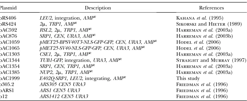
Plasmids used in this study
Plasmid Description References
pRS406 LEU2, integration,AMPR Kahanaet al.(1995)
pRS424 2m,TRP1,AMPR Sikorskiand Hieter(1989)
pAC592 RSL2,2m,TRP1,AMPR Harremanet al.(2003a)
pAC876 SRP1,CEN,URA3,AMPR Harremanet al.(2003b)
pAC1059 pMET25-BPSV40T3-NLS-GFP-GFP,CEN,URA3,AMPR Hodelet al.(2006)
pAC1065 pMET25-SV40-NLS-GFP-GFP,CEN,URA3,AMPR Hodelet al.(2006)
pAC1303 CSE1,2m,TRP1,AMPR Harremanet al.(2003a)
pAC1344 TUB1-GFP, integration,URA3,AMPR Straightand Murray(1997)
pAC1354 SRP1,CEN,TRP1,AMPR Harremanet al.(2003a)
pAC1385 NUP2,2m,TRP1,AMPR Harremanet al.(2003a)
pAC1999 E402Q-SRP1,LEU2, integrating,AMPR This study
p305.2 ARS305 CEN5 URA3 Friedmanet al.(1996)
pARS1 ARS1 CEN5 URA3 Friedmanet al.(1996)
p12 ARS1412 CEN5 URA3 Friedmanet al.(1996)
diploid strains were sporulated and tetrads were dissected to generatesrp1mutant strains expressing Cdc45-GFP, Yox1-GFP, or Mcm10-GFP (Table 1).
In vivo functional analysis: The function of importin-a variantsin vivowas assessed by examining the growth of the integrated allelessrp1-31,srp1-55, andsrp1-E402Q. As a control, each mutant was covered with a wild-typeSRP1 URA3plasmid (pAC876) to ensure that conditional phenotypes were com-plemented prior to the growth assays. Single colonies were grown to saturation in liquid culture, serially diluted (1:10), and spotted on minimal medium plates as a control or on fluoroorotic acid (5-FOA) plates. The drug 5-FOA eliminates theURA3plasmid-encoded wild-typeSRP1(pAC876) to reveal the phenotype of the mutants (Boekeet al.1987). Plates were
incubated at the indicated temperatures for 3–7 days.
Immunoblot analysis:Immunoblot analysis was performed using standard methods (Towbinet al.1979). Cultures were
grown to log phase in yeast extract peptone dextrose (YEPD) media at 25and then shifted to the indicated temperature. Cells were then harvested by centrifugation and washed twice in water and once in PBSMT [100 mmKH
2PO4, pH 7.0, 15 mm
(NH4)2SO4, 75 mmKOH, 5 mmMgCl2, 0.5% Triton X-100].
Cells were subsequently lysed in PBSMT with protease inhib-itors (0.5 mmphenylmethylsulfonyl fluoride, 3mg/ml each
of aprotinin, leupeptin, chymostatin, and pepstatin) by glass bead lysis. Equal amounts of total protein were resolved by SDS–PAGE and immunoblotted with polyclonal anti-importin-aantibody (1:5000 dilution) raised against recombinant GST– importin-afollowed by anti-rabbit secondary antibody (1:5000 dilution).
NLS–GFP import assay: The NLS–GFP import assay was performed as described previously (Shulgaet al.1996; Hodel
et al. 2006). Cells were resuspended in glucose-containing synthetic media pre-equilibrated to 25(permissive), 37(srp1-31, srp1-E402Q), or 18(srp1-55). For scoring, 2-ml samples were removed every 2.5 min and images were collected through a GFP-optimized filter (Chroma Technology) using an Olympus BX60 epifluorescence microscope. Cells were scored as ‘‘nuclear’’ if both the nucleus was brighter than the surround-ing cytoplasm and a nuclear-cytoplasmic boundary was visible. At least 100 cells were counted at each time point.
Microscopy:Direct fluorescence microscopy was performed to localize GFP fusion proteins in live cells. For all experi-ments, cultures were also labeled with Hoechst dye (1mg/ml) to visualize DNA and confirm the location of the nucleus. The localization of GFP fusion proteins was monitored by directly viewing the GFP signal in living cells through a GFP-optimized filter (Chroma Technology) using an Olympus BX60 epifluor-escence microscope equipped with a Photometrics Quantix digital camera. For localization of candidate cargoes, cells expressing Cdc45-GFP, Mcm10-GFP, or Yox1-GFP were grown to log phase at the permissive temperature and then shifted to the nonpermissive temperature for 3 hr.
High-copy suppressor analysis: For high-copy suppressor analysis, high-copy plasmids (2mTRP1) encoding the nuclear transport factors importin-b(pAC592), Cse1 (pAC1303), and Nup2 (pAC1385) were transformed intosrp1-55 (ACY641), srp1-E402Q(ACY1560), andsrp1-31(ACY1561) cells covered by anSRP1 URA3maintenance plasmid (pAC876). Genetic suppression was assessed by growing single colonies in liquid culture to saturation, serially diluting (1:10), and spotting on minimal medium plates as a control or on 5-FOA plates. Plates were incubated at the indicated temperatures for 3–6 days.
Cell cycle arrest and release:For cell cycle studies, cultures were synchronized by treatment witha-factor or hydroxyurea. For arrest witha-factor, cells were grown to early mid-log phase (OD6000.25–0.35) in YEPD media at 25. Cell cycle arrest was
accomplished by pelleting the cells, washing with YEPD (pH
3.9), and resuspending in fresh YEPD (pH 3.9) containing a 1:500 dilution of 5 mg/ml ofa-factor (Sigma) followed by a 90-min incubation at 25. Additionala-factor (1:1000 dilution) was added every 30 min. Cells were released froma-factor arrest by pelleting the cells, washing twice with YEPD, and resuspend-ing in fresh YEPD for release at the indicated temperatures for an additional 3 hr (Swaminathanet al.2007).
For hydroxyurea arrest, cells were arrested witha-factor as described above. Then cells were pelleted, washed twice with YEPD, and resuspended in YEPD containing 200 mm
hydroxy-urea. Cultures were incubated for an additional 2 hr at 25. Cells were then pelleted, washed twice with YEPD, and resuspended in fresh YEPD for release at the indicated temperatures for an additional 3 hr (Swaminathanet al.2007).
Flow cytometry analysis:Cells were prepared for flow cytom-etry analysis by staining with propidium iodide (Sazerand
Sherwood1990). Briefly, asynchronous or synchronous cultures
were ethanol fixed overnight at 4, washed, and resuspended in 50 mmsodium citrate. Cells were then treated with 0.1 mg/
ml RNase A for 2 hr at 37and 10 mg/ml of Proteinase K for 1 hr at 50. Cells were stained with 8mg/ml of propidium iodide. Each sample was analyzed with a FACSVantage SE (Becton Dickinson, Franklin Lakes, NJ). Data were analyzed using FloJo 7.2.2 software.
Plasmid loss: Plasmid loss was determined in wild type or mutant cells containing plasmids with an early (ARS305)-, middle (ARS1)-, or late (ARS1412)-firing autonomously replicating sequence (ARS) (Friedmanet al.1996). Cells were grown to log
phase after which at least 200 cells were plated on nonselective YEPD plates. Colonies that grew on YEPD plates were then replica plated to uraselective plates to determine plasmid loss. The percentage of plasmid loss was calculated as 100(the ratio of colonies on the uraselective plate/colonies on the YEPD plate)3100. The experiment was performed in triplicate.
Statistical analysis: Statistical methods were employed to determine if a significant difference was observed in the per-centage of plasmid loss in thesrp1mutant cells as compared to wild-type cells. Data were analyzed using a one-way analysis of variance (ANOVA) for origin of replication followed by a Dunnett’s multiple comparison test using Graph Pad Prism 3.0. The significance level (a) was set at 0.05 for all statistical tests. If the calculatedP-value was,a, then the difference in plasmid loss was reported as being statistically significant.
RESULTS
To examine links between the classical nuclear protein import pathway and cell cycle progression, we exploited two mutants of importin-a with specific molecular defects. Thesrp1-E402Qmutant alters a critical glutamic acid residue in the minor pocket of the cNLS cargo-binding domain to glutamine (Contiet al.1998; Fontes
et al.2000). This conservative amino acid substitution at position 402 creates the conditional allele of importin-a,
srp1-E402Q, which causes a decrease in bipartite cNLS cargo bindingin vitroand affects the steady-state local-ization of a bipartite NLS cargo in vivo (Leung et al.
2003).
As a complement to the analysis of the NLS cargo-binding pocket, a variant of importin-athat affects cargo delivery was also employed. A conserved NLS-like se-quence within the N-terminal IBB domain of importin-a,
54KRR56, is essential for the auto-inhibitory function of
substitution at position 55 within this auto-inhibitory sequence (KRR/KAR) creates a conditional allele of importin-a,srp1-55, which specifically affects cargo de-livery/release into the nucleus (Harremanet al.2003a). Functional analysis ofsrp1mutants:To compare the consequences of defects in cargo binding and cargo releasein vivo, we generated alleles of thesrp1-55 and
srp1-E402Q importin-a mutants that could be directly compared to one another (seematerials and methods).
As a control, we also generated the previously charac-terizedsrp1-31 mutant (Loebet al.1995) in the same
genetic background. As an initial characterization and comparison of these mutants, we analyzed their growth at various temperatures. To ensure that equal numbers of cells were grown and spotted, thesrp1-31,srp1-55, and
srp1-E402Qmutants were transformed with a wild-type
SRP1plasmid. To assay growth, 10-fold serial dilutions of the samples were spotted on control plates where wild-typeSRP1is maintained or on 5-FOA plates where the plasmid encoding wild-typeSRP1is lost. In comparison to wild-type cells, srp1-55 mutant cells show a growth defect at 18as previously reported (Harreman et al.
2003a). In contrast,srp1-31andsrp1-E402Qmutants show growth defects at 37(Figure 1B). Immunoblot analysis indicates no significant change in the level of any of the Srp1 mutant proteins at the nonpermissive
tempera-tures as compared to wild-type importin-a, indicating that the growth defects observed are not due simply to loss of the essential importin-aprotein (data not shown).
NLS–GFP import assay: Prior studies examined the cargo-binding properties of thesrp1-55andsrp1-E402Q
variantsin vitroas well as their impact on the steady-state localization of cNLS cargoin vivo(Harremanet al.2003a;
Leunget al.2003). To further characterize the impact
of each of these alleles as well as the srp1-31 mutant on nuclear protein import, we used a semiquantitative kinetic NLS–GFP import assay that assesses the initial rate of NLS cargo import (Shulgaet al.1996). For this
assay, import of both a monopartite SV40 and a bipartite BPSV40T3 NLS cargo was examined. The SV40 and BPSV40T3 cargoes were selected for the NLS–GFP im-port assay because both bind to imim-portin-awith a similar affinity (10 nm) and, importantly, the bipartite cargo is
engineered such that productive binding to importin-a
absolutely depends on the basic cluster of amino acids that binds to the minor NLS binding pocket (Hodel
et al. 2001). Experiments were carried out at the non-permissive temperatures in live cells as described in
materials and methods. In comparison to wild-type
cells, thesrp1-31andsrp1-55mutants showed a decrease in the initial rate of nuclear import of the monopartite cNLS cargo SV40-GFP (Figure 1C). As expected, there Figure1.—Functional analysis of
im-portin-amutantsin vivo. (A) Schematic of importin-awith the three major do-mains (IBB; NLS cargo-binding domain with major and minor NLS binding pockets; Cse1 binding domain) indi-cated. The position of the auto-inhibi-tory NLS-like sequence (54KRR56) is also indicated. The approximate loca-tion as well as the amino acid change for each of thesrp1mutant alleles em-ployed in the study is shown. (B) Growth of each srp1 mutant was as-sessed at 18, 25, and 37. Each mutant was covered with a wild-typeSRP1 URA3 plasmid (pAC876) and analyzed by se-rial dilution and spotting on control plates (where the wild-type SRP1 plas-mid is maintained) or on 5-FOA plates (where the wild-type SRP1 plasmid is lost). Plates were incubated at the indi-cated temperatures for 3–7 days. (C) Kinetic nuclear import assay for mono-partite SV40-GFP NLS and (D) bimono-partite BPSV40T3-GFP NLS import reporters. The initial import rates for the cNLS im-port reim-porters were analyzed using a kinetic import assay as described in
materials and methods(Shulgaet al.
1996). Cultures were grown to early mid-log phase at 25and then shifted to the nonpermissive temperature (37forsrp1-31 andsrp1-E402Qcells and 18forsrp1-55cells). Initial import kinetics was measured by determining the percentage of cells showing nuclear accumulation of the cNLS reporter at a given time. For scoring, 2-ml samples were removed every 2.5 min. Cells were scored as ‘‘nuclear’’ if both the nucleus was brighter than the surrounding cytoplasm and a nuclear–cytoplasmic boundary was visible. At least 100 cells were counted at each time point. Results are plotted as the percentage of cells showing nuclear cNLS reporter signalvs.time for wild type (WT) (¤),srp1-31(n),srp1-55(:), andsrp1-E402Q(X) cells.
was no observed change in the initial import rate of the monopartite cargo insrp1-E402Qcells (Figure 1C) because the E402Q amino acid substitution decreases bipartite cargo binding without impairing binding to monopartite cNLS cargo (Leunget al.2003). However,
all three mutants,srp1-31,srp1-55, andsrp1-E402Q, show a decrease in the initial rate of nuclear import of the bipartite cNLS cargo BPSV40T3-GFP (Figure 1D). These results confirm that cells expressing each variant of importin-ahave defects in cNLS protein import.
Importin-avariants differ in their in vivo molecular defects: Although all of the importin-a mutant cells showed defects in the kinetic NLS import assay, we hy-pothesized that the different engineered amino acid changes in each importin-a variant should impair nu-clear import through distinct mechanisms. To address this hypothesis, we tested whether each of the
importin-a mutants could be suppressed by overexpression of several nuclear transport factors with the assumption that suppression by specific factors could provide in-formation about what interactions are compromised
in vivo. For this analysis, we examined overexpression of importin-b, Cse1, and the nuclear pore protein, Nup2. Importin-binteracts with the IBB domain of importin-a
to target the import complex to the nuclear pore (Go¨ rlich
et al.1996; Weiset al.1996). Cse1 is the export receptor
for recycling importin-a to the cytoplasm (Hoodand
Silver1998; Solsbacheret al.1998) and both Cse1 and
Nup2 facilitate cargo delivery in the nucleus (Matsuura
et al. 2003; Matsuura and Stewart 2004; Liu and
Stewart2005). As controls, eachsrp1mutant was also
transformed with a vector alone or with a wild-type importin-a plasmid. To ensure equal growth and spot-ting, each mutant was transformed with wild-typeSRP1
on aURA3plasmid. Cultures were grown to saturation, serially diluted, and then spotted either on control plates where theSRP1maintenance plasmid is retained
or on plates containing 5-FOA where the maintenance plasmid is lost but the overexpression plasmids are retained (Figure 2). The 5-FOA plates were incubated at the nonpermissive temperature as previously deter-mined (see Figure 1B) for each mutant. Interestingly, we find that the temperature-sensitive growth defect of
srp1-31cells is suppressed by overexpression of impor-tin-b (Figure 2). As previously observed (Harreman
et al.2003a), the cold-sensitive growth defect ofsrp1-55
mutant cells is suppressed by overexpression of the nuclear export factor, Cse1 (Figure 2). In contrast, the temperature-sensitive growth defect ofsrp1-E402Qcells is not suppressed by any of the nuclear transport factors (Figure 2). Taken together, these results suggest that the
in vivodefects that underlie diminished protein import in each of these mutants are likely to be distinct from one another.
Mutants of importin-aaffect cell cycle progression:
To probe the link between importin-a and cell cycle events, we exploited thesesrp1 mutants that affect the classical nuclear protein import pathway through dis-tinct mechanisms. As a first step to determining whether all defects in importin-aimpair G2/M of the cell cycle as
previously described for thesrp1-31mutant (Loebet al.
1995), each importin-a mutant was spotted on plates containing either hydroxyurea, which inhibits DNA replication (Mitchisonand Creanor1971), or
beno-myl, which blocks mitosis prior to the onset of anaphase (Liand Murray1991). Consistent with previous reports,
thesrp1-31mutant shows sensitivity to growth on beno-myl, suggesting a defect in G2/M of the cell cycle (Figure
3A) (Loeb et al. 1995). In contrast, the srp1-55 and
srp1-E402Qmutant cells show no obvious sensitivity to benomyl. Surprisingly, in comparison to wild-type cells, srp1-31,srp1-55, and srp1-E402mutants all show sensi-tivity to hydroxyurea, suggesting a defect in processes critical to the G1/S cell cycle transition, including DNA
Figure2.—copy suppressor analysis.
replication, DNA repair, and/or checkpoint function (Figure 3A).
To begin to assess how the cell cycle is affected in each of thesrp1mutants, the DNA content of each mutant was measured in an asynchronous cell population using flow cytometry (Figure 3B). Samples were analyzed at 30-min intervals over 6 hr at both permissive and non-permissive temperatures. The cells are scored as having 1N (G1) or 2N (G2/M) DNA content. All importin-a
mutants show a distribution of 1N and 2N DNA content that is indistinguishable from wild-type (SRP1) cells at the permissive temperature (data not shown). In wild-type (SRP1) cells, the distribution of 1N and 2N DNA content is unchanged relative to the permissive temper-ature. Consistent with previous results (Loebet al.1995),
srp1-31 mutant cells show some accumulation of cells in the G2/M phase of the cell cycle as compared to the
G1phase. As withsrp1-31cells,srp1-55cells show some
increase in cells with 2N DNA content as compared to wild-type cells (SRP1), which is consistent with previous analysis of an asynchronous population of these cells (Harremanet al.2003a). In contrast to thesrp1-31and
srp1-55 mutants, srp1-E402Q mutant cells show an in-crease in the population of cells with 1N DNA content as compared to wild-type (SRP1) cells. While these results are consistent with impaired cell cycle transitions due to compromised classical nuclear protein import, defects at multiple points in the cell cycle are not readily un-covered by analysis of asynchronous cultures.
Both sensitivity to hydroxyurea and analysis of DNA content for the srp1-E402Q mutant suggest a role for importin-ain the G1/S cell cycle transition. To assess the
DNA content of synchronized cultures, cells were ar-rested in late G1phase of the cell cycle with the
mating-type pheromone,a-factor (Siedeet al.1989; Siedeand
Friedberg1990), and then analyzed by flow cytometry
over time following release froma-factor. Samples were analyzed at 30-min intervals over 3 hr at both permissive and nonpermissive temperatures. All importin-amutants show wild-type progression at the permissive temperature (data not shown). As expected, wild-type cells progress through the cell cycle over the time course of the ex-periment at all temperatures. As previously reported,
srp1-31 mutant cells display a defect in progression through G2/M (Figure 4A) (Loebet al.1995). The
srp1-31mutant also shows a slight defect in the G1/S phase
transition, which is consistent with growth sensitivity to hydroxyurea. Interestingly, bothsrp1-55andsrp1-E402Q
mutant cells show a profound defect in the G1/S
tran-sition (Figure 4A). These results link importin-a and hence the classical nuclear import pathway to the G1/S
transition of the cell cycle as well as the previously reported G2/M transition.
Using the synchronization method employed for flow cytometry, the spindle morphology of each mutant fol-lowing the shift to the nonpermissive temperature was assessed over time (Figure 4B). The percentage of spindle formation was defined as containing the following: single asters (no spindles), short spindles (spindle not extend-ing to the duplicated nucleus of the daughter cell), or anaphase spindles (spindle extending to the duplicated nucleus) (Goh and Surana 1999). In comparison to
wild-type cells, srp1-31cells show an increased number of short spindles and a decrease in the relative number Figure3.—Analysis of cell cycle
de-fects insrp1mutants. (A) Analysis of cell growth on hydroxyurea and benomyl plates. To determine whether any of the srp1 mutant alleles show hyper-sensitivity to growth on plates containing hydroxyurea or benomyl, cultures were grown to saturation at the permissive temperature and then serially diluted and spotted on control YEPD plates, YEPD containing 100 mmhydroxyurea,
or YEPD plates containing 10 mg/ml benomyl. Plates were incubated at the permissive temperature for 3–7 days. (B) Analysis of cell cycle progression in unsynchronized cells. Wild-type (SRP1) and mutant (srp1-31, srp1-55, and srp1-E402Q) cells were grown to early mid-log phase at the permissive temperature and then shifted to the nonpermissive temperature (37 for srp1-31and srp1-E402Q cells and 18 for srp1-55cells). Samples were collected every 30 min for 6 hr for flow cytometry to analyze DNA content. The positions of unrepli-cated DNA (1N) and repliunrepli-cated DNA (2N) are indicated below the graphs.
of anaphase spindles after 3 hr at the nonpermissive temperature (Figure 4B). In contrast,srp1-55and srp1-E402Qmutant cells show a constant level of single asters over time, which correlates with a defect in the G1/
S phase transition where spindle formation has not yet begun (Figure 4B).
Since a profound delay at the G1/S transition was
observed in srp1-55 and srp1-E402Q mutant cells, we wanted to determine whether these cells were defective not only in transition but also in progression through S phase. To examine progression through S phase, the mutant cells were arrested in G1using a-factor at the
permissive temperature and then released into media containing hydroxyurea to synchronize cells in early S phase. After 2 hr, cells were released at either the permissive or the nonpermissive temperature. Samples were then analyzed by flow cytometry. At the permissive temperature, wild-type and all mutant cells progress through the cell cycle in a comparable manner (data not shown). However, when mutants are released to
the nonpermissive temperature, bothsrp1-55and srp1-E402Qcells progress slowly through S phase in compar-ison to wild-type cells (Figure 5A). This experiment also provides further evidence thatsrp1-31cells have defects in S phase progression as well as in G2/M as they also
progress through S phase more slowly than wild-type cells (Figure 5A). To ensure that bothsrp1-55and srp1-E402Qcells were able to release froma-factor arrest, the DNA content of these mutant cells was also monitored at the permissive temperature (Figure 5B). Flow cytometry analysis shows that both srp1-55 and srp1-E402Q cells can release froma-factor arrest and enter the cell cycle at the permissive temperature, providing evidence that these cells, when first synchronized with a-factor and then released into media containing hydroxyurea, should be synchronized in early S phase. We also employed spindle morphology analysis for each mutant as inde-pendent confirmation of the cell cycle delays observed in the srp1-31, srp1-55, and srp1-E402Q cells (Figure 5C). As observed for thea-factor arrest, bothsrp1-55and Figure4.—Analysis of cell cycle
pro-gression in cells synchronized in G1
phase of the cell cycle. (A) Wild-type (SRP1) and mutant (srp1-31, srp1-55, andsrp1-E402Q) cells were grown to early mid-log phase at the permissive temper-ature and then arrested at late G1phase
srp1-E402Qcells showed nearly 100% of cells with single asters throughout the time course of the experiment. This spindle morphology is consistent with the failure to enter the cell cycle over the time course of the experi-ment. As suggested by the flow cytometry data, thesrp1-31
cells show a delay in spindle formation relative to wild-type (SRP1) cells, but spindles do begin to form and elongate in this mutant after90 min [compare to30 min for wild-type (SRP1) cells].
Results indicate that all of the importin-a mutants examined show defects in the G1/S transition and in
S phase progression. As an initial approach to un-derstanding howsrp1 mutants might affect DNA repli-cation, we utilized a plasmid loss assay (Friedmanet al.
1996; Dohrmannand Sclafani2006). This assay
mon-itors loss of plasmids containing an ARS, which function as replication origins in S. cerevisiae (Dohrmann and
Sclafani 2006). Although defects in many cell cycle
pathways can contribute to chromosome loss, mutants with general DNA replication defects exhibit loss re-gardless of the origin of replication used in the plasmid, whereas mutants specific for replication initiation often Figure5.—Analysis of cells synchronized
in S phase of the cell cycle. (A) Wild-type (SRP1) and mutant (srp1-31, srp1-55, and srp1-E402Q) cells were grown to early mid-log phase at the permissive temperature and then arrested at late G1phase witha
-fac-tor. Samples were then released into hy-droxyurea at the permissive temperature and incubated for 2 hr. Finally, the cells syn-chronized in early S phase were released to the nonpermissive temperature (37for srp1-31 and srp1-E402Q cells and 18 for srp1-55cells). Samples were collected every 30 min for 3 hr for analysis by flow cytometry to analyze DNA content. The positions of unreplicated DNA (1N) and replicated DNA (2N) are indicated below the graphs. (B) Mutant (srp1-31andsrp1-E402Q) cells were grown to early mid-log phase at the per-missive temperature and then arrested at late G1phase witha-factor. Samples were
then released to the permissive temperature to determine if the cells are released froma -factor arrest. Samples were collected every 30 min for 3 hr for flow cytometry to analyze DNA content. The positions of unreplicated DNA (1N) and replicated DNA (2N) are in-dicated below the graphs. (C) Spindle mor-phology. Wild-type (SRP1) and mutant (srp1-31,srp1-55, andsrp1-E402Q) cells ex-pressing Tub1-GFP to visualize microtu-bules were treated as described above in A. Spindles were visualized by examining inte-grated Tub1-GFP signal by direct fluores-cence microscopy. Results are plotted as the percentage of spindles in the total cell population scored as single asters (no spin-dles,¤), short spindles (spindle not extend-ing to the duplicated nucleus of the daughter cell, n), or anaphase spindles (spindle extending to the duplicated nu-cleus,d)vs. time.
exhibit differential loss dependent on the nature of the replication origin (Maineet al.1984; Hartwelland
Smith1985; Palmeret al.1990).
Therefore, we compared the rate of loss of plasmids containing an ARS that fires early in S phase (ARS305), in the middle of S phase (ARS1), or late in S phase (ARS1412) (Friedmanet al.1996). All results are
com-pared to wild-type (SRP1) cells (Figure 6). In comparison to wild-type (SRP1) cells, both thesrp1-31andsrp1-E402Q
mutant cells exhibit plasmid loss of the early firing origin of replication,ARS305. As described inmaterials and methods, statistical analysis reveals a significant
increase in plasmid loss in thesrp1-31mutant as com-pared to the wild-type (SRP1) cells (**P,0.01) (Figure 6A). We also observed increased plasmid loss in srp1
mutant cells with ARS1, which fires in the middle of S phase. Statistical analysis shows a significant increase in plasmid loss in both thesrp1-31(**P,0.01) and the
srp1-55(**P ,0.01) mutant cells (Figure 6B). Finally, the srp1-31 and srp1-E402Q mutants show an increase in plasmid loss for the late-firing origin of replication,
ARS1412. Statistical analysis reveals a significant in-crease in plasmid loss for both srp1-31 (**P , 0.01) andsrp1-E402Q(*P,0.05) mutant cells as compared to wild-type (SRP1) cells (Figure 6C). To rule out changes within the wild-type control, plasmid loss for early, middle-, and late-firing replication origins was com-pared in wild-type cells. This comparative analysis shows no significant difference in plasmid loss between the early, middle, and late replication origins in the wild-type cells (P¼0.8417) (data not shown). Interestingly, thesrp-31mutant shows a significant increase in plasmid loss at the early, middle-, and late-firing origins of repli-cation, suggesting a general chromosome loss phenotype. The plasmid loss defect in thesrp1-55mutant suggests a specific defect in the middle-firing origin of replica-tion,ARS1. In contrast, thesrp1-E402Qmutant shows a specific defect in the late-firing origin of replication,
ARS1412. Together, these results indicate that these
srp1 alleles have distinct effects on chromosome loss, which may result from differential import of key cell cycle proteins.
Bipartite cNLS candidates involved in G1/S of the cell cycle: While it is likely that numerous import cargoes are affected in thesrp1mutant cells, we wanted to determine whether there were key candidates most likely to be affected. To identify candidate cargoes whose import could be impaired in thesrp1-55andsrp1-E402Q
cells, a bioinformatic approach was taken to identify proteins containing a putative bipartite cNLS that are involved in G1/S of the cell cycle and are localized to
the nucleus (Figure 7A). To identify the entire comple-ment of candidate bipartite cNLS cargoes, the PSORT II algorithm for predicting bipartite cNLS motifs (Nakai
and Horton 1999) was used to query theS. cerevisiae
GenBank (Bensonet al. 2006). Of 5850 proteins, 968
contain a predicted bipartite cNLS. We then used the
PSORTII algorithm to query the pool of 1515 nuclear or nucleolar proteins as defined by localization of GFP fusion proteins (Huhet al.2003). Of these 1515 nuclear/
nucleolar proteins, 391 contain a predicted bipartite cNLS (Huhet al.2003). As a preliminary approach to Figure6.—Analysis of plasmid loss. To determine ifsrp1
mu-tant cells have defects in DNA replication, a plasmid loss assay was performed. Plasmid loss was determined in cells containing plas-mids with early (ARS305)-, middle (ARS1)-, or late (ARS1412 )-firing ARSs as described inmaterials and methods. Results
identifying a subset of these proteins linked to the G1/S
cell cycle transition, we searched for genes containing the terms G1, S, or G1/S in their Gene Ontology
defini-tion (http://www.geneontology.org/). Through this initial approach, we identified 93 proteins implicated in G1/S
of the cell cycle. The overlap between these three data sets identifies 15 proteins that contain a predicted bipartite cNLS, localize to the nucleus, and are already implicated in the G1/S transition (Figure 7A). These 15
candidate proteins that could be affected in cells that are defective in nuclear import of cargoes containing bipartite cNLS are listed in Table 3.
As an initial test of whether the nuclear import of any of these candidate cargoes is impaired in the importin-a
mutants, we selected three candidates that had pre-viously been visualized as nuclear GFP fusion proteins: Cdc45, Mcm10, and Yox1 (Huhet al.2003). Cdc45 is
in-volved in DNA replication and remains nuclear through-out theS. cerevisiaecell cycle (Moiret al.1982; Hopwood
and Dalton1996). Cdc45 interacts with the
minichro-mosome maintenance proteins, which regulate DNA replication (Hopwoodand Dalton 1996). Mcm10 is
a constitutively nuclear chromatin-associated protein that plays a role in DNA replication initiation by re-cruiting both the Mcm2-7 complex and DNA polymer-ase-ato DNA replication origins (Burichand Lei2003;
Rickeand Bielinsky 2004; Zhuet al.2007). Yox1 is a
homeodomain transcriptional repressor that regulates the expression of genes that are critical for the G1/S
transition (Pramila et al. 2002; Braun and Breeden
2007). We examined the localization of Mcm10-GFP, Yox1-GFP, and Cdc45-GFP in wild-type (SRP1),srp1-31, and srp1-55 cells (Figure 7, B–D). At the permissive temperature, Mcm10-GFP, Yox1-GFP, and Cdc45-GFP are localized to the nucleus in all cells examined (data not shown). Following a shift to the nonpermissive tem-perature, both srp1-31andsrp1-55cells show a decrease in nuclear localization of Mcm10-GFP (Figure 7B) and Yox1-GFP (Figure 7C) in comparison to wild-type (SRP1) cells. Interestingly, Cdc45-GFP remains nuclearly local-ized insrp1-55cells but is mislocalized to the cytoplasm insrp1-31cells (Figure 7D).
DISCUSSION
Results of this study define an important role for the NLS receptor, importin-a, in DNA replication. Specifi-cally, we report that two mutants of importin-a cause a profound delay in the G1/S cell cycle transition.
Although previous studies described defects in G2/M
associated with other importin-amutants,srp1-31(Loeb
et al.1995) andsrp1-55(Harremanet al.2003a), this is
the first report demonstrating that mutations in the NLS receptor and hence defects in the classical protein import pathway impair the G1/S transition.
Our study also reveals that thesrp1-31mutant shows a delay in both the G2/M and G1/S cell cycle transitions,
although the G1/S delay for thesrp1-31cells is not as
profound as observed forsrp1-55andsrp1-E402Qcells. Figure7.—Candidate bipartite proteins
impli-cated in G1/S of the cell cycle are not efficiently
targeted to the nucleus in importin-a mutants. (A) The PSORT II algorithm for bipartite cNLSs was used to query three sets of data: the 5850 proteins in theS. cerevisiaeGenBank (bipartite) (Bensonet al.
2006), the 1515 proteins localized to either the nu-cleus or the nucleolus in the global yeast GFP-fusion library (nuclear) (Huhet al.2003), and 93 proteins
implicated in G1/S of the cell cycle based on the
Gene Ontology definition (G1/S) (http://www.
geneontology.org/). The overlap between these three data sets identifies 15 proteins that contain a predicted bipartite cNLS, localize to the nucleus, and are already implicated in G1/S of the cell cycle.
Data from this analysis are presented as a Venn diagram. (B–D) Wild-type (SRP1) andsrp1mutant (srp1-31,srp1-55) cells expressing Mcm10-GFP (B), Yox1-GFP (C), or Cdc45-GFP (D) were analyzed by direct fluorescence microscopy. Cultures were grown to log phase at the permissive temperature and then shifted to the nonpermissive temperature (37forsrp1-31cells and 18forsrp1-55cells) for 3 hr. Wild-type cells (SRP1) were analyzed following shifts to both 37and 18and identical results were ob-tained with each of the GFP fusion proteins. Results are shown for the wild-type cells shifted to 37. Cor-responding DIC and Hoechst DNA-staining images are shown.
The G2/M cell cycle defect observed for the srp1-31
mutant is consistent with previous work (Loeb et al.
1995) and also with hypersensitivity to benomyl. Genetic analysis indicated that the temperature-sensitive growth defect ofsrp1-31is suppressed by the overexpression of the nuclear import receptor, importin-b. This finding suggests that the amino acid change in the srp1-31
variant protein could impair importin-b binding. As interaction between importin-a and -b is critical to targeting cargoes to the nuclear pore for import (Stewart2007), decreased interaction with
importin-bcould lead to profound defects in nuclear import of critical cargoes required for cell cycle transitions.
This study is distinct from the previous analysis of the
srp1-31allele because the importin-avariants employed here have well-characterized molecular defects. The Srp1-E402Q mutant protein is impaired in binding to cargoes that contain a bipartite cNLS (Leunget al.2003).
The Srp1-55 protein shows defects in cargo delivery into the nucleus (Harremanet al.2003a). We find that cells
that express each of these importin-avariants as the sole cellular copy of importin-ahave defects in the G1/S cell
cycle transition. The finding that importin-avariants with defects in cargo binding and cargo delivery cause similar cell cycle defects may be surprising; however, the most logical interpretation of this result may be that both variants are defective in the effective nuclear import of cargo proteins that contain a classical bipartite NLS. This conclusion is fairly obvious for thesrp1-E402Qprotein, which has an amino acid substitution in a key residue of the minor NLS-binding pocket that makes contact only with cargoes that contain a bipartite cNLS and, in fact, shows only defects in the import of bipartite cNLS cargoes (Contiet al.1998; Leunget al.2003). In the case
of the srp1-55 protein, the auto-inhibitory, and hence the inherent cargo release mechanism, is impaired
(Harremanet al. 2003a). However, in cells, there are
additional factors that facilitate cargo release, including the export receptor, Cse1, and the nuclear pore protein, Nup2 (Solsbacher et al. 1998, 2000; Gilchrist et al.
2002). Indeed, overexpression of Cse1 suppresses the growth defect of thesrp1-55mutant. Possibly, cargoes that bind tightly to the NLS receptor may be most affected
in vivo when the cargo delivery function inherent to importin-ais impaired. These tight-binding cargoes are most likely to be the cargoes that contain a bipartite cNLS as bipartite cargoes typically bind with a stronger affinity to the NLS receptor than monopartite cargoes (Hodel
et al.2001). Thus, bothsrp1-55andsrp1-E402Qcells may, logically, be most impaired in nuclear delivery of bipartite cNLS cargo proteins.
Althoughsrp1-55 and srp1-E402Qmutant cells show similar defects in the cell cycle, the plasmid loss assay provides further insight into the molecular defect. The plasmid loss assay indicates that a defect in cNLS cargo binding (srp1-E402Q) affects DNA replication late in S phase. Similarly, the plasmid loss assay suggests that a defect in cNLS cargo release (srp1-55) also affects DNA replication but in the middle of S phase. Furthermore, as withsrp1-31cells, our results suggest thatsrp1-55cells are impaired in both the G1/S and the G2/M cell cycle
transitions. Interestingly, these results suggest that differ-ent defects in the NLS receptor importin-aare likely to affect the import of distinct complements of nuclear import cargoes, including some that may be uniformly affected and some that may be distinct for each mutant. Presumably, which cargoes are affected in each mutant depends on both cargo recognition in the cytoplasm and release into the nucleus. Other factors that effect recognition and release could also influence the phe-notype of these mutants.
The use of variants of importin-awith specific molec-ular defects allowed us to identify a number of candi-date proteins that may be affected in these mutants. For three of these candidate proteins, Mcm10, Yox1, and Cdc45, we show that nuclear localization is differentially impaired in the importin-a mutants. While Mcm10 and Yox1 are mislocalized in each importin-a mutant examined, Cdc45 is mislocalized to the cytoplasm only insrp1-31cells. Overall, these results show that different defects in importin-a affect distinct cargoes. In the future, genetic approaches that exploit the conditional growth phenotypes of thesesrp1mutants may be useful in identifying additional cargo proteins affected in these mutants, including some that may be specific for each mutant.
Collectively, our data demonstrate a molecular role for the nuclear localization signal receptor, importin-a, during the G1/S stage of the cell cycle. This finding adds
to our previous understanding of the link between nu-clear transport and cell cycle progression as previously only defects in the G2/M transition had been linked to
mutants in the nuclear import receptor. Future studies
TABLE 3
Bipartite cNLS candidates involved in G1/S
Protein Function
Bck2 Molecular function unknown
Cdc40 RNA splicing factor activity
Cdc45 DNA replication
Mcm10 DNA replication and chromatin binding
Rsc3 DNA binding
Swi5 DNA replication and transcriptional
activator activity
Tos4 Transcription factor activity
Tye7 Transcription factor activity
Pog1 RNA POL II transcription factor activity
Taf1 RNA POL II transcription factor activity
Taf13 RNA POL II transcription factor activity
Taf14 RNA POL II transcription factor activity
Yap5 RNA POL II transcription factor activity
Yhp1 DNA binding
will be aimed at identifying key cargoes that must enter the nucleus to mediate these critical cell cycle transitions.
We are very grateful to Deanna Koepp for her help with cell cycle studies as well as her comments on the manuscript. We thank Ryan Mills for his help with bioinformatics analysis as well as members of the Corbett laboratory for helpful suggestions. This work was supported by a National Institutes of Health (NIH) grant to A.H.C. K.F.P. was supported by an NIH and Facilitating Academic Careers in Engineer-ing and the Sciences supplement.
LITERATURE CITED
Adams, A., D. E. Gottschling, C. A. Kaiser and T. Stearns,
1997 Methods in Yeast Genetics.Cold Spring Harbor Laboratory Press, Cold Spring Harbor, NY.
Bayliss, R., A. H. Corbettand M. Stewart, 2000 The molecular
mechanism of transport of macromolecules through nuclear pore complexes. Traffic1:448–456.
Beg, A. A., S. M. Ruben, R. I. Scheinman, S. Haskill, C. A. Rosen
et al., 1992 I kappa B interacts with the nuclear localization se-quences of the subunits of NF-kappa B: a mechanism for cytoplas-mic retention. Genes Dev.6:1899–1913.
Benson, D. A., I. Karsch-Mizrachi, D. J. Lipman, J. Ostelland D. L.
Wheeler, 2006 GenBank. Nucleic Acids Res.34:D16–D20.
Boeke, J. D., J. Truehart, G. Natsoulisand G. Fink, 1987
5-Fluo-roorotic acid as a selective agent in yeast molecular genetics. Methods Enzymol.154:164–175.
Braun, K. A., and L. L. Breeden, 2007 Nascent transcription of
MCM2-7 is important for nuclear localization of the minichromo-some maintenance complex in G1. Mol. Biol. Cell18:1447–1456. Briscoe, J., F. Kohlhuberand M. Muller, 1996 JAKs and STATs
branch out. Trends Cell Biol.6:336–340.
Burich, R., and M. Lei, 2003 Two bipartite NLSs mediate
constitu-tive nuclear localization of Mcm10. Curr. Genet.44:195–201. Burke, B., and J. Ellenberg, 2002 Remodelling the walls of the
nu-cleus. Nat. Rev. Mol. Cell Biol.3:487–497.
Conti, E., and J. Kuriyan, 2000 Crystallographic analysis of the
spe-cific yet versatile recognition of distinct nuclear localization sig-nals by karyopherin alpha. Structure8:329–338.
Conti, E., M. Uy, L. Leighton, G. Blobeland J. Kuriyan, 1998
Crys-tallographic analysis of the recognition of a nuclear localization signal by the nuclear import factor karyopherin alpha. Cell94:
193–204.
David-Pfeuty, T., F. Chakrani, K. Ory and Y. Nouvian-Dooghe,
1996 Cell cycle-dependent regulation of nuclear p53 traffic oc-curs in one subclass of human tumor cells and in untransformed cells. Cell Growth Differ.7:1211–1225.
Dohrmann, P. R., and R. A. Sclafani, 2006 Novel role for
check-point Rad53 protein kinase in the initiation of chromosomal DNA replication inSaccharomyces cerevisiae.Genetics174:87–99. Du, Q., L. Taylor, D. A. Comptonand I. G. Macara, 2002 LGN
blocks the ability of NuMA to bind and stabilize microtubules: a mechanism for mitotic spindle assembly regulation. Curr. Biol.
12:1928–1933.
Fontes, M. R., T. Tehand B. Kobe, 2000 Structural basis of
recog-nition of monopartite and bipartite nuclear localization se-quences by mammalian importin-alpha. J. Mol. Biol. 297:
1183–1194.
Friedman, K. L., J. D. Diller, B. M. Ferguson, S. V. Nyland, B. J.
Brewer et al., 1996 Multiple determinants controlling
activa-tion of yeast replicaactiva-tion origins late in S phase. Genes Dev.10:
1595–1607.
Gilchrist, D., and M. Rexach, 2003 Molecular basis for the rapid
dissociation of nuclear localization signals from karyopherin al-pha in the nucleoplasm. J. Biol. Chem.278:51937–51949. Gilchrist, D., B. Mykytkaand M. Rexach, 2002 Accelerating the
rate of disassembly of karyopherin.cargo complexes. J. Biol. Chem.
277:18161–18172.
Goh, P. Y., and U. Surana, 1999 Cdc4, a protein required for the
onset of S phase, serves an essential function during G(2)/M
transition in Saccharomyces cerevisiae. Mol. Cell. Biol. 19:
5512–5522.
Go¨ rlich, D., S. Kostka, R. Kraft, C. Dingwall, R. A. Laskeyet al.,
1995 Two different subunits of importin cooperate to recognize nuclear localization signals and bind them to the nuclear enve-lope. Curr. Biol.5:383–392.
Go¨ rlich, D., P. Henklein, R. A. Laskeyand E. Hartmann, 1996 A
41 amino acid motif in importin-alpha confers binding to importin-beta and hence transit into the nucleus. EMBO J.15:
1810–1817.
Gruss, O. J., R. E. Carazo-Salas, C. A. Schatz, G. Guarguaglini, J.
Kastet al., 2001 Ran induces spindle assembly by reversing the
in-hibitory effect of importin alpha on TPX2 activity. Cell104:83–93. Harreman, M. T., P. E. Cohen, M. R. Hodel, G. J. Truscott, A. H.
Corbettet al., 2003a Characterization of the auto-inhibitory
sequence within the N-terminal domain of importin alpha. J. Biol. Chem.278:21361–21369.
Harreman, M. T., M. R. Hodel, P. Fanara, A. E. Hodeland A. H.
Corbett, 2003b The auto-inhibitory function of importin
al-pha is essential in vivo. J. Biol. Chem.278:5854–5863. Hartwell, L. H., and D. Smith, 1985 Altered fidelity of mitotic
chromosome transmission in cell cycle mutants ofS. cerevisiae.
Genetics110:381–395.
Hetzer, M. W., T. C. Waltherand I. W. Mattaj, 2005 Pushing the
envelope: structure, function, and dynamics of the nuclear pe-riphery. Annu. Rev. Cell Dev. Biol.21:347–380.
Hodel, M. R., A. H. Corbettand A. E. Hodel, 2001 Dissection of a
nuclear localization signal. J. Biol. Chem.276:1317–1325. Hodel, A. E., M. T. Harreman, K. F. Pulliam, M. E. Harben, J. S.
Holmeset al., 2006 Nuclear localization signal receptor affinity
correlates with in vivo localization in Saccharomyces cerevisiae. J. Biol. Chem.281:23545–23556.
Hood, J. K., and P. A. Silver, 1998 Cse1p is required for export of
Srp1p/importin-alpha from the nucleus in Saccharomyces cere-visiae. J. Biol. Chem.273:35142–35146.
Hopwood, B., and S. Dalton, 1996 Cdc45p assembles into a
com-plex with Cdc46p/Mcm5p, is required for minichromosome maintenance, and is essential for chromosomal DNA replication. Proc. Natl. Acad. Sci. USA93:12309–12314.
Huh, W. K., J. V. Falvo, L. C. Gerke, A. S. Carroll, R. W. Howson
et al., 2003 Global analysis of protein localization in budding yeast. Nature425:686–691.
Jones, A. L., B. B. Quimby, J. K. Hood, P. Ferrigno, P. H. Keshava
et al., 2000 SAC3 may link nuclear protein export to cell cycle progression. Proc. Natl. Acad. Sci. USA97:3224–3229. Kahana, J. A., B. J. Schnappand P. A. Silver, 1995 Kinetics of
spin-dle pole body separation in budding yeast. Proc. Natl. Acad. Sci. USA92:9707–9711.
Kalderon, D., B. L. Roberts, W. D. Richardsonand A. E. Smith,
1984 A short amino acid sequence able to specify nuclear loca-tion. Cell39:499–509.
Kobe, B., 1999 Autoinhibition by an internal nuclear localization
signal revealed by the crystal structure of mammalian importin alpha. Nat. Struct. Biol.6:388–397.
Lange, A., R. E. Mills, C. J. Lange, M. Stewart, S. E. Devineet al.,
2007 Classical nuclear localization signals: definition, function, and interaction with importin alpha. J. Biol. Chem.282:5101–5105. Lee, S. J., Y. Matsuura, S. M. Liuand M. Stewart, 2005 Structural
basis for nuclear import complex dissociation by RanGTP. Nature
435:693–696.
Leung, S. W., M. T. Harreman, M. R. Hodel, A. E. Hodeland A. H.
Corbett, 2003 Dissection of the karyopherin alpha nuclear
lo-calization signal (NLS)-binding groove: functional requirements for NLS binding. J. Biol. Chem.278:41947–41953.
Li, R., and A. W. Murray, 1991 Feedback control of mitosis in
bud-ding yeast. Cell66:519–531.
Liu, S. M., and M. Stewart, 2005 Structural basis for the high-affinity
binding of nucleoporin Nup1p to the Saccharomyces cerevisiae importin-beta homologue, Kap95p. J. Mol. Biol.349:515–525. Loeb, J. D., G. Schlenstedt, D. Pellman, D. Kornitzer, P. A. Silver
et al., 1995 The yeast nuclear import receptor is required for mitosis. Proc. Natl. Acad. Sci. USA92:7647–7651.
Maine, G. T., P. Sinhaand B. K. Tye, 1984 Mutants ofS. cerevisiae
defective in the maintenance of minichromosomes. Genetics
106:365–385.
Matsuura, Y., and M. Stewart, 2004 Structural basis for the
assem-bly of a nuclear export complex. Nature432:872–877. Matsuura, Y., A. Lange, M. T. Harreman, A. H. Corbettand M.
Stewart, 2003 Structural basis for Nup2p function in cargo
re-lease and karyopherin recycling in nuclear import. EMBO J.22:
5358–5369.
Middeler, G., K. Zerf, S. Jenovai, A. Thulig, M. Tschodrich-Rotter
et al., 1997 The tumor suppressor p53 is subject to both nuclear import and export, and both are fast, energy-dependent and lectin-inhibited. Oncogene14:1407–1417.
Mitchison, J. M., and J. Creanor, 1971 Induction synchrony in the
fis-sion yeast. Schizosaccharomyces pombe. Exp. Cell Res.67:368–374. Moir, D., S. E. Stewart, B. C. Osmondand D. Botstein, 1982
Cold-sensitive cell-division-cycle mutants of yeast: isolation, properties and pseudoreversion studies. Genetics100:547–563.
Moll, T., G. Tebb, U. Surana, H. Robitsch and K. Nasmyth,
1991 The role of phosphorylation and the CDC28 protein kinase in cell cycle-regulated nuclear import of the S. cerevisiae transcrip-tion factor SWI5. Cell66:743–758.
Nachury, M. V., T. J. Maresca, W. C. Salmon, C. M. Waterman
-Storer, R. Healdet al., 2001 Importin beta is a mitotic target
of the small GTPase Ran in spindle assembly. Cell104:95–106. Nakai, K., and P. Horton, 1999 PSORT: a program for detecting
sorting signals in proteins and predicting their subcellular local-ization. Trends Biochem. Sci.24:34–36.
Nishitani, H., M. Ohtsubo, K. Yamashita, H. Iida, J. Pineset al.,
1991 Loss of RCC1, a nuclear DNA-binding protein, uncouples the completion of DNA replication from the activation of cdc2 protein kinase and mitosis. EMBO J.10:1555–1564.
Palmer, R. E., E. Hoganand D. Koshland, 1990 Mitotic
transmis-sion of artificial chromosomes incdcmutants of the yeast, Saccha-romyces cerevisiae.Genetics125:763–774.
Pines, J., and T. Hunter, 1991 Human cyclins A and B1 are
differ-entially located in the cell and undergo cell cycle-dependent nu-clear transport. J. Cell Biol.115:1–17.
Pramila, T., S. Miles, D. GuhaThakurta, D. Jemioloand L. L. Breeden,
2002 Conserved homeodomain proteins interact with MADS box protein Mcm1 to restrict ECB-dependent transcription to the M/G1 phase of the cell cycle. Genes Dev.16:3034–3045.
Ricke, R. M., and A. K. Bielinsky, 2004 Mcm10 regulates the
stabil-ity and chromatin association of DNA polymerase-alpha. Mol. Cell16:173–185.
Riddick, G., and I. G. Macara, 2007 The adapter importin-alpha
provides flexible control of nuclear import at the expense of ef-ficiency. Mol. Syst. Biol.3:118.
Robbins, J., S. M. Dilworth, R. A. Laskey and C. Dingwall,
1991 Two interdependent basic domains in nucleoplasmin nuclear targeting sequence: identification of a class of bipartite nuclear targeting sequence. Cell64:615–623.
Sambrook, J.,andD. Russell, 2001 Molecular Cloning:A Laboratory
Man-ual.Cold Spring Harbor Laboratory Press, Cold Spring Harbor, NY. Sazer, S., and M. Dasso, 2000 The ran decathlon: multiple roles of
Ran. J. Cell Sci.113(Pt. 7): 1111–1118.
Sazer, S., and P. Nurse, 1994 A fission yeast RCC1-related protein
is required for the mitosis to interphase transition. EMBO J.13:
606–615.
Sazer, S., and S. W. Sherwood, 1990 Mitochondrial growth and
DNA synthesis occur in the absence of nuclear DNA replication in fission yeast. J. Cell Sci.97(Pt. 3): 509–516.
Schroeder, A. J., X. H. Chen, Z. Xiaoand M. Fitzgerald-Hayes,
1999 Genetic evidence for interactions between yeast importin alpha (Srp1p) and its nuclear export receptor, Cse1p. Mol. Gen. Genet.261:788–795.
Shulga, N., P. Roberts, Z. Gu, L. Spitz, M. M. Tabbet al., 1996 In
vivo nuclear transport kinetics in Saccharomyces cerevisiae: a role
for heat shock protein 70 during targeting and translocation. J. Cell Biol.135:329–339.
Sidorova, J. M., G. E. Mikeselland L. L. Breeden, 1995 Cell
cycle-regulated phosphorylation of Swi6 controls its nuclear localiza-tion. Mol. Biol. Cell6:1641–1658.
Siede, W., and E. C. Friedberg, 1990 Influence of DNA repair
de-ficiencies on the UV sensitivity of yeast cells in different cell cycle stages. Mutat. Res.245:287–292.
Siede, W., G. W. Robinson, D. Kalainov, T. Malleyand E. C. Friedberg,
1989 Regulation of the RAD2 gene of Saccharomyces cerevisiae. Mol. Microbiol.3:1697–1707.
Sikorski, R. S., and P. Hieter, 1989 A system of shuttle vectors and
yeast host strains designed for efficient manipulation of DNA in
Saccharomyces cerevisiae.Genetics122:19–27.
Solsbacher, J., P. Maurer, F. R. Bischoffand G. Schlenstedt,
1998 Cse1p is involved in export of yeast importin alpha from the nucleus. Mol. Cell. Biol.18:6805–6815.
Solsbacher, J., P. Maurer, F. Vogeland G. Schlenstedt, 2000 Nup2p,
a yeast nucleoporin, functions in bidirectional transport of importin alpha. Mol. Cell. Biol.20:8468–8479.
Stewart, M., 2007 Molecular mechanism of the nuclear protein
im-port cycle. Nat. Rev. Mol. Cell Biol.8:195–208.
Straight, A. F., and A. W. Murray, 1997 The spindle assembly
checkpoint in budding yeast. Methods Enzymol.283:425–440. Swaminathan, S., A. C. Kile, E. M. MacDonaldand D. M. Koepp,
2007 Yra1 is required for S phase entry and affects Dia2 binding to replication origins. Mol. Cell. Biol.27:4674–4684.
Timney, B. L., J. Tetenbaum-Novatt, D. S. Agate, R. Williams, W.
Zhanget al., 2006 Simple kinetic relationships and nonspecific
competition govern nuclear import rates in vivo. J. Cell Biol.175:
579–593.
Towbin, H., T. Staehelin and J. Gordon, 1979 Electrophoretic
transfer of proteins from polyacrylamide gels to nitrocellulose sheets: procedures and some application. Proc. Natl. Acad. Sci. USA76:4350–4354.
Umeda, M., S. Izaddoost, I. Cushman, M. S. Mooreand S. Sazer,
2005 The fission yeastSchizosaccharomyces pombehas two impor-tin-a proteins, Imp1p and Cut15p, which have common and unique functions in nucleocytoplasmic transport and cell cycle progression. Genetics171:7–21.
Vetter, I. R., A. Arndt, U. Kutay, D. Go¨ rlich and A.
Wittinghofer, 1999 Structural view of the Ran-importinb
in-teraction at 2.3 A˚ resolution. Cell97:635–646.
Weis, K., U. Ryderand A. I. Lamond, 1996 The conserved
amino-terminal domain of hSRP1 alpha is essential for nuclear protein import. EMBO J.15:1818–1825.
Wiese, C., A. Wilde, M. S. Moore, S. A. Adam, A. Merdeset al.,
2001 Role of importin-beta in coupling Ran to downstream tar-gets in microtubule assembly. Science291:653–656.
Winston, F., C. Dollardand S. L. Ricupero-Hovasse, 1995
Con-struction of a set of convenient Saccharomyces cerevisiae strains that are isogenic to S288C. Yeast11:53–55.
Yano, R., M. L. Oakes, M. M. Tabband M. Nomura, 1994 Yeast
Srp1p has homology to armadillo/plakoglobin/beta-catenin and participates in apparently multiple nuclear functions including the maintenance of the nucleolar structure. Proc. Natl. Acad. Sci. USA91:6880–6884.
Zhu, W., C. Ukomadu, S. Jha, T. Senga, S. K. Dharet al., 2007 Mcm10
and And-1/CTF4 recruit DNA polymerase alpha to chromatin for initiation of DNA replication. Genes Dev.21:2288–2299.