pISSN 2320-1770 | eISSN 2320-1789
Research Article
Study of 50 cases of modern management of ectopic pregnancy
Rajita S. Jani1
*, Devangi S. Munshi
1, Shashwat K. Jani
2, Sanjay P. Munshi
2INTRODUCTION
Ectopic gestation
When fertilized ovum gets implanted at site other than normal position of uterine cavity it is known as ectopic gestation. Ectopic gestation is an unmitigated disaster of human production, and is the most important cause of
maternal morbidity and mortality in 1st trimester with major cause of reduced child bearing potential. 95 – 98 % of all ectopic pregnancies are tubal.1
Increase incidence of ectopic gestation seen due to2
Dramatic rise in PID (6-10 fold)
1Department of Obstetrics & Gynecology, L.G. Hospital, Maninagar, Ahmedabad, Gujarat, India
2Department of Obstetrics & Gynecology, V.S. General Hospital, Ellisbridge, Ahmedabad, Gujarat, India
Received: 23 March 2014
Revised: 1 April 2014
Accepted: 2 April 2014
*Correspondence:
Dr.Rajita S. Jani,
E-mail: [email protected]
© 2014 Jani RS et al. This is an open-access article distributed under the terms of the Creative Commons Attribution Non-Commercial License, which permits unrestricted non-commercial use, distribution, and reproduction in any medium, provided the original work is properly cited.
ABSTRACT
Background: In recent years, an increased incidence of ectopic gestation has been noted, which can be attributed to different reasons. Cases with ectopic gestation were studied and objectives were to study the incidence in various age groups, to study predisposing factors, to study different modes of clinical presentation, to study different sites of ectopic pregnancy and to study changing trends of modern management from radical surgical method to laparoscopic and medical management.
Methods: A study of 50 cases of tubal ectopic pregnancy was carried out from May 2009 to June 2011 in tertiary health centre. Patients managed with following treatment modalities were selected for study. (1) Medical management (MTX), (2) Laparoscopic management: salpingostomy, salpingectomy, (3) Laparotomy management: salpingostomy, segmental resection, fimbrial expression, salpingectomy.
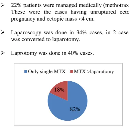
Results: Lower abdominal pain was most common presenting symptom of ectopic pregnancy in 96% cases. The classical triad of symptoms (amenorrhea, abdominal pain and vaginal bleeding) was present in only 28% cases. PID contributed 24% cases and previous abortion contributed 28% cases indicating these two as the common risk factors. Ampulla was the commonest site for ectopic pregnancy, in 52% cases. Salpingostomy performed mainly at this site. In 6% cases ectopic pregnancy in infundibulum were treated with fimbrial expression and fimbriectomy. 22% patients were managed medically (methotraxte). These were the cases having unruptured ectopic pregnancy and ectopic mass <4 cm. Laparoscopy was done in 34% cases, in 2 cases it was converted to laparotomy. While open laparotomy was done in 40% cases.
Conclusions: Ectopic pregnancy is a treatable problem. Ultrasonography plays central role in the diagnosis and management. Mode of therapy is determined by a combination of clinical symptoms, sonography findings and serum
-HCG values. Surgical management is still a cornerstone of management of ectopic pregnancy. But now scope of medical and laparoscopic management is also there. In recent years laparotomy has been replaced by laparoscopic surgery which is more conservative, minimally invasive and less time consuming which leads to quick recovery.
Keywords: Ectopic pregnancy, Laparoscopy
Popularity of intrauterine devices (relative 7 times more in in situ IUCD)
ART (5-7%)
Decrease in rate of mortality
The dramatic decrease in rate of mortality in patients of ectopic gestation can be directly attributable to earlier detection which allows us to pursue more conservative treatment modalities. High resolution USG and serum -HCG level are helpful in early detection which allows the use of minimally invasive surgery or medical treatment which will significantly enhance both survival and conservation of reproductive capacity.3
The objectives of this study are:
To study the incidence in various age groups.
To study predisposing factors
To study different modes of clinical presentation
To study different sites of ectopic pregnancy
To study changing trends of modern management from radical surgical method to laparoscopic and medical management
Course of ectopic pregnancy:4
Spontaneous resolution
Tubal abortion (Ampullary, fimbrial)
Resolution
Pelvic hematocele
Hematosalpinx
Tubal rupture (Isthmic, interstitial- at 12-16 wks)
Rupture followed by secondary abdominal pregnancy.
Diagnostic modalities for ectopic pregnancy
Serial B-HCG titre
- 66% rise in B-HCG titre is seen at 2 days in normal intrauterine pregnancy (IUP).
- 15 % of normal IUP has < 66 % rise at 48 hrs. - So 53 % rise is now considered to be normal for an
IUP (Barnhart).
- Discriminating zone 1500 IU/ml of βHCG 5 - At this level of βHCG, IUP must be located. - However 1000 IU & 2000 IU are also suggested. - There is a decrease of 21 - 35% if spontaneous
abortion occurs.
- Slower decrease or slower increase suggests ectopic pregnancy.
- In 17% patients with Ectopic Pregnancy βHCG doubling time is normal.
- Progesterone level5
o It has a poor diagnostic value.
o >25 ng/ml suggests normal IUP.
o <5 ng/ml suggests abortion.
o Ectopic Pregnancy can have range from 5-25 ng.
o Limitations include patients undergoing infertility treatment via IVF.
Features seen on TVS (Transvaginal ultrasound)5
- Ectopic cardiac activity Diagnosis is 100 % - Ectopic gestational. Sac strong evidence
- Ectopic mass & fluid in POD moderately strong evidence
Colour Doppler5
[image:2.595.348.506.560.720.2]Ring of fire sign seen around a cold uterus is diagnostic of ectopic gestation on color Doppler ultrasound.
Laparoscopy
Laparoscopy is rarely required for diagnosis. Findings of laparoscopy may be normal in very early stages of ectopic pregnancy. The advantage is that a diagnostic scopy can be easily being converted to therapeutic scopy and treatment of ectopic gestation can be done simultaneously.
Uterine curettage5
Curettage is usually among the least used diagnostic modalities for ectopic pregnancy. It can help to differentiate from nonviable intrauterine pregnancy. The confirmatory finding for IUP is the presence of chorionic villi in normal saline.
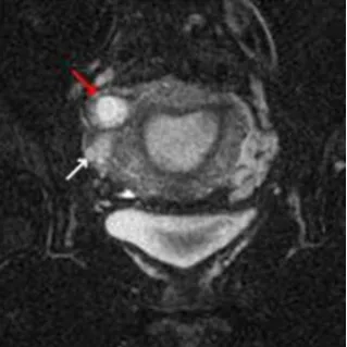
MRI
[image:3.595.88.248.374.534.2]For academic purposes, MRI scanning can be used to get an accurate diagnostic imaging for ectopic pregnancy. The MRI of choice is coronal section of T2-weighted fat-saturated MRI of the pelvis which clearly depicts the ectopic gestational sac with its exact location within the pelvic adnexa.
Figure 2:MRI of the pelvis.
Medical management6
Selection criteria
- Mass <3.5 cm - βHCG <4000 mIU/ml - <6 weeks gestational age - Absent cardiac activity - No hemoperitoneum
- Hemodynamically stable patient
- Patient must be compliant & well counselled
Methotrexate regimen
- Single dose - 50 mg/m2 IM - HCG on day 4 & day 7
- It should decrease by 15 % of the initial level - If it persists on day 7, repeat the dose (max. 4 doses) - If it decreases do weekly HCG till Ectopic
Pregnancy resolves (<10 mIU)
METHODS
A study of 50 cases of tubal ectopic pregnancy was carried out from May 2009 to June 2011 in tertiary health centre.
Patients managed with following treatment modalities were selected for study.
1) Medical management (MTX)
2) Laparoscopic management: Salpingostomy,
Salpingectomy
3) Laparotomy management: Salpingostomy, segmental resection, fimbrial expression, salpingectomy
RESULTS
Figure 3 shows age distribution during study.
Figure 3: Age distribution.
Table 1: Parity distribution.
Parity Number Percentage
0 30 60%
1 11 22%
2 07 14%
3 01 02%
4 01 02%
6%
48%
34%
8%
4% 0%
10% 20% 30% 40% 50% 60%
20 or less
21-25 26-30 31-35 >35
As per study, 60% patients were nullipara and 82% were primipara or nullipara. This suggests that low parity has high chances of ectopic pregnancy.
Table 2: Symptoms.
Symptoms Number Percentage
Gharono et al. (2002)
Lower abdominal pain 48 96% 83.6%
Bleeding per vagina 20 40% 73%
Amenorrhea 44 88% 77.6%
Classical triad 14 28% -
Syncope 07 14% 25.7%
Nausea & vomiting 3 6% 16.5%
Lower abdominal pain was most common presenting symptoms in 96% cases.
[image:4.595.56.281.391.531.2]88% cases had amenorrhea as the presenting symptom. Classical triad of symptoms (amenorrhea, abdominal pain and vaginal bleeding) was present in only 28% cases. Syncope was experienced by 14% patients, which was due to significant intraperitoneal hemorrhage.
Figure 4: Risk factors.
PID contributed 24% cases and previous abortion contributed 28% cases indicating these two as the common risk factors.
Infertility was also a contributory factor in 18% cases.
Figure 5: Presenting signs.
[image:4.595.311.531.529.747.2]Abdominal tenderness was present in 92% cases and cervical motion tenderness was present in 68% cases.
Figure 6:investigations done & its accuracy.
UPT was done in all cases, positive in 45 cases.
TVS done in all cases, of which 96% cases showed positive results.
In 2 cases in which UPT & TVS are negative, Laparoscopy done which shows 100% conclusive information.
Table 3: Mode of treatment.
Mode of treatment Number Percentage
Caminiti et al.
(2006)
Medical treatment 11 22% 21%
Laparoscopy 17 34% 26%
Laparotomy 20 40% 28%
Laparoscopy +
Laparotomy 02 04% 25%
MTX >laparotomy 02 04% -
22% patients were managed medically (methotraxte). These were the cases having unruptured ectopic pregnancy and ectopic mass <4 cm.
Laparoscopy was done in 34% cases, in 2 cases it was converted to laparotomy.
Laprotomy was done in 40% cases.
Figure 7: Success rate - Methotrexate (MTX).
50 50
2
45 48
2
90 96 100
UPT TVS Laparoscopy
Done Positive Accuracy
82% 18%
[image:4.595.68.268.626.747.2]In 82% cases single dose Methotrexate was successful.
Figure 8: Type of surgery (laparoscopic).
Above chart suggests that in 63% cases salpingectomy was preferred. While in 36% cases salpingostomy was done.
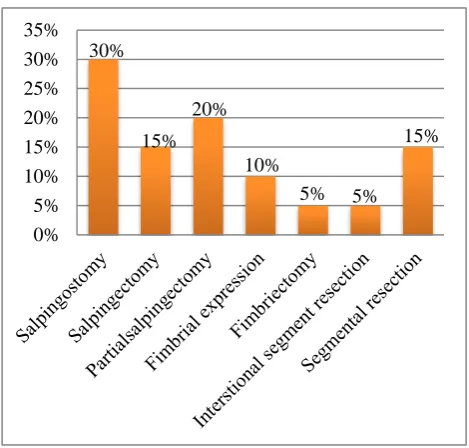
Figure 9: Type of laparotomy.
[image:5.595.52.286.91.241.2]Above data shows that in laparotomy, 30% patients had salpingostomy, while 20 % patients underwent partial salpingectomy.
Table 4: Success rate of treatment.
Treatment Success rate Sraj et al.
Methotrexate 81.81% 94.70%
Laparoscopy 89.40% 91.40%
Laparotomy 100% -
Success rate 81.81% for medical management by methotrexate
Laparoscopic management has a success rate of 89.4%.
Figure 10: Site of ectopic pregnancy.
Ampulla was the commonest site for ectopic pregnancy, in 52% cases. Salpingostomy performed mainly at this site.
[image:5.595.50.285.316.540.2]In 6% cases ectopic pregnancy in infundibulum were treated with fimbrial expression and fimbriectomy.
Table 5: Laparotomy vs. laparoscopy.
Complication Laparotomy Laparoscopy
Morbidity More Less
Postoperative adhesions More Less
Risk of future ectopic More Less!
Future fertility Same Same
Persistent ectopic Less! More
Experience / instruments Routine Special
Cochrane database review 2007
Salpingectomy vs. Salpingostomy?
Salpingectomy partial or total is only indicated when there is uncontrollable bleeding or future
childbearing is not desired.
Risk of recurrent ectopic & infertility are same.
Persistent trophoblastic activity is slightly more in salpingostomy.
DISCUSSION
Ectopic pregnancy is a treatable problem. Ultrasonography plays central role in the diagnosis and management.
Mode of therapy is determined by a combination of clinical symptoms, ultrasound findings and serum -HCG values.
Surgical management is still a cornerstone of management of ectopic pregnancy, but now scope of medical and laparoscopic management is also there. 62.63%
36.84%
10.52% 0.00%
10.00% 20.00% 30.00% 40.00% 50.00% 60.00% 70.00%
30%
15% 20%
10%
5% 5%
15%
0% 5% 10% 15% 20% 25% 30% 35%
12%
52%
6% 2%
[image:5.595.309.548.355.444.2]In recent years laparotomy has been replaced by laparoscopic surgery which is more conservative, minimally invasive and less time consuming which leads to quick recovery.
Funding: No funding sources Conflict of interest: None declared Ethical approval: Not required
REFERENCES
1. Surette AM, Dunham SM. Early pregnancy risks. In: AH DeCherney et al., eds. Current Diagnosis and Treatment Obstetrics & Gynecology. 11th ed. New York: McGraw-Hill; 2013: 234-249.
2. Varma R, Gupta J. Tubal ectopic pregnancy. BMJ Clin Evid (Online). 2009;2009:1406.
3. Cunningham FG et al. Ectopic pregnancy. In: Cunningham FG et al., eds. Williams Obstetrics.
23rd ed. New York, NY: McGraw-Hill; 2010: 238-256.
4. Leven ED et al. Ectopic pregnancy and spontaneous abortion. In: RG Nabel, eds. ACP Medicine. Hamilton, ON: BC Decker; 2010: Section 16, Chap. 6.
5. Fritz MA. Ectopic pregnancy. In: Fritz MA, Speroff L, eds. Clinical Gynecologic Endocrinology and Infertility. 8th ed. Philadelphia, PA: Lippincott Williams and Wilkins; 2011: 1383-1412.
6. American College of Obstetricians and
Gynecologists (2008, reaffirmed 2012). Medical management of ectopic pregnancy. ACOG Practice
Bulletin No. 94. Obstet and Gynecol.
2012;111(6):1479-85.
DOI: 10.5455/2320-1770.ijrcog20140618