University of Windsor University of Windsor
Scholarship at UWindsor
Scholarship at UWindsor
Electronic Theses and Dissertations Theses, Dissertations, and Major Papers
8-3-2017
Quantifying Forearm Soft Tissue Motion and Shock Attenuation
Quantifying Forearm Soft Tissue Motion and Shock Attenuation
following Hand Impacts Consistent with Forward Falls using
following Hand Impacts Consistent with Forward Falls using
Massless Skin Surface Markers
Massless Skin Surface Markers
Danielle Linda Gyemi University of Windsor
Follow this and additional works at: https://scholar.uwindsor.ca/etd
Recommended Citation Recommended Citation
Gyemi, Danielle Linda, "Quantifying Forearm Soft Tissue Motion and Shock Attenuation following Hand Impacts Consistent with Forward Falls using Massless Skin Surface Markers" (2017). Electronic Theses and Dissertations. 6599.
https://scholar.uwindsor.ca/etd/6599
This online database contains the full-text of PhD dissertations and Masters’ theses of University of Windsor students from 1954 forward. These documents are made available for personal study and research purposes only, in accordance with the Canadian Copyright Act and the Creative Commons license—CC BY-NC-ND (Attribution, Non-Commercial, No Derivative Works). Under this license, works must always be attributed to the copyright holder (original author), cannot be used for any commercial purposes, and may not be altered. Any other use would require the permission of the copyright holder. Students may inquire about withdrawing their dissertation and/or thesis from this database. For additional inquiries, please contact the repository administrator via email
Quantifying Forearm Soft Tissue Motion and Shock Attenuation following Hand Impacts Consistent with Forward Falls using Massless Skin Surface Markers
By
Danielle Gyemi
A Thesis
Submitted to the Faculty of Graduate Studies through the Department of Kinesiology in Partial Fulfillment of the Requirements for the Degree of Master of Human Kinetics at the
University of Windsor
Windsor, Ontario, Canada
2017
Quantifying Forearm Soft Tissue Motion and Shock Attenuation following Hand Impacts Consistent with Forward Falls using Massless Skin Surface Markers
By
Danielle Gyemi
APPROVED BY:
______________________________________________ W. Altenhof
Department of Mechanical, Automotive and Materials Engineering
______________________________________________ P. van Wyk
Department of Kinesiology
______________________________________________ D. Andrews, Advisor
Department of Kinesiology
iii
DECLARATION OF ORIGINALITY
I hereby certify that I am the sole author of this thesis and that no part of this
thesis has been published or submitted for publication.
I certify that, to the best of my knowledge, my thesis does not infringe upon
anyone’s copyright nor violate any proprietary rights and that any ideas, techniques,
quotations, or any other material from the work of other people included in my thesis,
published or otherwise, are fully acknowledged in accordance with the standard
referencing practices. Furthermore, to the extent that I have included copyrighted
material that surpasses the bounds of fair dealing within the meaning of the Canada
Copyright Act, I certify that I have obtained a written permission from the copyright
owner(s) to include such material(s) in my thesis and have included copies of such
copyright clearances to my appendix.
I declare that this is a true copy of my thesis, including any final revisions, as
approved by my thesis committee and the Graduate Studies office, and that this thesis has
iv ABSTRACT
The purpose of this thesis was twofold: 1) quantify planar (2D) displacement and
velocity of, and the amount of shock attenuated by, the forearm soft tissues following a
forward fall impact; and 2) compare two massless skin surface marker designs with
different uniformity and visual contrast (i.e., single layer, uniform (SLU) design; stacked,
non-uniform (SNU) design) in terms of how well they can be tracked over varying skin
pigmentation using automated motion capture software. Simulated forward fall impacts
were performed by two groups of participants (skin pigmentation: light – 9F, 8M; dark –
9F, 6M) using a torso-release apparatus, in which a high speed camera (5000 f/s)
captured planar motion of the right forearm. Automated motion tracking software
(ProAnalyst®) was used to quantify displacement, velocity, and shock attenuation
capacity of the forearm soft tissue from manually digitized markers. Overall, the greatest
mean peak soft tissue displacement (1.47 cm) and velocity (112.8 cm/s) occurred in the
distal direction in proximal regions of the forearm where more soft tissue is distributed.
Soft tissue displacement and velocity exhibited similar trends, increasing from distal to
proximal regions of the forearm, while impact shock accelerations were not attenuated in
the forearm, but instead increased by 76%. Apart from proximal rebound distance, soft
tissue kinematics between females and males did not significantly differ (p > 0.05).
Conversely, the effects of specific tissue masses (i.e., bone mineral content, fat mass, lean
mass, and wobbling mass) on tissue kinematics varied between the sexes. Significant
differences were found between marker designs for displacement, rebound distance, and
velocity (p ≤ 0.05), wherein the SLU design consistently produced higher values than the
v DEDICATION
To each and every person who has made a positive impact on my life, no matter how big
vi
ACKNOWLEDGEMENTS
First and foremost, I would like to express my sincerest gratitude to my Master’s
thesis advisor, Dr. Dave Andrews. Your ability to bring out the best in your students and
help foster their potential for success, even at times when they may doubt it, is a quality I
truly admire. I am very thankful to have had the opportunity to work with and learn from
you over the past couple years and cannot tell you how much I have come to appreciate
your unparalleled guidance and support.
To my committee members, Dr. William Altenhof and Dr. Paula van Wyk, thank
you for the time and resources you have invested towards the preparation, execution, and
completion of this thesis. Your insights and advice during this process have been
invaluable and I hold both of you in the highest regard.
A special thank you to Don Clarke, without whom this thesis project would not
have been possible. Your willingness to not only assist with numerous aspects of this
study, but also take the extra time to entertain my curiosity and answer my questions, no
matter how small or seemingly obvious, is greatly appreciated.
Thank you to the past and present faculty and staff in the Department of
Kinesiology for their kindness and support throughout my graduate degree, as well as my
fellow grad students and lab mates for listening to my frustrations and always offering
encouragement to persevere through many challenging and stressful times. You have all
contributed to making this experience an enjoyable one.
To my each of my friends who lent a helping hand during the many trials and
tribulations of my thesis project, regardless of the extent of the efforts, I am fortunate to
vii
Finally, I wish to thank my family, including my parents and sisters, for their
viii
TABLE OF CONTENTS
DECLARATION OF ORIGINALITY ... iii
ABSTRACT ... iv
DEDICATION ... v
ACKNOWLEDGEMENTS ... vi
LIST OF TABLES ... xii
LIST OF FIGURES ... xvi
LIST OF APPENDICES ... xx
LIST OF ABBREVIATIONS ... xxi
GLOSSARY ... xxii
1. INTRODUCTION ... 1
1.1. Hypotheses ... 9
2. REVIEW OF LITERATURE ... 11
2.1. Soft Tissue Motion Analysis ... 11
2.1.1. Soft Tissue Artifact ... 11
2.1.2. Stereo-photogrammetry ... 12
2.2. ProAnalyst® Motion Analysis Software ... 21
2.3. Massless Surface Markers ... 24
2.3.1. Marker Contrast ... 25
2.3.2. Marker Shape ... 26
2.4. In Vivo Test Specimen ... 28
2.4.1. Forearm Tissue Composition... 28
2.4.2. Human Skin ... 29
2.5. Forward Fall Impacts ... 30
2.5.1. Shock Wave Attenuation ... 32
2.5.2. Impact Apparatuses ... 34
2.5.3. Hand Impact Force ... 37
3. METHODS ... 39
3.1. Participants ... 39
3.1.1. Sample Size ... 39
ix
3.1.3. Exclusion Criteria ... 41
3.1.4. Consent ... 42
3.2. Instrumentation and Apparatus ... 42
3.2.1. High Speed Camera ... 42
3.2.2. Torso-Release Impact Apparatus... 43
3.2.3. Force Plates... 45
3.2.4. Laser Displacement Transducer ... 46
3.2.5. Markers ... 47
3.3. Procedures ... 49
3.3.1. Anthropometric Measurements ... 50
3.3.2. Participant Preparation ... 51
3.3.3. Impact Protocol... 52
3.3.4. Video Analysis ... 55
3.4. Data Analysis ... 58
3.4.1. Statistical Analysis ... 62
4. RESULTS ... 65
4.1. Purpose 1 ... 69
4.1.1. Soft Tissue Displacement ... 69
4.1.2. Soft Tissue Velocity ... 71
4.1.3. Soft Tissue Shock Attenuation ... 73
4.2. Purpose 2 ... 73
4.2.1. Sex and Soft Tissue Displacement ... 75
4.2.2. Region and Soft Tissue Displacement ... 76
4.2.3. Sex, Region, and Soft Tissue Displacement ... 80
4.2.4. Sex and Soft Tissue Velocity... 83
4.2.5. Region and Soft Tissue Velocity ... 83
4.2.6. Sex, Region, and Soft Tissue Velocity ... 87
4.2.7. Sex and Soft Tissue Shock Attenuation ... 88
4.3. Purpose 3 ... 90
4.3.1. Participant Tissue Masses ... 90
4.3.2. Displacement (Distal) Correlations ... 90
x
4.3.4. Rebound Distance (Proximal) Correlations ... 92
4.3.5. Displacement (Anterior) Correlations ... 93
4.3.6. Displacement (Posterior) Correlations ... 94
4.3.7. Rebound Distance (Posterior) Correlations ... 95
4.3.8. Velocity (Distal) Correlations ... 95
4.3.9. Velocity (Proximal) Correlations ... 96
4.3.10. Velocity (Anterior) Correlations ... 97
4.3.11. Velocity (Posterior) Correlations ... 98
4.3.12. Shock Attenuation Correlations ... 99
4.4. Purpose 4 ... 100
4.4.1. Marker Design, Skin Pigmentation, and Region on Soft Tissue Displacement ... 101
4.4.2. Marker Design, Skin Pigmentation, and Region on Soft Tissue Rebound Distance ... 104
4.4.3. Marker Design, Skin Pigmentation, and Region on Soft Tissue Velocity .... 106
4.4.4. Improvements to Automated Motion Tracking ... 109
5. DISCUSSION ... 110
5.1. Purpose 1 ... 110
5.2. Purpose 2 ... 112
5.2.1. Forearm Region and Tissue Movement ... 112
5.2.2. Sex and Tissue Movement ... 116
5.2.3. Sex and Shock Attenuation ... 117
5.3. Purpose 3 ... 118
5.3.1. Tissue Masses and Movement ... 118
5.4. Purpose 4 ... 121
5.4.1. Skin Pigmentation and Tissue Movement ... 121
5.4.2. Marker Design, Skin Pigmentation, and Tissue Movement ... 122
5.5. Limitations ... 125
6. FUTURE DIRECTIONS ... 128
6.1. Muscle Activation and Joint Angles ... 128
6.2. Forward Fall Impact Simulations... 129
6.3. Three-Dimensional Motion Capture ... 130
xi
6.5. Wrist Guards and Compliant Safety Flooring ... 131
7. CONCLUSIONS... 134
REFERENCES ... 138
APPENDICES ... 151
Appendix A ... 151
Appendix B ... 154
Appendix C ... 155
xii
LIST OF TABLES
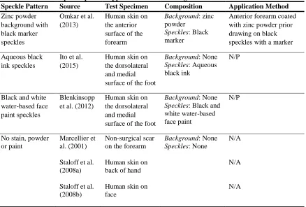
Table 1. Overview of speckle pattern approaches used on biological soft tissue in vivo... ... 17
Table 2. Overview of surface markers used with ProAnalyst® motion tracking software. ... 23
Table 3. Mean (±SD) age, height, and body mass of all participants. ... 40
Table 4. Prediction equations for bone mineral content (BMC), fat mass (FM), lean mass (LM), and wobbling mass (WM) tissues of the forearm (Modified from Arthurs et al., 2009). ... 50
Table 5. Mean (±SD) overall, female, and male peak IRFs (N) across the six trials for each hand. ICC values are included for between trials. No significant differences were found for any variable. ... 67
Table 6. Mean (±SD) overall, female, and male peak normalized IRFs as a percentage (%) of BW across the six trials for each hand. ICC values are included for between trials. No significant differences were found for any variable... 68
Table 7. Mean (±SD) overall, female, and male peak soft tissue displacement (cm) in the proximal, distal, anterior and posterior directions for each of the eight regions. ... 70
Table 8. Mean (±SD) overall, female, and male peak soft tissue velocity (cm/s) in the proximal, distal, anterior and posterior directions for each of the eight regions. ... 72
Table 9. Mean (±SD) overall, female, and male peak soft tissue distal and proximal accelerations (cm/s2) of the forearm and un-normalized calculations of shock attenuation (%)... 73
Table 10. Mean (±SD) bone mass (g), fat mass (g), lean mass (g), and wobbling mass (g) of all participants estimated using the tissue mass prediction equations from Arthurs et al. (2009). ... 90
xiii
Table 12. Pearson correlations (r-values) between male distal soft tissue displacement (cm) in each region (1-8), as well as the entire forearm (mean), and specific tissue masses: bone mineral content (BMC), fat mass (FM), lean mass (LM), and wobbling mass (WM)... 91
Table 13. Pearson correlations (r-values) between female proximal soft tissue
displacement (cm) in each region (1-8), as well as the entire forearm (mean), and specific tissue masses: bone mineral content (BMC), fat mass (FM), lean mass (LM), and
wobbling mass (WM). ... 91
Table 14. Pearson correlations (r-values) between male proximal soft tissue displacement (cm) in each region (1-8), as well as the entire forearm (mean), and specific tissue
masses: bone mineral content (BMC), fat mass (FM), lean mass (LM), and wobbling mass (WM)... 92
Table 15. Pearson correlations (r-values) between female proximal soft tissue rebound distance (cm) in each region (1-8), as well as the entire forearm (mean), and specific tissue masses: bone mineral content (BMC), fat mass (FM), lean mass (LM), and
wobbling mass (WM). ... 92
Table 16. Pearson correlations (r-values) between male proximal soft tissue rebound distance (cm) in each region (1-8), as well as the entire forearm (mean), and specific tissue masses: bone mineral content (BMC), fat mass (FM), lean mass (LM), and
wobbling mass (WM). ... 93
Table 17. Pearson correlations (r-values) between female anterior soft tissue
displacement (cm) in each region (1-8), as well as the entire forearm (mean), and specific tissue masses: bone mineral content (BMC), fat mass (FM), lean mass (LM), and
wobbling mass (WM). ... 93
Table 18. Pearson correlations (r-values) between male anterior soft tissue displacement (cm) in each region (1-8), as well as the entire forearm (mean), and specific tissue masses: bone mineral content (BMC), fat mass (FM), lean mass (LM), and wobbling mass (WM)... 94
Table 19. Pearson correlations (r-values) between female posterior soft tissue
displacement (cm) in each region (1-8), as well as the entire forearm (mean), and specific tissue masses: bone mineral content (BMC), fat mass (FM), lean mass (LM), and
xiv
Table 20. Pearson correlations (r-values) between male posterior soft tissue displacement (cm) in each region (1-8), as well as the entire forearm (mean), and specific tissue
masses: bone mineral content (BMC), fat mass (FM), lean mass (LM), and wobbling mass (WM)... 94
Table 21. Pearson correlations (r-values) between female posterior soft tissue rebound distance (cm) in each region (1-8), as well as the entire forearm (mean), and specific tissue masses: bone mineral content (BMC), fat mass (FM), lean mass (LM), and
wobbling mass (WM). ... 95
Table 22. Pearson correlations (r-values) between male posterior soft tissue rebound distance (cm) in each region (1-8), as well as the entire forearm (mean), and specific tissue masses: bone mineral content (BMC), fat mass (FM), lean mass (LM), and
wobbling mass (WM) ... 95
Table 23. Pearson correlations (r-values) between female distal soft tissue velocity (cm/s) in each region (1-8), as well as the entire forearm (mean), and specific tissue masses: bone mineral content (BMC), fat mass (FM), lean mass (LM), and wobbling mass (WM). ... 96
Table 24. Pearson correlations (r-values) between male distal soft tissue velocity (cm/s) in each region (1-8), as well as the entire forearm (mean), and specific tissue masses: bone mineral content (BMC), fat mass (FM), lean mass (LM), and wobbling mass (WM). ... 96
Table 25. Pearson correlations (r-values) between female proximal soft tissue velocity (cm/s) in each region (1-8), as well as the entire forearm (mean), and specific tissue masses: bone mineral content (BMC), fat mass (FM), lean mass (LM), and wobbling mass (WM)... 97
Table 26. Pearson correlations (r-values) between male proximal soft tissue velocity (cm/s) in each region (1-8), as well as the entire forearm (mean), and specific tissue masses: bone mineral content (BMC), fat mass (FM), lean mass (LM), and wobbling mass (WM)... 97
xv
Table 28. Pearson correlations (r-values) between male anterior soft tissue velocity (cm/s) in each region (1-8), as well as the entire forearm (mean), and specific tissue masses: bone mineral content (BMC), fat mass (FM), lean mass (LM), and wobbling mass (WM). ... 98
Table 29. Pearson correlations (r-values) between female posterior soft tissue velocity (cm/s) in each region (1-8), as well as the entire forearm (mean), and specific tissue masses: bone mineral content (BMC), fat mass (FM), lean mass (LM), and wobbling mass (WM)... 98
Table 30. Pearson correlations (r-values) between male posterior soft tissue velocity (cm/s) in each region (1-8), as well as the entire forearm (mean), and specific tissue masses: bone mineral content (BMC), fat mass (FM), lean mass (LM), and wobbling mass (WM)... 99
Table 31. Pearson correlations (r-values) between female soft tissue peak acceleration (cm/s2), as well as un-normalized shock attenuation, and specific tissue masses: bone mineral content (BMC), fat mass (FM), lean mass (LM), and wobbling mass (WM). .... 99
xvi
LIST OF FIGURES
Figure 1. Different optoelectronic camera system markers and formations: A) active light emitting markers; B) passive reflective markers; C) single markers and marker triads (Modified from Gao & Zheng, 2008; Qualisys, 2016a, 2016b, 2016c). ... 14
Figure 2. Examples of black and white paint combinations used to create speckle patterns for DIC analyses: A) black paint speckles with no background applied to a biomimetic elastomer; B) black paint speckles with a white painted background applied to swine brain tissue (Modified from Libertiaux et al., 2011; Mates et al., 2012). ... 18
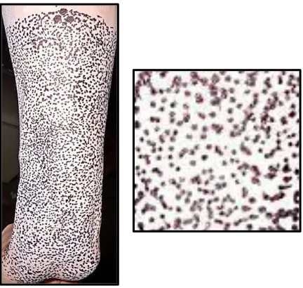
Figure 3. Zinc powder and black marker speckle pattern used on the anterior surface of the human forearm for in vivo DIC analysis of wrist extension (Modified from Omkar et al., 2013). ... 20
Figure 4. Square grid of uniform circular markers used for tracking soft tissue motion on the distal lower extremity (Modified from Brydges et al., 2015). ... 24
Figure 5. Different types of speckle patterns: A) random speckle; B) small black spots; C) large black spots; D) small black spots and random speckle; E) large black spots and random speckle (Modified from Haddadi and Belhabib, 2008). ... 27
Figure 6. Cross-section of human skin and subcutaneous layer (Modified from Tortora & Nielsen, 2014). ... 30
Figure 7. Schematic diagram representing the hand, wrist, and forearm biomechanics associated with a forward fall impact on the hand of an outstretched arm. ... 31
Figure 8. Bimodal shape of the measured ground reaction force on the hand during forward fall arrests with two force peaks: peak impact force (Fimp) and peak braking force
(Fbrk) (Modified from Chiu & Robinovitch, 1998). ... 37
Figure 9. Schematic diagram of the experimental test set-up: A) high-speed camera; B) primary flood light; C) secondary flood light; D) left hand force plate; E) right hand force plate. ... 43
Figure 10. Schematic diagram of the torso-release apparatus and the location of the high speed camera and force plates: A) sagittal view; B) posterior view. ... 44
xvii
Figure 12. Schematic diagram of the SLU marker design (2 x 2 cm square grid of circular black dots of 0.5 cm diameter) on the forearm from A) posterior and B) lateral views. .. 48
Figure 13. Schematic diagram of the SNU marker design (2 x 2 cm square grid of circular white dots of ~1 cm diameter with random black dots overlaid on top) on the forearm demonstrating the contrast for A) light and B) dark skin pigmentations. ... 48
Figure 14. Flowchart of the procedures. ... 49
Figure 15. Picture of the measurement scale used to standardize the distance participants stood from the force plates on the elevated platform. ... 53
Figure 16. ProAnalyst® calibration process performed for all videos... 56
Figure 17. Schematic diagram of the marker grid pattern (2 x 2 cm squares of dots) and the four analysis zones (0%, 25%, 50%, and 75%) on the forearm. ... 57
Figure 18. Screenshot from ProAnalyst® (zoomed in) showing the two columns of
markers (A and B) selected for the 0%, 25%, 50%, and 75% zones. ... 58
Figure 19. Schematic diagram of the marker grid (2 x 2 cm squares of dots) and the eight regions on the forearm. ... 59
Figure 20. Schematic diagram of the most distal and proximal markers used for
calculating shock attenuation in the forearm. ... 60
Figure 21. Onset point analysis procedures showing the graphical representation of the displacement (X), velocity (Vx), and acceleration (Ax) curves along the proximal-distal axis. ... 61
Figure 22. Sample displacement curves (proximal-distal axis) for each of the eight regions across the forearm for a single trial from one participant. The curves from each region have been aligned in time and displacement in order to show the relative
differences. ... 74
Figure 23. Mean (SE) peak soft tissue rebound distance in the proximal direction between females and males. ... 76
Figure 24. Mean (SE) peak soft tissue displacement in the distal direction for each region. ... 77
xviii
Figure 26. Mean (SE) peak soft tissue displacement in the anterior direction for each region.. ... 78
Figure 27. Mean (SE) peak soft tissue rebound distance in the proximal direction for each region. ... 79
Figure 28. Mean (SE) peak soft tissue rebound distance in the posterior direction for each region. ... 80
Figure 29. Interaction effect of Sex and Region (1-8) on proximal displacement. ... 81
Figure 30. Interaction effect of Sex and Region (1-8) on proximal rebound distance (* = statistically significant at p ≤ 0.05). ... 82
Figure 31. Mean (SE) peak soft tissue velocity in the distal direction for each region. ... 84
Figure 32. Mean (SE) peak soft tissue velocity in the proximal direction for each region. ... 85
Figure 33. Mean (SE) peak soft tissue velocity in the anterior direction for each region. 86
Figure 34. Mean (SE) peak soft tissue velocity in the posterior direction for each region. ... 87
Figure 35. Interaction effect of Sex and Region (1-8) on anterior velocity. ... 88
Figure 36. Interaction effect of Marker Design (SLU, SNU) and Region (1-8) on distal displacement (* = statistically significant at p ≤ 0.05). ... 102
Figure 37. Interaction effect of Marker Design (SLU, SNU) and Skin Pigmentation (light, dark) on distal displacement (* = statistically significant at p ≤ 0.05). ... 102
Figure 38. Interaction effect of Marker Design (SLU, SNU) and Skin Pigmentation (light, dark) on proximal displacement... 103
Figure 39. Interaction effect of Marker Design (SLU, SNU) and Region (1-8) on proximal rebound distance for the light skin pigmentation group (* = statistically
significant at p ≤ 0.05). ... 105
Figure 40. Interaction effect of Marker Design (SLU, SNU) and Region (1-8) on proximal rebound distance for the dark skin pigmentation group (* = statistically
xix
Figure 41. Interaction effect of Marker Design (SLU, SNU) and Region (1-8) on distal velocity (* = statistically significant at p ≤ 0.05)... 107
Figure 42. Interaction effect of Marker Design (SLU, SNU) and Region (1-8) on
proximal velocity (* = statistically significant at p ≤ 0.05). ... 107
xx
LIST OF APPENDICES
xxi
LIST OF ABBREVIATIONS
BMC –bone mineral content
CT – computed tomography
DIC – digital image correlation
FM –fat mass
ISO – International Organization for Standardization
LEDs – light emitting diodes
LM –lean mass
MRI – magnetic resonance imaging
PULARIS – Propelled Upper Limb fall ARrest Impact System
SLU – single layer, uniform SNU – stacked, non-uniform
STA – soft tissue artifact
UV – ultraviolet
xxii GLOSSARY
Anisotropic –Having a physical property that is directionally dependent and exhibits
different values when measured in different directions.
Anterior (displacement) – The downward motion of the forearm soft tissue
perpendicular to the long axis of the forearm following forward fall impact.
Attenuation –Weakening orreduction in force, intensity, or value that occurs as a result
of absorption, spreading, or distance.
BMC (bone mineral content) – The amount of bone material or mineral in a specific
bone site (measured in grams).
Calibration – The process of checking the experimental readings of a device or
instrument against a known standard to determine the correctness of its quantitative measurements.
Contrast –The ratio to which adjacent areas of an image differ in brightness.
Deformation –The action or process of changing in shape through the application of
mechanical loads.
Digital Image Correlation (DIC) – A non-contact optical technique that employs
tracking and image registration practices to acquire 2D and/or 3D measurements of deformation, displacement, and strain on the surface of a specimen.
Displacement – A vector value that refers to the change in position of a moving body
from an initial to final position in a given direction.
Displacement Field – A region in a body for which the displacement of all points is
defined.
Distal (displacement) – The motion of the forearm soft tissue towards the wrist joint
following forward fall impact.
Distal Upper Extremity –The furthermost section of the upper extremity relative to the
trunk, consisting of the forearm and hand.
Fall – (of a person) downward movement from a loss of balance, typically rapidly and freely without control, resulting in impact with the ground or other lower level.
FM (fat mass) – The total mass of the adipose tissue in the body or segment.
Impact –A transient event in which a high force or shock is applied when two or more
bodies collide.
In-plane motion –The motion of a body such that all its points move within (parallel to)
xxiii
In Vitro –The study of biological tissues outside of their living biological context.
In Vivo –The study of biological tissues within a living body.
LM (lean mass) –The total mass of all body tissues that does not contain fat (i.e.,
muscle).
Out-of-plane motion –The motion of a body such that its points move in additional
planes outside of the fixed plane of motion.
Pendulum – A body suspended from a fixed point that has the ability to swing freely
back and forth under the action of gravity.
Photogrammetry – The science of making measurements from photographs; high-speed
imaging is often employed to detect, measure, and record the exact positions of surface reference points on any moving object to quantify 2D and 3D motion fields.
Posterior (displacement) – The upward motion of the forearm soft tissue perpendicular
to the long axis of the forearm following forward fall impact.
Proximal (displacement) – The motion of the forearm soft tissue towards the elbow
joint following forward fall impact.
Shape Permutations – One of several possible variations in shape.
Shock (mechanical) –A sudden, transient acceleration of a system caused by an abrupt
change in force application, such as an impact event.
Shock attenuation – A reduction in the amplitude of the impact force that occurs as the
shock wave propagates through the tissues of the body.
Shock wave –The propagation of a stress wave through a medium such as the soft
tissues of the human body.
Soft Tissue –A generic term for tissues that are not bone that connect, support, or
surround various structures and organs of the body. This includes muscle, fat, skin, tendons, blood vessels, etc.
Spatial Resolution – The capacity of an imaging system to distinguish between small
details of adjacent points; it is dependent on the number of independent pixel values available per unit length.
Stereo-photogrammetry –An extension of photogrammetry that uses the process of
triangulation to estimate 3D points on the surface of an object using measurements made in two or more photographic images taken from different positions.
Strain –A measure of deformation that represents the displacement between particles in
a deformed body relative to the same particles in a reference (undeformed) body.
xxiv
Subset –A defined set consisting of elements within a larger, inclusive set.
Triangulation – The process of determining the location of a point by measuring the
angles relative to its position from known reference points at either end of a fixed baseline.
Viscoelastic –Having a combination of both viscous and elastic properties when
undergoing deformation.
WM (wobbling mass) –The non-rigid tissues of the body (lean and fat masses) that are
1
1. INTRODUCTION
A common reaction to a fall resulting from a loss of balance during recreational
and daily activities is to land on the hand of an outstretched arm to protect your head and
trunk from injury (Hsiao & Robinovitch, 1998; O’Neill et al., 1994). Forces sustained by
the distal upper extremity from this type of fall arrest impact have the potential to
jeopardize the structural integrity of the wrist, forearm, and elbow joint, leading to severe
injuries such as sprains, dislocations, and fractures (Nevitt & Cummings, 1993; Oskam et
al., 1998; Palvanen et al., 2000; Sasaki et al., 1999).
Forward falls and direct impacts to the hand and wrist have been highlighted in
the literature as being particularly problematic due to the high incidence of upper
extremity injury associated with them (Idzikowski et al., 2000; Nevitt & Cummings,
1993; Palvanen et al., 2000; Vellas et al., 1998). Among older adults, Vellas et al. (1998)
found that approximately 50% of falls occurred in the forward direction. Of those falls,
the hands were the part of the body that were impacted most frequently. Palvanen et al.
(2000) found that the majority of elderly patients sustaining a fracture to the upper
extremity (i.e., proximal humerus fracture: ~47%; elbow fracture: ~66%; wrist fracture:
~45%) reported that the fall occurred in a forward or forward oblique direction. Seventy
six percent of patients with a wrist fracture in this sample reported the main impact to be
directed straight to the hand and wrist, a finding which supports previous work by Nevitt
and Cummings (1993) for elderly women. In addition, young adults engaged in
recreational activities such as snowboarding (Idzikowski et al., 2000) and rollerblading
(Mirhadi et al., 2015) are also often subjected to forward falls involving impacts that
2
of all snowboarding upper extremity injuries were due to a fall, with 53.6% occurring in
the forward direction.
It has been estimated that the direct medical costs for non-fatal fall-related injuries
to the upper extremity among adults aged 65 years and older in the United States in 2000
was approximately $3 billion (Stevens et al., 2006). In Canada, falls were the leading
cause of all injury-related hospitalizations (55%) and emergency room visits (30%) in
2010, accounting for $8.7 billion or 34% of the total injury costs that year; this included
$6.7 billion and $2 billion in direct and indirect costs, respectively (Parachute, 2015).
Considering that the baby boomer population is expected to grow exponentially over the
next two decades (Parachute, 2015), and high-risk sporting activities like snowboarding
are continuing to rapidly grow in popularity each passing year (Canadian Ski Council,
2014), this raises major concerns for potentially significant increases in healthcare costs
associated with forward fall-related injuries. Therefore, given the magnitude of the
negative health outcomes linked to forward fall-related injuries and the economic burden
that they place on healthcare systems, if the mechanism by which rigid and soft tissues
interact to attenuate impact shock as it propagates through the hand and forearm can be
identified, improved injury prevention strategies, such as age-specific fall arrest strategies
and modified wrist guard designs, may then be realized.
Prior research concerning the injury mechanisms of a fall on the hand of an
outstretched arm has focused largely on the in vitro impact response of the distal radius
(i.e., bone) (Burkhart et al., 2012a; Muller et al., 2003; Myers et al., 1991), since it is a
very common fracture site for both young and older adult populations. Comprising
3
al., 2009), distal radius fractures are often due to high-energy trauma from sporting
activities and relatively low-energy trauma from accidental falls (Krishnan, 2002).
Although these studies demonstrate the capacity of the distal radius to dissipate high
levels of mechanical energy from dynamic impact loads, rigid tissues (bone) do not act
independently to protect the human body from injury. There is much evidence to support
that the response of soft tissues (muscle, fat, skin) relative to bone also plays an important
protective role in mitigating the potentially injurious effects of impact through shock
attenuation (Cole et al., 2006; Gittoes et al., 2006; Pain & Challis, 2001; Pain & Challis,
2002; Pain & Challis, 2006), despite often being viewed as error (or soft tissue artifact –
STA) that needs to be removed from biomechanical analyses (Peters et al., 2010). To
date, impact events involving the lower extremity (e.g., running, drop landings) have
received the most attention due to the frequency of their occurrence in everyday human
movement (Cole et al., 2006; Gittoes et al., 2006; Pain & Challis, 2006). However, Pain
and Challis (2002) demonstrated that soft tissue deformation of the forearm following a
downward hand striking task could account for approximately 70% of the dissipated
energy lost during these impacts.
The contribution of active mechanisms (i.e., muscle activation and joint angle) as
well as passive structures (soft and rigid tissue masses) for impact shock attenuation in
the body has been well documented for various foot impacts (Chu & Caldwell, 2004;
Coventry et al., 2006; Dufek et al., 2009; García-Pérez et al., 2014; Mercer et al., 2003;
Mercer et al., 2010; Schinkel-Ivy et al., 2012; Zhang et al., 2005). In contrast, with
respect to hand impacts, only the influence of active mechanisms has been examined
4
the role of different tissue masses, such as bone mineral content (BMC), fat mass (FM),
lean mass (LM), and wobbling mass (WM), for passive energy dissipation, has yet to be
assessed. Following controlled heel impacts with a human pendulum, Schinkel-Ivy et al.
(2012) were able to show that increases in absolute leg tissue masses corresponded to
decreases in tibial acceleration responses, with LM and BMC having the most significant
contributions. Therefore, quantifying soft tissue motion in the distal upper extremity may
help identify how the different tissue types, and amounts of specific tissue masses
between individuals, influence the motion of soft tissue following a forward fall impact.
Review of the literature has shown that a broad spectrum of motion tracking
techniques has been employed to quantify soft tissue motion (predominantly in the lower
extremity) for human movement analysis, each with their own set of limitations.
Three-dimensional (3D) optoelectronic systems (Fuller et al., 1997; Gao & Zheng, 2008; Wolf
& Senesh, 2010) as well as Magnetic Resonance Imaging (MRI) (Akbarshahi et al., 2010;
Sangeux et al., 2006) involve the use of complex and expensive equipment that often
requires the assistance of a trained professional to operate, whereas radiological methods
such as X-ray and video fluoroscopy can expose the participant to potentially harmful
radiation (Akbarshahi et al., 2010; Kuo et al., 2011; Sati et al., 1996; Südhoff et al., 2007;
Wrbaškić & Dowling, 2007). Consistent with each of these methods is also the
requirement for externally mounted devices (e.g., accelerometers, active or passive skin
surface markers, etc.) to be attached to the body segment in order to track soft tissue
motion; an action that has been found to alter the natural physiological movement of the
underlying soft tissue (Leardini et al., 2005; Stefanczyk et al., 2013). Consequently, if
5
measured using motion tracking techniques, eliminating the need to affix external devices
to the participant is key in order to avoid any non-physiological tissue responses
following impact. Photogrammetric motion tracking methods that utilize massless skin
surface markers (e.g., paint, marker pens) would thus be a preferred contact,
non-invasive, and radiology-free measurement tool to document and assess soft tissue motion
during dynamic loading.
One such method, digital image correlation (DIC), is a non-contact, optical
technique that is used to understand the deformation behavior of a wide range of
materials. Traditionally, DIC is used in the field of experimental solid mechanics to
assess the mechanical properties of inanimate structural materials (e.g., wood [Betts et
al., 2010; Samarasinghe & Kulasiri, 2004], concrete [Choi & Shah, 1997; Shih & Sung,
2013], and metals [Backman et al., 2006; Bewerse et al., 2013]). To date, the use of this
method for quantifying soft tissue motion in vivo during human movement analysis is
very limited. Instead of using discrete surface markers, DIC works by tracking unique,
stochastic details (i.e., random speckle patterns) on the surface of the specimen when in a
non-deformed and deformed state to provide full-field measurements of displacement and
strain (Sutton et al., 2009). With no mechanical interaction with the specimen required,
the capacity of DIC to provide accurate results is directly dependent on the quality of the
speckle pattern on the surface of the specimen, as it is the sole carrier of the deformation
information (Crammond et al., 2013; Hua et al., 2011; Lecompte et al., 2006; Pan et al.,
2010). Therefore, when a suitable textured pattern is not naturally present on the
specimen being evaluated, a high-quality artificial speckle pattern must be created in
6
Unfortunately, the feasibility of successfully implementing DIC for in vivo
human soft tissue motion and shock attenuation impact analysis is limited by several
factors. Primarily, methodological inconsistencies and insufficient procedural
information in the literature regarding speckle pattern application methods (i.e.,
application tools and strategies) and compositions (i.e., substance(s) applied and
background contrast) make it difficult to discern an optimal approach for creating a
high-quality speckle pattern on any surface, let alone a surface with physical properties as
complex as human skin. In addition, the expense associated with acquiring DIC
equipment and software, along with the tools necessary for applying a speckle pattern on
human skin, such as complete airbrush kits, is very costly. Time efficiency is also
another limitation, as the proper use of DIC requires extensive training, and being able to
consistently achieve a desired speckle pattern during specimen preparation has been
shown to require a tedious “trial-and-error” process (Betts et al., 2010; Lecompte et al.,
2006; Yavari et al., 2013).
A study by Brydges et al. (2015) demonstrated the success of an alternative
method in which position and velocity data of leg soft tissue motion following heel
impacts (i.e., pendulum, drop landing) could be quantified using a motion capture system
with automatic feature tracking capabilities (ProAnalyst®; Xcitex, Cambridge, MA,
USA). An appealing element of the marker system presented in this work was the use of
massless skin surface markers, wherein a flexible plastic stencil was used to apply a grid
of circular black surface markers onto the leg with a permanent black marker pen. Since
no supplementary external devices were attached to the body segment, the risk of
7
removed. Furthermore, compared to more traditional motion capture systems, software
systems with the capacity to perform automatic feature tracking, such as ProAnalyst®, are
relatively inexpensive and compatible with a variety of camera video formats. More
specifically, the use of ProAnalyst® to analyze high-speed camera images has been
validated against optical displacement transducers with respect to the measurement of
vertical wheel displacements of heavy mining vehicles, illustrating a maximum
difference of 4.05% between the two methods (Tonkovich et al., 2012). The adaptability
of the system also lends itself to a range of applications, as the number of markers that
can be used is specific to the research objectives (with a minimum of one marker needed
to measure tissue motion), and thus, can be expanded to investigate soft tissue motion of
other body segments (e.g., the distal upper extremity).
Despite the many advantages of using this experimental set-up for recording and
tracking soft tissue motion, it is not without its limitations. Position measurements were
reported to have good within- and between-measurer reliability; however, velocity
measurements were found to be slightly less reliable (Brydges et al., 2015). Although
this was shown to have a relatively small impact on the differences between the measured
kinematic variables (between-measurer: <0.8 cm for position, <3.7 cm/s for velocity;
within-measurer: <0.5 cm for position, <2.6 cm/s for velocity), modifications can be
made to improve both the accuracy and reliability of this method. For example, with
respect to marker contrast and shape, changes can be made to account for different
participant skin pigmentations and enhance the accuracy of automated tracking,
8
Therefore, utilizing the research methods conducted by Brydges et al. (2015) to
examine the impact response of forearm soft tissue following a forward fall on the hand
of an outstretched arm will provide the most authentic insight into how soft tissue motion
attenuates impact energy in the upper extremity. Taking into consideration the tissue
composition of the distal upper extremity will also better our understanding of the
individual role that each tissue mass (e.g., FM, LM, WM, and BMC) plays in dissipating
impact shock, which will help drive biomechanical modelling efforts. Additionally,
testing the use of a novel massless surface marker design with improved contrast and
shape variation on participants of varying skin pigmentation will benefit the automated
motion tracking process and broaden the inclusivity of this motion tracking technique.
Therefore, the purposes of this thesis are to:
1) quantify planar (2D) displacement and velocity of, and the amount of shock
attenuated by, the soft tissues of the forearm following a forward fall impact;
2) assess if there are differences in soft tissue motion and impact shock attenuation
due to sex, or as a function of the region of the forearm measured;
3) identify the relationship between the displacement, velocity, and shock
attenuation capacity of the forearm soft tissues and their individual tissue masses
(BMC, FM, LM, and WM);
4) determine if a stacked, non-uniform (SNU) marker design (non-uniform, ~0.5 cm
diameter black dots overlaid on top of a grid of contrasting ~1 cm diameter white
dots; 2 cm inter-marker distance) produces significantly different kinematic
9
pigmentations compared to the single layer, uniform (SLU) marker design (grid of
uniform, 0.5 cm diameter black dots; 2 cm inter-marker distance) previously
established by Brydges et al. (2015).
1.1. Hypotheses
It is hypothesized that:
1) it will be possible to quantify planar (2D) displacement and velocity of,
and the amount of shock attenuated by, the soft tissues of the forearm following a
forward fall impact through a combination of photogrammetric motion tracking
techniques and massless skin surface markers;
2a) males will have greater soft tissue displacements and velocities, and attenuate a
greater amount of shock following impact than females because, on average,
males have greater amounts of WM in the arms compared to females (Mazess et
al., 1990);
2b) WM in the proximal region of the forearm will demonstrate greater displacement
than the distal region, and WM in the anterior region will be greater than the
posterior region, respectively, since greater amounts of WM are distributed
proximally and anteriorly in the forearm;
3) the amount of impact shock attenuated by passive soft tissue movement will be
positively correlated with the absolute magnitude of the estimated forearm tissue
masses (FM, LM, WM, and BMC), similar to the results found by Schinkel-Ivy et
al. (2012) for the influence of leg soft tissue composition on tibial acceleration
10
will be positively correlated with the displacement and velocity of soft tissue in
the proximal-distal direction, but have no significant correlations in the
anterior-posterior directions.
4) the SNU marker design will produce similar kinematic results to the SLU marker
design for the light skin pigmentation group, but significantly different results for
the dark skin pigmentation group. This is because the SNU marker design will
improve the automated marker tracking process by 1) adding a localized white
background against the black dot marker to provide consistently high contrast for
marker detection across all skin pigmentations (especially those individuals with
darker skin pigmentations), and 2) using non-uniformly shaped markers to
enhance discrete marker recognition and tracking by providing a more unique
11
2. REVIEW OF LITERATURE
2.1. Soft Tissue Motion Analysis
2.1.1. Soft Tissue Artifact
In studies of human motion analysis using skin-based systems, the movement of
surface markers on the skin relative to the underlying bone that they are intended to
represent is a phenomenon commonly referred to as “soft tissue artifact” (STA) (Peters et
al., 2010). The occurrence of STA is the result of soft tissue deformation associated with
skin movement and inertial effects, especially around the joints (Cappozzo et al., 1996),
as well as muscular contractions (Leardini et al., 2005). It has also been shown that the
amount of STA observed is dependent on multiple factors, such as differing physical
characteristics between individuals (Holden et al., 1997), the location of surface markers
on the body (Schwartz et al., 2004), and the nature of the task being performed (Fuller et
al., 1997; Leardini et al., 2005; Manal et al., 2003).
Ultimately, STA is viewed as a major source of error in human movement
analysis that limits the ability to accurately quantify skeletal system kinematics and
detailed joint movements. For example, during a natural cadence walking task, Manal et
al. (2003) demonstrated that the movement of bone compared to soft tissue on the
proximal tibia had average differences of 7.4, 3.7, and 2.1 mm along the X
(medial-lateral), Y (anterior-posterior), and Z (superior-inferior) axes, respectively. Across a
variety of tasks (i.e., stationary bicycling, squatting, normal gait, voluntary swing
movement), Fuller et al. (1997) observed larger differences overall, in which magnitudes
12
calculated joint angles were found to have significantly different values from those that
were expected due to soft tissue motion. Consequently, many solutions have been
proposed in the literature to minimize the error related to STA when analyzing human
movement that involve a variety of complex estimation algorithms and skin marker set
techniques (e.g., point cluster technique) in an attempt to more accurately model the
motion of the underlying bone (Alexander and Andriacchi 2001; Cappello et al., 1997;
Cappello et al., 2005; Gao et al., 2007; Soderkvist & Wedin, 1993). However, with
regard to human impact analysis, measuring soft tissue motion is critical to understanding
the mechanisms by which the complex structure and non-linear, viscoelastic mechanical
behaviour of soft tissues work with rigid tissue to prevent injury through impact shock
attenuation. Therefore, removing soft tissue motion from biomechanical analyses would
eliminate an important contributor to how the human body attenuates shock during
impacts (Pain and Challis, 2002).
2.1.2. Stereo-photogrammetry
The combination of stereo-photogrammetry and skin markers is one of the most
commonly used measurement methods for 3D motion analysis of human movement
(Akbarshahi et al., 2010; Cappozzo et al., 2005; Fuller et al., 1997; Holden et al., 1997;
Houck et al., 2004; Leardini et al., 2005; Manal et al., 2003; Stagni et al., 2005). This
motion tracking technique utilizes the process of triangulation and photogrammetry to
estimate 3D coordinates of specific reference points on the surface of a moving object
from measurements based on two or more images taken from different, fixed positions.
13
markers along the length of the body segment being analyzed; therefore, making
stereo-photogrammetry an appealing alternative without the limitations of other motion tracking
techniques, such as the highly invasive nature of intra-cortical pins and percutaneous
skeletal trackers, potentially harmful radiological exposure of X-ray and fluoroscopy, and
limited static or quasi-static investigations of MRI (Peters et al., 2010).
2.1.2.1. Optoelectronic Systems
Depending on the type of marker that is applied, different stereo-photogrammetry
based motion capture systems and software can be used to track and measure soft tissue
motion. Optoelectronic (3D) camera systems work by means of light detection, and
involve the use of two basic types of systems (Figure 1): active, which use infrared light
emitting diodes (LEDs) as markers that actively emit light themselves (Ball, 2011; Fuller
et al., 1997; Houck et al., 2004; Scholz, 1989), and passive, which use retro-reflective
markers that passively reflect light off their surface (Akbarshahi et al., 2010; Chu et al.,
2010; Dufek et al., 2009; Gao & Zheng, 2008; Holden et al., 1997; Manal et al., 2003;
Pain & Challis, 2002; Stagni et al., 2005; Telfer et al., 2010). Single markers allow
variables such as displacement, velocity, and accelerations to be analyzed, whereas
marker triads can be used to acquire measures of rotation and translation of the skin
surface (Gao & Zheng, 2008). Each optoelectronic system uses multiple motion position
sensors or cameras to record and track 3D marker movement, with upwards of 12
cameras used for experimental setups in certain cases (Dufek et al., 2009).
Despite the progressions made by optoelectronic camera systems in human
14
capabilities are reliant on external devices (i.e., active or passive markers) typically
mounted to the skin using double-sided adhesive tape (Fuller et al., 1997; Gao & Zheng,
2008; Houck et al., 2004) or straps (Fuller, et al., 1997; Manal et al., 2003). A recent
investigation by Stefanczyk et al. (2013) found that the attachment of a 4 g accelerometer
to the distal lower extremity just distal to the knee joint using only a thin Velcro® strap
significantly altered the natural physiological motion of the underlying soft tissues of the
leg after heel impacts, especially in the proximal regions of the segment, closer to the
knee where the strap was fashioned. Moreover, affixing an external device to a body
segment can interfere with soft tissue motion by way of their mass (e.g., 4–7 g), size
(e.g., 10 mm diameter), and/or shape (Gao & Zheng, 2008). Therefore, when using
optoelectronic systems to quantify soft tissue movement following impact, researchers
cannot be certain that the true motion of soft tissue will remain undisrupted.
Another limitation of these optoelectronic systems is that the frame rate for data
collection decreases as the total number of markers used increases, and thus, are
Figure 1. Different optoelectronic camera system markers and formations: A) active light emitting markers; B) passive reflective markers; C) single markers and marker
15
commonly only able to capture images at relatively low frame rates. This can prove to be
problematic in terms of acquiring detailed kinematic data regarding soft tissue motion
from impact events, such as forward falls, due to the impulsiveness associated with these
events. Using the Optotrak Certus System (Northern Digital Inc., Waterloo, Canada) as
an example, if soft or rigid tissue movement were to be monitored, the sample rate would
be limited by the total number of markers, as calculated by Equation 1 (Northern Digital
Inc., 2016):
Sample rate = 4600/(𝑁 + 1.3) Hz (Eq. 1)
where N = number of markers.
Considering that prior investigations of shock attenuation in the distal upper
extremity following hand impacts has used as many as 28 reflective surface markers on
the forearm to measure soft tissue motion (Pain & Challis, 2002), using Equation 1, a
maximum sampling rate for this marker array would only be approximately 157 Hz.
With deceleration of the hand and arm following a forward fall observed to begin as
quickly as 20 ms after initial hand impact (Chiu & Robinovitch, 1998), a sampling rate of
this magnitude is not sufficient to collect a comprehensive view of the soft tissue
response that occurs during this time. Additionally, the expense associated with
optoelectronic systems is another limitation that must be taken into account as
asymmetrical or highly dynamic tasks may require the use of multiple cameras, in
addition to the active and passive marker sets, to optimally track the area of interest (Chu
16 2.1.2.2. Digital Image Correlation
Digital image correlation is a non-contact, optical method that employs tracking
and image registration techniques to measure full-field 2D or 3D surface displacement,
strain, and deformation of a specimen undergoing mechanical loading (Sutton et al.,
2009). A major advantage of this method is that no mechanical interaction with the
specimen is required, and thus, the need for external devices to measure kinematic
variables is eliminated. The basic principle of DIC involves identifying, matching, and
correlating target subsets from a recorded image of a specimen in a deformed state
relative to the corresponding subsets in an undeformed reference image (Reu, 2012a).
Typically, if the natural texture of the specimen does not have sufficient grey intensity
variation, this process is accomplished with the aid of an artificial speckle pattern applied
to the surface of the specimen. As a result, the resolution and accuracy of any DIC
analyses are conditional on the quality of the speckle pattern, whether naturally occurring
or artificially applied (Crammond et al., 2013; Hua et al., 2011; Lecompte et al., 2006;
Pan et al., 2010). Unfortunately, a general lack of practical instruction combined with a
limited number of references in the literature regarding artificial speckle pattern
application on the surface of biological soft tissues (e.g., human skin) in vivo make it
difficult to discern an optimal approach for utilizing DIC to track soft tissue motion.
Table 1 provides an overview of speckle pattern approaches that have been used on
17
Table 1. Overview of speckle pattern approaches used on biological soft tissue in vivo.
Speckle Pattern Source Test Specimen Composition Application Method Zinc powder
background with black marker speckles
Omkar et al. (2013)
Human skin on the anterior surface of the forearm
Background: zinc powder
Speckles: Black marker
Anterior forearm coated with zinc powder prior drawing on black speckles with a marker
Aqueous black ink speckles
Ito et al. (2015)
Human skin on the dorsolateral and medial surface of the foot
Background: None
Speckles: Aqueous black ink
N/P
Black and white water-based face paint speckles
Blenkinsopp et al. (2012)
Human skin on the dorsolateral and medial surface of the foot
Background: None
Speckles: Black and white water-based face paint
N/P
No stain, powder or paint
Marcellier et al. (2001)
Staloff et al. (2008a)
Staloff et al. (2008b)
Non-surgical scar on the forearm
Human skin on back of hand
Human skin on face
Background: None
Speckles: None
N/A
N/A
N/A
Note: N/A = Not applicable; N/P = Not provided
The use of painted speckle patterns has been observed in two recent studies that
utilized 3D DIC to assess the deformation of the human foot while running (Blenkinsopp
et al., 2012) and walking (Ito et al.,2015), respectively. Blenkinsopp et al. (2012)
reported that water-based face paint was used to produce a contrasting black and white
speckled pattern on the dorsal surface of the foot, whereas Ito et al.(2015) only used
speckles of aqueous black ink. The notion of using varying combinations of black and
white paint to create artificial speckle patterns is strongly supported in other experimental
domains, such as experimental solid mechanics and in vitro biological soft tissue DIC
studies (Figure 2), in which the most frequently used patterns include: black paint
speckles (Abanto-Bueno & Lambros, 2002; Choi & Shah, 1997; Gerhardt et al., 2012;
18
combination of black and white paint speckles (Samarasinghe & Kulasiri, 2004), and
black paint speckles over a solid base coat of white paint (Betts et al., 2010; Shih & Sung,
2013; Backman et al., 2006; Zhang & Arola, 2004; Libertiaux et al., 2011; Bruck et al.,
1989; Lan et al., 2014).
Although painted speckle patterns are capable of acting as virtually massless
marker systems for tracking soft tissue motion that (compared to external devices) allow
underlying tissues to move freely without interruption, notable limitations with this
approach are still present. First, in spite of evidence presented by Barranger et al. (2010)
supporting the superior accuracy of painted over powder speckle patterns at lower strains
(10%–50%), paint was still found to be susceptible to DIC measurement error following
the eventual occurrence of cracks within the patterns at strains greater than 50%, which
diminished their quality. Moreover, since this study used a flat, transparent silicone
specimen for testing, the findings may not be indicative of the additional complications
that the unique mechanical properties of human skin in vivo may pose under similar
Figure 2. Examples of black and white paint combinations used to create speckle patterns for DIC analyses: A) black paint speckles with no background applied to a biomimetic elastomer; B) black paint speckles with a white painted background applied to swine brain tissue (Modified
19
loading conditions. Second, while other motion tracking techniques, such as
optoelectronic systems, only measure localized points designated by discrete markers,
DIC software calculates global measures (i.e., full-field displacement and strain maps),
and thus, requires that the entire area of interest be covered in a speckle pattern. As a
result, it must be ensured that the applied speckle pattern does not alter the mechanical
properties of the soft tissue (e.g., increased stiffness and/or dehydration). Libertiaux et al.
(2011) used displacement-driven compression tests to demonstrate that the application of
a painted speckle pattern caused no significant statistical difference in the mechanical
response of brain tissue samples; the implications for human skin in vivo may be very
different.
Aside from painted speckle patterns, only two other speckle pattern approaches
have been used on human skin in vivoto execute DIC analyses. In an attempt to better
understand the etiology of carpal tunnel syndrome, Omkar et al. (2013) used a unique
speckle pattern approach to measure the strain of the superficial muscles and tendons in
the anterior compartment of the forearm during wrist extension in vivo, in which zinc
powder was coated on the surface of the right anterior forearm to improve the contrast
against a random black speckle pattern applied with a marker (Figure 3). However,
since no further detail was provided in the methods on this specimen preparation process
(e.g., brand of zinc powder, ease of application, cost, exposure time, etc.), the
reproducibility of this approach and its appropriateness for in vivoDIC research remains
questionable, especially given the notable adverse side effects associated with zinc-based
20
Figure 3. Zinc powder and black marker speckle pattern used on the anterior surface of the human forearm for in vivo DIC analysis of wrist extension (Modified from Omkar et al., 2013).
Alternatively, there have also been DIC studies on human skin that involve no
speckle pattern treatment, suggesting that the pores intrinsic to the skin form an ideal set
of markers for assessing its mechanical properties (Marcellier et al., 2001; Staloff et al.,
2008a; Staloff et al., 2008b). While this is an enticing option that would theoretically
provide the truest representation of soft tissue motion after impact without any form of
image artifact, each of these studies only tested very confined areas of the skin in which
the applied skin deformation was kept very subtle (e.g., wrinkles near the corner of the
eye when closing the eye lid) on participants with relatively light skin pigmentation.
Thus, further assessment of this technique would be necessary to determine if the contrast
would be sufficient for large impact deformations of the skin characteristic of entire body
21 2.2. ProAnalyst® Motion Analysis Software
ProAnalyst® is a motion analysis software package that employs photogrammetric
techniques to perform non-contact motion tracking analyses on a moving object. The
software allows users to measure and compute many kinematic variables associated with
specified reference points on the surface of the object throughout its motion pathway,
including position, velocity, acceleration, size, and location, in addition to other
characteristics. With both manual and automatic tracking capabilities, users have the
option to track reference points manually by continuously selecting the same feature
frame by frame, or automatically by selecting a feature in the initial video frame and then
using the automatic tracking tool within the software to locate and track the motion of
that feature in subsequent frames. ProAnalyst® is compatible with virtually any video
camera and format; however, it is often paired with high speed imaging systems to
document the motion pathway of an object for 2D or 3D analysis, depending on the
version being used. In addition, the comprehensive capabilities of this motion analysis
software make it a highly versatile tool that can be applied in laboratory simulations as
well as real-life tasks and activities across numerous fields of research (e.g., automotive,
ballistics, biomechanics, etc.). It is also worth noting that, as commercially available
premium motion analysis software, a large portion of the research performed with
ProAnalyst® software is conducted by companies that do not publish their findings to the
scientific community (e.g., NASA).
Although ProAnalyst® can be used for markerless motion tracking, in which
specific regions on the object of interest (e.g., edge length or diameter in the x-axis) are
22
2012; Audysho et al., 2013), researchers have also used a variety of surface markers in
combination with the motion analysis software to aid the automated marker tracking
process (see Table 2 for an overview of the different surface markers used with
ProAnalyst®). Reflective markers have been used with rats to track limb positions for
assessing locomotor compensation after peripheral nerve lesion (Bennet et al., 2012) and
to compute head rotational and lateral translational displacements in the coronal plane for
investigations on the pathology of diffuse axonal injury (Li et al., 2010). Neto and Magni
(2007) reported the use of high contrast markers affixed to the lateral surface of
participants’ forearms (2 cm apart) from elbow to wrist to analyze the kinematic
characteristics of Kung Fu Yau-Man palm strikes without impact. Furthermore,
Facchinello et al. (2015) reported that rigid markers were attached to vertebral bodies to
test the stabilization capacity of monolithic spinal rods with different flexural stiffness
and anchoring arrangement.
As opposed to using externally mounted surface markers, recent studies have also
been performed in which massless surface markers have been used. O'Neill et al. (2015)
applied nontoxic, water-soluble white paint markers over specific anatomical landmarks
to assess the kinematics of the chimpanzee pelvis and hindlimb during bipedal walking.
Similarly, white paint dots were also utilized in a study by Tonkovich et al. (2012) to
investigate tyre deformation behaviour on heavy mining vehicles. In contrast, Crowley et
al. (2015) reported the use of black ink to track the limb position of specific anatomical
landmarks on rats in relation to the effect of intrathecal neurochemical excitation of
thoracic propriospinal neurons on locomotion performance. Finally, a recent study by