ABSTRACT
QIU, RUOYI. Dynamics of DNA Mismatch Repair Initiation Complexes Revealed by Single Molecule Fluorescence. (Under the direction of Dr. Keith Weninger).
Dynamics of DNA Mismatch Repair Initiation Complexes Revealed by Single Molecule Fluorescence
by Ruoyi Qiu
A dissertation submitted to the Graduate Faculty of North Carolina State University
in partial fulfillment of the requirements for the degree of
Doctor of Philosophy
Physics
Raleigh, North Carolina 2012
APPROVED BY:
_______________________________ ______________________________ Dr. Keith Weninger Dr. Robert Riehn
BIOGRAPHY
Ruoyi Qiu was born in Guangzhou, China in 1985 and then moved to Shenzhen, a coastal city and the first special economic zone in China. In high school, he was attracted by the way how physics applies mathematics to solve practical problems. He started to read popular science books, such as ‘Fearful Symmetry’ and ‘The Arrow of Time’. In 2003, he entered the
ACKNOWLEDGMENTS
Committee members: Keith Weninger (NC State, Physics), Robert Riehn (NC State, Physics), Hans Hallen (NC State, Physics), Alexander Nevzorov (NC State, Chemistry)
Weninger Lab: Brandon Choi, Laura Wessels, John Sakon, Trevor Anderson, Elizabeth Sacho
TABLE OF CONTENTS
LIST OF FIGURES ... viii
LIST OF ABBREVIATIONS ... ix
Chapter 1Introduction to DNA Mismatch Repair ... 1
1.1 Biological significance of mismatch repair ... 1
1.2 The methyl-directed Mismatch Repair in E.coli ... 2
1.2.1 The hemimethylated GATC site as a strand signal ... 3
1.2.2 Bidirectional mismatch provoked excision ... 3
1.3 Mismatch Repair in Eukaryotes ... 4
1.4 Structural and Biochemical Information about MutS ... 5
1.4.1 Crystal Structure of MutS ... 5
1.4.2 MutS scanning homoduplex DNA for mismatches ... 6
1.4.3 DNA bending by MutS ... 7
1.4.4 MutS forming sliding clamp after mismatch recognition ... 8
1.4.5 Adenosine nucleotide binding and ATPase of Taq MutS ... 9
1.5 Structural and Biochemical Information about MutL ... 9
1.5.1 Crystal Structures of E.coli MutL ... 10
1.5.2 ATP-driven conformational changes in MutL ... 10
1.6 Coupling of Mismatch Recognition and Strand Discrimination ... 11
Figures... 13
References ... 20
Chapter 2Overview of experiment methods used in this thesis ... 29
2.2 Single molecule fluorescence microscopy ... 31
2.3 Surface immobilization methods ... 32
2.3.1 Liposome encapsulation... 32
2.3.2 Streptavidin-lipid bilayer surface ... 32
2.4 Protein preparation and dye labeling ... 33
2.4.1 Taq MutS expression and purification ... 33
2.4.2 Taq MutL expression and purification ... 33
2.4.3 Protein labeling strategies ... 34
Figures... 35
References ... 38
Chapter 3 Large conformational changes in MutS during DNA scaning, mismatch recognition and repair signaling ... 40
3.1 Summary ... 40
3.2 Large conformational changes in MutS during DNA scaning, mismatch recognition and repair signaling ... 42
3.3 Supplementary Content within this Article... 42
Chapter 4Conformational Dynamics of MutS-MutL Complex Formation ... 43
4.1 Introduction ... 43
4.2 Results ... 43
4.2.1 MutS sliding clamp formation is inhibited by MutL ... 43
4.2.2 MutS interconverts between two conformations when interacting with MutL ... 45
4.2.3 Multiple MutS and MutL are recruited to form a large complex ... 45
4.2.4 MutS domains I are open in MutS-MutL complex ... 47
4.4 Biological Implications ... 52
Figures... 54
References ... 59
Chapter 5Summary ... 61
LIST OF FIGURES
Figure 1.1 Mechanism of E.coli methyl-directed mismatch repair. ... 13
Figure 1.2 Possible role of MutL latent endonuclease activity in bidirectional eukaryotic mismatch-provoked excision systems... 14
Figure 1.3 Structures of Taq MutS. ... 15
Figure 1.4 Single molecule studies of DNA bending by MutS. ... 16
Figure 1.5 The composite model of full length MutL ... 17
Figure 1.6 ATPase regulation of MutL ... 18
Figure 1.7 Models for coupling between mismatch recognition and strand discrimination signal ... 19
Figure 2.1 Förster resonance energy transfer. ... 35
Figure 2.2 Prism-type TIRFM setup for single molecule FRET measurement. ... 36
Figure 2.3 Schematic diagram of microscope slide and immobilization methods ... 37
Figure 4.1 Dynamical differences in MutS-mismatch interaction between the absence and presence of MutL ... 54
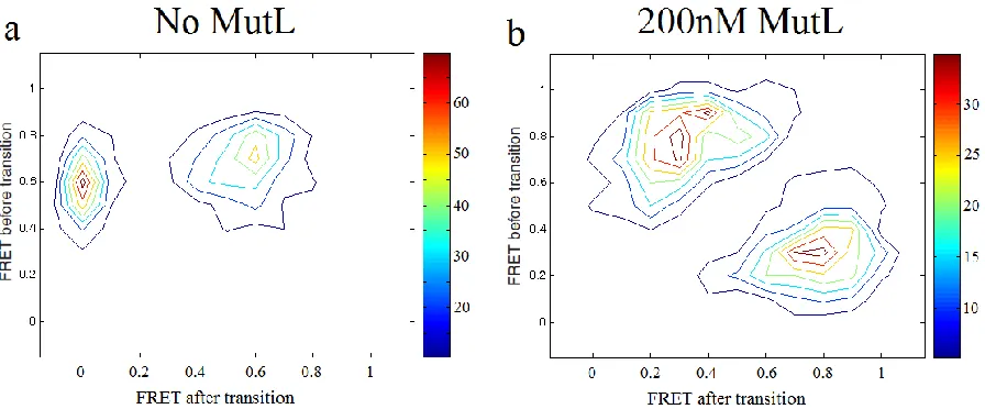
Figure 4.2 Transition density plots of donor-MutS and acceptor-DNA FRET. ... 55
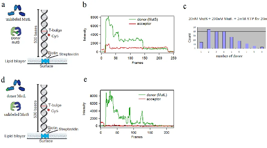
Figure 4.3 Counting the number of MutS and MutL by photobleaching of donor ... 56
Figure 4.4 FRET measurement of donor-acceptor labeled MutS in the MutS-MutL assemblies. ... 57
LIST OF ABBREVIATIONS
% percent
alpha
beta
gamma
delta
μ micro
~ approximately
°С Celsius degree
3’ three prime end
5’ five prime end
6His 6 histidine tag
Å angstrom
ADP adenosine diphosphate AFM atomic force microscopy Arg arginine
ATP adenosine-5’-triphosphate ATPase adenosine triphosphatase DNA deoxyribonucleic acid
dsDNA double stranded deoxyribonucleic acids E.coli Escherichia coli
IDL insertion-deletion loop in vitro outside a living system in vivo inside a living system MMR DNA mismatch repair
nM nanomolar
PCNA proliferating cell nuclear antigen PCR polymerase chain reaction
Phe phenylalanine
RFC replication factor C
smFRET single molecule Förster resonance energy transfer Taq Thermus aquaticus
Chapter 1
Introduction to DNA Mismatch Repair
1.1 Biological significance of mismatch repair
DNA replication is a primary cellular process that occurs in all living organisms that allows a cell to produce a copy of the genomic DNA for cell division. The integrity of the genetic information is dependent on the fidelity of DNA replication combined with several DNA repair processes. Replication occurs with a small but finite error rate. The DNA mismatch repair (MMR) system is responsible for correcting base-base mismatches and small nucleotide insertion/deletion (IDL) mismatches that arise from polymerase misincorporation. The activity of the MMR system elevates the fidelity of replication 50-1000 fold (Kunkel and Erie 2005; Iyer, Pluciennik et al. 2006; Li 2008). Inactivation of MMR causes greatly increased rate of genome-wide point mutations and is linked to hereditary nonpolyposis colorectal cancer (HNPCC) as well as to sporadic tumors in a variety of tissues (Peltomaki 2003; Peltomaki 2005).
The activities of MMR are best characterized in Escherichia coli, although substantial information on yeast and human systems is also available. The complete MMR process can be generally divided into several steps: mismatch recognition by MutS homologues; nascent strand identification; strand excision and mismatch removal; DNA resynthesis and ligation (Kunkel and Erie 2005; Iyer, Pluciennik et al. 2006; Li 2008).
Interactions between MMR proteins MutS and MutL are responsible for accomplishing this task. The strand discrimination signal can be transient in time, remotely located from the mismatch and can be on either side of the mismatch. The mechanism by which MMR proteins identify parent and daughter strand identity is still quite controversial. In this thesis, we present experimental characterization of the conformational dynamics of MMR protein-DNA complexes in the early steps of MMR to shed light on these questions.
1.2 The methyl-directed Mismatch Repair in
E.coli
coupled to DNA replication (Lopez de Saro and O'Donnell 2001; Jeruzalmi, O'Donnell et al. 2002).
1.2.1 The hemimethylated GATC site as a strand signal
The hemimethylated GATC sites only exist transiently before they are modified by the Dam methylase (Lyons and Schendel 1984). As expected, heteroduplex DNA without Dam modification on either strand has little or no strand bias for repair, and DNA with modification on both strands is not fixed (Lu, Clark et al. 1983; Pukkila, Peterson et al. 1983). Interestingly, strains that overproduce Dam methylase are mutators, probably due to a shorter time window during the existence of the unmodified GATC sites (Herman and Modrich 1981). Although hemimethylated GATC sites play a major role in strand identification, a pre-existing strand break is also enough to conduct strand specific mismatch repair (Längle-Rouault, Maenhaut-Michel et al. 1987; Lahue, Au et al. 1989). In fact, studies show that the GATC modification is responsible for only a subset of repair events, and the rest might be attributed to natural strand discontinuities on the nascent strand during DNA replication (Claverys and Mejean 1988).
1.2.2 Bidirectional mismatch provoked excision
1.3 Mismatch Repair in Eukaryotes
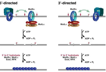
MMR in eukaryote has many features in common with E.coli MMR (Modrich 2006). DNA mismatches are first located by a MutS homologue (MutS Msh6) or Mut (Msh2-Msh3)). MutSis primarily responsible for repair of single base-base and IDL mismatches, and Mut is primarily responsible for repairing IDL mismatches containing up to 16 extra nucleotides on one strand (McCulloch, Gu et al. 2003). Next, a MutL homologue (MutL (Mlh1-Pms2 in humans, Mlh1-Pms1 in yeast)) is recruited in an ATP dependent manner. There is no known MutH homologue in eukaryote, so the signal for identifying the newly synthesized strand is less clear. A single strand nick or gap is sufficient to direct mismatch repair in vitro, and strand discontinuities associated with replication have been suggested as the signal in vivo (Umar, Buermeyer et al. 1996; Pavlov, Mian et al. 2003).
A similar mode of nick-directed excision has been observed, with single stranded gap spanning from the nick to a 90~170 bases beyond the mismatch (Fang and Modrich 1993). Different from the methyl-directed MMR in E.coli, there is no helicase involvement in eukaryotic mismatch repair (Bennett, Umar et al. 1997). And only one exonuclease, EXOI, has been convincingly related to excision reaction. A simple reconstituted system, which contains only four human proteins MutS, MutL, EXOI and RPA, supports mismatch removal with a pre-existing nick 5’ to the mismatch (Genschel, Bazemore et al. 2002; Genschel and Modrich 2003). EXOI hydrolyzes duplex DNA with only 5’ to 3’ polarity, but adding PCNA and RFC to the reaction surprisingly activates the ability of 3’ to 5’ mismatch removal (Waga and Stillman 1998). Also, depletion of ExoI activity attenuated not only the 5’ to 3’ excision but also 3’ to 5’ excision (Genschel, Bazemore et al. 2002).
After the mismatch is removed, the MMR is completed by resynthesis of the gap by DNA polymerase and ligation by DNA ligase (Longley, Pierce et al. 1997; Kunkel and Erie 2005).
1.4 Structural and Biochemical Information about MutS
As mentioned above, MMR is initiated by biniding of MutS to mismatches. MutS homologues are homodimers in prokaryotes and heterodimers in eukaryotes. The behaviors and functionality of MutS appear to be conserved from bacteria to human. All MutS homologues have both DNA binding and ATPase activities. MutS has several different tasks including searching for mismatches on double stranded DNA and signaling downstream steps with other MMR cofactors. In light of these different activities, the relation between structures and functions in MutS is very important for understanding the early stages of MMR.
1.4.1 Crystal Structure of MutS
Structures of Escherichia coli MutS,Thermus aquaticus (Taq) MutS, human MutSand human MutS bound to various mismatch DNA bases and base insertion/deletions have been solved with x-ray crystallography (Lamers, Perrakis et al. 2000; Obmolova, Ban et al. 2000; Alani, Lee et al. 2003; Natrajan, Lamers et al. 2003; Warren, Pohlhaus et al. 2007; Gupta, Gellert et al. 2012). Taq MutS will be mainly described here, although all MutS structures are very similar.
mismatched base and the glutamate forms a hydrogen bond with the N3 of the mismatched thymine or N7 of the mismatched purine. Domain V contains the dimerization interface and adenosine nucleotide binding site. Domain III is directly connected to domain II, IV and V and is suggested to relate the nucleotide binding of domain V and the DNA binding of domain I. This Phe-X-Glu motif is conserved in prokaryotes and in the Msh6 subunit of eukaryotic MutS(Warren, Pohlhaus et al. 2007).
When MutS is bound to a mismatched DNA, the homodimer forms a ‘’ shape structure with two channels. The mismatched DNA is bound in the lower channel and is kinked by ~60°at the mismatch. All the structures with different mismatches reveal similar degree of DNA bending. The upper channel is large enough and has electrostatic potential to bind DNA, but no evidence of DNA binding has been found. The crystal structure of ADP occupied Taq MutS bound to T-bulge DNA is very similar to the one with no nucleotide, but the domain I of subunit A has higher mobility and the position of domain I of subunit B is altered (Alani, Lee et al. 2003).
1.4.2 MutS scanning homoduplex DNA for mismatches
machinery removes nucleosomes and makes ~260bp of naked DNA behind the progressing fork (Gasser, Koller et al. 1996). Association with replication positions MutS in the ideal location to track mismatches, allowing the MMR to initiate before loss of the strand discrimination signal. Interestingly, some Msh2-Msh6 proteins were found in an immobile state, and they can be activated to move with addition of ATP. In physiological conditions, all MutS are bound to ATP or ADP due to biochemical data. Taq MutS labeled with cy3 dye was also found to slide on homoduplex (Jeong, Cho et al. 2011; Cho, Jeong et al. 2012).
1.4.3 DNA bending by MutS
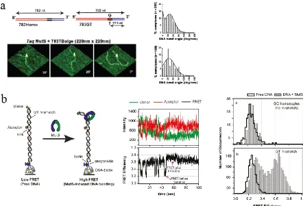
While crystallography has revealed high resolution structures of MutS-DNA complexes, there remain questions about how the DNA bending is related to the MMR pathways. Atomic force microscopy (AFM) and single molecule FRET have been used to visualize the conformations of mismatched and homoduplex DNA bound with MutS (Wang, Yang et al. 2003; Tessmer, Yang et al. 2008; DeRocco, Anderson et al. 2010; Sass, Lanyi et al. 2010). AFM provides snapshots of every MutS-DNA complex after they are ‘frozen’ by deposition on a surface (Figure 1.4a). The bending angle of DNA bound to MutS can be extracted from each image. The bending angle histograms show that in MutS-homoduplex complexes are all bent, but both bent and unbent populations are observed at mismatched sites (Figure 1.4). These observations indicate the unbent state is a result of specific interaction between MutS and the mismatched base, and the bent state may be an intermediate in the formation of the unbent state.
lifetime of each state, identifies 6 MutS bound states besides the free-DNA state and the transition probabilities between any two states. Among these MutS bound states, a bent state (B) and an unbent state (U) are most populated and thought to be the major states in the functional pathways. When a free mismatched DNA is bound by MutS, it is most likely to get into the B state first, then it transits to the U state (42%) or the MutS dissociates (35%) for most cases. This transition sequence is consistent with the suggestion that the bent state is an intermediate to the unbent state, which leads to the downstream reactions.
1.4.4 MutS forming sliding clamp after mismatch recognition
After recognition of a mismatch, the mechanism of how MutS signals initiation of MMR and identifies the strand signal is still under debate. Some studies suggest that MutS is activated to a sliding clamp by ADP->ATP exchange after recognition of a mismatch (Gradia, Subramanian et al. 1999; Schofield, Nayak et al. 2001). This idea is mainly based on the observations that E.coli MutS and human MutS dissociate from a mismatched DNA with unblocked ends by the challenge of ATP but stay on a mismatched DNA with blocked ends in the same condition. Multiple MutS loading on a circular DNA with a single mismatch further strengthens this suggestion (Gradia, Subramanian et al. 1999; Acharya, Foster et al. 2003). The ATP-activated sliding clamp is stable, with binding lifetime longer than 10mins, on a DNA without free ends (Acharya, Foster et al. 2003; Jeong, Cho et al. 2011). ATPS, which is a ATP analog with extremely slow hydrolysis rate, can trigger sliding clamp formation at the same efficiency, supporting the hypothesis that MutS functions as a molecular switch instead of a motor (Kadyrov, Dzantiev et al. 2006).
2010) and chemical cross-linking studies (Winkler, Marx et al. 2011) both revealed that the DNA binding is coupled to the ATPase domain, and both the DNA binding domain and connector domain undergo structural changes during sliding clamp formation. However, the lacking of time resolved picture of these conformational changes is obstacle for better understanding the mechanism.
1.4.5 Adenosine
nucleotide binding and ATPase of Taq MutS
Corresponding to the asymmetric structure in mismatched DNA binding, Taq MutS also shows asymmetric nucleotide binding affinities and ATPase activities between the two subunits. One subunit (S1) binds ADP or ATPS 10-fold tighter than the other subunit (S2) (Antony and Hingorani 2004). S1 binds to ADP or ATPS with binding constant of 0.5~3M and S2 binds both nucleotides with binding constant about 30S1 hydrolyzes ATP rapidly (10.4 s-1 at 40°C) while S2 hydrolyzes ATP at a 30 to50-fold slower rate. Binding to mismatched DNA inhibits the ATP hydrolysis at S1 but the slow hydrolysis continues at S2. The nucleotide binding states also affect the mismatch binding stability of MutS. In the absence of nucleotide, Taq MutS is bound to mismatched DNA stably with Koff 0.07 s-1. Taq
MutS dissociates from a mismatch much faster (Koff =1.7 s-1) when both subunits are
occupied by ADP (Jacobs-Palmer and Hingorani 2007). The nucleotide binding states of MutS dimer (9 possible modes) are believed to control the MutS functions in MMR pathways, although the exact mechanism is still not clear due to the lack of direct observation of nucleotide binding in real time.
1.5 Structural and Biochemical Information about MutL
present. They are known as MutL (MLH1-PMS2), MutL (MLH1-PMS1) and MutL (MLH1-MLH3). However, MutL is the only one that is essential for MMR. Unlike other MMR proteins, MutL does not have easily measured biochemical properties. Its functions are mostly accessed by its effects on the activities of its partner proteins.
1.5.1 Crystal Structures of
E.coli
MutL
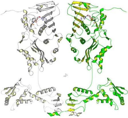
Full length MutL protein has not been crystalized, likely due to the long flexible linkers between its N-terminal domains and C-terminal domains. Instead, the N-terminal domain (LN40) and C-terminal domain (LC20) have been crystalized separately (Ban and Yang 1998; Kosinski, Steindorf et al. 2005). Based on these structures, a full length model has been constructed (Figure 1.5).
The N-terminal domain contains ATPase activities and is highly conserved. The 40 kDa N-terminal domain is a monomer in the absence of any nucleotide, but it forms a dimer with ADP or a non-hydrolyzable ATP homologue (AMPPNP).
The C-terminal domain forms dimer in the presence or absence of nucleotide and thus mediates dimerization of full length MutL homologues in all conditions. In contrast to the N-terminal domain, the C-N-terminal domain shares little sequence similarity among MutL homologs. A model of full length MutL is constructed based on these two separated crystalized structures (Polosina and Cupples 2010).
1.5.2 ATP-driven conformational changes in MutL
Studies of yeast Mlh1-Pms1 revealed different nucleotide-induced conformational changes (Figure 1.6b) (Sacho, Kadyrov et al. 2008). The heterodimer shows differential folding of the two N-terminal domains during the ATP cycle. This may be due to differences in nucleotide binding affinities and different ATPase rates between the Mlh1 and Pms1.
1.6 Coupling of Mismatch Recognition and Strand Discrimination
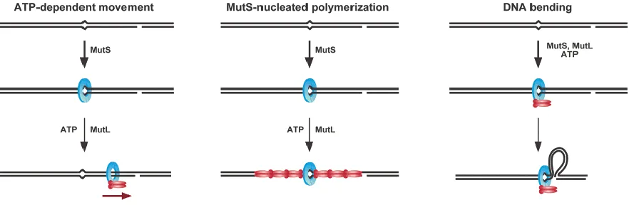
The MutS-MutL-heteroduplex ternary complex is believed to be an important intermediate in MMR, which is responsible for the communication between the mismatch and the daughter strand signal (Kunkel and Erie 2005; Iyer, Pluciennik et al. 2006). This task is difficult because the strand identification signal can be distantly located, transient in time and on either side of the mismatch. Controversy exists about the precise molecular mechanisms by which MutS and MutL achieve this function. Based on the available biochemical evidence, three types of models have been suggested.
The molecular-switch model posits that MutS, docked with MutL, forms an ATP-activated sliding clamp and diffuses from the mismatch to the strand signal along the DNA (Acharya, Foster et al. 2003). This model is mainly based on the observation that MutS, in the absence of MutL, can be triggered into a sliding clamp after mismatch recognition. One-dimension diffusion can overcome the difficulty of bidirectional long distance searching. Losing the specific interaction with the mismatch leaves the open question of how the excision stops after mismatch removal.
The DNA bending/verification model proposes that a MutS-MutL complex remains in the vicinity of a mismatch and the mismatch and strand marker sites communicate by looping out the intervening DNA (Junop, Obmolova et al. 2001). This mechanism requires two DNA binding sites, one near or at the mismatch and one searching for the distal strand signal. This model explains how repair can occur across physical blocks placed on the DNA between the mismatch and the strand discrimination marker (Junop, Obmolova et al. 2001; Schofield, Nayak et al. 2001).
large amount of proteins to participate especially when the two DNA sites are far away. Also, other DNA binding proteins in the cell might affect the polymerization since bare DNA is rare in vivo.
Figures
References
Acharya, S., P. L. Foster, et al. (2003). "The coordinated functions of the E. coli MutS and MutL proteins in mismatch repair." Molecular cell 12(1): 233-246.
Alani, E., J. Y. Lee, et al. (2003). "Crystal structure and biochemical analysis of the MutS.ADP.beryllium fluoride complex suggests a conserved mechanism for ATP interactions in mismatch repair." The Journal of biological chemistry 278(18): 16088-16094.
Antony, E. and M. M. Hingorani (2004). "Asymmetric ATP binding and hydrolysis activity of the Thermus aquaticus MutS dimer is key to modulation of its interactions with mismatched DNA." Biochemistry 43(41): 13115-13128.
Ban, C. and W. Yang (1998). "Crystal structure and ATPase activity of MutL: implications for DNA repair and mutagenesis." Cell 95(4): 541-552.
Ban, C. and W. Yang (1998). "Structural basis for MutH activation in E.coli mismatch repair and relationship of MutH to restriction endonucleases." The EMBO journal 17(5): 1526-1534.
Bennett, S. E., A. Umar, et al. (1997). "Mismatch repair in extracts of Werner syndrome cell lines." Cancer research 57(14): 2956-2960.
Biswas, I. and R. Vijayvargia (2000). "Heteroduplex DNA and ATP induced conformational changes of a MutS mismatch repair protein from Thermus aquaticus." The Biochemical journal 347 Pt 3: 881-886.
Burdett, V., C. Baitinger, et al. (2001). "In vivo requirement for RecJ, ExoVII, ExoI, and ExoX in methyl-directed mismatch repair." Proceedings of the National Academy of Sciences of the United States of America 98(12): 6765-6770.
Cho, W. K., C. Jeong, et al. (2012). "ATP alters the diffusion mechanics of MutS on mismatched DNA." Structure 20(7): 1264-1274.
Claverys, J. P. and V. Mejean (1988). "Strand targeting signal(s) for in vivo mutation avoidance by post-replication mismatch repair in Escherichia coli." Molecular & general genetics : MGG 214(3): 574-578.
Cooper, D. L., R. S. Lahue, et al. (1993). "Methyl-directed mismatch repair is bidirectional." The Journal of biological chemistry 268(16): 11823-11829.
Dao, V. and P. Modrich (1998). "Mismatch-, MutS-, MutL-, and helicase II-dependent unwinding from the single-strand break of an incised heteroduplex." The Journal of biological chemistry 273(15): 9202-9207.
DeRocco, V., T. Anderson, et al. (2010). "Four-color single-molecule fluorescence with noncovalent dye labeling to monitor dynamic multimolecular complexes." BioTechniques 49(5): 807-816.
Fang, W. H. and P. Modrich (1993). "Human strand-specific mismatch repair occurs by a bidirectional mechanism similar to that of the bacterial reaction." The Journal of biological chemistry 268(16): 11838-11844.
Genschel, J., L. R. Bazemore, et al. (2002). "Human exonuclease I is required for 5' and 3' mismatch repair." The Journal of biological chemistry 277(15): 13302-13311.
Genschel, J. and P. Modrich (2003). "Mechanism of 5'-directed excision in human mismatch repair." Molecular cell 12(5): 1077-1086.
Gorman, J., A. Chowdhury, et al. (2007). "Dynamic basis for one-dimensional DNA scanning by the mismatch repair complex Msh2-Msh6." Molecular cell 28(3): 359-370.
Gradia, S., D. Subramanian, et al. (1999). "hMSH2-hMSH6 forms a hydrolysis-independent sliding clamp on mismatched DNA." Molecular cell 3(2): 255-261.
Grilley, M., J. Griffith, et al. (1993). "Bidirectional excision in methyl-directed mismatch repair." The Journal of biological chemistry 268(16): 11830-11837.
Gupta, S., M. Gellert, et al. (2012). "Mechanism of mismatch recognition revealed by human MutSbeta bound to unpaired DNA loops." Nature structural & molecular biology 19(1): 72-78.
Hall, M. C., J. R. Jordan, et al. (1998). "Evidence for a physical interaction between the Escherichia coli methyl-directed mismatch repair proteins MutL and UvrD." The EMBO journal 17(5): 1535-1541.
Hall, M. C. and S. W. Matson (1999). "The Escherichia coli MutL protein physically interacts with MutH and stimulates the MutH-associated endonuclease activity." The Journal of biological chemistry 274(3): 1306-1312.
Herman, G. E. and P. Modrich (1981). "Escherichia coli K-12 clones that overproduce dam methylase are hypermutable." Journal of bacteriology 145(1): 644-646.
Iyer, R. R., A. Pluciennik, et al. (2006). "DNA mismatch repair: functions and mechanisms." Chemical reviews 106(2): 302-323.
Jacobs-Palmer, E. and M. M. Hingorani (2007). "The effects of nucleotides on MutS-DNA binding kinetics clarify the role of MutS ATPase activity in mismatch repair." Journal of molecular biology 366(4): 1087-1098.
Jeong, C., W. K. Cho, et al. (2011). "MutS switches between two fundamentally distinct clamps during mismatch repair." Nature structural & molecular biology 18(3): 379-385.
Jeruzalmi, D., M. O'Donnell, et al. (2002). "Clamp loaders and sliding clamps." Current opinion in structural biology 12(2): 217-224.
Joshi, A., S. Sen, et al. (2000). "ATP-hydrolysis-dependent conformational switch modulates the stability of MutS-mismatch complexes." Nucleic acids research 28(4): 853-861.
Junop, M. S., G. Obmolova, et al. (2001). "Composite active site of an ABC ATPase: MutS uses ATP to verify mismatch recognition and authorize DNA repair." Molecular cell 7(1): 1-12.
Kadyrov, F. A., L. Dzantiev, et al. (2006). "Endonucleolytic function of MutLalpha in human mismatch repair." Cell 126(2): 297-308.
Kleczkowska, H. E., G. Marra, et al. (2001). "hMSH3 and hMSH6 interact with PCNA and colocalize with it to replication foci." Genes & development 15(6): 724-736.
Kosinski, J., I. Steindorf, et al. (2005). "Analysis of the quaternary structure of the MutL C-terminal domain." Journal of molecular biology 351(4): 895-909.
Kunkel, T. A. and D. A. Erie (2005). "DNA mismatch repair." Annual review of biochemistry 74: 681-710.
Lahue, R. S., K. G. Au, et al. (1989). "DNA mismatch correction in a defined system." Science 245(4914): 160-164.
Lahue, R. S., S. S. Su, et al. (1987). "Requirement for d(GATC) sequences in Escherichia coli mutHLS mismatch correction." Proceedings of the National Academy of Sciences of the United States of America 84(6): 1482-1486.
Lamers, M. H., A. Perrakis, et al. (2000). "The crystal structure of DNA mismatch repair protein MutS binding to a G x T mismatch." Nature 407(6805): 711-717.
Längle-Rouault, F., G. Maenhaut-Michel, et al. (1987). "GATC sequences, DNA nicks and the MutH function in Escherichia coli mismatch repair." The EMBO journal 6(4): 1121-1127.
Lee, S. D. and E. Alani (2006). "Analysis of interactions between mismatch repair initiation factors and the replication processivity factor PCNA." Journal of molecular biology 355(2): 175-184.
Longley, M. J., A. J. Pierce, et al. (1997). "DNA polymerase delta is required for human mismatch repair in vitro." The Journal of biological chemistry 272(16): 10917-10921.
Lopez de Saro, F. J., M. G. Marinus, et al. (2006). "The beta sliding clamp binds to multiple sites within MutL and MutS." The Journal of biological chemistry 281(20): 14340-14349.
Lopez de Saro, F. J. and M. O'Donnell (2001). "Interaction of the beta sliding clamp with MutS, ligase, and DNA polymerase I." Proceedings of the National Academy of Sciences of the United States of America 98(15): 8376-8380.
Lu, A. L. (1987). "Influence of GATC sequences on Escherichia coli DNA mismatch repair in vitro." Journal of bacteriology 169(3): 1254-1259.
Lu, A. L., S. Clark, et al. (1983). "Methyl-directed repair of DNA base-pair mismatches in vitro." Proceedings of the National Academy of Sciences of the United States of America 80(15): 4639-4643.
Lyons, S. M. and P. F. Schendel (1984). "Kinetics of methylation in Escherichia coli K-12." Journal of bacteriology 159(1): 421-423.
McCulloch, S. D., L. Gu, et al. (2003). "Bi-directional processing of DNA loops by mismatch repair-dependent and -independent pathways in human cells." The Journal of biological chemistry 278(6): 3891-3896.
Modrich, P. (2006). "Mechanisms in eukaryotic mismatch repair." The Journal of biological chemistry 281(41): 30305-30309.
Modrich, P. and R. Lahue (1996). "Mismatch repair in replication fidelity, genetic recombination, and cancer biology." Annual review of biochemistry 65: 101-133.
Mukherjee, S. and M. Feig (2009). "Conformational change in MSH2-MSH6 upon binding DNA coupled to ATPase activity." Biophysical journal 96(11): L63-65.
Natrajan, G., M. H. Lamers, et al. (2003). "Structures of Escherichia coli DNA mismatch repair enzyme MutS in complex with different mismatches: a common recognition mode for diverse substrates." Nucleic acids research 31(16): 4814-4821.
Obmolova, G., C. Ban, et al. (2000). "Crystal structures of mismatch repair protein MutS and its complex with a substrate DNA." Nature 407(6805): 703-710.
Pavlov, Y. I., I. M. Mian, et al. (2003). "Evidence for preferential mismatch repair of lagging strand DNA replication errors in yeast." Current biology : CB 13(9): 744-748.
Peltomaki, P. (2003). "Role of DNA mismatch repair defects in the pathogenesis of human cancer." Journal of clinical oncology : official journal of the American Society of Clinical Oncology 21(6): 1174-1179.
Peltomaki, P. (2005). "Lynch syndrome genes." Familial cancer 4(3): 227-232.
Polosina, Y. Y. and C. G. Cupples (2010). "Wot the 'L-Does MutL do?" Mutation research 705(3): 228-238.
Pukkila, P. J., J. Peterson, et al. (1983). "Effects of high levels of DNA adenine methylation on methyl-directed mismatch repair in Escherichia coli." Genetics 104(4): 571-582.
Sacho, E. J., F. A. Kadyrov, et al. (2008). "Direct visualization of asymmetric adenine-nucleotide-induced conformational changes in MutL alpha." Molecular cell 29(1): 112-121.
Sass, L. E., C. Lanyi, et al. (2010). "Single-molecule FRET TACKLE reveals highly dynamic mismatched DNA-MutS complexes." Biochemistry 49(14): 3174-3190.
Schofield, M. J., S. Nayak, et al. (2001). "Interaction of Escherichia coli MutS and MutL at a DNA mismatch." The Journal of biological chemistry 276(30): 28291-28299.
Tessmer, I., Y. Yang, et al. (2008). "Mechanism of MutS searching for DNA mismatches and signaling repair." The Journal of biological chemistry 283(52): 36646-36654.
Tran, P. T., J. A. Simon, et al. (2001). "Interactions of Exo1p with components of MutLalpha in Saccharomyces cerevisiae." Proceedings of the National Academy of Sciences of the United States of America 98(17): 9760-9765.
Umar, A., A. B. Buermeyer, et al. (1996). "Requirement for PCNA in DNA mismatch repair at a step preceding DNA resynthesis." Cell 87(1): 65-73.
Wang, H., Y. Yang, et al. (2003). "DNA bending and unbending by MutS govern mismatch recognition and specificity." Proceedings of the National Academy of Sciences of the United States of America 100(25): 14822-14827.
Warren, J. J., T. J. Pohlhaus, et al. (2007). "Structure of the human MutSalpha DNA lesion recognition complex." Molecular cell 26(4): 579-592.
Winkler, I., A. D. Marx, et al. (2011). "Chemical trapping of the dynamic MutS-MutL complex formed in DNA mismatch repair in Escherichia coli." The Journal of biological chemistry 286(19): 17326-17337.
Yamaguchi, M., V. Dao, et al. (1998). "MutS and MutL activate DNA helicase II in a mismatch-dependent manner." The Journal of biological chemistry 273(15): 9197-9201.
Chapter 2
Overview of experiment methods used in this thesis
2.1 FRET
Förster resonance energy transfer (FRET) is a powerful tool with the capability of measuring distance in nanometer scale, which makes it a perfect ruler for revealing conformational information of macromolecules (Pawley 2006; Harris 2010).
FRET occurs between a donor molecule in the excited state and an acceptor molecule in the ground state. The donor molecule typically emits shorter wavelength light that overlaps with the absorption spectrum of the acceptor. Energy transfer occurs without the creation of a photon and is the result of near-field dipole-dipole interaction between the donor and acceptor. The rate of energy transfer for a donor and acceptor separated by a distance r is given by (Lakowicz 2006)
( ) ( (ln ) n ) ∫ ( ) ( ) (2.1)
where is the quantum yield of the donor in the absence of acceptor, n is the refractive index of the medium, N is Avogadro’s number, r is the distance between the donor and acceptor, and is the lifetime of the donor in the absence of acceptor. ( ) is the corrected fluorescence intensity of the donor in the wavelength range to with the total intensity normalized to unity. ( ) is the extinction coefficient of the acceptor at . The term is a factor describing the relative orientation in the space of transition dipoles of the donor and acceptor. is usually assumed to be equal to 2/3, which is appropriate for dynamic random averaging of the donor and acceptor orientations.
The efficiency of energy transfer (E) is the fraction of photons absorbed by the donor which are transferred to the acceptor. This fraction is given by
- ( )
which is the ratio of the transfer rate to the total decay rate of the donor in the presence of acceptor. The distance at which FRET is 50% is defined as Förstor distance. From (2.1) and (2.2), we can derive
(ln )
n ∫ ( ) ( ) (2.3)
and
(2.4) The Förstor distance is typically in the range of 40 to 60 Å in our studies. The distance dependence of FRET allows measurement of the distances between the donors and acceptors (Figure 2.1).
When converting transfer efficiencies into distances, orientation factor is generally assumed equal to 2/3. Since the sixth root is taken to calculate the distance, the variation of gives limited errors in distance unless is close to 0. Low can only happen when the donor and acceptor are rigid and perpendicular to each other. By measurements of the fluorescence anisotropy of the labeling sites on proteins, one can set the limits on and thereby minimize uncertainties in the calculated distance. We measured the anisotropy of alexa555 at the M88C site on Taq MutS and obtained value of 0.26 whereas unconjugated alexa555 had anisotropy of 0.20, which indicates that the labeled dyes are not rigidly immobilized (McCann, Zheng et al. 2012).
In single molecule FRET experiments, we measure the intensities of donor emission and acceptor emission to calculate the transfer efficiency:
(2.5) The differences in quantum yield (φ) and detection efficiencies (η) between the donor and acceptor can result in errors in calculating E in (2.5). A correction factor γ, which is defined as
(2.7)
2.2 Single molecule fluorescence microscopy
In traditional ensemble measurements, information of heterogeneous behaviors is buried by averaging of many molecules. Single molecule techniques provide us an opportunity to obtain information of heterogeneous populations and catch rare events even in equilibrium conditions (Ha 2001). Single molecule FRET has achieved great success in resolving protein dynamics and protein-DNA interactions. Although most of smFRET measurements have been carried out in vitro, smFRET in live cell has also been realized recently (Sakon and Weninger 2010).
A challenge in making single molecule fluorescence measurements is to extract the relatively low signal of fluorescence emission from one molecule from the background and noise. Total internal reflection fluorescence microscope (TIRFM) is a typical method to reduce background (Reck-Peterson, Derr et al. 2010). The evanescence wave generated by total internal reflection confines the excitation in a very thin layer (100~200nm) where the target molecules are immobilized. An emCCD is used as a detector so that hundreds of molecules can be monitored in parallel (Figure 2.2). This approach allows simultaneous observations with typical time resolution of 10-100ms. Higher time resolution can be achieved by a confocal system and avalanche photodiode detectors, but the efficiency of data collection has to be sacrificed because it can only measure one molecule at one time.
Quartz microscope slides are used to minimize the auto-fluorescence background. Flow chambers are formed between the quartz slide and a glass coverslip (Figure 2.3a). Two holes are made to allow the sample injection and buffer exchange, while all the edges are sealed with double sided tape and epoxy glue (Roy, Hohng et al. 2008).
2.3 Surface immobilization methods
Fluorophore labeled samples are usually immobilized on the surface of quartz microscope slides so they can be excited by the evanescence wave generated by TIRF microscopy. In this thesis, two different surface immobilization methods are used as described below.
2.3.1 Liposome encapsulation
Interaction between immobilized proteins and a surface can affect the behavior of the protein and can alter conclusions about the functional dynamics. Encapsulation of dye labeled proteins into liposomes minimizes the unwanted interaction with the glass surface in our flow cells (Boukobza, Sonnenfeld et al. 2001). To trap proteins into liposomes, dry lipid is rehydrated in protein containing solutions and then is extruded through membranes with pores size between 50nm and 200nm. Gel filtration (Sepharose CL4B) column is used to remove unencapsulated proteins from liposomes. If proteins do not interact with lipid bilayers, they can rotate freely inside the liposomes. Single molecule measurement can be achieved when a single labeled protein is trapped in one liposome (Figure 2.3b).
Liposomes containing a mixture of phosphatidylcholine (Egg PC) and 0.5% head group biotinylated 1,2-Dioleoyl-sn-Glycero-3-Phosphoethanolamine (biotin-PE) lipids are incubated with biotinylated BSA/streptavidin surface. In this way, the liposomes are immobilized to the streptavidin on slide surface and stay in the excitation layer.
2.3.2 Streptavidin-lipid bilayer surface
molecules are first nonspecifically deposited on the slide surface and then lipid bilayer is formed in the area among the streptavidin islands. The lipid bilayer blocks nonspecific interaction between the proteins and the slide surface (Figure 2.3c). Biotinylated DNA molecules are immobilized on the surface by binding to streptavidin islands in the supported lipid bilayer. The MMR proteins used in our study are injected in the flow chamber to interact specifically with the DNA. These proteins generally resist binding to the lipid bilayer. The DNA-protein binding events can be monitored by the fluorescence signals. One of the advantages of this method is that multiple buffer conditions (such as different nucleotide concentrations) can be tested in the same flow chamber by buffer exchange. The liposome encapsulation method makes buffer exchange challenging since many buffer components do not cross lipid membranes.
2.4 Protein preparation and dye labeling
2.4.1 Taq MutS expression and purification
An E. coli expression vector for Taq MutS mutant C42A/M88C with a single cysteine in the DNA binding domain I was prepared by Quickchange (Agilent). This protein was expressed in E. coli and purified as described previously (Antony and Hingorani 2004).
2.4.2 Taq MutL expression and purification
Wildtype Taq MutL with 6-his tag in the N-terminal domain was over-expressed in E.coli. Cell lysis was completed by sonication or repeated freeze-thaw cycles. The lysate was removed by ultracentrifugation.
2.4.3 Protein labeling strategies
Purified proteins are labeled with maleimide derivatives of Alexa dyes (Invitrogen) after purification steps mentioned above (Weninger, Bowen et al. 2003). Protein is incubated with 10 fold excess reducing agent TCEP at room temperature for 10 minutes. Then 10 fold excess maleimide-dyes are added and allowed to react for more than 12 hours at 4°C. Sephadex G50 columns are used to separate the labeled protein from free dye. Protein concentration and labeling efficiency are determined by absorbance spectroscopy. When both donor and acceptor are mixed with protein containing two cysteines, some molecules have two donors and some have two acceptors. The molecules with exactly one donor and one acceptor are selected by single-step photobleaching in single molecule measurements.
The method of Tris-NTA fluorescent dye labeling to 6his-tagged protein has been described before (DeRocco, Anderson et al. 2010). 10nM Tris-NTA Oregon Green is first incubated with 30nM NiCl2 for 1 hour at room temperature. After MutS-MutL complexes are
Figures
References
Antony, E. and M. M. Hingorani (2004). "Asymmetric ATP binding and hydrolysis activity of the Thermus aquaticus MutS dimer is key to modulation of its interactions with mismatched DNA." Biochemistry 43(41): 13115-13128.
Boukobza, E., A. Sonnenfeld, et al. (2001). "Immobilization in surface-tethered lipid vesicles as a new tool for single biomolecule spectroscopy." Journal of Physical Chemistry B 105(48): 12165-12170.
DeRocco, V., T. Anderson, et al. (2010). "Four-color single-molecule fluorescence with noncovalent dye labeling to monitor dynamic multimolecular complexes." BioTechniques 49(5): 807-816.
Graneli, A., C. C. Yeykal, et al. (2006). "Organized arrays of individual DNA molecules tethered to supported lipid bilayers." Langmuir : the ACS journal of surfaces and colloids 22(1): 292-299.
Ha, T. (2001). "Single-molecule fluorescence resonance energy transfer." Methods 25(1): 78-86.
Harris, D. C. (2010). Quantitative chemical analysis. New York, W.H. Freeman and Co.
Helms, V. (2008). Principles of computational cell biology : from protein complexes to cellular networks. Weinheim, Wiley-VCH.
McCann, J. J., L. Zheng, et al. (2012). "Supertertiary structure of the synaptic MAGuK scaffold proteins is conserved." Proceedings of the National Academy of Sciences of the United States of America 109(39): 15775-15780.
Pawley, J. B. (2006). Handbook of biological confocal microscopy. New York, NY, Springer.
Periasamy, A. (2001). "Fluorescence resonance energy transfer microscopy: a mini review." Journal of biomedical optics 6(3): 287-291.
Reck-Peterson, S. L., N. D. Derr, et al. (2010). "Imaging single molecules using total internal reflection fluorescence microscopy (TIRFM)." Cold Spring Harbor protocols 2010(3): pdb top73.
Roy, R., S. Hohng, et al. (2008). "A practical guide to single-molecule FRET." Nature methods 5(6): 507-516.
Sakon, J. J. and K. R. Weninger (2010). "Detecting the conformation of individual proteins in live cells." Nature methods 7(3): 203-205.
Chapter 3
Large conformational changes in MutS during DNA
scaning, mismatch recognition and repair signaling
3.1 Summary
Our study of the conformational changes in MutS during DNA scanning, mismatch recognition and repair signaling was published in the April 2012 issue of EMBO Journal. The publication is reproduced in the appendix of this dissertation. Here, I will briefly summarize the results in this paper.
MutS initiates DNA mismatch repair by binding to a mismatched pair on double stranded DNA. MutS first searches for mismatch by diffusing on homoduplex DNA. Once MutS recognizes a mismatch, it is stabilized at the mismatch and undergoes an ATP-dependent formation to sliding clamp. The MutS dimer contains two ATPase sites with nucleotide binding capacity. Nucleotide binding to the MutS plays an important role in all steps of the MMR process. Although X-ray crystal structures of MutS proteins bound to various DNA mismatches have been solved, no clear, time-resolved picture of MutS conformations throughout the MMR pathway has been generated.
Here, we use single molecule fluorescence resonance energy transfer (smFRET) to monitor the conformational dynamics of MutS during these processes. A single cysteine mutant Taq MutS (C42A/M88C) was constructed, so each MutS homodimer has two cysteine bases. The M88C is in the DNA binding domain I. By labeling a donor dye and an acceptor dye on the MutS dimer, we were able to measure the relative distance between the domains I of the two subunits. Control experiements show that labeling the M88C with dyes does not negatively alter DNA binding or ATPase activities.
DNA. When we labeled the MutS with just donor dye and attached a dye on the DNA near the mismatch, we were able to monitor the relative position of MutS on the DNA as well as subtle conformational changes of the MutS-DNA complex.
We first determined the effect of nucleotide binding to MutS in the absence of DNA. We found that domain I of MutS was very mobile with a very broad range of FRET signals. Binding of either ADP or ATP, stabilized domains I into populations of 2 states: a low FRET state and a high FRET state. The similarity of the ATP and ADP results might arise from ATP hydrolysis. Therefore, the results report the state of ADP bound MutS in both cases. Binding to ATPS (a non-hydrolysable ATP analogue) locked the MutS in a single high FRET state, which is a state with little DNA binding capacity.
Specific contributions of co-authors on this published work: In this study, Vannessa DeRocco and Credle Harris worked out the protocol of making the 500bp DNA substrates. Anushi Sharma and Manju Hingorani provided proteins and performed ensemble DNA-binding and ATPase measurements. Dorothy Erie and Keith Weninger designed the research. All authors contributed to interpretation and manuscript preparation.
3.2 Large conformational changes in MutS during DNA scaning,
mismatch recognition and repair signaling
Available online at http://www.nature.com/emboj/journal/v31/n11/full/emboj201295a.html
or in Appendix.
3.3 Supplementary Content within this Article
Available online at
Chapter 4
Conformational Dynamics of MutS-MutL Complex
Formation
4.1 Introduction
The MutS-MutL complex on mismatched DNA has attracted much attention and thought because it is expected to be a key intermediate in MMR. MutL is a matchmaker in MMR. It interacts with MutS and most of the proteins in downstream pathways (Polosina and Cupples 2010). Yet, the precise interactions of MutL that control DNA MMR are debated.
In E.coli, DNA helicase II (UvrD) unwinds the nicked strand towards the mismatch. This directional unwinding is activated by MutS and MutL, inferring that the MutS-MutL complex plays an important role in the coupling between the mismatch and the strand discrimination signal (Yamaguchi, Dao et al. 1998). Despite of its importance, only a few details about this protein complex are known. As mentioned in Chapter I, three different models have been suggested to explain the communication between the two distal sites on DNA: 1) protein diffusion; 2) DNA bending and looping; 3) protein polymerization (Kunkel and Erie 2005; Iyer, Pluciennik et al. 2006). We designed experiments to provide time resolved structural information about the complex formed between Taq MutS and MutL on mismatched DNA in order to attempt to understand the role of MutL in signaling DNA mismatch repair.
4.2 Results
4.2.1 MutS sliding clamp formation is inhibited by MutL
sliding, the donor on MutS and the acceptor near the mismatch will be farther than the FRET range (~8nm), resulting in a zero FRET state. The presence of donor signal but with zero FRET (zero acceptor signal) indicates that MutS is on the DNA but far from the mismatch. The MutS sliding clamp stays on the 500bp DNA for about 2 seconds before leaving the free end. With this lifetime, we were able to estimate the diffusion constant of the sliding clamp.
mismatch directly. These experiments strongly suggest that sliding clamp formation of Taq MutS is inhibited in the presence of 200nM MutL.
4.2.2 MutS interconverts between two conformations when interacting with
MutL
During sliding clamp formation, MutS undergoes large conformational changes in DNA binding domain I. In the donor-MutS acceptor-DNA labeling strategy, MutS typically binds to the mismatch at a FRET ~0.7 state. The FRET transitions to ~0.5, and then transitions to 0 when MutS slides away the mismatch (Figure 4.1b). The transition density plot, which was assembled by collecting thousands of transitioning events for MutS interacting with mismatched DNA in the absence of MutL, showed that these two-step sequential transitions are the dominant pathway (Figure 4.2a). In the presence of 200nM MutL, a different sequence of transitions is clearly seen in both individual time-traces and in the transition density plot accumulated from many molecules (Figure 41.d, 4.2b). Instead of sequentially transitioning from high FRET to medium FRET then to zero FRET as in the absence of MutL, the FRET levels in the presence of MutL switched between a high FRET state and a low FRET state. The dwell time of each state lasted for a few hundred milliseconds to a few seconds, which can be well resolved in our time resolution (100ms). In the transition density plot, two major populations representing 0.3->0.8 and 0.8->0.3 transitions were dominant and they were almost equally populated. The complete absence of a FRET zero state in population histograms confirms the conclusion that MutS does not convert to a sliding state in the presence of MutL. In figure 4.1c, the 0.3 FRET peak and the 0.8 FRET peak were also about equal in population, which indicated that MutS-MutL complex did not dissociate preferentially in either conformation.
4.2.3 Multiple MutS and MutL are recruited to form a large complex
20% of the proteins were observed to form tetramers in a sample with 20nM Taq MutS monomer equivalent concentration. If a tetramer binds to the mismatch, up to 4 donors can be seen. In a single molecule measurement, the number of dyes can be revealed by photobleaching steps or can be estimated by the emission intensity compared with a single dye. On a DNA with one free (un-blocked) end, no more than 4 donors were observed in the absence of MutL, although multiple sliding clamps can be formed and stay on the DNA when the DNA end was blocked by an anti-DIG.
yeast MMR protein (DeRocco, Anderson et al. 2010). When 100nM MutS, 20nM MutL and 2mM ATP were incubated with the mismatched DNA substrate and Tris-NTA-Oregon Green, we observed multiple dyes on the same DNA (Figure 4.3e). The colocalization of up to 4 dyes strongly suggested the multiple MutL were involved in the complex. Control experiment that omitted MutS resulted in no MutL binding on the DNA reported by the Tris-NTA-Oregon Green. The MutL binding is also ATP dependent. When ATP was exchanged to ADP, MutL binding was not seen. A future improvement will seek alternate labeling strategies to estimate the precise number of MutL proteins and the ratio of MutS and MutL assemblies on mismatched DNA.
4.2.4 MutS domains I are open in MutS-MutL complex
With donor-acceptor labeled C42A/M88C Taq MutS dimers, we are able to measure the distance between the domains I of the two subunits. This is the system used to report the conformations of MutS in the two-step sliding clamp formation in chapter 3. However, when there were more than one MutS dimer in the MutS-MutL complex, the existence of multiple donors and acceptors can complicate the interpretation of the donor and acceptor signals. To overcome the difficulty of measuring the conformation of MutS in the complex containing multiple proteins, we mixed 10% donor/acceptor labeled MutS with 90% unlabeled MutS and then formed the large MutS-MutL complexes on mismatched DNA. From Figure 4.3c and the known labeling efficiency of 75% of the labeled MutS sample, we estimated the number of MutS in the complex ranged from 3 to 7. Under these conditions, use of 10% fraction of labeled MutS would give a high probability of having one donor-acceptor labeled MutS in the complex. In this situation, single molecule FRET can be accurately measured, allowing the conformation of MutS to be effectively sampled within this assembly of multiple copies of MutS protein.
FRET) but they were both unstable on DNA with an unblocked end. The small populations in low FRET and medium FRET are corresponding to the sliding clamp and the intermediate state, respectively. These results are consistent with our findings in chapter 3.
When 200nM MutL was added and incubated for 20 minutes, multiple MutS and MutL molecules assembled into large complexes in the presence of ATP. We selected the complexes which only had one donor and one acceptor and measured the FRET values. In the histogram, we found that about 70% of the MutS were in a low FRET state (~0.2) and 30% stayed at high FRET (~0.9). The higher population in low FRET indicates that MutS is more stable in a conformation in which the domains I of the two subunits are opened up. This outward movement of domain I might arise from a physical interaction between MutS and MutL. The high FRET state, since it was similar to the state in experiments without MutL, might arise from single MutS bound the mismatch that did not form MutS-MutL complex. Another possibility is that heterogeneous conformations of the MutS proteins are present in the complex. The high FRET state could also involve the special MutS molecules within a large filament of MutS/MutL that remains directly interacting with the mismatch. If correct, this result indicates that there is always one MutS remaining in close proximity to the mismatch.
4.3 Discussion
Our understanding of DNA mismatch repair has been provided by many structural, biochemical and genetic studies and has largely developed since the successful reconstitution of human and E.coli systems with purified proteins (Grilley, Griffith et al. 1993; Zhang, Yuan et al. 2005). This large body of work has confirmed the required components of MMR and suggested roles for each protein. Yet, important questions are still waiting to be answered.
Furthermore, studies show that the repair can be accomplished even when the mismatch and strand signal are as far as 1000 DNA bases apart (Pluciennik and Modrich 2007).
Based on the available evidence, distinct models have been proposed. The molecular switch model and the polymerization model suggest a cis mechanism (Hall, Wang et al. 2001; Acharya, Foster et al. 2003). In cis models, proteins translocate or polymerize along the DNA helix to enable communication between the two sites. The DNA verification/bending model postulates a trans mechanism (Junop, Obmolova et al. 2001). In this model, contacts occur between the two separated DNA sites by bending or looping.
There is evidence for both types of signaling mechanisms. Some examples include the study that provided the first evidence of a trans model, where it was observed that MutH can be activated when the GATC and mismatch sites are on separate DNA substrates (Schofield, Nayak et al. 2001). However, another study demonstrated that binding of a catalytically inactive mutant EcoRI to an EcoRI recognition site located between the mismatch and hemimethylated GATC site significantly reduced the activation of MutH (Pluciennik and Modrich 2007). This result fell strongly on the side of a cis mechanism for MutH activation. These sorts of contradictory results hinder us from understanding MMR in more detail.
MutL has been found to increase the dwell time of MutS on a linear mismatched DNA (Schofield, Nayak et al. 2001). The increase of dwell time is dependent on the DNA length. As intriguing as these results are, they offer no hint about where and when the MutS-MutL complex is formed. MutL might interact with MutS before the sliding clamp formation at the mismatch, or might block the diffusion of activated MutS on the DNA after MutS slides away from the mismatch. Determining the details about these macroscopic behaviors is crucial in modeling the mechanism of communication between the mismatch and the strand signal.
form a sliding clamp in a two-step transition. This clamp then freely diffuses along the DNA helix. Once MutS starts sliding, the distance between the donor on MutS and the acceptor near the mismatch quickly becomes larger than the FRET range (~8nm) resulting in zero FRET. Addition of 200nM Taq MutL strongly inhibits the sliding clamp formation. The FRET value does not drop to zero when MutS is bound to the DNA, which indicates that MutS stays at the mismatch until it directly dissociates from the DNA. MutL interacts with MutS at the mismatch and before the sliding clamp is formed. Without MutL exposure, MutS transitions to an intermediate state before beginning to slide away from a mismatch. This MutL-free intermediate state involves movement of domain I and lasts for about 1.5 seconds. It is possible that MutL interacts with a new surface that is exposed by this movement of domain I in the intermediate state. Consistency with this idea can be found in an earlier chemical crosslinking study which suggested that MutS-MutL complex is formed via the connector domain of MutS, which is adjacent to the DNA binding domain I.
Most single MutS-MutL complexes dissociate from the mismatch after a few seconds. Stable complexes containing multiple MutS and multiple MutL can be observed on some DNA substrates. The number of MutS and MutL can be estimated by counting the donor dyes on each DNA. The labeling efficiency of the MutS is about 0.75 dye per cysteine, so there are 1.5 dyes on each MutS dimer on average. For some MutS/MutL complexes bound to mismatched DNA substrates, as many as 9 dyes can be found, which indicates at least five MutS dimers are in the complex. Interestingly, there are always one or two donors within the FRET range (8nm) of the acceptor that is near the mismatch, while the other donors are out of this range. Therefore, the large protein complex is probably stabilized at the mismatch by specific interaction of one of the MutS dimer.
allow stronger conclusions about the relative abundance of MutS and MutL in these assemblies.
FRET from donor-acceptor labeled MutS can reveal the relative position of domains I in the two subunits of a MutS dimer. In chapter 3, we found three different conformations of MutS corresponding to the 3 states in sliding clamp formation. We examined these same FRET reporters to address the conformation of MutS in the MutS/MutL complex. In these larger complexes, the presence of multiple donors and multiple acceptors will confuse the FRET analysis if they are all within FRET coupling range to each other. As an attempt to find conditions that can assemble the large MutS/MutL assemblies on mismatched DNA while simultaneously ensuring that no more than donor and one acceptor are present in the complex, we mixed 10% double-labeled MutS with 90% unlabeled MutS to form the complex. In this way, some complexes would contain only one double labeled MutS and its conformation can be measured by smFRET. With this approach, we found that about 70% of the MutS adopt a low FRET conformation in which domains I are far from each other. This conformation is very similar to the sliding clamp, but evidently not mobile as it was trapped on unblocked DNA. The sliding clamp can diffuse along the DNA helix with 1D random walk and dissociate from the DNA ends. MutS in the MutS-MutL complex stayed on the unblocked DNA for more than ten minutes. The other 30% MutS are in a high FRET state similar to the mismatch recognition state of single MutS. Since we used 90% unlabeled MutS, we are not able to confidently determine that this state is present in the large filaments. It seems likely it is, and allows us to speculate that one MutS dimer within the large complex maintains specific interactions with the mismatched base.
4.4 Biological Implications
yields a model of MutS-MutL interactions (Figure 4.5). Our data revealed that the ATP- and mismatch- provoked Taq MutS sliding clamp formation was inhibited by MutL. Initial MutS-MutL complexes are formed at the mismatched sites and recruit more MutS and MutS-MutL into large assemblies. The large protein assemblies are stable on DNA.
Figures
References
Acharya, S., P. L. Foster, et al. (2003). "The coordinated functions of the E. coli MutS and MutL proteins in mismatch repair." Molecular cell 12(1): 233-246.
DeRocco, V., T. Anderson, et al. (2010). "Four-color single-molecule fluorescence with noncovalent dye labeling to monitor dynamic multimolecular complexes." BioTechniques 49(5): 807-816.
Elez, M., M. Radman, et al. (2012). "Stoichiometry of MutS and MutL at unrepaired mismatches in vivo suggests a mechanism of repair." Nucleic acids research 40(9): 3929-3938.
Grilley, M., J. Griffith, et al. (1993). "Bidirectional excision in methyl-directed mismatch repair." The Journal of biological chemistry 268(16): 11830-11837.
Hall, M. C., H. Wang, et al. (2001). "High affinity cooperative DNA binding by the yeast Mlh1-Pms1 heterodimer." Journal of molecular biology 312(4): 637-647.
Hombauer, H., C. S. Campbell, et al. (2011). "Visualization of eukaryotic DNA mismatch repair reveals distinct recognition and repair intermediates." Cell 147(5): 1040-1053.
Iyer, R. R., A. Pluciennik, et al. (2006). "DNA mismatch repair: functions and mechanisms." Chemical reviews 106(2): 302-323.