ABSTRACT
MACKENZIE, ELIZABETH LOUISE. Regulation of Ferritin H in the Cellular Response to Stress. (Under the direction of Yoshiaki Tsuji.)
Cells are exposed to a constant onslaught of diverse stressors from exogenous and endogenous sources. Due to the ubiquitous nature of such insults, cells have evolved elaborate systems of defense. Phase II genes encode enzymes that are responsible for the reduction in toxicity of xenobiotics and other stressors and for cytoprotection. Ferritin H is one such phase II gene that protects the cell from oxidative stress by tightly regulating the levels of free intracellular iron that are available to catalyze the formation of potent reactive oxygen species (ROS). The purpose of this research was to elucidate the mechanisms of ferritin H regulation in response to oxidative stress that results from mitochondrial complex I inhibition by rotenone, and also from elevated intracellular calcium concentrations.
Increased intracellular calcium concentrations and ROS are concordantly produced in activated Jurkat T-cells. Calcium ionophore induced ferritin H mRNA and protein expression; however, calcium did not activate ferritin H transcription. In addition, induction of ferritin H was independent of activation of Nuclear Factor of Activated T-cells (NFAT). Interestingly, elevated calcium led to retarded degradation of ferritin H mRNA, suggesting mRNA stability was enhanced. In addition, an au-rich element, which is a putative target for RNA binding proteins involved in regulation of mRNA stability, was identified in the 3’untranslated region of ferritin H mRNA. Taken together, these results indicate that ferritin H induction by calcium is regulated through a posttranscriptional mechanism involving mRNA stabilization.
REGULATION OF FERRITIN H IN THE
CELLULAR RESPONSE TO STRESS
by
ELIZABETH LOUISE MACKENZIE
A dissertation submitted to the Graduate Faculty of North Carolina State University
in partial fulfillment of the requirements for the Degree of
Doctor of Philosophy
TOXICOLOGY
Raleigh, North Carolina 2007
APPROVED BY:
_________________________ _________________________ Dr. Jun Ninomiya-Tsuji Dr. B. Alex Merrick
DEDICATION
I dedicate this work to my mom, who has filled my life with unconditional love, frequent phone calls, and unwavering support. She is my touchstone.
BIOGRAPHY
ACKNOWLEDGEMENTS
TABLE OF CONTENTS
Page
LIST OF FIGURES...vii
CHAPTER ONE: ROTENONE INDUCES TRANSCRIPTION OF THE FERRITIN H GENE BY OXIDATIVE STRESS-MEDIATED ACTIVATION OF AN ANTIOXIDANT RESPONSE ELEMENT... 1
ABSTRACT ... 2
INTRODUCTION ... 3
EXPERIMENTAL PROCEDURES ... 6
RESULTS... 12
DISCUSSION... 16
FIGURES... 21
REFERENCES ... 27
CHAPTER TWO: ELEVATED INTRACELLULAR CALCIUM ENHANCES FERRITIN H EXPRESSION THROUGH AN NFAT-INDEPENDENT MECHANISM32 ABSTRACT ... 33
INTRODUCTION ... 34
EXPERIMENTAL PROCEDURES ... 37
RESULTS... 40
DISCUSSION... 45
FIGURES... 48
REFERENCES ... 54
CONCLUSION... 60
LIST OF FIGURES
CHAPTER ONE
Page Fig. 1. Rotenone induces ferritin H mRNA... 21 Fig. 2. Rotenone treatment results in increased ferritin H
protein synthesis ... 22 Fig. 3. Transcriptional activation of ferritin H by rotenone ... 23 Fig. 4. Rotenone alters transcription factor binding to the ARE ... 24 Fig. 5. ROS production by rotenone mediates ferritin H
mRNA induction ... 25 Fig. 6. Ferritin H knockdown by siRNA results in increased ROS
production and apoptosis following rotenone treatment... 26
CHAPTER TWO
Fig. 1. Ferritin protein expression is increased by calcium
ionophore treatment... 48 Fig. 2. Calcium ionophore treatment increases ferritin H mRNA ... 49 Fig. 3. NFAT is activated by calcium ionophore treatment ... 50 Fig. 4. NFAT is not involved in ferritin H transcriptional
activation by calcium ionophore ... 51 Fig. 5. Inhibition of NFAT by cyclosporin A does not block
ionomycin-mediated ferritin H mRNA induction ... 52 Fig. 6. Ionomycin treatment results in the stabilization of
Rotenone Induces Transcription of the Ferritin H Gene by Oxidative Stress-Mediated Activation of an Antioxidant Responsive Element
Elizabeth L. MacKenzie and Yoshiaki Tsuji
From the Department of Environmental and Molecular Toxicology, North Carolina State University, Campus Box 7633, Raleigh, North Carolina 27695, USA.
Running title: Ferritin regulation by rotenone
ABSTRACT
INTRODUCTION
Iron is a vital element mandatory for metabolic processes and the function of many enzymes, including cytochrome P450s and ribonucleotide reductase (1); however, a surplus of free iron leads to the formation of reactive oxygen species (ROS) via the Fenton reaction, and is potentially harmful to the cells (2). Thus, cells have an intricate system of control of intracellular iron levels. Ferritin is a nanocage protein that functions to sequester free intracellular iron that may become toxic to the cells (3). In vertebrates, there are 2 ferritin subunits, heavy and light, which coassemble in 24 subunit bundles to form a channel that encloses iron (4). The heavy, or H, subunit is catalytically active, inducing oxidation of ferrous iron (Fe(II)) to ferric iron (Fe(III)), and causing aggregation of the oxidized iron inside the core, while the light (L) subunit does not have ferroxidase activity and may serve a structural function (5,6). In this manner, ferritin H functions to protect cells against iron mediated oxidative stress (7,8). Because of its important role, ferritin is tightly regulated at both the transcriptional and translational levels. Levels of ferritin protein are controlled by a well-characterized translational repression system in response to free iron levels in the cells (9). The 5’ untranslated region of ferritin message contains an iron responsive element, to which the iron regulatory proteins, IRP1 and IRP2, bind in low iron concentrations, thereby blocking translation when necessary (10,11). In addition to iron, ferritin levels are also altered by other stimuli. TNFα was shown to stimulate ferritin H expression, but not ferritin
Hemin (16-18). A similar ARE element was also identified in ferritin L gene (19). The ARE is a highly conserved enhancer element in various phase II genes involved in detoxification or with antioxidant properties (20,21). It allows for the activation of a battery of antioxidant genes, including glutathione-s-transferases, NADH quinone oxidoreductase 1 and heme oxygenase 1 (20,22-25), under chemical and oxidative stress conditions. The ferritin H ARE contains a bidirectional AP1-like and AP-1/NFE2 sequence, to which basic leucine zipper (bZip) transcription factors including JunD and NFE2-related factor 2 (Nrf2) bind (16,18,26). This facilitates activation of ferritin H transcription in conditions of oxidative stress. Our recent studies demonstrated that JunD was phosphorylated following oxidative stress, and subsequently its binding to the ARE increased (18).
A large body of evidence indicates a correlation between increased oxidative stress and Parkinson’s disease (PD) incidence (27,28). Recent studies have shown that iron chelators can block 6-hydroxydopamine and 1-methyl-4-phenyl-1,2,3,6-tetrapyridine (MPTP) induced dopaminergic cell death (29,30). Excess free iron may be the catalyst in the production of deleterious ROS that are responsible for damage to DNA, lipids, and proteins, and may ultimately lead to the deletion of dopaminergic neurons (31). Furthermore, the insults that induce neuronal degeneration are intimately connected with oxidative stress (32). Rotenone, a mitochondrial complex I inhibitor, induces oxidative stress; however, despite its universal effect, it leads to the specific deletion of dopaminergic neurons (33,34), suggesting that they are highly sensitive to oxidative stress.
EXPERIMENTAL PROCEDURES
Cell culture.
NIH3T3 mouse fibroblasts were obtained from American Type Culture Collection. NIH3T3 cells were cultured at 37οC and 5% CO2 in a humidified atmosphere in Dulbecco’s Modified
Eagle’s Medium containing 1mM sodium pyruvate, 4 mM L-glutamine, and 4.5g/L glucose with 10% bovine calf serum (Hyclone), and penicillin/ streptomycin. Rotenone and tert-butylhydroquinone (tBHQ) were purchased from Sigma and were dissolved in DMSO. H2O2 and N-acetylcysteine (NAC) were purchased from Calbiochem. H2O2 was diluted in sterile phosphate-buffered saline (PBS), and NAC was reconstituted in media and adjusted to pH 7.4 with NaOH.
Northern Blotting.
Total RNA from NIH3T3 cells was isolated using TRIzol (Invitrogen). 5-10 ug RNA was
separated on a 1.1% agarose, formaldehyde gel, and separated RNA was transferred to a 0.45 micron nitrocellulose Protran BA85 membrane (Whatman, Schleicher & Schuell) by capillary transfer. Northern blotting was performed using an α-32P-dCTP random primer
labeled 0.9 kb fragment of ferritin H human cDNA probe.
Plasmids and DNA transfections.
pGL3–0.22 kb mouse ferritin H luciferase (mFH-Luc) was constructed by digesting pGL3–
mFH-Luc plasmid (below) following oligonucleotide purification by urea denaturing polyacrylamide gel electrophoresis, Sephadex G-25 column purification, and subsequent annealing. The sequences are:
wt ARE SENSE:
CATGACAAAGCACTTTTGGAGCCCAACCCCTCCAAAGGAGCAGAATGCTGAGTC ACGG
wt ARE ANTISENSE:
CCGTGACTCAGCATTTCTGCTCCTTTGGAGGGGTTGGGCTCCAAAAGTGCTTTGT CATG
mARE SENSE:
CACAACAAAGCACTTTTGGAGCCCAACCCCTCCAAAGGAGCAGAACACTGAGTC
ACGG
mARE ANTISENSE:
CCGTGACTCTAGTGTTCTCTCCTTTGGAGGGGTTGGGCTCCAAAAGTGCTTTGTT GTG
24 hours, and preparation of cell lysates and luminometry were performed with luciferase assay kit (Promega).
Gel retardation Assay.
Nuclear extract preparation, binding reactions, and electrophoretic mobility shifts were previously described (35). JunD (sc-74X), JunB (sc-46X), and cJun (sc-45X) antibodies were obtained from Santa Cruz Biotechnology, Inc.
ChIP Assay.
Chromatin immunoprecipitation (ChIP) assays were performed according to a minor modification of Upstate Biology's ChIP assay protocol as previously described (16). A total of 1X10⁶ cells/100-mm plate were treated with 1uM rotenone for 4 hours, followed by formaldehyde cross-linking of chromatin and preparation of lysates. (Upstate Biology). 2 ul of the following antibodies were used for immunoprecipitation: rabbit IgG (alpha diagnostics), anti-Nrf2 (sc-722X), anti-JunD (sc-074X), (Santa Cruz Biotechnology). Quantitative PCR was performed in the presence of 1 uCi α-32P dCTP/reaction, while using
specific primers designed to amplify a 230 bp region within the mouse ferritin H promoter that contains the ARE:
Forward:
5’GGCCCCTCTGTTCTGTACAATACTAGCTC-3’ Reverse:
5’TAACCACAAAACCACAGCCCTCCAG-3’
Western blotting.
Total cell lysates were subjected to electrophoresis with a 15% or 12.5% sodium dodecyl sulfate-polyacrylamide gel (SDS-PAGE), followed by wet transfer of separated proteins to an Immobilon-P membrane (Millipore). Membranes were blocked for one hour in 5% skim milk in Tris buffered saline with 0.1% Tween 20, and subsequently incubated overnight at 4°C with anti-ferritin H antibody (Abcam 1:20,000). Recombinant human ferritin H and L were purchased from Calbiochem.
35S-Translabeling/immunoprecipitation.
Exponentially growing NIH3T3 cells were treated with either rotenone or ferric ammonium
citrate for the indicated times. Following treatment, media was removed and methionine/cysteine deficient DMEM containing 10% dialyzed bovine calf serum was added. Simultaneously, 10 uCi/ml of 35S methionine/cysteine (GE Healthcare) was added to each dish. Cells were incubated under normal culture conditions for one hour. Total cell lysates were prepared, and pre-cleared with rabbit serum (CAPEL) and protein A agarose (Calbiochem) overnight. Total incorporation of 35S was measured using TCA precipitation and scintillation counting. Input protein for immunoprecipitation was determined by adding equal counts (1X10⁶ cpm) for each immunoprecipitation reaction. 6 ul of Anti-Ferritin antibody (DAKO, A133) and 20 ul of protein A agarose were utilized for overnight immunoprecipitation at 4οC. Finally, the resulting immunoprecipitates were subjected to
SDS-PAGE as described above, the gel was dried, and exposed to film for 4-10 days at –86ο
SiRNA transfection.
Ferritin H siRNA was purchased from DHARMACON, Inc, siGENOME duplex D-045965-01 mouse FTH, NM_D-045965-010239.
Sense:5’CAAGAAUGAUCCCCACUUAUU-3’
Antisense: 5’PUAAGUGGGAUCAUCAUUCUUGUU-3’
Briefly, 1X10⁵ cells/35 mm dish were plated and cultured under normal conditions in serum-containing, antibiotic-free media (2 ml) until cells reached approximately 80% confluency. 16 ul DharmaFECT3 (Dharmacon) in a final volume of 200 ul OPTI-MEM media (Invitrogen) was added to 7.5ul of 20 uM (150 pMol) siRNA dissolved in 200 ul of OPTIMEM, and the mix was incubated for 20 minutes. After formation of RNA complexes, the 400 ul mix was added to dishes containing 1.6 ml of fresh, antibiotic-free DMEM with 10% bovine calf serum, and the cells were cultured under normal conditions. After 24 hours, cells were split into a 6-well plate for treatment. Whole cell extracts were obtained, and subjected to Western blotting as described above to examine ferritin H protein expression. In other cases, cells were trypsinized and assessed for ROS production and apoptosis using flow cytometry.
Detection of ROS. NIH3T3 cells in 35mm dishes were incubated with 5 uM
unincorporated dye was washed out with phenol red-free complete medium, and rotenone (5 uM) and H2O2 (50 uM) were added for 0.5 hours. Following treatment, cells were trypsinized and resuspended in phenol red complete medium, and cell pellets were collected after centrifugation at 2000 rpm for 3 minutes. The resultant pellets were suspended in 500 ul phenol red free complete medium for flow cytometry on a Becton-Dickinson FACSCalibur.
Detection of Apoptosis. To detect apoptosis, Annexin V conjugated with AlexaFluor647
RESULTS
Rotenone induces ferritin H- To investigate the effect of the mitochondrial complex I inhibitor, rotenone, on ferritin H expression, we examined the levels of ferritin H mRNA following rotenone exposure by Northern blotting. As shown in Fig. 1, treatment of NIH3T3 cells with increasing concentrations of rotenone for 24 hours increased ferritin H message in a dose dependent manner. The inductive effect of the highest dose used was similar to that of tBHQ, an activator of ferritin transcription (17,36). Our previous studies indicated that H2O2 inhibits ferritin translation within 1-2 hours due to transient IRP activation (17); therefore, we next investigated whether ferritin H protein synthesis was transiently repressed following rotenone treatment. We exposed NIH3T3 cells to 1 uM rotenone for various times over a period of 24 hours, and subsequently subjected them to 35S-methionine/cysteine translabeling. As shown in Fig. 2, increased ferritin protein was observed 1 hour following treatment and was maintained for 24 hours. This suggests that IRP (translational repression system) is not activated by rotenone treatment, since there was no transient reduction in the level of ferritin H protein synthesized at any time point following the initiation of treatment. In addition, ferritin protein exhibited a dose responsive increase following 24-hour treatment with 0.1, 0.5, and 1 uM rotenone (Fig. 2b).
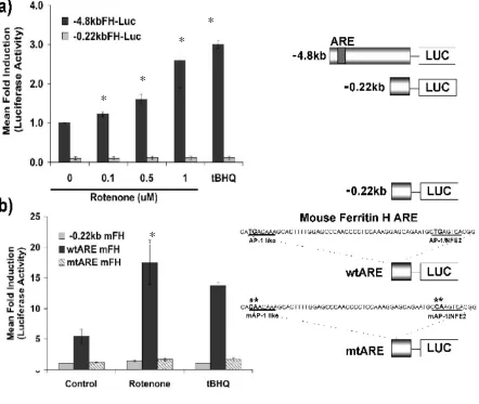
rotenone. Increasing concentrations of rotenone specifically activated the -4.8 kb promoter in a dose-dependent manner, but had no effect on the -0.22 kb promoter (Fig. 3). This suggests that the increase in ferritin H mRNA may be result of transcriptional activation.
The -4.8 kb region of the mouse ferritin H promoter that was activated by rotenone contains an ARE, located 4.1 kb upstream of the transcription start site (37). Next, we asked whether or not the ARE was involved in transcriptional activation of ferritin H by rotenone. We cloned the wt ARE or double mt ARE, in which both the AP-1 like and AP-1/NFE2 sites contain critical mutations that abrogate transcription factor binding, into the minimum 0.22 kb promoter reporter, and employed them for transient transfection assays in NIH3T3 cells. Fig. 3b shows that insertion of the wt ARE was sufficient for promoter activation by rotenone treatment, while introduction of mutations in the AP-1 like and AP-1/NFE2 sites abrogated rotenone mediated promoter activation. This indicates that not only is the ARE activated by rotenone treatment, but that the AP-1 binding sequences are also critical for rotenone mediated transcriptional activation of the ferritin H gene.
Nrf2 and JunD binding to the ferritin H ARE is enhanced by rotenone treatment- Given that
ARE ChIP assay. As shown in Fig. 4b, in vivo binding of Nrf2 and JunD to the ARE is increased following rotenone treatment, suggesting that they are involved in the transcriptional activation of the ferritin H gene in response to rotenone treatment.
Ferritin H knockdown increases ROS production and sensitizes NIH3T3 cells to
DISCUSSION
Maintenance of iron homeostasis is critically important in preventing oxidative cell damage via the Fenton reaction. Ferritin is a major protein involved in the regulation of intracellular free iron levels. It sequesters iron within its shell, thereby blocking iron from participating in reactions that generate free radicals. In some instances where cellular oxidative load is increased, cells may be more susceptible to iron mediated production of ROS and damage. We, along with others, demonstrated that ferritin is upregulated in response to a battery of different oxidative stressors (16,17,19,26). In mouse erythroleukemia cells, overexpression of ferritin H reduced free iron and decreased production of ROS post-treatment with H2O2 (39,40). HeLa cells overexpressing ferritin H were deficient in labile iron and resistant to oxidative stress (41). Deficiencies in ferritin lead to cellular profiles of oxidative stress and iron accumulation (7).
Oxidative stress and iron also have both been implicated in the pathogenesis of Parkinson’s disease. Increased iron concentrations and oxidative damage have been observed in damaged regions of the substantia nigra in both human cases of Parkinson’s disease (PD) and in animal models (42-44). Taken together with rotenone-induced experimental models of Parkinson’s disease (33), we hypothesized that rotenone may increase levels of ferritin H. In fact, we observed that ferritin H is induced following rotenone exposure at the mRNA and protein synthesis levels (Figs. 1, 2).
this study demonstrate that ferritin H synthesis was not reduced at any time point following rotenone exposure, suggesting that IRP was not activated by rotenone. Like rotenone, tBHQ also does not activate IRP (unpublished data). This may potentially be due to the differences in the ROS produced by each stressor. tBHQ and rotenone produce ROS indirectly by redox cycling and leakage of electrons from the electron transport chain, respectively (45). H2O2, on the other hand, is a ROS of its own right, and may directly interact with IRP, leading to its activation.
(Fig 5), suggesting that oxidative stress is a necessary cue in ferritin H activation by rotenone. Recently, two studies demonstrated a protective function of Nrf2 in mitochondrial dysfunction and oxidative stress caused by the complex II inhibitor, 3-nitropropionic acid (47,48). Interestingly, tBHQ has been shown to stabilize Nrf2 and enhance Nrf2-mediated ARE activation. Furthermore, tBHQ conferred cytoprotection against 6-hydroxydopamine induced oxidative stress in neuronal cells (49). These findings, along with our results in this study suggest that ferritin H may be a target gene in the response to mitochondrial dysfunction and oxidative stress produced by rotenone.
Interestingly, in brain tissue from individuals afflicted with PD, the Fe(II)/Fe(III) ratio is 1:2, compared to 2:1 in normal substantia nigra pars compacta (50). This indicates that an increased amount of Fenton reactions occur during the progression of PD. Also, regions of iron accumulation colocalize with those of neuronal death (51), and iron chelators prevent alpha synuclein translocation and mitochondrial aggregation, two hallmark events in the pathogenesis of PD (52). It is also interesting to note that increased levels of a number of antioxidant enzymes such as glutathione peroxidase and catalase are evident in the PD tissues (43,53), while there are also increases in byproducts of lipid peroxidation in PD affected structures (54).
ACKNOWLEDGEMENTS
Fig. 3 Transcriptional activation of ferritin H by rotenone. a) NIH3T3 cells were transfected via electroporation with 0.5 ug of either -0.22 kb FH or -4.8 kb FH firefly luciferase promoter reporter constructs. After a 24 hour recovery period, cells were treated with 0, 0.1, 0.5, or 1.0 uM rotenone, or 10 µM tBHQ and incubated for 24 hours. Lysates were collected and assessed for transcriptional activation via luminometry. –4.8 kb FH, 0 uM rotenone treatment was set to 1 to calculate mean fold induction. Standard errors of means (S.E.M.) are shown, where n=5 independent experiments. b) NIH3T3 cells were transfected as above with -0.22 kb FH, wt ARE insertion, or mt ARE insertion fused to a firefly luciferase reporter construct. Cells were treated in duplicate for 24 hours with either 1.0 µM rotenone or 10 µM tBHQ. Samples were normalized for recovery and transfection efficiency differences using a co-transfected internal control pRL-EF (elongation factor-renilla luciferase plasmid). The -0.22 kb FH-Luc control value was set to 1 to calculate mean fold induction. S.E.M. shown, n=4 independent experiments. A * denotes statistical significance compared to 0 uM rotenone (a) or control (no treatment) (b), defined as p<0.001, as
*
* * X
Fig. 6 Ferritin H knockdown increases ROS production and sensitizes NIH3T3 cells to rotenone-induced apoptosis. a) NIH3T3 cells were transfected with non-targeted (NT) siRNA or ferritin H (FH) siRNA. Resulting ferritin H protein levels were assessed by Western blotting with anti-ferritin antibody. Recombinant human ferritin H (rFH) was included as a Western blotting control. A representative Western blot is shown. NT and FH siRNA transfectants were also treated with 0, 12.5, and 25 uM rotenone for 0.5 hours following loading for 0.5 h with CM-H2DCFDA and ROS levels were analyzed by flow cytometry. Resulting histograms showing counts (y-axis) vs. ROS FITC fluorescence (x-axis) are shown at right. The shaded area represents the histogram of NT siRNA transfectants, and the thick black line is the FH siRNA transfectants. The mean fold increase in ROS positive cells NT and FH siRNA transfectants is summarized at left. The difference between NT and FH siRNA transfectants treated with 0 uM rotenone was set to 1. Standard error is shown for n=3 independent experiments. b) NT and FH siRNA transfectants were treated with 0, 12.5, or 25 uM rotenone for 8 hours, and apoptosis was quantified using flow cytometry to measure the fluorescence of AlexaFluor 647 conjugated to Annexin V. Standard error is shown for n=3 independent experiments. Average percent annexin positive cells are summarized in table, at right. A * denotes statistical significance p<0.005, as determined by Student’s t-test for both (a) and (b).
REFERENCES
1. Le, N. T., and Richardson, D. R. (2002) Biochim Biophys Acta 1603, 31-46
2. Papanikolaou, G., and Pantopoulos, K. (2005) Toxicol Appl Pharmacol 202, 199-211 3. Theil, E. C., Matzapetakis, M., and Liu, X. (2006) J Biol Inorg Chem 11, 803-810 4. Harrison, P. M., and Arosio, P. (1996) Biochim Biophys Acta 1275, 161-203
5. Santambrogio, P., Levi, S., Cozzi, A., Corsi, B., and Arosio, P. (1996) Biochem J 314 ( Pt 1), 139-144
6. Lawson, D. M., Treffry, A., Artymiuk, P. J., Harrison, P. M., Yewdall, S. J., Luzzago, A., Cesareni, G., Levi, S., and Arosio, P. (1989) FEBS Lett 254, 207-210
7. Cozzi, A., Corsi, B., Levi, S., Santambrogio, P., Biasiotto, G., and Arosio, P. (2004) Blood 103, 2377-2383
8. Arosio, P., and Levi, S. (2002) Free Radic Biol Med 33, 457-463 9. Eisenstein, R. S., and Blemings, K. P. (1998) J Nutr 128, 2295-2298
10. Hentze, M. W., and Kuhn, L. C. (1996) Proc Natl Acad Sci U S A 93, 8175-8182 11. Klausner, R. D., Rouault, T. A., and Harford, J. B. (1993) Cell 72, 19-28
12. Torti, S. V., Kwak, E. L., Miller, S. C., Miller, L. L., Ringold, G. M., Myambo, K. B., Young, A. P., and Torti, F. M. (1988) J Biol Chem 263, 12638-12644
13. Kwak, E. L., Larochelle, D. A., Beaumont, C., Torti, S. V., and Torti, F. M. (1995) J Biol Chem 270, 15285-15293
15. Pham, C. G., Bubici, C., Zazzeroni, F., Papa, S., Jones, J., Alvarez, K., Jayawardena, S., De Smaele, E., Cong, R., Beaumont, C., Torti, F. M., Torti, S. V., and Franzoso, G. (2004) Cell 119, 529-542
16. Iwasaki, K., Mackenzie, E. L., Hailemariam, K., Sakamoto, K., and Tsuji, Y. (2006) Mol Cell Biol 26, 2845-2856
17. Tsuji, Y., Ayaki, H., Whitman, S. P., Morrow, C. S., Torti, S. V., and Torti, F. M. (2000) Mol Cell Biol 20, 5818-5827
18. Tsuji, Y. (2005) Oncogene 24, 7567-7578
19. Hintze, K. J., and Theil, E. C. (2005) Proc Natl Acad Sci U S A 102, 15048-15052 20. Prestera, T., Holtzclaw, W. D., Zhang, Y., and Talalay, P. (1993) Proc. Natl. Acad.
Sci., USA 90, 2965-2969
21. Owuor, E. D., and Kong, A. N. (2002) Biochem Pharmacol 64, 765-770
22. Rushmore, T. H., Morton, M. R., and Pickett, C. B. (1991) J. Biol. Chem. 266, 11632-11639
23. Xie, T., Belinsky, M., Xu, Y., and Jaiswal, A. K. (1995) J. Biol. Chem. 270, 6894-6900
24. Alam, J., Camhi, S., and Choi, A. M. K. (1995) J Biol. Chem. 270, 11977-11984 25. Jaiswal, A. K. (2004) Methods Enzymol 378, 221-238
26. Pietsch, E. C., Chan, J. Y., Torti, F. M., and Torti, S. V. (2003) J Biol Chem 278, 2361-2369
27. Przedborski, S., and Ischiropoulos, H. (2005) Antioxid Redox Signal 7, 685-693 28. Bossy-Wetzel, E., Schwarzenbacher, R., and Lipton, S. A. (2004) Nat Med 10 Suppl,
29. Ben-Shachar, D., Eshel, G., Finberg, J. P., and Youdim, M. B. (1991) J Neurochem 56, 1441-1444
30. Kaur, D., Yantiri, F., Rajagopalan, S., Kumar, J., Mo, J. Q., Boonplueang, R.,
Viswanath, V., Jacobs, R., Yang, L., Beal, M. F., DiMonte, D., Volitaskis, I., Ellerby, L., Cherny, R. A., Bush, A. I., and Andersen, J. K. (2003) Neuron 37, 899-909
31. Berg, D., Gerlach, M., Youdim, M. B., Double, K. L., Zecca, L., Riederer, P., and Becker, G. (2001) J Neurochem 79, 225-236
32. Andersen, J. K. (2004) Nat Med 10 Suppl, S18-25
33. Betarbet, R., Sherer, T. B., MacKenzie, G., Garcia-Osuna, M., Panov, A. V., and Greenamyre, J. T. (2000) Nat Neurosci 3, 1301-1306
34. Sherer, T. B., Betarbet, R., Stout, A. K., Lund, S., Baptista, M., Panov, A. V., Cookson, M. R., and Greenamyre, J. T. (2002) J Neurosci 22, 7006-7015 35. Tsuji, Y., Torti, S. V., and Torti, F. M. (1998) J Biol Chem 273, 2984-2992
36. Wasserman, W. W., and Fahl, W. E. (1997) Proc Natl Acad Sci U S A 94, 5361-5366 37. Tsuji, Y., Akebi, N., Lam, T. K., Nakabeppu, Y., Torti, S. V., and Torti, F. M. (1995)
Mol Cell Biol 15, 5152-5164
38. Itoh, K., Wakabayashi, N., Katoh, Y., Ishii, T., Igarashi, K., Engel, J. D., and Yamamoto, M. (1999) Genes & Dev. 13, 76-86
39. Epsztejn, S., Glickstein, H., Picard, V., Slotki, I. N., Breuer, W., Beaumont, C., and Cabantchik, Z. I. (1999) Blood 94, 3593-3603
41. Orino, K., Lehman, L., Tsuji, Y., Ayaki, H., Torti, S. V., and Torti, F. M. (2001) Biochem J 357, 241-247
42. Faucheux, B. A., Martin, M. E., Beaumont, C., Hunot, S., Hauw, J. J., Agid, Y., and Hirsch, E. C. (2002) J Neurochem 83, 320-330
43. Gerlach, M., Ben-Shachar, D., Riederer, P., and Youdim, M. B. (1994) J Neurochem 63, 793-807
44. Thompson, K., Menzies, S., Muckenthaler, M., Torti, F. M., Wood, T., Torti, S. V., Hentze, M. W., Beard, J., and Connor, J. (2003) J Neurosci Res 71, 46-63
45. Tretter, L., Sipos, I., and Adam-Vizi, V. (2004) Neurochem Res 29, 569-577 46. Grunblatt, E., Mandel, S., Maor, G., and Youdim, M. B. (2001) J Neurochem 78,
1-12
47. Shih, A. Y., Imbeault, S., Barakauskas, V., Erb, H., Jiang, L., Li, P., and Murphy, T. H. (2005) J Biol Chem 280, 22925-22936
48. Calkins, M. J., Jakel, R. J., Johnson, D. A., Chan, K., Kan, Y. W., and Johnson, J. A. (2005) Proc Natl Acad Sci U S A 102, 244-249
49. Hara, H., Ohta, M., Ohta, K., Kuno, S., and Adachi, T. (2003) Brain Res Mol Brain Res 119, 125-131
50. Sofic, E., Riederer, P., Heinsen, H., Beckmann, H., Reynolds, G. P., Hebenstreit, G., and Youdim, M. B. (1988) J Neural Transm 74, 199-205
51. Moos, T., and Morgan, E. H. (2004) Ann N Y Acad Sci 1012, 14-26
53. Bharath, S., Hsu, M., Kaur, D., Rajagopalan, S., and Andersen, J. K. (2002) Biochem Pharmacol 64, 1037-1048
54. Jenner, P., and Olanow, C. W. (1996) Neurology 47, S161-170
55. Ebadi, M., Brown-Borg, H., El Refaey, H., Singh, B. B., Garrett, S., Shavali, S., and Sharma, S. K. (2005) Brain Res Mol Brain Res 134, 67-75
Elevated Intracellular Calcium Enhances Ferritin H Expression Through an NFAT-Independent Mechanism
Elizabeth L. MacKenzie and Yoshiaki Tsuji*
Department of Environmental and Molecular Toxicology, North Carolina State University, Raleigh, North Carolina 27695, USA
ABSTRACT
Upon stimulation by antigen, a rise in intracellular Ca2+ is one of the initiating events in
T-cell activation. A calcium-mediated signaling cascade that involves activation of calcineurin
and the dephosphorylation and translocation of Nuclear Factor of Activated T-cells (NFAT)
is triggered, and results in the transcriptional activation of gene targets, including IL-2.
During this process, oxidative stress is produced. In the present study, we report that
increased intracellular calcium leads to induction of the antioxidant protein ferritin H
through an NFAT-independent pathway. Treatment of Jurkat T-cells with the calcium
ionophore, ionomycin, led to increased ferritin H mRNA, and enhanced ferritin protein
expression. Though NFAT translocated to the nucleus and bound and activated a consensus
NFAT binding sequence located in the IL-2 promoter following ionomycin treatment, it did
not activate ferritin H transcription despite a putative NFAT consensus sequence in the
ferritin H antioxidant responsive element. In addition, cyclosporin A treatment did not
abrogate the ionomycin-mediated increase in ferritin H mRNA. Analysis of mRNA stability
following actinomycin D treatment revealed that ionomycin prolongs ferritin H mRNA
half-life. Taken together, these results suggest that ionomycin-mediated induction of ferritin H
INTRODUCTION
An elevation in intracellular calcium incites a signaling cascade, leading to the activation of
T-cells in the immune response [1]. Increased calcium is responsible for the activation of
calcineurin, a calcium-calmodulin dependent phophatase that is involved in transcriptional
regulation of cytokine genes [2]. Studies have shown that calcineurin is integrally involved
in diverse signaling pathways, which correlate with those controlled by calcium
concentration, especially pathways that dictate lymphocyte function [3]. Calcineurin’s
major effector protein, NFAT, was originally described for its role in T-cell activation [4].
Classically, NFAT binds a highly conserved promoter sequence originally found in the
upstream region of IL-2 gene; however, recently it has been shown to bind many other
promoter targets, including a number of other interleukins, GM-CSF, IFN-gamma,
TNF-alpha, and e-selectin [5].
During T-cell activation, the population of T-cells increases and reactive oxygen species are
produced; however, following these events a large proportion of the population dies via an
apoptotic pathway [6]. Calcium may lead directly to activation of the apoptotic pathway [7].
Recently, some evidence has shown that calcium may be released from the ER or taken up
through channels during generalized stress [8-10]. It is thought that this calcium increase in
the cell may be the compulsory signal for the cell to die [11]. It has also been proposed that
ROS produced during T-cell activation may sensitize T-cells to undergo apoptosis [12].
ROS are potentially damaging to the cell, but because they are ubiquitous, cells have
to oxidative stress, phase II enzymes are activated and upregulated. At the transcriptional
level, such phase II genes, including glutathione-s-transferase,
NADPH:quinone-oxioreductase-1, and heme-oxygenase-1, are induced via a conserved cis-element in their
promoter, aptly named antioxidant responsive element (ARE) [14, 15]. Recently, our
studies revealed an ARE in the 5’ region of the human ferritin H promoter that is responsible
for activation of transcription in response to such stimuli as hydrogen peroxide, tBHQ, and
hemin [16, 17].
Ferritin H serves a cytoprotective role against iron-catalyzed formation of ROS under
conditions of oxidative stress, where labile Fe2+ (Fe(II)) becomes an enabler of the
production of the hydroxyl radical, which is capable of producing the most deleterious
effects in the cell [18]. Ferritin is a nanobox protein designed to sequester free intracellular
iron that may become toxic to the cell. In vertebrates, there are 2 subunits, heavy and light,
which assemble in 24 subunit collections to create a channel that encloses the iron. The
heavy, or H, subunit has ferroxidase activity, and is thereby able to interact with, and
oxidize, Fe(II), causing it to aggregate inside the core [19]. Ferritin H functions to protect
the cell against iron mediated oxidative stress [20, 21]. We, and others, have demonstrated
that the transcriptional activation of ferritin H and other phase II genes through the ARE is
mediated by AP-1 family member transcription factors, including JunD and Nrf2 [17,
22-25]. Ferritin translation, in contrast, is controlled by a well known mechanism involving the
binding of an iron regulatory protein (IRP) to an iron responsive element (IRE) in the
3’UTR of ferritin mRNA [26]. Binding of IRP to IRE blocks translation, and occurs under
Very little is known about the responses that allow a cell to survive the concurrent
intracellular calcium concentration and ROS production during T-cell activation, which may
prevent apoptosis in selected cells in the population. Here, we show that the antioxidant
protein, ferritin H is upregulated in response to elevated intracellular calcium. Enhanced
ferritin H expression was independent of NFAT activity, and occurred through a
transcription independent pathway. Interestingly, we identified an au-rich element, which is
a putative binding site for a number of mRNA binding proteins involved in control of
mRNA stability, in the 3’ UTR of ferritin H mRNA, and increased calcium appeared to
confer ferritin H mRNA stabilization. Taken together these results suggest that increased
ferritin H expression after ionomycin treatment is mediated by the posttranscriptional
stabilization of ferritin H mRNA. Furthermore, this upregulation may be part of a
cytoprotective response to the concomitant ROS production or subsequent apoptotic
EXPERIMENTAL PROCEDURES
Cell culture: NIH3T3 and Jurkat E6-1 cells were purchased from American Type Culture
Collection and cultured in Dulbecco’s Modified Eagle's Medium with 10% Bovine Calf
Serum and RPMI with 10% Fetal Bovine Serum, 4.5 mM glucose, 1 mM sodium syruvate,
and penicillin/streptomycin, respectively. Cells were maintained in a humidified, 5% CO2
incubator at 37oC. A23187, Ionomycin free acid, actinomycin D (Calbiochem), tBHQ, and
TPA (Sigma Aldrich) were dissolved in DMSO. Cyclosporin A (Calbiochem) was
dissolved in EtOH.
Promoter reporter constructs and DNA transfection: pGL3-ARE mouse-ferritin H and –
0.22kb mouse-ferritin H-Luc were constructed by Sma I digestion of pGL3-4.8 kb m-ferritin
H [28] followed by either self ligation or insertion of double stranded ARE oligo.
pBluescript SK(-)ARE-TATA human ferritin H-luciferase reporter plasmids were
previously described [17]. Jurkat cells were transiently transfected via electroporation
(BioRad Gene Pulser X-Cell) utilizing a manufacturer presetting for each cell type. As an
internal control for transfection efficiency, 0.1 ug of pRL-EF (elongation factor
promoter-renilla luciferase reporter, Promega) was simultaneously cotransfected. Following
electroporation of luciferase promoter-reporter constucts into 1x10⁷ cells, cells were seeded
at an initial density of 4-5 x 10⁵ cells per 35 mm dish containing 2 ml culture medium. After
incubation for 48 hours, and addition of 2 ml fresh RPMI media, the cells were treated with
the indicated stressors for 24 hours. Preparation of cell extracts and luciferase assays were
luciferase expression driven by the ARE of the ferritin H gene was normalized to the Renilla
luciferase activity.
Western blotting: Western blots were performed using either whole cell lysates prepared
using Reporter Lysis Buffer (Promega, Madison, WI), Lysis Buffer A Solution containing
10 mM Na2HPO4, 150 mM NaCl, 1% TritonX100, 0.5% sodium deoxycholate, 0.1% SDS,
and 0.2% Na azide, pH 7.4, or cytosolic and nuclear extracts obtained using Nuclear Extract
Kit (Active Motif, Carlsbad, CA). Unless otherwise mentioned, 50 ug total protein was
subjected to SDS-PAGE, and all primary antibodies were incubated overnight at 4oC.
NFAT family antibody (k-18X, SCBT) and NFATc1 specific antibody (7a6X, SCBT) were
used with a working dilution of 1:500 in phosphate-buffered saline (PBS) containing 0.1%
tween and 5% skim milk. Ferritin H specific antibody (H-53, SCBT) was diluted 1:1000.
Secondary antibodies (anti-rabbit IgG, anti-mouse IgG (Alpha Diagnostic), and anti-Goat
(Calbiochem)), were used at 1:5000 working dilutions. Signals were visualized via ECL
Western Blotting Detection Reagents and ECL Advance Western Blotting Detection
Reagents (Amersham-GE Healthcare).
Northern blotting: Jurkat cells were treated for 24 hours with 0, 1, 5 uM ionomycin, or an
equimolar amount of DMSO vehicle. NIH3T3 cells were treated for 24 hours with 1 uM
A23187 or ionomycin. In some cases, cells were pretreated with cyclosporin A. Total RNA
was isolated using Tri Reagent (Invitrogen) or TriZol (Sigma). 5-15 ug total RNA was
applied to a 1.1% agarose formaldehyde-containing gel. The separated RNA was blotted to
ferritin H mRNA was detected using an α-32P-dCTP labeled 0.9 kb fragment of ferritin H
human cDNA as a probe.
Gel Retardation Assay: Nuclear and cytosolic extracts were prepared using the nuclear
extraction kit from Active Motif. Binding reaction and separation of retarded bands by
polyacrylamide gel electrophoresis was described previously [17]. NFAT antibody, along
with wild type and mutant NFAT consensus oligonucleotides, was obtained from Santa Cruz
Biotechnology.
35S Translabeling /ferritin immunoprecipitation: Jurkat cells were treated with either 2.5 uM
ionomycin for 0, 1, 2, 4, 8, or 24 hours, or 1, 2.5, 5 uM ionomycin, or tBHQ for 24 hours.
After treatment, cells were pelleted at 1000 rpm for 5 min, and normal growth media was
replaced with methione/cysteine deficient media. Simultaneously, 10 uCi/ml of 35S
methionine/cysteine (GE Healthcare) was added to each pellet and incubated for 1 hour.
Total cell lysates were prepared with Lysis Buffer A Solution, and cleared using
non-immunized rabbit serum (CAPEL) and protein A agarose (Calbiochem) overnight at 4oC.
35S incorporation was measured by TCA precipitation and scintillation counting. The
amount of input for each immunoprecipitation was determined by adding equal radioactive
counts to each immunoprecipitation reaction. 6 ul of anti-ferritin antibody (DAKO, A133)
and 20 ul of protein A agarose were utilized for overnight immunoprecipitation at 4oC.
Finally, the resulting immunoprecipitates were subjected to SDS-PAGE as described above,
RESULTS
Calcium ionophore increases ferritin H expression in Jurkat cells. We previously
demonstrated that ferritin H is induced by various oxidative stressors and during K562
erythroid differentiation by hemin [16, 17, 22, 29]. Because T-cells are exposed to high
levels of ROS during activation and also may subsequently undergo apoptosis [30, 31], we
investigated whether the initiating signal for T-cell activation, intracellular calcium increase,
may induce ferritin H. Jurkat acute leukemic T-cells were treated with the calcium
ionphore, ionomycin, 12-O-tetradecanoylphorbol-13-acetate (TPA), or TPA and ionomycin
together. TPA is a tumor promoter, which also activates PKC signaling [32]. Co-treatment
of TPA and ionomycin is a prototypical model for T-cell activation [33]. Treatment of
Jurkat cells with 1 and 2.5 uM ionomycin resulted in a dose-dependent increase in ferritin H
protein (Fig. 1a). Neither TPA nor TPA/ ionomycin induced ferritin H protein; in fact,
ferritin H protein expression appears to be slightly decreased compared to control. Another
phase II enzyme, glutathione s-transferase-pi (GST-pi), was unaffected by ionomycin or
TPA treatment, and is shown as a loading control.
Ferritin is regulated by iron regulatory proteins (IRPs) at the translational level [27]. We
previously reported that H2O2 transiently represses ferritin H protein translation [29]. To
examine the translational regulation of ferritin by ionomycin, we performed 35S methionine
translabeling and ferritin immunoprecipitation. Treatment of Jurkat cells for 1, 2, 4, 8, and
24 hours resulted in enhanced translation of ferritin protein (Fig. 1b). We also observed that
ferritin H protein appears to be preferentially upregulated in comparison to ferritin L. In
dose responsive increase in ferritin H protein synthesis. The amount of synthesized ferritin
H following ionomycin treatment is similar to that of tBHQ, a prototypical phase II gene
activator [34]. The highest dose of ionomycin still resulted in a modest increase in ferritin H
protein synthesis in a similar manner to that observed in our initial Western blotting result.
This may be due to some toxicity observed at higher concentrations of ionomycin.
In addition to ferritin translational regulation, ferritin H can also be activated at the
transcriptional level [35]. To address whether or not ionomycin treatment may regulate the
ferritin H gene, we exposed both Jurkat and NIH3T3 cells to either 1 or 5 uM ionomycin or
vehicle control for 24 hours and assessed ferritin H mRNA by Northern blotting (Fig. 2).
Ferritin H mRNA was increased by ionomycin treatment in a dose responsive manner in
both Jurkat and NIH3T3 cells.
NFAT transcription factor is activated by ionomycin treatment. NFAT is one of the major
transcription factors activated by increased intracellular calcium during T-cell activation.
Our previous result showed that ferritin H mRNA increased; therefore, we hypothesized that
the increase may be due to transcriptional activation of the ferritin H gene by NFAT. First,
we confirmed that NFAT was activated by ionomycin treatment. Nuclear and cytoplasmic
fractions of Jurkat cells treated with ionomycin, TPA, or TPA/ ionomycin were subjected to
Western blotting to detect the translocation of NFAT to the nucleus. Both ionomycin and, to
a greater extent, TPA/ionomycin treatment resulted in an increase in the amount of NFAT
detected in the nuclear fraction, suggesting that NFAT is activated and available to bind to
cellular fractions were utilized for gel retardation assay using an NFAT consensus sequence
(derived from the IL-2 promoter) labeled with γ32P-ATP for gel retardation assay. A shifted
band was detected in the ionomycin and TPA/ ionomycin treated nuclear fractions,
suggesting that the translocated NFAT is capable of binding to specific DNA target
elements. A shifted band was not detected after treatment with the TPA alone, which is in
agreement with our previous result that TPA does not induce ferritin H protein (Fig. 1).
Ferritin H mRNA induction following ionomycin treatment is independent of
NFAT-mediated transcriptional activation. Because NFAT is activated by ionomycin and is
involved in the transcription of cytokine genes during T-cell activation [1], and ferritin H
mRNA was induced by ionomycin, we hypothesized that NFAT may target the ferritin H
promoter for activation following ionomycin treatment. Upon examination of the mouse
and human ferritin H promoter sequence, we identified two putative NFAT binding sites
adjacent to the AP-1/NFE2 sites found in the antioxidant responsive element (Fig. 4a).
Though the putative NFAT binding site was not an exact match, the presence of a
contiguous AP-1 binding site increases the likelihood of NFAT binding because NFAT
cooperatively binds and functions with AP-1 family transcription factors [35]. First, we
examined whether or not protein binding to the NFAT-ARE sequence following ionomycin
treatment was increased. A probe containing the putative NFAT site and AP-1/NFE2 site
was used for gel retardation assay with nuclear extracts obtained from Jurkat cells treated
with ionomycin. We observed no increase in the amount of protein binding after ionomycin
treatment compared to the no treatment control (Fig. 4b). TPA and TPA/Ionomycin
This is likely due to the stimulation of AP-1 transcription factor binding by TPA. This
suggests that NFAT may not bind to the putative NFAT site identified in the human ferritin
H ARE, since ionomycin treatment did not stimulate a protein binding increase. Next, we
asked whether or not the ferritin H promoter is activated by ionomycin. We employed 5.2
kb of the upstream region of the human ferritin H promoter fused to a luciferase reporter
[17] for transient transfection. Ionomycin did not activate the ferritin H promoter; it did,
however, strikingly induce an NFAT-Luciferase promoter reporter construct (Fig. 4c). In
addition, the antioxidant tBHQ was included as a postive control, and activated the ferritin H
promoter. Taken together, these results suggest that ionomycin does not activate ferritin H
transcription, and also that NFAT does not play a role in ferritin H regulation by ionomycin.
It is possible that some repressor element may be present in the entire –5.2 kb region of the
ferritin H gene, so we employed a wild type ARE consensus oligonucleotide inserted into a
TATA-luciferase reporter for transient transfection assay. Similarly, the ARE was also not
activated by ionomycin, though it was induced by tBHQ. Thus, our results suggest that
ferritin H is not transcriptionally activated by ionomycin.
To confirm that NFAT is not involved in ferritin H mRNA induction, we employed
cyclosporin A. Cyclosporin A is an immunosuppressant that blocks the activation of
calcineurin and also prevents downstream calcium mediated signaling events. To confirm
that cyclosporin A abrogates ionomycin-mediated activation of NFAT, we examined the
amount of NFAT present in the nuclear fraction following treatment. NFAT was increased
in the nuclear fraction after ionomycin treatment alone, but the addition of cyclosporin A
examined, cyclosporin A treatment actually enhanced basal expression (Fig. 5b). It did not
appear to abrogate ionomycin-mediated increase in ferritin H mRNA. We also showed that
cyclosporin A had no effect on ferritin L mRNA, which was decreased dose-dependently by
ionomycin treatment.
Ionomycin mediated induction of ferritin H occurs via a posttranscriptional mechanism
involving mRNA stabilization. Our previous results suggested that ferritin H is not
transcriptionally activated by ionomycin, and indicated that ferritin H induction is
independent of NFAT activation. We were interested in whether or not the increase in
ferritin H mRNA following ionomycin treatment may be due to a posttranscriptional
mechanism. Enhanced mRNA stability is one of the major posttranscriptional mechanisms
that can increase mRNA levels. One of the main mechanisms of mRNA stabilization is the
binding RNA binding proteins to target sequences in the RNA, which allows them to
modulate the stability of the transcript. Upon examination of the 3’UTR of ferritin H
mRNA, we identified several consecutive au-rich elements. Au-rich elements are one of the
main binding sequences for proteins involved in control of mRNA stability [36]. To test
whether or not ferritin H mRNA stability is enhanced by ionomycin treatment, Jurkat cells
were treated with either vehicle or ionomycin, and then treated for up to 8 hours with the
transcription inhibitor, actinomycin D. Ionomycin treatment prolonged ferritin H mRNA
half-life and appeared to confer mRNA stabilization (Fig. 6b). Taken together, the presence
of au-rich elements in the ferritin H mRNA 3’-UTR and the fact that ionomycin treatment
retarded ferritin H mRNA degradation, it is likely that ionomycin posttranscriptionally
DISCUSSION
T-cells are subjected to elevated intracellular calcium and increased ROS during activation
[2, 30]. Our previous studies and others have demonstrated that ferritin H is an iron
sequestering protein that is transcriptionally activated by a number of oxidative stress
inducing compounds through an antioxidant responsive element in the far upstream region
of the promoter [16, 17, 24, 29, 37]. We hypothesized that increased intracellular calcium
would induce ferritin H. We observed that, indeed, ferritin H mRNA and protein are
increased following treatment with calcium ionophore (Figs. 1, 2). Interestingly, the potent
T-cell activating stimulus of co-treatment with TPA and ionomycin did not induce ferritin
H. This may be a result of the activation of AP-1 transcription factors by TPA that are
involved in the repression of the ferritin H gene, a suggestion that is supported by our
finding that protein binding to the ferritin H ARE was increased by both TPA and
TPA/Ionomycin treatment, but not by ionomycin alone.
NFAT is a critical transcription factor involved in the activation of cytokine genes during
T-cell activation [4]. We identified a putative NFAT binding site adjacent to the AP-1/NFE2
binding site in the ferritin H ARE. Though the NFAT binding sequence homology is rather
low, the presence of a contiguous AP-1 binding site enhances the potential for NFAT
binding and transactivation. Fos and Jun family transcription factors enhance NFAT
mediated transcriptional activation in cytokine genes [38]. Though we confirmed the
translocation and transactivating potential of NFAT on a consensus binding site derived
from its classical target, the IL-2 promoter, following ionomycin treatment, the 5.2kb
addition, ionomycin did not activate the wild-type ARE inserted into a TATA-luciferase
promoter reporter (Fig. 4c). Finally, cyclosporin A blocked NFAT activation by ionomycin,
but actually enhanced basal levels of ferritin H mRNA (Fig. 5). It is possible that the
inhibition of the phosphatase, calcineurin, may prevent the dephosphorylation of some
transcription factors that positively regulate ferritin H transcription. Interestingly,
calcineurin is inhibited by ROS [39], suggesting that inhibition of phosphatase activity is a
pan-activating action that allows for the activation or maintenance of activation of many
transcription factors that may be important to a stress response. In the case of NFAT, it is
positively regulated by dephosphorylation by calcineurin [40]. Our results indicate that the
ionomycin-mediated induction of ferritin H mRNA appears to occur through a pathway
distinct from calcineurin-NFAT mediated transcriptional activation. Interestingly,
ionomycin decreased ferritin L mRNA in a dose dependent manner (Fig 5b). Ferritin L also
contains an ARE [41]; however, many prooxidants preferentially activate ferritin H [29].
Furthermore, ferritin H and not ferritin L confers cytoprotection against oxidative stress
[42].
Since ferritin H did not appear to be transcriptionally activated by ionomycin treatment, we
investigated whether or not ferritin H may be posttranscriptionally regulated. Examination
of the 3’UTR of ferritin H mRNA revealed the presence of two contiguous au-rich elements,
suggesting that RNA binding proteins may target ferritin H mRNA for posttranscriptional
regulation. Au-rich elements are localized in the UTRs of mRNA of many short-lived
transcripts, including those of immediate early genes and cytokines [36, 43]. Interestingly,
stabilization may be one event in calcium signaling [45, 46]. When we examined the
stability of ferritin H mRNA following ionomycin exposure and actinomycin D treatment,
we found that the half-life of ferritin H mRNA was extended by ionomycin treatment. The
overall degradation of ferritin H mRNA appeared to retarded by ionomycin, supporting our
hypothesis that mRNA stabilization may be the mechanism of ferritin H induction by
ionomycin. Of the RNA-binding proteins that are involved in mRNA stability and that
target au-rich elements, most are involved in the destabilization of mRNA. Few, including
p38, HurR, and tristetraprolin have been implicated to play a role in conferring stability to
message [47-49]. The mechanism by which transcripts are stabilized is unclear. The poly-A
tail does provide a degree of protection from degradation machinery and ubiquitination
pathways that have been proposed to be involved in the destabilization and degradation of
mRNA [50]. Future studies that elucidate the responsible RNA binding protein and
mechanism of enhanced mRNA stability will provide more insight into this unique pathway
of posttranscriptional regulation of the ferritin H gene in response to increased intracellular
calcium.
ACKNOWLEDGEMENTS
Fig. 1 Ferritin protein expression is increased by calcium ionophore treatment.
a) Jurkat cells were treated with 0, 1, 2.5 and 5 uM ionomycin (IO), TPA, TPA+IO, or tBHQ (as a positive control for ferritin H induction) for 24 hours, and whole cell extracts were subjected to Western blotting using anti-Ferritin H specific antibody. Recombinant ferritin H and L (recFH, recFL) were included to assess specificity. Ferritin H specific band is indicated with an arrow. The membranes were re-probed with anti-GST pi as a loading control. b) In vivo 35S methionine/cysteine labeling was performed following incubation with 2.5 uM ionomycin for 0-24 hours, or 24 hours incubation with 0-5 uM ionomycin.
Lysates equaling 1x10⁷ TCA insoluble counts/reaction were subjected to
Fig. 2 Calcium ionophore treatment increases ferritin H mRNA.
Jurkat (a) or NIH3T3 cells (b) were treated for 24 hours with 0, 1, 5 uM ionomycin, or an
equimolar concentration of DMSO vehicle was added. Northern blots were performed using
5-15 ug total RNA and α32P-dCTP random primer labeled human ferritin H cDNA probe.
The ferritin H specific band and migration of the 18 and 28s ribosomal RNA subunits are indicated. EtBr staining of RNA was performed to assess equal loading. Representative
Fig. 3 NFAT is activated by calcium ionophore treatment. Nuclear and cytoplasmic fractions from Jurkat cells treated with ionomycin (IO), TPA, or ionomycin + TPA
(IO+TPA) for 3 hours were prepared. a) Nuclear and cytoplasmic fractions were used for
Western blotting with anti-NFAT antibody. b) Fractions were used for gel retardation assay
with a γ32P-ATP labeled NFAT consensus binding sequence as a probe (see materials and
methods). c) Jurkat cells were treated with ionomycin, TPA, tBHQ, alone or in combination
Fig. 4 NFAT is not involved in ferritin H transcriptional activation by calcium ionophore. a) Schematic of the human and mouse ARE sequences, including critical AP-1
like, AP-1/NFE2 sites (underlined), and putative NFAT binding sites (box). b) Nuclear and
cytosplasmic extracts were prepared from cell treated with ionomycin (Iono), TPA, or Iono
+TPA for 3 hours. 50 ug of each fraction was utilized for gel retardation assay with a γ
32P-ATP labeled extended AP-1/NFE2 probe, containing the putative NFAT site. +Competitor
Fig. 5 Inhibition of NFAT by cyclosporin A does not block ionomycin mediated ferritin H mRNA induction. a) Nuclear and cytoplasmic fractions were prepared from Jurkat cells treated with either ionomycin or ionomycin + cyclosporin A. Western blotting for NFAT was performed to assess nuclear translocation of NFAT using an NFAT-1 specific
antibody (Santa Cruz). b) Jurkat calls were treated with 0, 1, or 2.5 uM ionomycin with or
without cyclosporin A for 24 hours. Total RNA was harvested and ferritin mRNA levels
were assessed by Northern blotting, using 32P-dCTP labeled human ferritin H or ferritin L
REFERENCES
1. Feske, S., et al., Gene regulation mediated by calcium signals in T lymphocytes. Nat
Immunol, 2001. 2(4): p. 316-24.
2. Crabtree, G.R., Contingent genetic regulatory events in T lymphocyte activation.
Science, 1989. 243(4889): p. 355-61.
3. Clipstone, N.A. and G.R. Crabtree, Identification of calcineurin as a key signalling
enzyme in T-lymphocyte activation. Nature, 1992. 357(6380): p. 695-7.
4. Shaw, J.P., et al., Identification of a putative regulator of early T cell activation genes. Science, 1988. 241(4862): p. 202-5.
5. Rao, A., C. Luo, and P.G. Hogan, Transcription factors of the NFAT family:
regulation and function. Annu Rev Immunol, 1997. 15: p. 707-47.
6. Owen, J.J. and E.J. Jenkinson, Apoptosis and T-cell repertoire selection in the
thymus. Ann N Y Acad Sci, 1992. 663: p. 305-10.
7. Jambrina, E., et al., Calcium influx through receptor-operated channel induces
mitochondria-triggered paraptotic cell death. J Biol Chem, 2003. 278(16): p. 14134-45.
8. Gorlach, A., P. Klappa, and T. Kietzmann, The endoplasmic reticulum: folding,
calcium homeostasis, signaling, and redox control. Antioxid Redox Signal, 2006. 8(9-10): p. 1391-418.
9. Rao, R.V., H.M. Ellerby, and D.E. Bredesen, Coupling endoplasmic reticulum stress
to the cell death program. Cell Death Differ, 2004. 11(4): p. 372-80.
10. Scorrano, L., et al., BAX and BAK regulation of endoplasmic reticulum Ca2+: a
11. Kass, G.E. and S. Orrenius, Calcium signaling and cytotoxicity. Environ Health Perspect, 1999. 107 Suppl 1: p. 25-35.
12. Tripathi, P. and D. Hildeman, Sensitization of T cells to apoptosis--a role for ROS?
Apoptosis, 2004. 9(5): p. 515-23.
13. Davies, K.J., Oxidative stress, antioxidant defenses, and damage removal, repair,
and replacement systems. IUBMB Life, 2000. 50(4-5): p. 279-89.
14. Jaiswal, A.K., Regulation of antioxidant response element-dependent induction of
detoxifying enzyme synthesis. Methods Enzymol, 2004. 378: p. 221-38.
15. Nguyen, T., P.J. Sherratt, and C.B. Pickett, Regulatory mechanisms controlling gene
expression mediated by the antioxidant response element. Annu Rev Pharmacol Toxicol, 2003. 43: p. 233-60.
16. Iwasaki, K., et al., Hemin-mediated regulation of an antioxidant-responsive element
of the human ferritin H gene and role of Ref-1 during erythroid differentiation of K562 cells. Mol Cell Biol, 2006. 26(7): p. 2845-56.
17. Tsuji, Y., JunD activates transcription of the human ferritin H gene through an
antioxidant response element during oxidative stress. Oncogene, 2005. 24(51): p. 7567-78.
18. Arosio, P. and S. Levi, Ferritin, iron homeostasis, and oxidative damage. Free Radic
Biol Med, 2002. 33(4): p. 457-63.
19. Theil, E.C., M. Matzapetakis, and X. Liu, Ferritins: iron/oxygen biominerals in
protein nanocages. J Biol Inorg Chem, 2006. 11(7): p. 803-10.
20. Epsztejn, S., et al., H-ferritin subunit overexpression in erythroid cells reduces the
oxidative stress response and induces multidrug resistance properties. Blood, 1999.
94(10): p. 3593-603.
21. Orino, K., et al., Ferritin and the response to oxidative stress. Biochem J, 2001.
22. Hintze, K.J. and E.C. Theil, DNA and mRNA elements with complementary responses to hemin, antioxidant inducers, and iron control ferritin-L expression.
Proc Natl Acad Sci U S A, 2005. 102(42): p. 15048-52.
23. Lee, J.M., et al., Nrf2-dependent activation of the antioxidant responsive element by
tert-butylhydroquinone is independent of oxidative stress in IMR-32 human neuroblastoma cells. Biochem Biophys Res Commun, 2001. 280(1): p. 286-92.
24. Tsuji, Y., S.V. Torti, and F.M. Torti, Activation of the ferritin H enhancer, FER-1, by
the cooperative action of members of the AP1 and Sp1 transcription factor families.
J Biol Chem, 1998. 273(5): p. 2984-92.
25. Pietsch, E.C., et al., Nrf2 mediates the induction of ferritin H in response to
xenobiotics and cancer chemopreventive dithiolethiones. J Biol Chem, 2003. 278(4): p. 2361-9.
26. Eisenstein, R.S. and K.P. Blemings, Iron regulatory proteins, iron responsive
elements and iron homeostasis. J Nutr, 1998. 128(12): p. 2295-8.
27. Eisenstein, R.S., Iron regulatory proteins and the molecular control of mammalian
iron metabolism. Annu Rev Nutr, 2000. 20: p. 627-62.
28. Tsuji, Y., et al., FER-1, an enhancer of the ferritin H gene and a target of
E1A-mediated transcriptional repression. Mol Cell Biol, 1995. 15(9): p. 5152-64.
29. Tsuji, Y., et al., Coordinate transcriptional and translational regulation of ferritin in
response to oxidative stress. Mol Cell Biol, 2000. 20(16): p. 5818-27.
30. Sekkat, C., J. Dornand, and M. Gerber, Oxidative phenomena are implicated in
human T-cell stimulation. Immunology, 1988. 63(3): p. 431-7.
31. Williams, M.S. and J. Kwon, T cell receptor stimulation, reactive oxygen species,
32. Strulovici, B., et al., Activation of distinct protein kinase C isozymes by phorbol esters: correlation with induction of interleukin 1 beta gene expression.
Biochemistry, 1989. 28(8): p. 3569-76.
33. Delia, D., et al., Characterization of the response of human thymocytes and blood
lymphocytes to the synergistic mitogenicity of 12-O-tetradecanoylphorbol-13-acetate (TPA)-ionomycin. Eur J Immunol, 1984. 14(8): p. 720-4.
34. Rushmore, T.H., M.R. Morton, and C.B. Pickett, The antioxidant responsive
element. Activation by oxidative stress and identification of the DNA consensus sequence required for functional activity. J Biol Chem, 1991. 266(18): p. 11632-9.
35. Pham, C.G., et al., Ferritin heavy chain upregulation by NF-kappaB inhibits
TNFalpha-induced apoptosis by suppressing reactive oxygen species. Cell, 2004. 119(4): p. 529-42.
36. Winzen, R., et al., Distinct domains of AU-rich elements exert different functions in
mRNA destabilization and stabilization by p38 mitogen-activated protein kinase or
HuR. Mol Cell Biol, 2004. 24(11): p. 4835-47.
37. Theil, E.C., Ferritin: at the crossroads of iron and oxygen metabolism. J Nutr, 2003.
133(5 Suppl 1): p. 1549S-53S.
38. Jain, J., et al., The T-cell transcription factor NFATp is a substrate for calcineurin
and interacts with Fos and Jun. Nature, 1993. 365(6444): p. 352-5.
39. Lee, J.E., et al., Hydrogen peroxide triggers the proteolytic cleavage and the
inactivation of calcineurin. J Neurochem, 2007.
40. Luo, C., et al., Recombinant NFAT1 (NFATp) is regulated by calcineurin in T cells
and mediates transcription of several cytokine genes. Mol Cell Biol, 1996. 16(7): p. 3955-66.
41. Theil, E.C., Integrating iron and oxygen/antioxidant signals via a combinatorial