ABSTRACT
DAVIS, MICHAEL FOSTER. Structural Studies of Inhibitor and Substrate Binding in the Hemoglobin Dehaloperoxidase. (Under the direction of Stefan Franzen).
Dehaloperoxidase (DHP) is a dual function heme protein found in the marine polychaete Amphitrite ornata. A. ornata is filter feeding worm that inhabits estuary inlets
alongside other annelids such as Notomastus lobatus and Thelepus crispus, which secrete
various haloaromatics theoretically as a means of territorial protection. N. lobatus, in
particular, expels mono-, di-, and trihalogentated phenols. Even though DHP is one of two hemoglobins found in A. ornata, the protein possesses significant peroxidase activity and is
capable of oxidatively dehalogenating certain halophenols found in its environment. The ability of DHP to bind monohalogenated phenols in an internal distal cavity separates the protein from any other known globin. A variety of spectroscopic and enzymatic techniques have been utilized to probe halophenol binding in DHP. In order to perform these
Structural Studies of Inhibitor and Substrate Binding in the Hemoglobin
Dehaloperoxidase
by
Michael Foster Davis
A dissertation submitted to the Graduate Faculty of North Carolina State University
in partial fulfillment of the requirements for the degree of
Doctor of Philosophy
Chemistry
Raleigh, North Carolina 2009
APPROVED BY:
_______________________________ ______________________________
Dr. Stefan Franzen Dr. Edmond Bowden
Committee Chair
________________________________ ________________________________
DEDICATION
BIOGRAPHY
Michael Foster Davis, the second child of Marie and Arthur Davis, was born on December 18th, 1978. He and his older sister, Heather, were raised in Rock Hill, SC, where they both attended and graduated from Northwestern High School. Upon graduation Mike enrolled at the University of South Carolina (USC). While attending USC, he taught
ACKNOWLEDGMENTS
Sincerest thanks go to all who have given guidance and encouragement throughout my research. Thank you to Dr. Hanna Gracz and Dr. Sean Decatur for helping me
understand and appreciate NMR spectroscopy. Dr. Franck A. P. Vendeix and Dr. Benjamin Bobay were also invaluable mentors for the NMR experiments. Thank you to Dr. Tim Sit and Dr. Steve Lommel for allowing use of their lab to perform the molecular biology work. Special thanks to Dr. Sit for all his guidance and advising through my graduate work. Thank you to my graduate committee: Dr. Edmond Bowden, Dr. Tatyana Smirnova, and Dr. Steve Lommel for their advice and encouragement. Most sincere thanks to Dr. Stefan Franzen for allowing me the opportunity to perform research in his lab. His insight and objective
TABLE OF CONTENTS
List of Tables ... viii
List of Schemes... ix
List of Figures... x
List of Abbreviations ... xiii
Chapter 1 Dehaloperoxidase Structure and Function: A Mini-Review... 1
Introduction... 2
DHP Structure... 3
DHP Activity ... 5
DHP Mutagenesis ... 8
FT-IR Experiments on DHPCO... 10
X-band EPR on metaquo DHP and Detection of Protein Radicals ... 13
NMR Investigations of metcyano DHP ... 15
References... 19
Figures... 24
Chapter 2 Codon Optimization of the DHP Gene... 27
Introduction... 28
Materials and Methods... 30
Results... 31
Discussion... 32
References... 35
Figures... 37
Chapter 3 Different Binding Modes of Mono-, Di- and Trihalogenated Phenols to the Hemoglobin Dehaloperoxidase from Amphitrite ornata... 43
Abstract... 44
Introduction... 45
Materials and Methods... 48
Results... 50
Discussion... 55
Conclusion ... 59
References... 61
Tables, Schemes, and Figures... 67
Chapter 4 A V59W Mutation Blocks the Distal Binding Pocket of the Hemoglobin Dehaloperoxidase from Amphitrite ornata... 77
Abstract... 78
Introduction... 79
Materials and Methods... 81
Results... 83 Discussion... 85 Conclusions... 89 Acknowledgements... 90 References... 91 Figures... 95 Chapter 5 Inhibition of DHP Activity by a Native Monohalogenated Phenol... 102
Introduction... 103
Materials and Methods... 105
Results... 106
Discussion... 107
References... 110
Figures... 112
Chapter 6 Separation of Inhibitor and Substrate Binding Locations in the Globin Dehaloperoxidase... 117
Abstract... 118
Introduction... 119
Materials and Methods... 121
Results... 123
Discussion... 125
Conclusion ... 130
Acknowledgements... 132
References... 133
Figures... 136
Appendix 1
Supporting Information for Chapter 3... 143 Supporting Information... 144
Appendix 2
Supporting Information for Chapter 4... 163 Supporting Information... 164
Appendix 3
LIST OF TABLES
Chapter 3
Table 1. H and C NMR assignments as well as T measurements for selected resonances
of DHPCN at 25 °C, pH 7.0
1 13
1
... 67
Chapter 4
Table 1. Time constants and amplitude of NO rebinding to DHP and DHP V59W ... 101
Appendix 3
LIST OF SCHEMES
Chapter 3
LIST OF FIGURES
Chapter 1
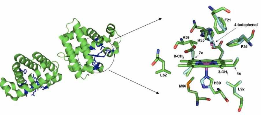
Figure 1. X-ray structure of DHP dimer and active site. ... 24
Figure 2. The Poulos-Kraut mechanism. ... 25
Figure 3. Electronic configuration of low-spin Fe(III). ... 26
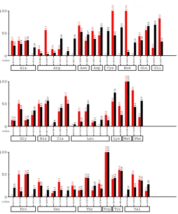
Chapter 2 Figure 1. Codon usage found in A. ornata DHP vs. codon usage preference in E. coli... 37
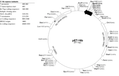
Figure 2. Map of the pET 16b plasmid cloning vector... 38
Figure 3. The 5’ to 3’ DNA sequence of the DHP... 39
Figure 4. Gel illustrating the results of the first round of mutagenesis... 40
Figure 5. Comparison of Arg codon usage by DHP (left), DHP2R (middle), and DHP4R (right) vs. codon usage in E. coli... 41
Figure 6. Comparison of cell pellets from w.t. DHP growth (left) and DHP2R mutant growth (right). ... 42
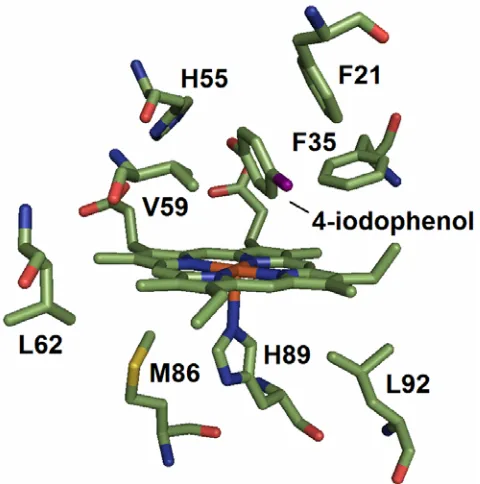
Chapter 3 Figure 1. The X-ray crystal structures of the local heme environment and substrate binding residues of DHP are shown with (light blue) and without (green) bound substrate 4-iodophenol... 69
Figure 2. H NMR spectrum of DHPCN1 ... 70
Figure 3. WEFT-NOESY map of metcyano DHP... 71
Figure 4. C- H HSQC spectrum of DHPCN.13 1 ... 72
Figure 5. High frequency hyperfine-shifted resonances of DHPCN in 100mM potassium phosphate, pH 7.0, 25 °C. A) Without (top) and with a 15 fold excess of 4-bromophenol (bottom). B) Without (top) and with excess 2,4-dichlorophenol (bottom). C) Without (top) and with excess 2,4,6-trifluorophenol (bottom)... 73
Figure 6. F NMR spectra of the 2,4,6-TFP substrate analog in pH 7, 100 mM potassium phosphate, 99.9% D O buffer (top) and titration of this substrate to metcyano DHP at 1:1, 3:1, and 15:1 concentration ratios 19 2 ... 75
Figure 7. A) T relaxation curves and B) T relaxation curves for the meta-protons of 2,4,6-trifluorophenol, alone and in the presence of DHPCN.1 2 ... 76
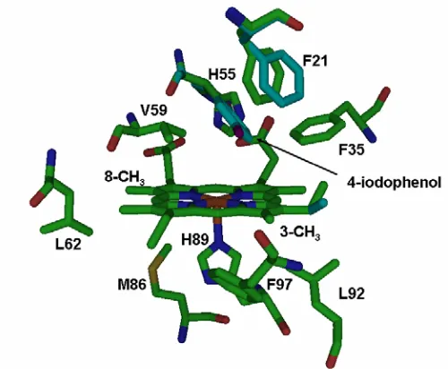
Chapter 4 Figure 1. The X-ray crystal structure (1EWA) of the DHP active site is shown with bound monohalogenated 4-iodophenol... 95
Figure 3. Product formation in both DHP and DHP V59W ... 97 Figure 4. High frequency hyperfine-shifted resonances of DHPCN and V59WCN in 100mM potassium phosphate, pH 7.0, 25 °C. A: Without (top) and with a 15x excess of
4-bromophenol (bottom). ... 98 Figure 5. Kinetic components of the transient differential spectra of NO rebinding to (A) DHP with and without excess 4-BP and (B) DHP V59W with and without excess 4-BP. .. 100
Chapter 5
Figure 1. UV-Vis assay of DHP activity toward monohalogenated 4-BP... 112 Figure 2. UV-Vis assay illustrating the inhibition of DHP activity toward trihalogenated 2,4,6-TCP upon addition of 4-BP ... 113 Figure 3. UV-Vis assay showing inhibition of DHP activity toward “native” substrate 2,4,6-TBP in the presence of 4-BP... 114 Figure 4. Lineweaver-Burk plot illustrating competitive inhibition of the enzyme by mixing 4-BP and 2,4,6-TCP... 115 Figure 5. Inhibition of compound II formation in ferric DHP (420 nm) at pH 7 due to
increasing concentrations of 4-BP. ... 116
Chapter 6
Figure 1. UV-Vis enzymatic assay illustrating 2,4,6-TCP and 4-BP activity ... 136 Figure 2. X-ray structure of the distal binding pocket of DHP... 137 Figure 3. N- H HSQC of DHPCN collected at 298K15 1 ... 138 Figure 4. N- H HSQC spectrum of DHPCN titrated with 4-BP (left column) and 2,4,6-TCP (right column)
15 1
... 139 Figure 5. 15N- H HSQC spectrum of DHPCN titrated with 2,4,6-TCP1 ... 140 Figure 6. The residues experiencing the largest chemical shift deviations mapped onto an existing X-ray structure... 141
Appendix 1
Figure S7. 1D NOE difference spectra of DHPCN in 90% H2O / 10% D2O, 100mM
potassium phosphate, pH 7.0 at 25 °C………... 150 Figure S8. NOESY and COSY overlay showing the connectivity between the two protons which exhibit NOEs to the exchangeable H89 Nε2H resonance………... 151 Figure S9. Titration of 2,4,6-TFP to DHPCN, pH 6.0, 25 °C……….. 152 Figure S10. Titration of 2,4,6-TFP to DHPCN (full spectra shown), pH 6.0, 25 °C……... 153 Figure S11. Variable temperature 1H NMR spectra of the hyperfine-shifted region of
DHPCN……….. 154 Figure S12. Titration of substrate 2,4-DCP to fully formed DHPCN, 100mM potassium phosphate, 25 °C……… 155 Figure S13. 1H NMR spectra of the hyperfine shifted region of DHPCN, pH 8.4, 7.0, and 6.0, at 25 °C………... 156 Figure S14. Analysis of the relaxation data for 1H NMR inversion recovery and spin echo experiments within the assumption of fast exchange……… 159 Figure S15. Spectra of 4-BP and DHP ranging form 0 s (red) to 60 s (blue) following
addition of H2O2 in 100 mM potassium phosphate, pH 7.0………. 160 Figure S16. Spectra of 2,4-DCP and DHP ranging form 0 s (red) to 60 s (blue) following addition of H2O2 in 100 mM potassium phosphate, pH 7.0………. 161 Figure S17. Spectra of 2,4,6-TFP and DHP ranging form 0 s (red) to 60 s (blue) following addition of H2O2 in 100 mM potassium phosphate, pH 7.0………. 162
Appendix 2
Figure S1. NOESY and COSY overlay map of DHPCN V59W………. 164 Figure S2. NOESY and COSY overlay map of DHPCN V59W showing dipolar connectivity between the heme methyls and the adjacent propionate or vinyl substituent……… 166
Appendix 3
LIST OF ABBREVIATIONS
2,4,6-TBP 2,4,6-tribromophenol
2,4,6-TCP 2,4,6-trichlorophenol
2,4,6-TFP 2,4,6-trifluorophenol
2,4-DCP 2,4-dichlorophenol
2,6-DBQ 2,6-dibromo-1,4-benzoquinone
2,6-DCQ 2,6-dichloro-1,4-benzoquinone
4-BP 4-bromophenol
1H NMR proton nuclear magnetic resonance
Ap ampicillin
Cam chloramphenicol
CcP cytochrome c peroxidase
COSY correlation spectroscopy
DHP dehaloperoxidase
DHPCN cyanide-ligated dehaloperoxidase
DHPCO CO-ligated dehaloperoxidase
EPR electron paramagnetic resonance
FT-IR Fourier transform infrared spectroscopy
HRP horseradish peroxidase
HSQC heteronuclear single quantum coherence
HYSCORE hyperfine sublevel correlation spectroscopy
IPTG isopropyl-beta-D-thiogalactopyranoside
Mb myoglobin
MCPBA meta-chloroperbenzoic acid
NOESY nuclear Overhauser effect spectroscopy
CHAPTER 1
Introduction
Dehaloperoxidase (DHP) from the marine polychaete Amphitrite ornata is a dual
function protein which exhibits both globin and peroxidase characteristics (1-2). Structural,
kinetic, and spectroscopic analysis of the protein have detailed the binding and oxidative
dehalogenation of various halophenols upon addition of co-substrate H2O2 (2-5). While the
peroxidase activity of DHP towards trihalogenated phenols is well established, the impact
that other naturally occurring halophenols, such as mono- and dihalogenated phenols, have
on this process has received little attention. Earlier reports illustrated and emphasized the
unique internal distal binding pocket of the protein (3, 6). X-ray data clearly indicate the
ability of monohalogenated phenols to bind in this pocket (3). Why monohalogenated
phenols bind at this site, given the lack of observable product conversion of these molecules,
is a fundamental question consistently glossed over in earlier reports. Moreover, the
molecules that exhibit highest product conversion rates (2,4,6-trihalophenols) have never
been shown to bind in the distal binding pocket under ambient conditions.
Current research shows that binding of monohalogenated phenols in the distal binding
pocket leads to inhibition of DHP peroxidase activity (see Chapter 5). In addition,
halophenols with high product conversion rates (2,4,6-trihalophenols) are now
experimentally shown to bind at a separate external location under ambient conditions (see
Chapters 3, 4, and 6). Herein we review key structural, kinetic, and spectroscopic data which
led to the current discoveries regarding the inhibition of peroxidase activity in the dual
DHP Structure
The heme protein DHP is one of two hemoglobins found in the terebellid polychaete
Amphitrite ornata (1). The dimeric hemoglobin consists of two identical 15.5 kDa subunits,
each containing a heme prosthetic group. To date only four structures of wild type DHP are
available in the PDB (1EW6, 1EWA, 2QFK, and 3DR9). Six unreleased structures, for
which coordinate files have been deposited, include those of DHP with bound
monohalogenated 4-bromophenol and 4-chlorophenol, and a structure of cyanide-ligated
DHP. X-ray structures currently available show the general globin fold of DHP, with 8 alpha
helices and the protein backbone ligated to the heme prosthetic group via a proximal histidine
(2-4). DHP is structurally homologous to myoglobin (Mb) and appears as a dimer in all
current X-ray structures. In the structure the dimer interface appears to involve interactions
of three key residues: D72, V74, and R122. There is also a single surface cysteine (C73)
located near the interface. Formation of a disulfide bond between the surface cysteine of
each subunit is unlikely given the ~11 Å of separation between the two sulfur atoms (3).
Dual conformations of the distal histidine were observed in the first DHP X-ray
structure. The structure indicated that H55 was found in both a “closed” and “open”
conformation (3). The “closed” conformation, typically seen in Mb at basic pH, shows H55
inside the distal pocket but not close enough for hydrogen bonding of the H55 Nε to an axial
ligand. In the “open” conformation, observed under acidic conditions in Mb, H55 is swung
out of the distal pocket. More recent X-ray studies indicate the presence of a single “closed”
H55 conformation in metaquo or ferrous oxy DHP (4). The recent X-ray structures, collected
than in previous studies (4). This places the distal histidine at a distance, with respect to the
heme iron, similar to that observed in Mb. Consequently, hydrogen bonding is observed in
these more recent structures between the distal histidine H55 Nε and coordinated axial
ligands, such as H2O or O2 (4). While only one conformation of H55 was observed in the
metaquo or ferrous oxy state of the protein, X-ray structures of the deoxy form indicated
different results. The deoxy X-ray structures showed the histidine in two distinctly different
“open” orientations when there is no coordinated axial ligand (5).
Perhaps the most frequently emphasized structural feature of DHP is the novel distal
binding pocket. X-ray structures show the ability of DHP to bind a monohalogenated phenol
in this hydrophobic distal cavity (3). The internal binding of 4-iodophenol (4-IP) in this
cavity distinguishes DHP from any other known globin (6). All observable structural
changes induced by binding of the monohalogenated phenol occur at the heme or in the distal
binding pocket. The location of monohalogenated binding in the crystal structure and
binding pocket resides of the active site of DHP can be seen in Figure 1. Upon 4-IP binding,
the distal histidine is observed solely in the “open” conformation. The location of 4-IP in the
pocket necessitates this change in conformation, as “closed” H55 and 4-IP appear to vie for
the same space in the distal pocket. As seen in Figure 1, the three closest residues to bound
4-IP are V59, F35, and F21 with nearest C-C distances of 2.98, 2.98, and 3.41 Å,
respectively. Notable conformations changes are seen in the distal pocket residues H55,
F35, and F21. In addition, small changes in orientation are observed in the 2-vinyl, 4-vinyl,
6-propionate, and 7-propionate heme substituents. Binding of 4-IP does not, however,
DHP Activity
Activity in peroxidases normally occurs through a reactive intermediate known as
compound I (7). One of the most well known and commonly studied peroxidases is
horseradish peroxidase (HRP). Activation of the enzyme occurs through formation of an
active intermediate known as compound I. Formation of this intermediate occurs via the
Poulos-Kraut mechanistic activation of peroxidases, as seen in Figure 2 (8). In this
mechanism, H2O2 binds to the ferric heme iron and is stabilized by hydrogen bonding to the
Nε of the distal histidine. Proton transfer between the -oxygen of H2O2 to the -oxygen,
subsequent heterolytic cleavage of the O-O bond, and release of H2O leads to formation of
the high valence Fe(IV)=O species which contains a π-cation radical. This reactive
intermediate is unknown as compound I. Loss of the π-cation radical results in a second
reactive intermediate known as compound II, which is the referred to as a Fe(IV)=O or
Fe(IV)-OH species. Regeneration of the Fe(III) resting state from compound I is achieved by
oxidation of a substrate molecule via two sequential one-electron oxidation steps.
Initial enzymatic analysis probing the peroxidase activity of DHP was reported along
with initial spectroscopic and crystallization of the protein in 1996 (2, 9). The activity of
DHP was found to be similar to that of HRP (10). DHP was shown to oxidatively
dehalogenate trihalophenols in a hydrogen peroxide dependent manner. Specifically, the
peroxidase activity of DHP towards bromophenol (TCP) and
2,4,6-tribromophenol (2,4,6-TBP) was demonstrated and initial product characterization was
spectrometry resulted with molecular ions characteristic of 2,6-dibromo-1,4-benzoquinone
(2,6-DBQ) and 2,6-dichloro-1,4-benzoquinone (2,6-DCQ), respectively (9). These initial
enzymatic assays set precedent for further enzymatic reports which included using
co-substrates other than H2O2. Using an alternate oxygen donating co-substrate,
meta-chloroperbenzoic acid (MCPBA), resulted in an approximate 3 fold increase in 2,4,6-TCP
turnover (11). The increased dehalogenation activity of DHP using MCPBA may indicate
the natural co-substrate is an oxidant other than H2O2 (11).
Further investigations into DHP activity revealed a correlation between 2,4,6-TBP
turnover and solvent pH. DHP activity and kinetic analysis of product formation was
performed using UV-Vis and stopped-flow experiments in pH ranging from 4.0 to 8.0.
Highest turnover rates for the “native” substrate, 2,4,6-TBP, were observed at pH 7.5. The
second highest turnover for the substrate occurred at pH 7.0 (12). Since 2,4,6-TBP exists in
the phenolate form at pH > 6.8, the data indicate binding and oxidation of substrate in the
phenolate form is favorable for protein activity. This is not the case with other peroxidases,
such as HRP, which does not oxidize substrates in the phenolate form (13). The high activity
of DHP between pH 7.0-7.5 may be due to the physiological conditions in which the protein
is found. DHP is the coelomic hemoglobin of A. ornata where pH is ~ 7.4. Even though
2,4,6-TBP turnover is highest at pH > 7.0, faster kinetics are seen at pH < 7.0. An increase in
product formation kinetics comes at the expense of protein stability. An increase in heme
degradation is observed lower pH values. This observation may explain why the greater rate
As DHP activity was investigated in detail, questions about the internal binding site
began to arise. Monohalogenated phenols are clearly bound in the distal cavity as shown in
the X-ray data. However, binding in this location effectively removes the only residue
capable of supporting proton transfer needed for compound I formation upon H2O2 binding.
To complicate this matter, studies found that order of addition of substrate, 2,4,6-TBP, and
co-substrate, H2O2, greatly affected protein activity (14). Mixing substrate and protein prior
to H2O2 addition resulted in typical substrate oxidation. However, if DHP and H2O2 were
incubated for more than about 30 seconds prior to substrate addition, protein activity was
inhibited. The results indicated that DHP activity is highest when substrate is added first.
This implied that the active form of H55 was solely in the “open” conformation and out of
the distal pocket. Removal of H55 means removal of the sole residue capable of the acid
base catalysis needed for compound I to form (14). These results raised important questions
about the relationship between substrate binding local and protein activity. Based on the
results, one of two conclusions can be reached. First, the substrate molecule, now bound in
the distal pocket, would have to act as a trigger for the protein to switch from globin to
peroxidase activity. Given the displacement of H55 out of the pocket, the mechanism of
compound I would also have to be completely different than that dictated by the
Poulos-Kraut mechanism. Second, substrate could be binding at a location other than the distal
binding pocket. This scenario would allow H55 to remain in the “closed” conformation,
thereby facilitating proton transfer needed during compound I formation. Depending on the
inhibit protein activity due to heme degradation or cause it to become “stuck” in an inactive
compound II state as indicated by current and previous investigations (11, 12, 14).
DHP Mutagenesis
Site-directed mutagenesis of the distal and proximal histidines, and residues
comprising the distal binding was performed to better ascertain the roles of active site amino
acids in DHP peroxidase activity. The relative activity of the following DHP mutants has
been compared at pH 7.0 using UV-Vis spectrophotometric assays: Y38F, H55R, H55V,
H55V/V59H, V59W, and H89G. The residue tyrosine 38 is important in regards to the distal
binding pocket, because it is close enough to form a hydrogen bond to the hydroxyl group of
bound monohalogenated phenol (3). This is believed to stabilize monohalogenated phenol
binding in the distal pocket. Mutation of this residue to phenylalanine will eliminate the
hydrogen bond and provide insight into how monohalogenated phenols bind in the distal
pocket. V59 is the closest residue to bound monohalogenated phenol in the X-ray structure,
with the nearest C-C distance being 2.98 Å (3). Mutation of this residue to a tryptophan
(V59W, as discussed in Chapter 4) will introduce a very bulky side chain into the distal
binding pocket. The hypothesis is that this mutation will “block” binding of
monohalogenated phenol in the distal cavity.
The distal (H55) and proximal (H89) histidines are extremely important residues in
both globins and peroxidases. The H89 provides the only covalent linkage between the heme
and protein backbone. In addition, the Nδ of H89 hydrogen bonds to the backbone carbonyl
hydrogen bonding of the proximal histidine Nδ to an aspartate side chain. This makes the
proximal histidine a better electron donor and therefore creates a stronger “push” to the heme
iron than observed in globins (15). The distal histidine, H55, acts to stabilize the binding of
H2O2 to the heme iron. The residue is also responsible for transferring a proton from the
-oxygen to the -oxygen of H2O2 and assists in “pulling” apart the O-O bond which leads to
formation of compound I (8).
Of the six mutations, only Y38F, H55R, and V59W showed significant activity
toward 2,4,6-TBP and 2,4,6-TCP (16, 17). The Y38F mutant, which eliminates any
hydrogen bonding between internal halophenol and the protein, actually shows an ~ 5 fold
increase in 2,4,6-TBP product turnover rate compared to DHP. Conversely, the H55R
mutation results in an ~10 fold decrease in product turnover (16). The V59W mutant
exhibits kinetics and product turnover rates similar to DHP. The wild type DHP only
exhibited 1.2x greater product formation than V59W mutant (17). The H55V, H55V/V59H,
and H89G mutants all resulted in negligible protein activity. The activity, albeit lowered, of
H55R and the lack of activity in H55V indicates the necessity for a residue capable of
acid-base catalysis at amino acid 55. The results of the V59W mutation indicate that having a
bulky side chain in the internal cavity does little to affect protein activity. The internal
location of V59 and the necessity for a “closed” H55 conformation needed for compound I
formation inherently dictate a crowded internal distal cavity. Alternatively, binding of
substrate on the external heme edge, as is typical with HRP, would account for the observed
activity in V59W. The enzymatic properties of the Y38F mutant also corroborate an exterior
was thought this mutant would lead to a decrease in activity. Since Y38 has the potential to
form a hydrogen bond with H55, it was thought that it could be part of a hydrogen bond
network needed for peroxidase function. However, the results indicate that removal of the
putative internal hydrogen bond network between monohalogenated phenol and the protein
enhances activity. Collectively, the mutagenesis data indicate a separate mode of binding of
trihalophenols and suggest that such binding may occur at site other than the
monohalogenated phenol binding pocket.
FT-IR Experiments on DHPCO
For the past 40 years myoglobin (Mb) has served as a model system for studying the
structure-function relationship of proteins. Myoglobin, the protein responsible for oxygen
storage and transportation in muscle, reversibly binds diatomic ligands such as oxygen (O2),
nitric oxide (NO), and carbon monoxide (CO). DHP shares these characteristically globin
features with Mb and, given the strong structural homology between DHP and Mb, any
structural analysis used for Mb should translate well to DHP experiments. For example,
monitoring the rebinding of diatomic ligands after laser photolysis provides excellent insight
into the dynamic structure-function relationship of residues near heme iron, which in DHP
also happens to be the monohalogenated phenol binding site (18).
If ligand rebinding were to involve a single process, then the kinetics should be
single exponential. However, one of the early experiments in biophysical studies of Mb was
the demonstration that CO rebinding was actually non-exponential in time below 200K (19).
conformational states each with its own rebinding rate (19, 20). Therefore when a ligand,
such as CO, is introduced to the protein the ligand acts as a probe of the internal protein
cavity around the heme iron. If Mb only had one general conformation then the CO
stretching vibration (vC-O) of this ligand in the protein would inherently have only one
frequency. However, different conformational substates of the protein cause the vC-O to
resonate at different frequencies. Upon laser photolysis at cryogenic temperatures, the ligand
remains trapped inside the protein for some period of time before escaping into the solvent.
After photodissociation, the ligand becomes trapped slightly above the heme group in a so
called primary docking site “B” (21, 22) and does not recombine by a thermal mechanism,
but rather by tunneling. This recombination is sufficiently slow that it can be observed in an
X-ray structure. X-ray experiments indicate that after photodissociation, at liquid-helium
temperatures, the photodissociated CO moves to this primary docking site (B) just above
pyrrole C and the iron shifts slightly out of the heme plane (22).
In wild type Mb, three heme bound CO stretching bands resonate at ~1965, ~1945,
and ~1933 cm-1 respectively. These three substates differ primarily in the position of the
distal histidine residue. If the distal histidine swings out of the pocket (“open” conformation)
the result is a more apolar environment in the distal pocket. This gives rise to the A0 band at
~1965 cm-1. If the distal histidine is in the “closed” conformation within the pocket the
partial positive charge of the Nε-H interacts with bound CO and hence gives rise to the A1
and A3 bands at ~1945 and ~1933 cm-1 (18). Upon photolysis the CO molecule can populate
the primary docking site in opposite orientations with respect to the heme iron. These two
third, smaller, photodissociated band, B0, at ~2150 cm-1 is characteristic of the
photodissociated CO at the docking site with the protein in the A3 conformation (18).
There are currently two cryogenic FT-IR studies on ferrous CO ligated DHP
(DHPCO). The initial report found strong similarities between the CO stretching frequencies
of MbCO and DHPCO. The typical heme-bound CO stretching vibrations of DHPCO were
found to be ~1950 cm-1 with H55 in the “closed” conformation (corresponding to A1 = 1945
cm-1 in MbCO) and ~1965 cm-1 for H55 in the “open” conformation (corresponding to A0 =
1965 cm-1 in MbCO) (23). This illustrates the relative similarities between the two globins in
regards to distal histidine conformational dynamics. Additional experiments also showed the
pKa of H55 protonation, and likewise rotation of the side chain the “open” conformation,
occurs around pH 4.5 (24).
The FT-IR experiments marked the first time a trihalogenated phenol had been used
for binding studies. Given the mM concentrations of protein required for the experiments,
the small, highly soluble trihalogenated 2,4,6-trifluorophenol substrate (2,4,6-TFP) was used.
The substrate was added to the protein at pH 5.5, 7.0, and 10.0. A new CO stretching band
was observed at 4K upon the addition of 2,4,6-TFP. The band, As, occurs at ~1972 cm-1 and
indicates that ~80% of all DHP molecules internally bind 2,4,6-TFP at 4K, pH 5.5 (23). This
band is not readily observed at pH 7, or pH 10, indicating a preference for internal binding
under acidic conditions. The As = 1972 cm-1 band represents a nonbonding, electrostatic
interaction between the aromatic substrate ring and the heme-bound CO ligand. However,
the 1972 cm-1 band completely disappears as the sample is warmed up, indicating a loss of
substrate is removed from the distal binding pocket. The removal of 2,4,6-TFP from the
internal pocket occurs at temperatures above 160K. No observable bands corresponding to
bound 2,4,6-TFP are observed at ambient temperatures. The results indicated for the first
time that trihalogenated phenols (active substrates) could bind in the “substrate” binding site
in the distal cavity. However, such binding was only observed at pH 5.5 and temperatures <
160K. Given the activity of DHP toward 2,4,6-TFP it is peculiar that no binding events are
observed under ambient conditions (25, 26).
X-band EPR on metaquo DHP and Detection of Protein Radicals
Recent investigations using X-band EPR and hyperfine sublevel correlation
spectroscopy (HYSCORE) have provided insight into the heme iron ligation state and the
nature of 2,4,6-TFP binding (27). Additional experiments probing internal substrate binding
is necessary in light of recent FT-IR data indicating the internalization of substrate 2,4,6-TFP
(23, 24). Current X-ray structures of metaquo DHP indicate a coordinated axial water ligand
(28). Analysis of resonance Raman data indicates a mixture of 5-coordinate and 6-coordinate
heme in ferric DHP (28). Upon addition of monohalogenated phenol, both resonance Raman
and X-ray data show a predominately 5-coordinate heme. This indicates expulsion of the
coordinated water molecule. EPR HYSCORE experiments provide an opportunity to probe
interactions between the iron and coordinated water. More importantly HYSCORE will help
determine the fate of bound water upon addition of trihalogenated 2,4,6-TFP.
HYSCORE is a 2D spectroscopic technique used to correlate electron spins to nearby
ring and the nearby iron-coordinated water protons were observed during the experiment.
The strongly coupled water protons at ~6 MHz were assigned based on previous studies of
metmyoglobin where the ligated water protons exhibited a 6.0 ± 0.1 MHz hyperfine coupling
(27). Addition of a 10x excess of 2,4,6-TFP at 4.5K causes the coupled water signal to
disappear, indicating displacement of the ligand and a transition from six- to five-coordinate
iron. Examination of the X-band EPR spectra of ferric DHP with and without 2,4,6-TFP
clearly show the g = 6 and gjj = 2 features characteristic of high spin (S = 5/2) heme iron
(27). The collective data indicate a change in coordination state (6-coordinate heme to
5-coordinate heme) with no alteration in spin state upon addition of 2,4,6-TFP at 4.5K. The
HYSCORE data corroborate FT-IR studies on DHPCO and indicate that substrate 2,4,6-TFP
is internalized at low temperatures.
EPR experiments were also used to detect protein radical formation in DHP upon
addition of H2O2. Protein radicals are generated by rapid electron transfer of the compound I
π-cation radical to the protein, which can include long range electron transfer of the radical to
distant sites within the protein (30, 31). Protein radicals have been shown to act as catalytic
intermediates, a site for subunit cross-linkage, or as a species used to divert an oxidizing
equivalent away from the active site (31). Recent investigations using EPR on rapid-freeze
quenched protein have detected the presence of a protein radical shortly after the addition of
H2O2 (32). This intermediate is similar in nature to the compound ES of cytochrome c
peroxidase, which arises from a ferryl heme and tryptophan radical (33). The exact location
of the radical has yet to be determined, but analysis of the signal g factor and hyperfine
radical in DHP activity is also undetermined but it may act as a second oxidizing equivalent
or as the site of substrate oxidation itself (34).
NMR Investigations of metcyano DHP
Heme proteins such as DHP contain the prosthetic group iron protoporphyrin IX,
which can exist in three spin states in the ferric form: high spin (S=5/2), intermediate spin
(S=3/2), and low spin (S=1/2). The introduction of a strong ligand such as CN- creates a
strong repulsion in the five d electrons of Fe(III). This strong repulsion is favored if the five
d electrons position themselves in an orbital with the lowest possible energy. In this strong
ligand field there is only one unpaired electron, and the iron is in a low-spin state (S=1/2).
Figure 3 illustrates the electronic configuration of low-spin Fe(III). In relation to NMR, the
magnetic moment of an unpaired electron is approximately 660 times greater than that of a
proton (35). If CN- ligand is bound to theheme iron the effect is a low spin Fe(III)
paramagnetic center with S = 1/2. This has dramatic affects on the NMR spectrum. The
magnetic moment of the unpaired electron causes paramagnetic relaxation of protons on the
heme and nearby residues (36). The protons near the unpaired electron experience
paramagnetic relaxation which results in large hyperfine-shifting of the resonances. The
hyperfine shifted resonances appear outside of the diamagnetic protein envelope, which do
not exhibit a paramagnetic contribution. In metMbCN, protons that lie at a distance greater
than 7.5 Å from the heme iron exhibit negligible paramagnetic relaxation (37). The
hyperfine shifts of the heme environment protons enable their NMR signals to be easily
Given the close proximity of the distal binding site to the heme iron, initial 1H NMR
experiments utilized the paramagnetic properties of the heme iron to specifically focus efforts
on active site resonances (25). The low-spin metcyano adduct of DHP was chosen because
the hyperfine-shifted resonances are much sharper and less dispersed in the low-spin form
than in the high-spin, metaquo form. Using primarily Water Eliminated Fourier
Transform-Nuclear Overhauser Enhancement Spectroscopy (WEFT-NOESY) and natural abundance
13C-1H Heteronuclear Single Quantum Coherence (13C-1H HSQC), the majority of
hyperfine-shifted resonances were assigned. Using the chemical shifts and spacing of the heme
methyls, the projection angle, Φ, of the proximal histidine on the heme plane was found to be
different than the angle observed in the X-ray structure (3, 4). According to X-ray structures
this angle is ~113°, in solution, however, the angle was found to be ~90° based on the NMR
data.
Assignment of the active site resonances allowed for substrate binding studies geared
toward detection of distal biding site interactions. Upon titration of monohalogenated 4-BP,
dihalogenated 2,4-dichlorophenol (2,4-DCP), and trihalogenated 2,4,6-TFP varying
perturbations were found. 4-BP and 2,4-DCP both induced line broadening in the 3-CH3 and
Phe97 CζH resonances, which are separated by ~4.5 Å in the X-ray structure (3, 4). In
addition, chemical shift deviations were observed in the 7α propionate protons. The line
broadening and chemical shift deviations were observed over the pH range 5.5 – 9.0.
Although the greatest degree of change was observed at pH 5.5, the changes were still
observable at the alkaline pH 9.9. Addition of 2,4,6-TFP did not result in observable changes
on 2,4,6-TFP alone and in the presence of DHPCN, evidence was found indicating possible
binding of 2,4,6-TFP at a separate site (25). Because the substrates were found to be in fast
exchange and no substrate-protein NOEs were detectable, additional 13C/15N labeled NMR
experiments were conducted to help locate any possible secondary binding locations.
After high levels of DHP expression in 13C/15N enriched minimal media were
obtained the protein backbone 13Cα, 13Cβ, carbonyl 13C, amide 1H and amide 15N resonances
were assigned in metcyano DHP using 15N-1H HSQC, HNCO, HCACOCANH, HNCOCA,
HNCA, and HNCACB NMR experiments. 125 of the 137 residues in DHP were readily
assigned as a result of the NMR data. Assignment of the backbone 13C, 15N, and 1H nuclei
allowed for detection of binding perturbations throughout the protein upon titration of
various halophenols. Isotopically labeled protein also allowed for rapid 15N-1H HSQC data
collection upon stoichiometric addition of halogenated phenols. Upon addition of the
monohalogenated phenol 4-BP, the largest deviations in amide 1H chemical shift were
observed in the three closest residues to bound 4-IP in the X-ray structure: F35, V59, and F21
(38). Titration of excess substrate 2,4,6-TCP induced the largest chemical shift deviations in
the exchangeable H55 NεH and local residues surrounding W120 , which is ~15 Å away
from H55. No significant changes were observed in distal binding pocket resonances F35,
V59, and F21 upon addition of 2,4,6-TCP. Addition of the smaller 2,4,6-TFP substrate
caused the largest changes in chemical shift deviation at the exchangeable H55 NεH, distal
binding pocket resides F35, V59, and F21, and the local area around W120. This indicates
2,4,6-TFP may be small enough to bind at both the 4-BP and 2,4,6-TCP site. The 2,4,6-TCP
interface near W120, or the substrate binds directly near the W120 residue. Binding at either
site could account for oxidation of the substrate. Oxidation could occur along the heme edge,
as seen in HRP (39), or at a tryptophan radical, as observed in lignin peroxidase (34, 40, 41).
Site-directed mutagenesis of the W120 residue will help elucidate whether or not the residue
References
(1) Weber, R. E., Magnum, C. P., Steinman, H., Bonaventura, C., Sullivan, B., and Bonaventura, J. (1977) Hemoglobins of two terebellid polychaetes:Enoplobranchus sanguineus and Amphitrite ornata. Comparative Biochemistry and physiology56A, 179-187.
(2) Zhang, E., , Chen, Y. P., Roach, M. P., Lincoln, D. E., Lovell, C. R., Woodin, S. A., Dawson, J.H., and Lebioda, L. (1996) Crystallization and initial spectroscopic
characterization of the heme-containing dehaloperoxidase from the marine polychaete
Amphitrite ornataActa Cryst. D 52, 1191-1193.
(3) LaCount, M. W., Zhang, E., Chen, Y.P., Han, K., Whitton, M. M., Lincoln, D. E., Woodin, S. A., and Lebioda, L. (2000) The crystal structure and amino acid sequence of dehaloperoxidase from Amphitrite ornata indicate a common ancestry with
globins. J. Biol. Chem. 275, 18712-18716.
(4) de Serrano, V., Chen, Z., Davis, M. F., and Franzen, S. (2007) X-ray crystal structural analysis of the binding site in the ferric and oxyferrous forms of the recombinant heme dehaloperoxidase cloned from Amphitrite ornata. Acta Crysta. D 63, 1094-1101.
(5) Chen, Z., de Serrano, V., Betts, L., and Franzen, S. (2009) Distal histidine
conformational flexibility in dehaloperoxidase from Amphitrite ornata. Acta Crysta.
D 65, 34-40.
(6) Lebioda, L., LaCount, M. W., Zhang, E., Chen, Y. P., Han, K., Whitton, M. M., Lincoln, D. E., and Woodin, S. A. (1999) Protein structure: An enzymatic globin from a marine worm. Nature. 401, 445.
(7) Hewson, W. D., and Dunford, H. B. (1976) Oxidation of p-cresol by horseradish peroxidase compound I. J. Biol. Chem. 251, 6036-6042.
(8) Poulos, T. L., and Kraut, J. (1980) The stereochemistry of peroxidase catalysis. J.
(9) Chen, Y. P., Woodin, S. A., Lincoln, D. E., and Lovell, C. R. (1996) An unusual dehalogenating peroxidase from the marine terebellid polychaete Amphitrite ornata.
J. Biol. Chem.271, 4609-4612.
(10) Ferrari, R. P., Laurenti, E., and Trotta, F. (1999) Oxidative 4-dechlorination of 2,4,6-trichlorophenol catalyzed by horseradish peroxidase, J. Biol. Inorg. Chem. 4, 232-237.
(11) Osborne, R. L., Taylor, L. O., Han, K. P., Ely, B., and Dawson, J. H. (2004) Amphitrite ornata dehaloperoxidase: enhanced activity for the catalytically active globin using MCPBA. Biochem. Biophys. Res. Comm.324, 1194-1198.
(12) Franzen,S., Gilvey, L.B., and Belyea, J. (2007) The pH dependence of the activity of dehaloperoxidase from Amphitrite ornata. Biochim. Biophys. Acta: Prot. Struct. Mol.
Enz.1774, 121-130.
(13) Patel, P. K., Mondal, M. S., Modi, S., and Behere, D. V. (1997) Kinetic studies on the oxidation of phenols by the horseradish peroxidase compound II. Biochim. Biophys.
Acta-Prot. Struct. Mol. Enz. 1339, 79-87.
(14) Belyea, J., Gilvey, L. B., Davis, M. F., Godek, M., Sit, T. L., Lommel, S. A., and Franzen, S. (2005) Enzyme function of the globin dehaloperoxidase from Amphitrite ornata is activated by substrate binding. Biochemistry44, 15637-15644.
(15) Dawson, J. H. (1988) Probing structure-function relations in heme-containing oxygenases and peroxidases. Science, 433-439.
(16) Franzen, S., Belyea, J., Gilvey, L. B., Davis, M. F., Chaudhary, C. E., Sit, T. L., and Lommel, S. A. (2006) Proximal cavity, distal histidine, and substrate hydrogen-bonding mutations modulate the activity of Amphitrite ornata dehaloperoxidase.
Biochemistry, 9084-9094.
(18) Nienhaus, K., Deng, D., Olsen, J. S., Warren, J. J., and Nienhaus, G. U. (2003) Structural dynamics of myoglobin: Ligand migration and binding in valine 68 mutants. J. Biol. Chem. 278, 42532-42544.
(19) Frauenfelder, H., Sligar, S. G., and Wolynes, P. G. (1991) The energy landscapes and motions of proteins. Science 254, 1598-1603.
(20) Austin, R. H., Benson, K. W., Eisenstein, L., Frauenfelder, H., and Gunsalus, I. C., (1975) Dynamics of ligand binding to myoglobin. Biochemistry14, 5355-5373. (21) Kriegl, J. M., Nienhaus, K., Deng, P., Fuchs, J., and Nienhaus, G. U. (2003) Ligand
dynamics in a protein internal cavity. Proc. Natl. Acad. Sci. U. S. A.100, 7069-7074. (22) Ostermann, A., Washipky, R., Parak, F. G., and Nienhaus, G. U. (2000) Ligand
binding and conformation motions in myoglobin. Nature404, 205-208.
(23) Nienhaus, K., Deng, P. C., Belyea, J., Franzen, S., and Nienhaus, G. U. (2006) Spectroscopic study of substrate binding to the carbonmonoxy form of
dehaloperoxidase from Amphitrite ornata. J. Phys. Chem. B110, 13264-13276. (24) Nienhaus, K., Nickel, E., Davis, M. F., Franzen, S., and Nienhaus, F. U. (2008)
Determinants of substrate internalization in the distal pocket of dehaloperoxidase hemoglobin of Amphitrite ornata. Biochemistry 47, 12985-12994.
(25) Davis, M. F., Gracz, H., Vendeix, F. A. P., de Serrano, V., Somasundaram, A., Decatur, S. M., and Franzen, S. (2009) Different modes of binding of mono-, di, and tri-halogenated phenols to the hemoglobin dehaloperoxidase from Amphitrite ornata.
Biochemistry 48, 2164-2174.
(26) Chaudhary, C. (2003) Point Mutagenesis and Spectroscopic Probing of
Dehaloperoxidase: Characterizing the Mechanism and Activity of the Heme Active Site of the Native Protein. MS Thesis, NC State Univ.
(28) Thompson, M. K., Davis, M. F., de Serrano, V., Nicoletti, F. P., Howes, B. D., Smulevich, G., and Franzen, S. Two-site competitive inhibition in dehaloperoxidase from Amphitrite ornata. (In preparation)
(29) Hofer, P., Grupp, A., Nebenfuhr, H., and Mehring, M. (1986) Hyperfine sublevel correlation (HYSCORE) spectroscopy: A 2D ESR investigation of the squaric acid radical. Chem. Phys. Lett. 132, 279-282.
(30) Lardinois, O. M., and Ortiz de Montellano, P. R. (2001) H2O2-mediated cross-linking between lactoperoxidase and myoglobin: Elucidation of protein-protein radical
transfer reactions. J. Biol. Chem. 276, 23186-23191.
(31) Lardinois, O. M., and Ortiz de Montellano, P. R. (2003) Intra- and intermolecular transfers of protein radicals in the reactions of sperm whale myoglobin with hydrogen peroxide. J. Biol. Chem. 278, 36214-36226.
(32) Feducia, J., Dumarieh, R., Gilvey, L., B., G., Smirnova, T., Franzen, S., and Ghiladi, R. A. (2009) Characterization of dehaloperoxidase compound ES and its reactivity with trihalophenols. Biochemistry48,995-1005.
(33) Sivaraja, M., Goodin, D. B., Smith, M., and Hoffman, B. M. (1989) Identification by ENDOR of Trp191 as the free-radical site in cytochrome c peroxidase. Science 245, 738-740.
(34) Ruiz-Dueñas, F. J., Pogni, R., Morales, M., Giansanti, S., Mate, M. J., Romero, A., Martínez, M. J., Basosi, R., and Martínez, A. T. (2009) Protein radicals in fungal versatie peroxidase: Catalytic tryptophano radical in both compound I and compound II and studies of W164Y, W164H, and W164S variants. J. Biol. Chem. 284, 7986-7994.
(35) Dunford, B. H. (1999) in Heme Peroxidases, pp 135-190, Wiley-VCH, New York. (36) La Mar, G. N., and de Ropp, J. S. (1993) in Biological Magnetic Resonance, pp 1-73,
Plenum Press, New York.
(38) Davis, M. F., Bobay, B. G., and Franzen, S. (2009) Separation of inhibitor and substrate binding locations in the globin dehaloperoxidase. (In preparation)
(39) Ator, M. A., and Ortiz de Montellano, P. R. (1987) Protein control of prosthetic heme reactivity: Reaction of substrates with the heme edge of horseradish peroxidase. J.
Biol. Chem.262, 1542-1551.
(40) Hammel, K. E., and Tardone, P. J., (1988) The oxidative 4-dechlorination of polychlorinated phenols is catalyzed by extracellular fungal lignin peroxidase.
Biochemistry 27, 6563-6568.
(41) Doyle, W. A., Blodig, W., Veitch, N. C., Piontek, K., and Smith, A. T. (1998) Two substrate interaction sites in lignin peroxidase revealed by site-directed mutagensis.
Figures
Figure 1. The DHP dimer is seen on the left and the heme active site including the distal
binding pocket are enlarged on the right. The green sticks shows the X-ray structure without
Figure 2. The Poulos-Kraut mechanism illustrating the formation of the reactive intermediate
xy xz, yz
z2 x2-y2
Low Spin Fe(III), S=1/2
CHAPTER 2
Introduction
As an alternative to extracting and isolating DHP from its natural host, the marine
worm Amphitrite ornata, we have used the bacterium Escherichia coli as a host organism for
the expression of the DHP protein. High levels of DHP expression in E. coli is vital given
the large quantities of protein needed for many spectroscopic experiments (e.g. X-ray
crystallography, NMR and EPR spectroscopy). NMR experiments particularly, if isotopic
labeling is involved, require an optimal expression system. In E. coli, as well in most other
organisms, there is a preference for usage of certain codons. There is a correlation between
the frequency of certain codons and the level of its respective tRNA in the cell (1). This
correlation dictates that expression of eukaryotic sequences containing codons rarely used in
E. coli may cause translational problems leading to reduced quantity or fidelity of protein
synthesized (1, 2). The arginine codons AGG and AGA are the two least frequently used
codons in E. coli (3). Numerous studies have shown that usage of the arginine codon AGG in
E. coli had adverse effects on protein expression (2, 4, 5). Additionally, studies have shown
that having consecutive AGG codons in the reading frame leads to a high level of
frameshifting (6). In addition, it has been shown that a processivity error in translation of a
sequence with just two consecutive AGG codons can lead to a 50% decrease in protein yield
(4-6).
Figure 1 shows that the protein coding region for DHP uses AGG to encode for
arginine with a frequency of 57%. In E. coli, however, AGG typically occurs at a frequency
coli host strain. The host strains of E. coli currently used are the BL21 (DE3) and Rosetta™
(DE3) cell lines. The Rosetta™ (DE3) cell line is a BL21 host derivative that was
specifically designed to enhance the expression of eukaryotic proteins by supplying
additional amounts of low frequency E. coli tRNAs, specifically the 6 rarely used tRNAs
AGG, AGA, AUA, CUA, CCC, and GGA. Even though the Rosetta™ (DE3) cell line
provides additional tRNAs for rare codons such as AGG, the fidelity and productivity of
DHP expression is hypothesized to increase still via mutation of these AGG codons to the
more amenable CGC arginine codons.
Initial experiments involved cloning the DHP gene ino the pUC 19 vector (7).
Cloning the gene into the pET 16b expression vector with the Rosetta™ (DE3) cell line as
the expression host was perfomed in order to enhance expression levels. The pET system is
a high level protein expression system, which utilizes the highly specific T7 RNA
polymerase promoter. A chromosomal copy of the gene for T7 RNA polymerase is under the
control of the inducible lac promoter. Upon induction of a non-hydrolysable lactose analog,
isopropyl-beta-D-thiogalactopyranoside (IPTG), T7 RNA polymerase is synthesized leading
to expression of DHP via the T7 promoter (8). Upon full induction, the T7 RNA polymerase
is so active that it focuses nearly all of the cells resources toward expression of the target
gene. The pET 16b vector is shown in Figure 2 (9). The protein coding region for DHP is
located between the restriction endonuclease sites Nco I and BamH I.
The DHP gene contains four AGG codons. As discussed earlier there is a strong bias
in E. coli against the usage of AGG for arginine. Although the Rosetta™ (DE3) cell line
the AGG codons within DHP leads us to believe that silent mutations of these codons to
CGC could enhance DHP expression. Figure 3 shows the coding sequence of DHP. AGG
codons are located at positions 14, 32, 33, and 122. Translation of a sequence with two or
more consecutive AGG codons, such as R32 and R33 in DHP, could lead to errors, such as
frameshifting, thereby leading to a decrease in both protein fidelity and productivity in E. coli
(4).
Mutation of the four AGG codons to a more frequently used condon, such as CGC
should have a substantial impact on DHP expression levels. Introduction of the four
mutations was conducted using the QuikChangeTM Multi Site-Directed Mutagenesis Kit
(Stratagene). The kit offers a method for site-directed mutagenesis of up to five sites
simultaneously and consists of three main steps (10). The first step involves the annealing of
mutagenic primers to a denatured DNA template via thermal cycling followed by extension
of the mutagenic strand via a PfuTurboTM DNA polymerase. The resultant double stranded
DNA molecule contains a parental strand and a strand with the introduced mutations. The
second step involves the use of the restriction endonuclease DpnI, which specifically cleaves
methylated DNA, for digestion of the parental strand leaving only the mutated strand. The
third and final step is transformation of the multi-mutated DNA into XL10-GoldTM
ultracompetent cells, which allow for transformation of single stranded DNAs.
Materials and Methods
The pET 16b-DHP plasmid DNA used for the mutations was transformed into E. coli
37° C. Single colonies were isolated from the plates and inoculated into 2mL LB Ap broth
for overnight growth at 37 °C with shaking at 300 RPM. DNA from the growths was
purified using QIAprepTM plasmid mini prep spin kits and then sent to MWG-Biotech, Inc.
for sequencing. All DNA used for the mutagenic experiments were sequenced prior to use.
The mutations were performed using the QuikChangeTM Multi Site-Directed Mutagenesis Kit
from Stratagene. After a round of mutation the DNA was transformed into the provided
XL10-GoldTM ultracompetent cells. DNA was purified from transformants as discussed
previously. The DHP2R and DHP4R mutants were transformed into competent RosettaTM
(DE3) cells and plated onto Ap/Chloroamphenicol (Cam) plates and grown overnight at 37°
C. Single colonies were isolated and used to inoculate 2mL 2xYT broth containing both
Ap/Cam which was subsequently grown at 37° C overnight with shaking at 300 RPM. This
growth was used to inoculate a scaled up growth from which the cell pellets were harvested.
The resulting soluble DHP protein was purified as describe elsewhere (11).
Results
Three mutagenic primers were used for the mutations. The R32R and R33R silent
mutations (AGG to GCG) were incorporated onto the same primer. This primer also
contained a silent Y28Y (TAC to TAT) mutation which incorporated a recognition site for
the restriction endonuclease BspE I. This would allow for screening of the transformants by
restriction analysis to see if the second and most important primer annealed. No silent
mutations could be made to introduce restriction sites near the R14R and R122R mutation
colonies were isolated and the DNA of each was purified and cut with BspE I. The result is
shown in Figure 4. The BspE I analysis indicated that the second primer annealed in 4 out of
the 12 colonies isolated. After sending these 4 samples for sequencing only 2 of the 4
actually contained the desired R32R/R33R mutations. Sequencing also revealed that neither
R14 nor R122 were successfully mutated. A second round of mutagenesis was then
performed using the newly synthesized R32R/R33R DHP mutant (DHP2R) as the template.
Out of this round 12 colonies were isolated and their DNA was extracted, purified, and sent
for sequencing. Again neither of the two remaining mutations was present. A third round of
mutagenesis was performed again using DHP2R as the template, but the Dpn I incubation
period was increased by 50%. 16 colonies were then isolated and the DNA was purified as
earlier. Sequencing results for these 16 colonies showed that the remaining two AGG
mutations (R14R/R122R) had been successfully made for 2 of the 16 colonies. Expression
levels of the DHP2R (R32R/R33R) and DHP4R (R14R/R32R/R33R/R122R DHP) mutant
DHP genes were then compared.
Discussion
Percent usage of respective Arg codons for the DHP2R mutant appears to be the most
comparable to that of E. coli (Figure 5). Even though DHP4R has the majority of its Arg
codons encoded by the more frequently used CGC codon its usage frequency of CGC is now
much higher than that in E. coli. The RosettaTM cell line supplies additional tRNAs for
AGG, AGA, AUA, CUA, CCC, and GGA codons. It does not supply additional tRNAs for
than w.t., it may not express as well as DHP2R due to the large excess of CGC codon usage.
The advantage of the DHP2R or DHP4R mutant is possible expression of the protein from E.
coli host strains other than RosettaTM without adverse effects on protein levels.
The DHP2R mutant was grown as an initial comparison to see if the AGG to CGC
mutations effected DHP protein expression. The DHP2R mutant contained the mutations for
the two consecutive AGG codons at positions 32 and 33. The result was a cell pellet that was
much redder in color than typical cell pellets from wild type DHP growths. A comparison
between the two is shown in Figure 6. The red color in the cell pellet is an indicator of
higher levels of the red DHP holoprotein. It is apparent from visualizing the cell pellets that
the 2R mutant expresses better than w.t. DHP in RosettaTM cells. It appears that mutating the
two consecutive AGG codons to CGC has had a dramatic effect on the amount of DHP
expressed. A comparison of the arginine codon usage in DHP, DHP2R, and DHP4R vs. the
usage in E. coli can be seen in Figure 5. Preliminary data suggested that DHP protein yields
from the DHP2R mutant showed expression levels near ~15 mg/L. This may be ~5x that of
normal yields from w.t. DHP. Analysis of the complete DHP4R mutant indicate expression
levels that average ~20 mg/L, indicating at least a 6x or 7x increase in protein expression
levels. Additional studies have resulted in yields as high as ~50 mg/L with optimization of
growth conditions, as seen elsewhere (11). In nutrient deficient minimal media, expression
of the DHP4R gene has resulted in protein yields as high as 24 mg/L, indicating the
effectiveness of the codon optimization (12). Up to the present, however, we have not used
induction of the pET system in the BL21 (DE3) cell line. The fact that we still get protein
There is still low level expression of T7 RNA polymersase from the lac promoter in DE3
References
(1) Del Tito, B. J., Ward, J. M., Hodgson, J., Gershater, J. L., Edwards, H., Wysocki, L. A., Watson, F. A., Sathe, G., and Kane, J. F. (1995) Effects of a minor isoleucyl tRNA on heterologous protein translation in Esherichia coli. J. Bacteriol.177, 7086-7091.
(2) Kane, J. F. (1995) Effects of rare condon clusters on high-level expression of heterologous proteins in Escherichia coli. Curr. Opin. Biotech.6, 494-500.
(3) Zhang, S., Zubay, G., Goldman, E. (1991) Low-usage condons in Escherichia coli, yeast, fruit fly and primates Gene105, 61-72.
(4) Spanjaard, R. A., van Duin, J. (1988) Translation of the sequence AGG-AGG yields 50% ribosomal frameshift. Proc. Natl. Acad. Sci. U. S. A. 85, 7967-7971.
(5) Kurland, C., Gallant, J. (1996) Errors of heterologous protein expression. Curr. Opin.
Biotech.7, 489-493.
(6) Rosenberg, A. H., Goldman, E., Dunn, J. J., Studier, F. W., Zubay, G. (1993) Effects of consecutive AGG codons on translation in Escherichia coli, demonstrated with a versatile codon test system. J. Bacteriol. 175, 716-722.
(7) Osborne, R. L., Taylor, L. O., Han, K. P., Ely, B., and Dawson, J. H. (2004) Amphitrite ornata dehaloperoxidase: enhanced activity for the catalytically active globin using MCPBA. Biochem. Biophys. Res. Comm.324, 1194-1198.
(8) pET-16b vector map, Cat. No. 69662-3, (Novagen, Inc.) (9) pEt system manual 10th ed. (2002) (Novagen, Inc.)
(10) QuikChangeTM Multi Site-Directed Mutagenesis Kit, Instruction Manual (Stratagene) Cat. No. 200515, Rev. No. 124003a
(11) Davis, M. F., Gracz, H., Vendeix, F. A. P., de Serrano, V., Somasundaram, A., Decatur, S. M., and Franzen, S. (2009) Different modes of binding of mono-, di, and tri-halogenated phenols to the hemoglobin dehaloperoxidase from Amphitrite ornata.
Figures
Figure 1. Codon usage in A. ornata DHP vs. codon usage in E. coli. The red bars represent
Figure 2. Map of the pET 16b plasmid cloning vector. The protein coding region for DHP is
ATGGGGTTTAAACAAGATATTGCCACCATCCGCGGTGATCTCAGGACCTAT M G F K Q D I A T I R G D L R T Y
GCACAGGACATTTTCCTCGCATTTTTGAATAAGTACCCGGACGAGAGGAGG A Q D I F L A F L N K Y P D E R R
TACTTCAAAAACTATGTCGGCAAATCTGACCAAGAGCTCAAGTCAATGGCC Y F K N Y V G K S D Q E L K S M A
AAGTTCGGTGATCACACTGAGAAAGTGTTCAACCTGATGATGGAAGTTGCG K F G D H T E K V F N L M M E V A
GACCGAGCCACCGATTGTGTCCCCCTTGCGTCCGACGCCAACACACTCGTC D R A T D C V P L A S D A N T L V
CAGATGAAACAGCATTCCAGCCTGACGACTGGAAACTTCGAGAAACTGTTC Q M K Q H S S L T T G N F E K L F
GTGGCATTGGTGGAGTATATGAGAGCGTCTGGCCAGTCCTTCGACTCTCAA V A L V E Y M R A S G Q S F D S Q
AGCTGGGATAGGTTCGGCAAGAATTTGGTCTCCGCGCTGAGCAGCGCAGGC S W D R F G K N L V S A L S S A G
ATGAAGTAG M K *
Figure 3. The 5’ to 3’ DNA sequence of the DHP. All AGG codons are labeled with red
text.
Figure 4. Gel showing the results of the first round of mutagenesis cut with BspE I. Lanes 1,
5, 7, and 9 showed successful cuts at the restriction site introduced near R32 and R33. The
Figure 5. Comparison of Arg codon usage by DHP (left), DHP2R (middle), and DHP4R
(right) vs. codon usage in E. coli. DHP codons usage is colored red. E. coli codon usage is
colored black. Note the change in % usage AGG and CGC codons as the various Arg
mutations are introduced. The DHP2R appears to be the most similar in codon usage to E.
Figure 6. Comparison of cell pellets from w.t. DHP growth (left) and DHP2R mutant growth
(right). The red color of the DHP2R growth is an indicator of the presence of increased
CHAPTER 3
Abstract
The hemoglobin dehaloperoxidase (DHP), found in the coelom of the terebellid
polychaete Amphitrite ornata, is a dual function protein that has the characteristics of both
hemoglobins and peroxidases. In addition to oxygen transport function, DHP readily
oxidizes halogenated phenols in the presence of hydrogen peroxide. The peroxidase activity
of DHP is high relative to wild type myoglobin or hemoglobin, but the most definitive
difference in DHP is a well-defined substrate-binding site in the distal pocket, which was
reported for 4-iodophenol in the X-ray crystal structure of DHP. The binding of
2,4,6-trihalogenated phenols is relevant since 2,4,6-tribromophenol is considered to be the native
substrate and 2,4,6-trichlorophenol also gives high turnover rates in enzymatic studies. The
most soluble trihalogenated phenol, 2,4,6-trifluorophenol, acts as a highly soluble structural
analog to the native substrate 2,4,6-tribromophenol. To better understand substrate binding,
we compared the most soluble substrate analogs, 4-bromophenol, 2,4-dichlorophenol and
2,4,6-trifluorophenol using 1H and 19F NMR to probe substrate-binding interactions in the
active site of the low-spin metcyano adduct of DHP. Both mono- and dihalogenated phenols
induced changes in resonances of the heme prosthetic group and an internal heme edge side
chain, while 1H NMR, 19F NMR, and relaxation data on a 2,4,6-trihalogented substrate
Introduction
The terebellid polychaete Amphitrite ornata inhabits estuarine mudflats with other
marine annelids, such as Notomastus lobatus, Saccoglossus kowalevskii, and Thelepus
crispus, which secrete brominated aromatic compounds as a means of territorial protection
(1-3). While such repellents would be deterrents for some organisms, A. ornata has
developed a novel defense mechanism in the hemoglobin dehaloperoxidase (DHP). DHP is
found in the coelom of A. ornata and is one of two hemoglobins in the organism (4).
Structurally, the DHP monomer is homologous to myoglobin containing the globin fold with
8 helices and a heme prosthetic group ligated to the protein backbone via a proximal histidine
(5, 6). The novelty of DHP lies in its ability to oxidatively dehalogenate haloaromatics found
in its environment while simultaneously maintaining an oxygen storage function consistent
with its hemoglobin structure (7-9). DHP has the highest turnover rate for
2,4,6-trihalogentated phenols, which have been shown to be extremely toxic to marine life (10, 11).
The first X-ray crystal structure of substrate-bound DHP revealed that 4-iodophenol
binds in the distal pocket, but is not coordinated to the heme iron (Figure 1) (5, 6). This
internal binding site distinguishes DHP, not only from other globins, but also from other
heme peroxidases that typically bind substrates on the heme periphery (12). According to
this X-ray structure (1EWA) the average occupancy of the substrate analog 4-iodophenol in
the internal binding site is only ~23% (6). Examination of the substrate-bound DHP crystal
structure shows primary structural changes occurring solely at the heme and surrounding