Copyright 0 1995 by the Genetics Society of America
P-Element-Induced Male Recombination Can Be Produced in
LkosqPhiZu
melanogaster
by Combining End-Deficient Elements in tram
Yasmine H. M. Svoboda, Merry1 K. Robson
and
John A. Sved
School of Biological Sciences, University of Sydney, New South Wales 2006, Australia
Manuscript received June 16, 1994
Accepted for publication November 26, 1994
ABSTRACT
Male recombination, not normally present in Drosophila melanogaster, can be produced at high rates
when target P elements at homologous sites are combined in the presence of transposase protein. We
have produced a set of elements by in situ deletion of a particular insertion and have found elements
that have deletions stretching into either end. Elements were tested in pairs to see whether they comple- ment each other in their ability to induce recombination. The combination of elements that are deficient for the same end produces very little recombination, but the combination of a right-end and a left-end
element can generate recombination values higher than given by two complete P [ CaSpeR] elements at
homologous sites. This strongly suggests that “hybrid” P elements, containing ends from two different
elements, can be recognized by transposase protein. We have also examined genotypes containing a
normal and an enddeficient element and found that they yield reasonably high levels of recombination.
We interpret the resultant gametes from such genotypes as showing that the majority of events in this genotype derive from the association of complementary ends from the same element, whereas the
complementary ends from elements in trans associate in only a minority of cases.
P
elements in Drosophila induce recombination as one manifestation of their activity (see ENGELS1989). This activity was originally manifested in crosses involving wild-derived strains of Drosophila melanogaster
( HIMZUMI 1971 )
.
Such crosses typically involve large numbers of elements, including complete and incom- plete elements, and generate something of the order of 1% recombination per chromosome.The development of genetically engineered P ele- ments has allowed the introduction of much simpler systems for producing recombination ( M C C ~ O N et al. 1989; SVED et al. 1990)
.
The system used by SVED et al. (1990) uses two elements and generates around 1%recombination. A P [ CuSpeR] transposon ( PIRROTTA 1988) acts as a target element in this system. This is a nonautonomous element, and transposase protein is provided by the P [
A2-?](99B)
element (ROBERTSON et al. 1988 ).
The twoelement system was modified by SVED et al.
( 1991 ) by adding a second homologous target element.
P [ CaSpeR] insertions were combined onto a marker chromosome by female recombination in the absence of transposase protein, thereby creating a genotype in which a large part of the chromosome, including the region of the element, was homozygous. It was found that male recombination in these genotypes increased by an order of magnitude to levels of -20%.
Corresponding author: Yasmine H. M. Svoboda, School of Biological Sciences A12, University of Sydney, NSW 2006, Australia.
E-mail: yasmine@extro.ucc.su.oz.au
Genetics 1 3 9 1601-1610 (April, 1995)
The mechanism whereby this recombination is pro- duced is not currently understood nor is it clear whether the same mechanism applies in the case of the low recombination levels given by a single target element and of the higher recombination levels given by homologous target elements. However, a consider- able amount is now known about the manner in which Pelement mobilization occurs, and as a first hypothesis the possibility must be considered that recombination is a byproduct of this mobilization.
ENGELS et ul. (1990) have hypothesized that the nor- mal mode of P element increase consists of excision of the element at the four-strand stage, followed by repair of the double-stranded gap using the sister chromatid as a template and insertion of the excised element else- where in the genome. In this way the copy number of the element rises, creating extra burden on the host’s repair machinery. This hypothesis remains unproven, because an element repaired by copying a sister ele- ment has no detectable differences from the original element. However, the process of repair against non- sister templates is well documented ( e . g . , GLOOR et al. 1991; NASSIF et al. 1994). If repair occurs in 80-90%
of cases against the sister strand, this would satisfactorily account for observed excision and insertion rates.
H. A.
P[CaSpeR](SOC)
w a1
"
X
"
w a1 I CY 0 I
I
Select Cy' Sb' d offspringI
orange-
'
a1eyed
white- eyed
1
Make homozygous lines
FIGURE 1.-Production of P [ D~kted-CnSp~R] (50C) lines.
long been known ( e.g., see SANKARANARAYANAN a n d
SORELS 1976) that male recombination in D. mehnogm-
tr?r is stimulated by X irradiation, which also creates double-stranded breaks. If recombination occurs be- tween a strand from which the element has been ex- cised and the homologous strand that does not contain an element, it is expected that recombinant chromo-
somes should not contain an element. Contrary to this prediction, many recombinant gametes contain a copy of the element at its original site.
O n e way of obtaining more information on the re- combination process is to combine target elements that are modifications of the original element. JOHNSON- SCHLITZ a n d ENCELS ( 1993) have shown that such mod- ifications can be produced in vivo by exposing an origi- nal element to the transposase source and selecting for derivative elements that have acquired deletions. We have applied this principle to modifying one particular insertion of the P [ CaSpeR] element, at location 50C.
P [ CaSpeR] contains a functional copy of the while gene
that enables the element to be detected phenotypically in a 7 u mutant background. Thus we selected for chro-
mosomes coming from an original P [ CnSpeR] (50C) element that were white eyed in a 7u background and that were therefore potentially deleted for part of the element. From among these deleted elements we found several that appeared to have deletions stretching into the ends of the element. It is these elements, and their recombination properties, that form the basis for the present paper.
We have fully characterized four partially deleted
P [ CnSpeR] (50C) elements deficient in either the left o r right end of the element while retaining most if not
all Pelement sequences at the other end. When two leftend or two rightend elements are combined on homologous chromosomes, little if any male recombi- nation is produced. However, the combination of a left- end element and a rightend element in trans results in high recombination frequency. This recombination rate is of the same order of magnitude as that produced by the combination of two complete P [ CaSpeR] (50C) elements.
METHODS AND MATERIALS
All of the result? of this paper use a single insertion of the P [ CnSpeR] element at site 50C on chromosome 2R. The method of selecting for modification of the P [ CuSpeR] (50C) element is shown in Figure 1. Individual males containing this element and P [ A2-3](99B) were backcrossed to white females. About 10% of G2 males with the original P [ CnSpeR]
-
containing chromosome showed the whiteeyed phenotype, indicating some sort of deletion, and these were crossed to astock containing SMl ( ~ 1 ' Cy) for the purpose of producing lines homozygous for the deleted element. In view of the tendency of dysgenic events to be clustered due to the premei- otic origin ( ENGELS 1989), no more than two males con- taining deletions were used from each G1 male parent to reduce the number of lines containing deletions attributable to a single event.
Genomic DNA was extracted from single flies as described by Gl.OOR and ENCEIS ( 1992) and used in studies of individ-
ual progeny from dysgenic crosses. Bulk genomic DNA from homozygous lines was prepared by grinding 100-200 flies
with mortar and pestle in liquid nitrogen, placing the powder
I
3' CGGCTTCACACGATAATTCT 5'I
15' TCGCTGTCTCACTCAGACTC 3'1B
f-
C
+
+
"""""""""-
...
. . .
+
t-
A
D
1
5' GCTGCAAACAGAAGGAAATA 3'I I
3' TTCCTCGG-ITCAGTCTGCC 5'I
FIGURE 2.- P [ CnSpeR] primers ( B and C ) and 50C primers (A and D ) for detec- tion of the P [ CuSpeR] (50C) element by PCR. When intact P [ CnS#eR] (50C) ele- ment ends are present, products of 331 bp
(A-B) and 43.5 bp (C-D) are expected.
A single product of 410 bp ( A - D ) is ex- pected when no element is present at this site. Primers A-D are also predicted to produce a 5401-bp fragment when an in-
Hybrid P-Element Activity 1603
TABLE 1
PCR screening of putative deletion elements
PCR fragments
A-B C-D A-D
Class (331 bp) (435 bp) (410 bp) N Description
1
+
+
42 Internal deletion2
+
3 4
5 ? ? ? 7 Other
-
- - 6 End deleted?
6 End deleted?
-
+
-- -
+
3 Complete excisionTotal 64
+,
Presence of defined amplification product; -, absence of the fragment. Class 5 represents homozygous lines containing aberrant PCR products either in conjunction with defined fragments or alone.in 2 ml TENS buffer (100 mM Tris, pH 7.6, 50 mM EDTA, pH 8.0, 100 mM NaCI, 200 mM sucrose) with 0.2% Sarkosyl and incubating at 60" for 15 min with occasional gentle inver- sion. Proteinase K was added to 0.1 mg/ml final concentra- tion, and the mixture was incubated at 50" for 48 hr with gentle inversion every 12 hr. RNase was added to a final con- centration of 0.1 mg/ml and incubation was continued at 37" for 2 hr. DNA was isolated by gentle phenol and chloroform extractions, ethanol precipitated and dissolved in 100 pI TE. Homozygous lines were screened by PCR using four prim- ers. These primers (Figure 2 ) give characteristic fragments for the left end (A-B) , the right end (C-D) and the empty site (A-D) . Because primer pairs A-B and C-D each involve an internal and an external primer, the screen using these primers is expected to detect elements with deletions affecting -39kb D
->:3kb { D E I
-2.6kb (DRI, DR2) the end of the element and also the flanking region. -2.4kb (DL2
-
Cy) To be able to measure recombination, elements selectedfor further study were recombined with the chromosome 2 markers b (48.5), cn (57.5) and bw (104.5). This was
-
1.3kb ( D R I ) achieved by backcrossing females of genotype:+
b n ug.
bw/+ + + +
Cu+
to males of genotype ul b n vg. b w , selectingfor individual double-recombinant offspring of phenotype
+
b cn+
Cu? bw and producing homozygous lines. The uglocus is located at 67.0, whereas the 50C site is estimated to be at approximately map position 70 (LINDSLEY and ZIMM
1992), giving a significant chance that any double recombi- nant will not contain the P[ CuSpeR] (50C) insertion. Each recombinant line was therefore screened to confirm acquisi- tion of the expected PCR fragments associated with particular deleted elements.
Southern hybridization was carried out using as a probe a
FIGURE 3.-Southern blot ofwhole genomic DNA, digested with EcoRI restriction endonuclease, from D. melanogaster stocks used in this study. Fragments A-B and C-D (Figure 2 ) were labeled with aR'P-dATP and used as a probe. ul b n
bw and P[ CuSpeR] (50C) stocks produced bands at 2.4 kb and 6.1 kb/ 1.3 kb, respectively, as expected. DL1 produced a single band at 3.3 kb, predicting a deleted P[ CuSpeR] ele-
ment of -900 bp with the P[ CuSpeR] EcoRI site deleted. DRI produced two bands at 2.6 kb/ 1.3 kb, predicting a deleted P[ CuSpeR] element of -1.5 kb with the P[ CuSpeR] EcoRI site retained. Heterozygote DL2 produced two bands, one at 2.4 kb from the SM1( Cy) balancer chromosome and another band at 3.9 kb, predicting a deleted P[ CuSpeR] element of -1.5 kb with the P[ CaSpeR] EcoRI site deleted. DR2produced a single band at 2.6 kb, predicting a deleted P[ CuSpeR] ele- ment of -200 bp with the P[ CuSpeR] EcoRI site deleted.
mixture of the two radioactively labeled PCR fragments A-B and C-D (Figure 2 ) produced from the phage clone, 1C1, containing the P[ CuSpeR] (50C) element and surrounding region. Absence of an element in the genomic DNA gives a single 2.4-kb band when the DNA is cut with EcoRI and hybrid- ized with either probe. There is a single EcoRI site in the P[ CuSpeR] element, and Southern hybridization with a com- plete P [ CaSpeR] element leads to 6.1- and 1.3-kb bands.
P element
0 non-coding deleted
E E E
2
4991 I II ' ~ G I . S ~ ~ I ~ K / ~ . ~ O C )
E E
I""""""""""""" I
"""""""""""""
x99 DL1 II
E E
""""""""""_
" " " " " " " " " " - ~
I1s2s DL2 52
E E E
"""""""""""
I I
~""""""""""-
I14 DRI I'l6R
E E
I
""""""""""""""-
I~ . _ " " " " " " " " " " " " " " ~
I6 DR2 207
FKXW 4.-Thc structure of P [ C a S p R ] and cnd-deficient derivatives of /'[ CcrSpR] (90C)
.
E, LmRI sites. Numbers o f base pairs remaining on cach end of the P [ CnSpR] elementare shown. All but DR2 have clean breaks at the deletion site, whereas DR2 has an additional five bases added between the two breakpoints.
RESULTS
Altogether, 64 lines were produced using the proce- dure of Figure 1 . These lines were screened using the primers of Figure 2 and gave the result5 shown in Table 1. Although most of the putative deletion stocks pro- duced expected PCR fragments, some did produce novel products (class 5 ) . Because the purpose of the
(a)
analysis was to establish deletion derivatives of the origi- nal P [ CnSpeR] (5OC) element rather than complex re- arrangements, these seven lines were not included in any further study.
A feature of the results is the low number of elements in class 4. The model of EN(;EIS d nl. (1990) predicts that if the homologous strand is available for repair, then complete loss of the element will occur frequently. Fortu- nately, use of the CJIO balancer as the homologous c h r e mosome appears to have reduced the frequency of this class. The inversion in this chromosome involves a break in the 50GD region (LINDSLEY and ZIMM 1992), which presumably accounts for the low conversion frequency.
The major class of elements, class 1, showed no changes from the amplification pattern given by
I>[ CnSpeR] (5OC). This class is described as "internal deletion" because of the loss of the 7 0 h d P phenotype
but may include some cases where the phenotype has been lost for reasons other than deletion.
Attention for the remainder of the paper is focused on classes
2
and 3, in which one or other of the end fragments is missing. Because these elements may in- clude cases where the end of an element has been de- leted a n d / o r where the flanking region has been de- leted, additional data were needed to fdly describe the deletion event. We have concentrated on four elements where sequencing shows that the deletion involves the end of the element itself.Southern blots shown in Figure 3 were used to esti- mate the size of the deleted region and to confirm that only the single element at 50C was present in the genome. We have reconfirmed the Southern data by a series of overlapping amplification products for each of the four elements (data not shown), including in
h
,,
cn
b
WS h
V n
+
+
"""_
"""_
(b)
P[A2-3](99B)
h
,
,
cn
h
WSb
V
+
n
+
+
+
""""
FI(;I!KE 5.--Gcnotypes of test males. Parts of chromosomes 2 and 3 are shown. ( a ) Combination of a left-end element with
Hybrid P-Element Activity 1605
SCHLITZ and ENGEI.S (1993) also found similar struc- tures after in vivo modification of Pelements.
Combinations of end-deficient elements: The four elements were combined in pairs, as shown in Figure 5. Single nonaged males of these genotypes were back-
were scored for the en and
In0
markers. Recombination frequencies between e n and In0 are shown in Figure 6. DR2 In each case, elements sharing the same end producelittle o r n o recombination. The combination of a left element with a right element in all cases leads to recom- bination frequencies of the same order of magnitude as given by two complete P [ C a S p R ] (5OC) elements.
FIGURE 6.-Percent recombination (vertical axis) given by It should be noted that although the DL2chromosome was homozygous lethal on i t s original chromosome, the procedure of recombining the DL2 element onto the en
Percent crossed to females of genotype /) m /nu. All progeny
Recombination
various combinations of end-deficient elements.
each case the original element and the element recom- bined onto the e n /nu marker chromosome.
The internal structure of the four elements as deter- mined by sequencing is shown in Figure 4. These ele- ments are designated as D I J , DL2, DICl and DR2, where
I, and R indicate the end that is p m n t . The chromo- some carrying the DL2 element is lethal in homozygous condition, and this element was therefore kept in het- erozygous condition against the balancer chromosome SMl ( Cy). Elements are drawn approximately to scale. In each case, a large internal deletion has occurred, stretching into one end. Small pieces of the deleted end are present. However, the deletion in all cases takes out most of the 139 bp of the left end or the 163 b p of the right end required for element activity and transpo- sase binding ( O’HARE and RURIN 1983; KAUFMAN et al. 1989; MUILINS et al. 1989).
The finding of breaks within the 31-bp repeated re- gion is consistent with the findings of SEARLES d al. (1982) and TSUROTA and SCHEDI. (1986), who re- ported similar events in studies of reversion. JOHNSON-
67u chromosome allowed the production of genotypes that were homozygous in the region of the element, suggesting
that the lethality lies outside of this region.
Table 2 shows the numbers observed in the different backcross classes for the four combinations of left- and rightend elements that produced the high recombina- tion frequencies of Figure 6. Both recombinant ga- metes, cn+ and +/nu, are seen a t similar frequency in all four combinations of Table 2A. T h e relative fre- quency of the two recombinant types among the few cases seen in Table 2B is not significantly different from those in Table 2A ( P = 0.26 by Fisher’s exact test). T h e excess among the parental gametes of
++
over enbw
and among the recombinant gametes of + / n u over cn+ are presumably d u e to viability differences.Backcross progeny were classified for PCR fragment pattern using the PCR primers of Figure 2. Results are given in Table 3 for all four classes ofgametes from the pair of elements D I , l / D R I , combined in the genotype shown in Figure 5a. A representative sample is shown in Figure 7, together with PCR results from parental
TABLE 2
Recombination results from (A) left- and rightend elements combined in trans and (B) leftend elements only or rightend elements only combined in trans
Parent cn htr
+ +
n ++
hu Total Rec. %191 1 1148 1218 61 1
859 968 549 357 1781 1579
261 3 1556 1577 794
1142 1374 653 472 2378 21 14
733 520 28 1
194
1099 728 332 24 1
6356 3952 3408 1840
2003 2343 1205 829 4165 3694
28.8 31.6 18.0 23.6
TABLE 3
PCR results from the four classes of gametes produced by the genotype with combined enddeficient
elements as in Figure 4
AB CD AB CD AB CD AB CD
+ +
- +
+ -
- _
rn h u 0 57 2 4
cn
+
0 30 46 4+
h u 0 30 26 11+ +
0 0 5.5 3The expected PCR products from primer pair AD was 410 bp, as shown in Figure 2, because female parents did not contain a p[CuSpR] (50C) insertion. This fragment was ampli- fied in all gametes tested.
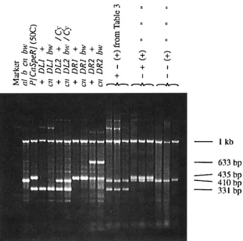
chromosomes. Note that the results of Table 3 are shown only for primer pairs A-B and C-D. All individu- als scored were heterozygous for the chromosome from the D L I / D R I parent and a chromosome from the b cn
h u parent. The 0 cn Inu chromosome does not contain an element at 50C and is therefore expected to amplify the 410-bp A-D primer fragment, as shown in Figure
7.
This was confirmed for all progeny classified. Three features of these results should be noted. First, recombinant gametes are approximately evenly divided between the two parental patterns. There is no prefer- ential tendency, for example, for the deficient end of either element to segregate with the locus with which it is linked. Second, no recombinant gamete was found having both ends present. Third, and most important, the presence of the class lacking both ends shows that there have been changes affecting the elements.PCR fragments were also classified in the case of pairs of either two leftend or two rightend elements. Al- though small numbers of recombinant gametes were found in such crosses (Table 2B), no recombinant ga- metes were present in the subset of flies available for PCR analysis. In a total of 67 parental gametes studied, the expected parental pattern was present in all cases. This contrasts with the results from Table 9, where 24 altered PCR patterns were found in a total of 268 ga- metes. All those showing
"--"
and the two cn h u prog- eny showing"-+"
PCR patterns are considered altered because the parental chromosomes cannot account for those of the progeny. Of these, 2 2 2 of these 24 repre-+
5
"
"
.
-
I lib633 bp
410 bp
-
-
435bp331 hp
-
-
FKXWI 7.--PCR amplification of single fly DNA using prim- ers A-D from Figure 2. Individual examples of each of the parental stocks used in this study are shown along with a representative sample of progeny from males of the genotype shown in Figure 5a. Heterozygote DL2 contained a 410-bp amplification product from primers A to D from the SM1( Cy)
balancer chromosome. The 633-bp amplification in DR2 was caused by amplification with primers A-D, including the 223 bp of remaining P [ CuSpR] sequence. A positive control am- plification product of 1 kb was obtained hy addition of oligo- nucleotides from the singd locus 5 'GCGCAACTTCGCTCGC
3
'
and 5 'CTGGAAGTAGAGATGGTCGC 3 '.
sented events from different male parents. Accepting this lower number for a conservative test, the number of altered elements is still significantly higher than that given by two left- or rightend elements ( P
<
1% by Fisher's exact test). Thus the PCR results are in agreement with the results from the visible markers in indicating element mobility when complementary ends are present but not when only one end is present.Combmations of enddeficient and complete
P[
GzS,beR]elements Genotypes were produced with an end-defi- cient element combined with a complete
P [
CuSpR] (50C) element (Figure 8 ) . Single males of these genotypes were backcrossed to females of genotype b mhu.
These produced less recombination than the combination ofPlA2-31(99B)
n
U
+
+
""""
Hybrid P-Element Activity
TABLE 4
Recombination results for backcross of genotypes with enddeficient and complete P [ caSpeR](5OC) elements
1607
Parent cn bw
+ +
m ++
bw Total Rec. %DLI/CU(5OC) 592 809 75 78 1554 9.8
DL2/CU(5OC) 357 537 40 33 967 7.5
DRI/CU(5OC) 1901 3238 330 448 5917 13.1
DR2/CU(5OC) 448 634 34 65 1181 8.4
end-deficient elements (Table 4 compared with Table
2A)
.
Both reciprocal recombinants were again found at similar frequencies.Results of the PCR classification of the backcross progeny are shown in Table 5. This table, for conve- nience, shows the combined results from all left- and rightend elements. A left-end element is depicted in Figure 8. In this case, the class labeled “Parental” in Table 5 has the [
+-
3
PCR pattern, whereas the class labeled “New” has the[ - + I
pattern. These symbols are reversed for a right-end element.A salient feature of the results in Table 5 is the homo- geneity of PCR patterns associated with the chromo- some carrying the end-deficient element ( cn bw in Ta- ble 5 ) . This indicates the stability of the end-deficient element in this genotype. Such a result is predicted by the “cut-and-paste” model of ENCELS et al. (1990) for a single normal element. In this model, the primary event is expected to be excision of the normal element, followed by repair against the end-deficient element. The expectation is that the opposite event, excision of the incomplete element, will be rare. This has pre- viously been shown to occur in the context of gene conversion ( JOHNSON-SCHLITZ and ENCELS 1993 )
By contrast, changes in PCR fragment patterns are frequent in the other three phenotypes. This applies particularly to the recombinant chromosomes, in which there is also a strong bias toward the incomplete rather than the complete P [ CuSpeR] element. There is ample
evidence in these genotypes for a considerable amount of mobility of the complete element, in contrast to the relative lack of mobility associated with the chromo- some containing the end-deficient element. This latter result therefore suggests that in genotypes such as in Figure 8 it is rare that the complete end of an end- deficient element associates with the complementary end of the normal element.
To confirm that the parental [
+-I
and [ -+
3
PCR patterns with++
phenotypes (Table 5, row 4 ) were in fact due to repair against the end-deficient element and not due to novel deletion events, we amplified 19 of the individual progeny across the deleted region ( + +phenotype gametes in Figure 9 )
.
These data show that in 14 of the 19 progeny the end-deficient element re- placed the complete P [ CaSpeR] element on the++
chromosome. The remaining five cases may be due to incomplete gap repair ( GLOOR et ul. 1991 ) with loss of one or both primer sites.Combinations of complete P [ ChSpeR] (50C ) ele- ments: For comparison with the end-deficient ele- ments, we also carried out a PCR analysis of the frag- ments present from crosses involving two complete
P [ CuSpeR] (50C) homologous elements (Table 6 ) .
The expected pattern of PCR products in this case, in the absence of any P-element structural changes, is
[ + + I
,
indicating that the element is present at the original site. A high proportion of gametes (41/
114) show a change from this pattern. Recombinant gametesTABLE 5
PCR results from the four classes of gametes produced by genotypes with enddeficient and complete fiCuSpeRI(5OC) elements such as in Figure 8
Parental New
AI3 CD AI3 CD AI3 CD AB CD
+ +
+ -
(left)- +
(right)
+ -
- +
- _
+ +
55+bw 10
cn
+
13cn bw 1
36 55 40 111
Again the A-D amplification product expected from the maternal cn bw chromosome was confirmed for
all progeny. The enddeficient element is shown here on the
+ +
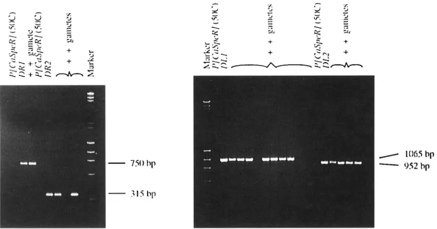
chromosome and the complete P[ CuSpeR]FI(;I'KI. 9 . - I Y X amplilication across putative deleted regions o f ++ progeny from males o f the genotype shown in Figure
8. Oligonucleotitle primcrs used were as follows. D I J , primer L) from Figure I! and 5' CTGC:AAAGCTC;TGACTGGAG J', which
were expccretl t o produce a fragment of 106.5 bp for DL1 sequences and 5124 bp for complete P [ C ~ S ~ P R ] (50C) elements.
111-2, primer D and 5' ATGAAAATAAGAGCTTGAGG 3', which were expected to produce a fragment of952 bp for DL2 sequences
and 4433 bp for complete P [ O'dij,~R] (50C) elements. D R I , primer A from Figure 2 and 5' AGGCAGCAAACACCCATCTG 3',
which were expected to produce a fragment of 750 bp for DR1 sequences and 4294 b p for complete P [ C ~ S ~ P R ] (50C) elements.
DR2, primer A and 5' AACATAAGGTGGTCCCGTCG 3', which were expected to produce a fragment of 315 bp for DR2
sequenccbs and 508.5 bp for complete I J [ C M ~ , P R ] (50C) elements. PCR conditions did not allow for amplification of complete
I.'[ C ~ I . S / J C . ~ ( ] ( S O ( : ) element fragments for any of the primer pairs shown here.
show a slightly, but not significantly, higher frequency of changes ( 2 0 / 4 6 = 0.43) compared with nonrecom- binant gametes (21 / 6 8 = 0.31 )
.
These results are in contrast t o those of SVEI) et nl. (1991 ) , who failed to find any changes i n Southern hybridization patterns in six lines arising from recombinant chromosomes. The present results show that such changes must occur mod- erately often.DISCUSSION
Approximate recombination frequencies from all classes ofcomplete and end-deficient P [ CnSpeR] (50C) elements are summarized in Figure 10. Where a left- end element is shown, classes A, C and E, the data also include results from equivalent right-end elements. The recombination frequencies are only order of magnitude estimates, particularly because they are variable he- hveen different end-deficient elements ( ~ . g . , Table 2 ) and between individual males, due to age and clustering effects. The recombination frequencies given by geno- types A and C , with only end-deficient elements, are
close to zero. It is not clear whether the recombination frequencies i n these cases are higher than those given by P [ A2-31 (99R) by itself ( MCCARRON et nl. 1989,
1994).
As expected from the known behavior of Pelements
( e . g . , MULLINS et nl. 1989), both a left and a right end must be present to induce P-element activity. Classes A and C contain only one type of end and induce little, if any, activity, whereas all the other genotypes contain both ends and induce significant activity. The novel finding from this paper comes from class D, showing that the two ends need not be on the same element or even on the same chromosome to induce male recombi- nation.
Figure 11 shows diagrammatically the situation ex- pected in class D genotypes. The model is based on the cut-and-paste model in which the primary event that initiates P-element transposition is the excision of an
TABLE 6
PCR patterns given by complete P[ CaSpeR] (50C)
homologous elements
AB
CD AB CD AB CD AB CD+ +
+ -
- +
cn /no 11 6 6 3
cn
+
14 4 7 0+
/no 12 3 4 2+ +
36 1 4 1- _
The genotype of this cross was as described for complete
I ~ C ~ S ~ , P R ] ( ~ O C ) elements in Figures 1 and 2 of S\W> el nl.
Hybrid P-Element Activity 1609
Class A B C D E F
Percent
FIGURE 10.-Approximate recombination frequencies for
various genotypes involving enddeficient and complete P[ CaSpeR] (50C) elements and P[ A2-?](99B). [Results
for class B, SVED et al. (1990); results for class F, SVED et al.
(1991) .]
element ( ENCELS et al. 1990). The top left panel shows the two end-deficient elements at the four-strand stage. In the top right panel, the chromosomes contort to allow association of a left and right end. This association is envisaged as being similar to what happens when the two ends of a normal element associate. In the bottom right panel, double-strand breaks are produced, in the same manner as would normally result in excision of an element, possibly accompanied by digestion back of the flanking DNA ( GLOOR et al. 1991 )
.
In the present case, however, no element is liberated. As seen in the bottom left panel, the result is that double-stranded breaks are produced on chromatids of both homolo- gous chromosomes. It is envisaged that these breaks lead to recombination, in a manner as yet unspecified.Competition for end association: Accepting that left and right ends from different elements can function
together, the next issue is to what extent this mode of association occurs when the possibility of association of ends of a single element also exists. The evidence from
MULLINS et ul. (1989) is relevant here. These authors constructed a P element with a left end and two right ends. They showed that the left end would preferen- tially pair with the proximal right end in preference to the distal one. However, when the proximal end was mutated beyond recognition, the distal end was used. MULLINS et al. suggested that, based on their data, pair- ing of P-element ends may occur in a processive man- ner, whereas the results from the enddeficient ele- ments presented here suggest that this mode of pairing is not a necessary one.
Based on the results of MULLINS et al., our expecta- tion is that association of ends from a single element, followed by cut-and-paste ( ENGELS et al. 1990)
,
should be the usual event and that association of ends from different elements should only occur as a secondary possibility. Two aspects of the results from class E geno- types (Table5 )
support this argument. ( 1 ) Chromo- somes containing the end-deficient element are much more stable than chromosomes containing the com- plete P [ CuSpeR] element. Only 2 of 113 progeny with the parental end-deficient phenotype ( c n bw in Table 5 ) show any change in PCR pattern as compared with 39 of 94 for the parental complete P [ CuSpeR] element phenotype (+
+
in Table 5 ).
(2
) A large number ofprogeny ( 3 6 ) with the parental complete P [ CaSpeR]
element phenotype show the PCR pattern expected
cn
”:::::”:&
bw
FIGURE 11.-Model showing “excision” of left and right ends to produce chromosome breakage. Thick lines indicate P
elements, with left-end elements being indicated by dashed lines and right-end elements by solid lines. Flanking chromosomal
DNA is shown by thin solid lines. 5’ to 3’ and 3
’
to 5’
strands of both sister chromatids of the two homologous chromosomesfrom gap repair after excision of a complete element.
As mentioned previously, 19 of these 36 were tested, of which 14 were confirmed gap repairs (Figure 9 ) . Thus where a right end can pair with a left end either on the same element (in cis) or on the homologous element (in trans)
,
the evidence indicates that the cis pairing is chosen in most cases.Given the conclusion that the pairing of ends from different elements is a secondary event compared with the pairing of ends in a single element, the comparison of recombination frequencies is, initially, a surprising one. In particular, class D leads to more recombination than classes E and F in which complete P [ CaSfieR] ele- ments are present. However, class D is unique in that all element-induced breaks are predicted to involve both chromosomes. Thus the lower frequency with which breaks occur in class D, as opposed to class E and F genotypes, could be compensated by the higher fre- quency with which such breaks lead to recombination. These arguments may be carried further to explain why recombination in classes D, E and F is an order of magnitude higher than in class B, in which no Pele- ment sequences are available on the homologous chro- mosome. Association of ends in trans is possible in each of classes D, E and F, leading to breaks on both chromo- somes, whereas in class B the only element-induced break will be on one chromosome.
Finally, it should be noted that the events postulated in this paper are closely related to what has recently been shown to occur in the production of chromosome breaks by the Ac-Ds system in tobacco and maize (EN- GLISH et al. 1993; WEIL and WESSLER 1993). These events were detected through the production of break- age-fusion-bridge cycles rather than recombination, but the postulated association of element ends on different chromatids is very similar to the sequence of events described in Figure 11.
We thank CARLOS FLORES, GREG GLOOR and CHRIS PRESTON for their helpful comments in discussion, and especially BILL ENGELS for his comments both in discussion and in critical reading of the manuscript. We also gratefully acknowledge the Mid-America Stock Center in Bowling Green, Ohio, for provision ofD. melanogasterstocks.
LITERATURE CITED
ENGEM, W. R., 1989 P element in Drosophila melanogaster, pp. 437- 484 in Mobile DNA, edited by D. BERG and M. HOW,. American Society for Microbiology, Washington, DC.
ENGELS, W. R., D. M. JOHNSON-SCHLITZ, W. B. EGGLESTON and J.
A. SVED, 1990 High-frequency P element loss in Drosophila is homologdependent. Cell 62: 515-525.
ENGLISH, J., K. HARISSON and J. D. G. JONES, 1993 A genetic associa-
tion of DNA sequence requirements for Dissociation State I activity in Tobacco. Plant Cell 5: 501-514.
GI.OOR, G. B., and W. R. ENGELS, 1992 Single fly DNA preps for PCR. Dros. Inf. Serv. 71: 148-149.
GLOOR, G. B., N. A. NASSIF, D. M. JOHNSON~CHLITZ, C. R. PRESTON and W. R. ENGELS, 1991 Targeted gene replacement in Dro- sophila via P element-induced gap repair. Science 253: 1110-
1117.
HIRAIZUMI, Y., 1971 Spontaneous recombination in Drosophila mela- nogaster males. Proc. Natl. Acad. Sci. USA 68: 268-270. JOHNSON-SCHLITZ, D. M., and W. R. ENGELS, 1993 Pelement-in-
duced interallelic gene conversion of insertions and deletions in
Drosophila melanogaster. Mol. Cell. Biol. 13: 7006-7018.
KAUFMAN, P.D., R. F. DOLI. and D. C. Rio, 1989 Drosophila P ele- ment transposase recognizes internal Pelement DNA sequences. LINDSIXY, D. L., and G. ZIMM, 1992 The Genome of Drosophila melano-
gaster. Academic Press, New York.
MCCARRON, M. Y., A. DUTrAROY, G. A. DOUGHTY and A. CHOVNICK, 1989 P element transposase induces male recombination in
Dro.sophila melanogaster. Genet. Res. 54: 137-141.
MCCARRON, M. Y., A. DUTTAROY, G. A. DOUGHTY and A. CHOVNICK, 1994 Drosophila P element transposase induces male recombi- nation additively and without a requirement for Pelement exci- sion or insertion. Genetics 136: 1013-1023.
MULLINS, M. C., D. C. RIO and G. M. RUBIN, 1989 cis-acting DNA sequence requirements for P element transposition. Genes Dev.
3: 729-738.
NASSIF, N. A,, J. PENNEY, S. PAL, W. R. ENGELS and G. B. GLOOR, 1994 Efficient copying of nonhomologous sequences from ectopic sites via Pelement-induced gap repair. Mol. Cell. Biol. 14: 1613-
1625.
O’HARE, K., and G. M. RUBIN, 1983 Structure of P transposable elements and their sites of insertion and excision in the Drosophila melanogaster genome. Cell 34: 25-35.
PIRROTTA, V., 1988 Vectors for P-mediated transformation Drosoph- ila, pp. 437-456, in Vectors, edited by R. L. RODRIGUEZ and D. T. DENHARDT. Butterworth, Stoneham, MA.
ROBERTSON, H. M., C. R. PRESTON, R. W. PHILLIS, D. M. JOHNSON-
SCHI.ITZ, W. K. BENZ et al., 1988 A stable genomic source of P
element transposase in Drosophila melanogaster. Genetics 118:
SANKARANARAYANAN, K., and F. SOBELS 1976 Radiation genetics, pp. 1090-1250, in The Genetics and MolecularBiology ofDrosophila, Vol.
I C , edited by M. ASHBURNER and E. NOVITSIU. Academic Press,
London.
SEARLES, L. L., R. S. JOKERST, P. M. BINGHAM, R. A. VOELKER and
A. L. GREENI.EAF, 1982 Molecular cloning of sequences from Cell 59: 359-371.
461-470.
Drosophila polymerase I1 locus by P e1emen;transposon tagging. Cell 31: 585-592.
Swm, J. A,, W. B. EGGLESTON and W. R. ENGELS, 1990 Germ-line and somatic recombination induced by in vitro modified P ele- ments in Drosophila melanogaster. Genetics 124: 331-337. Swm, J. A,, I,. M. BIACKMAN, A. S . GILCHRIST and W. R. ENGFAS, 1991
High levels of recombination induced by homologous Pelements in Drosophila melanogaster. Mol. Gen. Genet. 225: 443-447. SVED, J. A,, L. M. BIA(:KMAN, Y. H. M. SVOBODA and R. COL.LESS, 1994
Generation of high levels of recombination by P elements at homologous sites in Drosophila melanogaster. Mol. Gen. Genet. (in press).
TSUBOTA, S., and P. SCHEDL, 1986 Hybrid dysgenesisinduced re- vertants of insertions at the 5’ end of the rudimentary gene in
Urosqphila melanogaster: transposon-induced control mutations. Genetics 114 165-182.
WEIL, C. F., and S. R. WESSI.ER, 1993 Molecular evidence that chro- mosome breakage by Dselements is caused by aberrant transposi- tion. Plant Cell 5: 515-522.
![FIGURE 1.-Production of P [ D~kted-CnSp~R] (50C) lines.](https://thumb-us.123doks.com/thumbv2/123dok_us/1700793.1215648/2.590.45.279.77.302/figure-production-p-d-kted-cnsp-r-lines.webp)


![FIGURE 10.-Approximate various genotypes involving P[ for (1991) recombination frequencies for enddeficient and complete CaSpeR] (50C) elements and P[ A2-?](99B)](https://thumb-us.123doks.com/thumbv2/123dok_us/1700793.1215648/9.590.50.545.464.702/approximate-genotypes-involving-recombination-frequencies-enddeficient-complete-elements.webp)
